Article

Polyvinyl Alcohol Coatings Containing Lamellar Solids with Antimicrobial Activity

1
Prolabin & Tefarm s.r.l., Via dell’Acciaio 9, 06134 Perugia, Italy
2
International Laboratory Ionomer Materials for Energy and Department of Industrial Engineering, Tor Vergata University of Rome, Via del Politecnico 1, 00133 Roma, Italy
3
Medical Microbiology Unit, Department of Medicine and Surgery, University of Perugia, Piazzale Severi, 1/8, 06129 Perugia, Italy
*
Authors to whom correspondence should be addressed.
Physchem 2024, 4(3), 272-284; https://doi.org/10.3390/physchem4030019
Submission received: 20 May 2024 / Revised: 12 July 2024 / Accepted: 18 July 2024 / Published: 1 August 2024
(This article belongs to the Section Surface Science)

Abstract

:
The design of an antimicrobial coating material has become important in the prevention of infections caused by the transmission of pathogens coming from human contact with contaminated surfaces. With that aim, layered single hydroxides (LSHs) and layered double hydroxides (LDHs) containing Zn and Cu intercalated with antimicrobial molecules were synthesized and characterized. Cinnamate and salicylate anions were chosen because of their well-known antimicrobial activity. Several coatings based on polyvinyl alcohol (PVA) and LDHs or LSHs with increasing amounts of filler were prepared and filmed on a polyethylene terephthalate (PET) substrate. The coatings were characterized, and their antimicrobial activity was evaluated against several pathogens that are critical in nosocomial infections, showing a synergistic effect between metal ions and active molecules and the ability to inhibit their growth.

Graphical Abstract

1. Introduction

Over the 21st century, emerging infectious diseases (EIDs) have increased their incidence in the population, and there seems to be an upward tendency in the future. EIDs refer to emerging or drug-resistant infectious diseases that affect the human population. These diseases have their origin in the spread of many microorganisms, viruses, and other pathogens; their growth in all environments and their consequent inhibition have become a challenge [1].
The World Health Organization (WHO) classified antimicrobial resistance (AMR) as one of the most critical medical issues to be resolved. The bacteria responsible for AMR have been defined by the Infectious Disease Society of America and classified as ESKAPE pathogens: Enterococcus faecium, Staphylococcus aureus, Klebsiella pneumoniae, Acinetobacter baumannii, Pseudomonas aeruginosa, and Enterobacter spp. [2]. ESKAPE pathogens belong both to Gram-positive and Gram-negative bacteria, and they are often responsible for serious nosocomial infections [3].
Reducing the spread of the infection could help fight bacterial resistance against antibiotics. Coating the surfaces directly in contact with human daily life with antimicrobial and/or antiviral substances can be considered one of the most promising solutions. The global antimicrobial coatings market was valued at USD 8.34 billion in 2020 and is expected to grow at a compound annual growth rate (CAGR) of 13%, reaching USD 20.71 billion by 2028 [4]. Nanoparticles of Cu and Zn are already used in contact-killing surfaces with great efficacy. The antibacterial effect of Cu and Zn surfaces is attributed to a combination of the types of damage caused by Cu(II) and Zn(II) ions released from the surface. They interact with reactive oxygen species (ROS), leading to lipid peroxidation and the subsequent loss of membrane integrity, protein damage, DNA damage, and cell death.
The aim of this work is to synthesize and characterize layered single (LSHs) or double hydroxides (LDHs) containing Cu and/or Zn and to use them to convey antimicrobial molecules in the interlayer region. Lamellar solids have been widely used in the literature as carriers of antimicrobial molecules in different applications, as shown in references [5,6,7]. In that case, the idea is to combine the proven efficacy of the metals in the layer with that of the conveyed molecules, reaching a synergistic effect. In detail, ZnOH-LSH, ZnAl-LDH, and ZnCuAl-LDH in nitrate form were synthesized as starting materials.
Both cinnamate (CA) [8] and salicylate (SAL) [9] are well known for their antimicrobial efficacy. They were selected for their acidic nature, which gives them the ability to be easily intercalated as anions in both LSHs and LDHs.
Both molecules were conveyed in all the solids, obtaining six materials labelled as: ZnOH-CA, ZnAl-CA, ZnCuAl-CA, ZnOH-SAL, ZnAl-SAL, and ZnCuAl-SAL. The pristine solids and the intercalated ones have been used as functionalizing fillers in the preparation of coating resins based on polyvinyl alcohol (PVA). Water-based PVA resin is considered to be an eco-friendly synthetic petroleum-based resin. It is an ideal resin for a coating because it is biocompatible, transparent, chemo-resistant, tough, and a good barrier agent, and it can be industrially produced by non-petroleum routes, implying low-price production [10]. In the literature, PVA has already been functionalized with inorganic filler, obtaining interesting performances [11,12]. Moreover, it has also already been functionalized with ZnOH-SAL, demonstrating a good compatibility between the polymer and the filler [13]. However, the antimicrobial functionality was not explored.
LDH/PVA resin and LSH/PVA resin composites were successfully prepared and filmed on a polyethylene terephthalate (PET) substrate. Weight percentages of 1%, 5%, and 10% of the LSHs and LDHs were used. The films were characterized by X-ray diffraction spectroscopy (XRD) and Fourier transform infrared spectroscopy (FT-IR) for their antimicrobial properties. The main stages of the project are reported in Figure 1, in order to clarify the aim of the work.

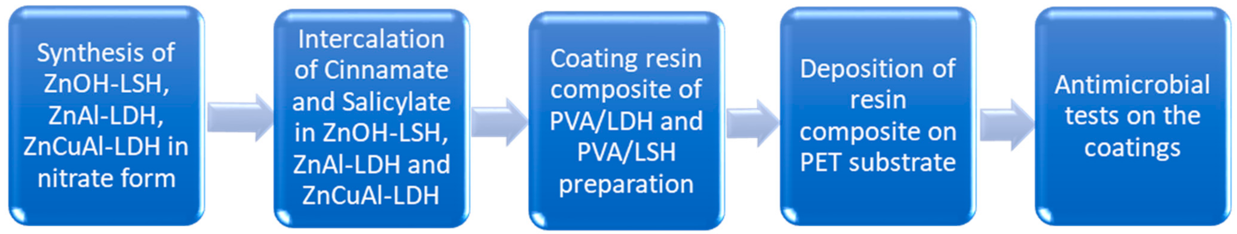
2. Materials and Methods

2.1. Materials

Zinc nitrate hexahydrate [Zn(NO3)2 × 6H2O] (98%), copper nitrate hemi-pentahydrate [Cu(NO3)2 × 2.5H2O] (98%), aluminum nitrate nonahydrate [Al(NO3)3 × 9H2O] (98%), trans-cinnamic acid (97%), urea (99%), and sodium hydroxide (NaOH) (98%) were purchased from Sigma-Aldrich. Sodium salicylate (99.83%) was purchased from Polichimica S.R.L (Bologna, Italia). Polyvinyl alcohol resin (Sealcoat HS 25, %) was purchased from Baumeister. All the reagents were used as they arrived and without further purification.

2.2. Synthesis of ZnOH-LSH, ZnAl-LDH, ZnCuAl-LDH in Nitrate Form

The synthesis of ZnOH-LSH in nitrate form was conducted according to Li et al.’s method [14]. The molar ratio of OH/Zn was equal to 1.6; 80 mL of a 0.2 M NaOH solution was dropped into a 50 mL solution containing 10 mmol of Zn(NO3)2 × 6H2O, under mechanical stirring at room temperature. A white precipitate was obtained. The syntheses of ZnAl-NO3 and ZnCuAl-NO3 were conducted following the urea hydrolysis method [15]. For ZnAl-NO3, an aqueous solution containing Al3+ and Zn2+ with the molar fraction of Al3+/(Al3+ + Zn2+) equal to 0.33 was prepared by dissolving Al(NO3)3 × 9H2O and Zn(NO3)2 × 6H2O in distilled water at a 1 M concentration. To this solution, solid urea was added until the molar ratio of urea/Al3+ reached 4.0. The clear solution was refluxed for 24 h, and a white precipitate was obtained. For ZnCuAl-NO3, an aqueous solution containing Al3+, Zn2+, and Cu2+, with the molar fraction of Al3+/(Al3+ + Zn2+ + Cu2+) equal to 0.33 and a ratio of Zn2+/Cu2+ equal to 2.5, was prepared by dissolving Al(NO3)3 × 9H2O, Zn(NO3)2 × 6H2O and Cu(NO3)2 × 6H2O in distilled water at a 1 M concentration. To this solution, solid urea was added until the molar ratio of urea/Al3+ reached 4.0. The obtained solution was poured into a reactor under mechanical stirring; the temperature of the reaction was fixed at 90 °C for 24 h. A light blue precipitate was obtained. After the syntheses, all the precipitates were centrifuged, washed twice to remove excess urea and ions, dried in the oven at 60 °C until a constant weight, and ground using a knife mill. The obtained solids were sieved with an 80-micron mesh sieve.

2.3. Intercalation of Cinnamate and Salicylate in ZnOH-LSH, ZnAl-LDH, and ZnCuAl-LDH

The intercalation of cinnamic acid was carried out using the following procedure: cinnamic acid was salified by the addiction of a stoichiometric amount of NaOH 1 M solution. The as-obtained salt was dissolved in a mixture of ethanol/water 1:1 to obtain a concentration of 0.1 M. ZnOH-LSH, ZnAl-LDH, and ZnCuAl-LDH were equilibrated with the cinnamate solution under mechanical stirring for 24 h, to obtain the following intercalation products: ZnOH-cinnamate (ZnOH-CA), ZnAl-cinnamate (ZnAl-CA), and ZnCuAl-cinnamate (ZnCuAl-CA). The mass/volume ratios used were 1 g/54 mL for ZnOH-LSH and 1 g/56 mL for ZnAl-LDH and ZnCuAl-LDH, respectively. For the preparation of the second set of intercalation products, a water solution of sodium salicylate 0.5 M was prepared. The solids in nitrate form were equilibrated with the salicylate solution under mechanical stirring for 24 h, to obtain the respective intercalation compounds: ZnOH-salicylate (ZnOH-SAL), ZnAl-salicylate (ZnAl-SAL), and ZnCuAl-salicylate (ZnCuAl-SAL). The solid/volume ratios used were: 1 g/10.4 mL for ZnOH-LSH and 1 g/11.2 mL for ZnAl-LDH and ZnCuAl-LDH, respectively. The intercalations were carried out at room temperature; the resulting compounds were recovered by centrifugation, washed twice with deionized water, dried in the oven at 60 °C, and ground using a knife mill. The obtained solids were sieved with an 80-micron mesh sieve.

2.4. Coating Resin Composite of PVA/LDH and PVA/LSH Preparation

Composite coatings were prepared mixing PVA resin dissolved in water with a weighted amount of the LDH and LSH powders. The exact amounts of PVA, water, and filler for the indicated percentages are reported in Table 1. The dispersions were prepared using the following as fillers: ZnOH-LSH, ZnAl-LDH and ZnCuAl-LDH, ZnOH-CA, ZnAl-CA, ZnCuAl-CA, ZnOH-SAL, ZnAl-SAL, and ZnCuAl-SAL at the following weight percentages (wt%): 1, 5, and 10. The films were labelled using the filler name and the relative percentage used, i.e., ZnAl-LDH 1%, etc. A PVA film reference was also prepared. After mixing using magnetic stirring, ultrasound was applied to assure a homogenous dispersion of the lamellar solid into the matrix. A few drops of 1M NaOH solution were poured to catalyze the polymerization of PVA and to avoid the dissolution of the LSHs and LDHs.

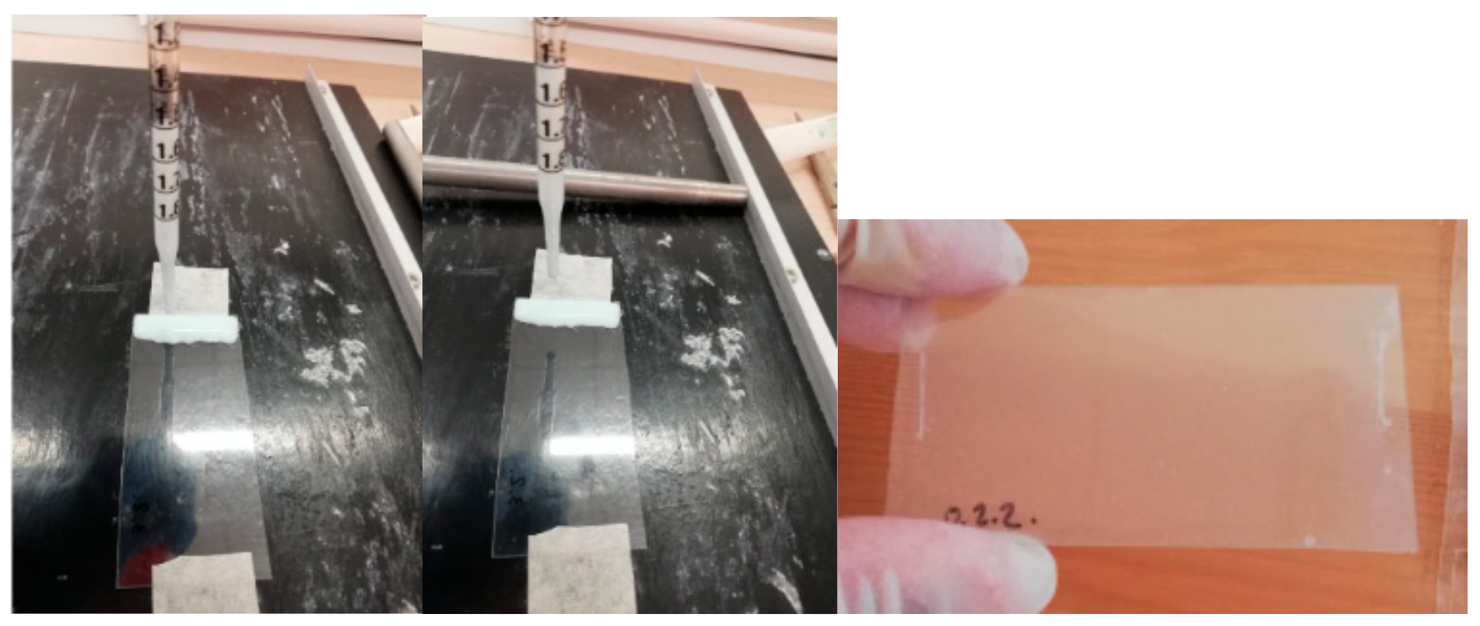
2.5. Deposition of Resin Composite on PET Substrate

PET samples, with dimensions of 7.5 × 3.6 cm, were coated with the as-prepared resins. Five milliliters of the dispersion was poured onto the substrate. The resin was filmed by manually rolling the product with Mayer’s bar number 25. Two layers of the resins containing fillers were deposed on each substrate. After the adhesion of both layers, each sample was dried in an oven at 60 °C for 7–10 min. The films obtained were characterized by XRD and FT-IR spectroscopies.

2.6. Microorganisms

The antimicrobial tests were carried out on the following pathogens: (a) two Gram-positive bacteria, Staphylococcus aureus (ATCC 29213) and the clinical isolate Enterococcus faecalis; (b) two Gram-negative clinical isolate bacteria, Acinetobacter baumannii and Klebsiella pneumoniae; and (c) the yeast Candida albicans (SC5314). The bacteria were grown in Muller Hinton agar (MHA), whereas the Candida cells were grown in Sabouraud agar. One colony of each microorganism was inoculated in the appropriate culture broth medium (Muller Hinton broth MHB or Sabouraud broth) and maintained for 24 h at 37 °C. Then, after centrifugation, the recovered microorganisms were washed twice in sterile saline solution and counted by spectrophotometric analysis. The suspensions were then diluted in a sterile saline solution to the concentration required in the following assays.

2.7. Antimicrobial Susceptibility Test

An antimicrobial susceptibility test was performed using the Kirby–Bauer disk diffusion method following the guidelines of the Clinical and Laboratory Standard Institute (CLSI) [16]. Bacterial sensitivity was tested for the films, and erythromycin and fluconazole were used as positive controls for inhibiting the bacteria strains and yeast, respectively. The films were cut into 5 mm diameter disks and sterilized under UV rays for 60 min. Muller Hinton agar plates were used for testing the different bacteria. After adjusting the bacterial suspension to the concentration of 0.5 McFarland in sterile saline solution, the suspension was spread on the agar plate by streaking a swab three times over the entire agar surface, rotating the plate approximately 60 degrees each time to ensure a homogeneous distribution of the inoculum. The films were placed on the top of the culture plates and incubated for 24 h at 37 °C. Using a ruler, each inhibition zone was measured. The results are expressed as zone of inhibition (ZOI), measured in mm. The data are the mean ± SD of three different experiments.

2.8. Analytical Procedures and Instrumentation

2.8.1. Inductively Coupled Plasma–Optical Emission Spectrometer

The solids were dissolved in water using a proper amount of HNO3, in order to determine the Zn, Al, and Cu contents. The solutions were analyzed after a proper dilution by ICP-OES Perkin Elmer, Avio 200 Waltham, MA, USA.

2.8.2. X-ray Diffraction Spectroscopy

X-ray powder diffraction (XRD) patterns were recorded with a Bruker D2 Phaser diffractometer operating at 30 kV and 15 mA, a step size of 0.02 (2θ degrees), and a time per step of 1 s, using the Cu Kα radiation (1.54 Å) and multistrip LYNXEYE SSD160 detector.

2.8.3. FT-IR Analysis

The FT-IR spectra were recorded at room temperature using an FT-IR-ATR Nicolet 380 (Thermo Fisher, Waltham, MA, USA). Typically, each spectrum was obtained in the spectral region from 400 to 4000 cm−1. For data collection, an attenuated total reflection crystal in SeZn was used.

2.8.4. Thermal Analysis

Thermogravimetric analysis (TGA) of the samples was carried out with a Perkin Elmer STA 8000, Waltham, MA, USA, in an air flow at a heating rate of 10 °C min−1.

2.8.5. UV-Vis Analysis

UV-visible analyses were carried out by a UV-visible double-beam spectrophotometer (Jasco V-750, Oklahoma City, OK, USA). The salicylate and cinnamate content in the samples was determined by UV-Vis spectroscopy. A weighed amount of the sample (~20 mg) was completely dissolved in 100 mL of a 0.85 M aqueous solution of HCl. Salicylate and cinnamate in the samples were determined by monitoring the maximum absorption value of the molecules after calibration and proper dilution. A suitable calibration was carried out by dissolving known amounts of pure salicylate or cinnamate in a proper volume of a 0.85 M aqueous solution of HCl. Five standards and a blank sample were then prepared and analyzed to obtain the calibration curve.

2.8.6. SEM Analysis

The morphology of the sample ZnOH-LSH 10% was investigated with a FEG LEO 1525 scanning electron microscope (FE-SEM). FE-SEM micrographs were collected by depositing the sample on a stub holder and after a sputter coating with chromium for 20 s.

3. Results and Discussion

3.1. Characterization of the Pristine Powders

The aim of this work is to synthesize and characterize lamellar solids for the vehiculation of antimicrobial molecules and to test their antimicrobial activity. In the literature, these intercalation compounds have been widely demonstrated to possess interesting properties [6,7,17], especially in the fields involved in the preparation of active polymer composites for applications in wound healing and food packaging. We explore for the first time the application of these intercalation compounds for the preparation of antimicrobial coatings. ZnOH-LSH, ZnAl-LDH, and ZnCuAl-LDH were chosen as fillers, due to the presence of Zn and Cu, which have well-documented antimicrobial activity [4]. The powders were prepared as described in the experimental part and characterized by XRD and ICP-OES. The XRD spectra are reported in Figure 2.
The first reflex of ZnOH-LSH is at 2θ = 9.2, which corresponds to the (200) diffraction peak and to an interlayer distance of 9.6 Å. These values are compatible with the presence of nitrate in the interlayer region [14]. For ZnAl-LDH and ZnCuAl-LDH, the strongest peak at 2θ = 9.9, due to (003) reflection [15], can be related to an interlayer distance of 8.9 Å, corresponding in both cases to the gallery height attributed to nitrate [15]. In the case of ZnCuAl-LDH, a very small amount of a second phase, that has been underlined with *, is present, and could be probably assigned to garhardtite [18], with the chemical formula Cu2(NO3)(OH)3. The ICP-OES results allowed the calculation of the following chemical formulas: Zn5(OH)8(NO3)2 × 2H2O, Zn0.70Al0.30(OH)2(NO3)0.30 × 0.5H2O, and Zn0.55Cu0.05Al0.40(OH)2(NO3)0.40 × 0.5H2O. An interesting observation is that the interlayer space of ZnOH-NO3 has the biggest value, which can probably be associated with the higher number of water molecules coordinated directly with the structure [19]. The FT-IR spectra of the obtained powders are reported in Figure S1 of the SI. The ZnOH-NO3 spectrum (black line) is similar to the one reported by Nabipour et al. [20]. The bands at 3573 cm−1 and 3462 cm−1 correspond to the O-H stretching vibrations of the lattice and water molecules occupying the interlayer region, respectively. The sharp band at 1636 cm−1 can be assigned to the bending mode of intercalated water molecules. The broad band located at 1367 cm−1 is due to the asymmetric stretching vibration of nitrate groups. The ZnAl-NO3 spectrum (red line) presents a signal at 3378 cm−1, indicating the presence of water molecules [21]. The band broadening is due to the H-bond stretching vibrations of the OH groups in the brucite-like layers and intercalated water molecules. The presence of water in the interlayer region is confirmed by the signal at 1636 cm−1, which is associated with the bending mode of water molecules occupying the interlamellar region [22]. A sharp band at 1341 cm−1 is characteristic of the asymmetric stretch vibration of nitrate groups. Regarding the ZnCuAl-NO3 spectrum (blue line), the nitrate band is observed at 1346 cm−1 while the vibration at 1415 cm−1 probably corresponds to a reduction in the symmetry of the carbonate anions. As reported by Palacio et al. [23], a broad peak appears at around 3481 cm−1, which is always associated with the H-bond stretching mode of the vibrations from OH groups in the structure and from water molecules between the layers and the physiosorbed area. The band at 1646 cm−1 is due to the intercalated water bending.

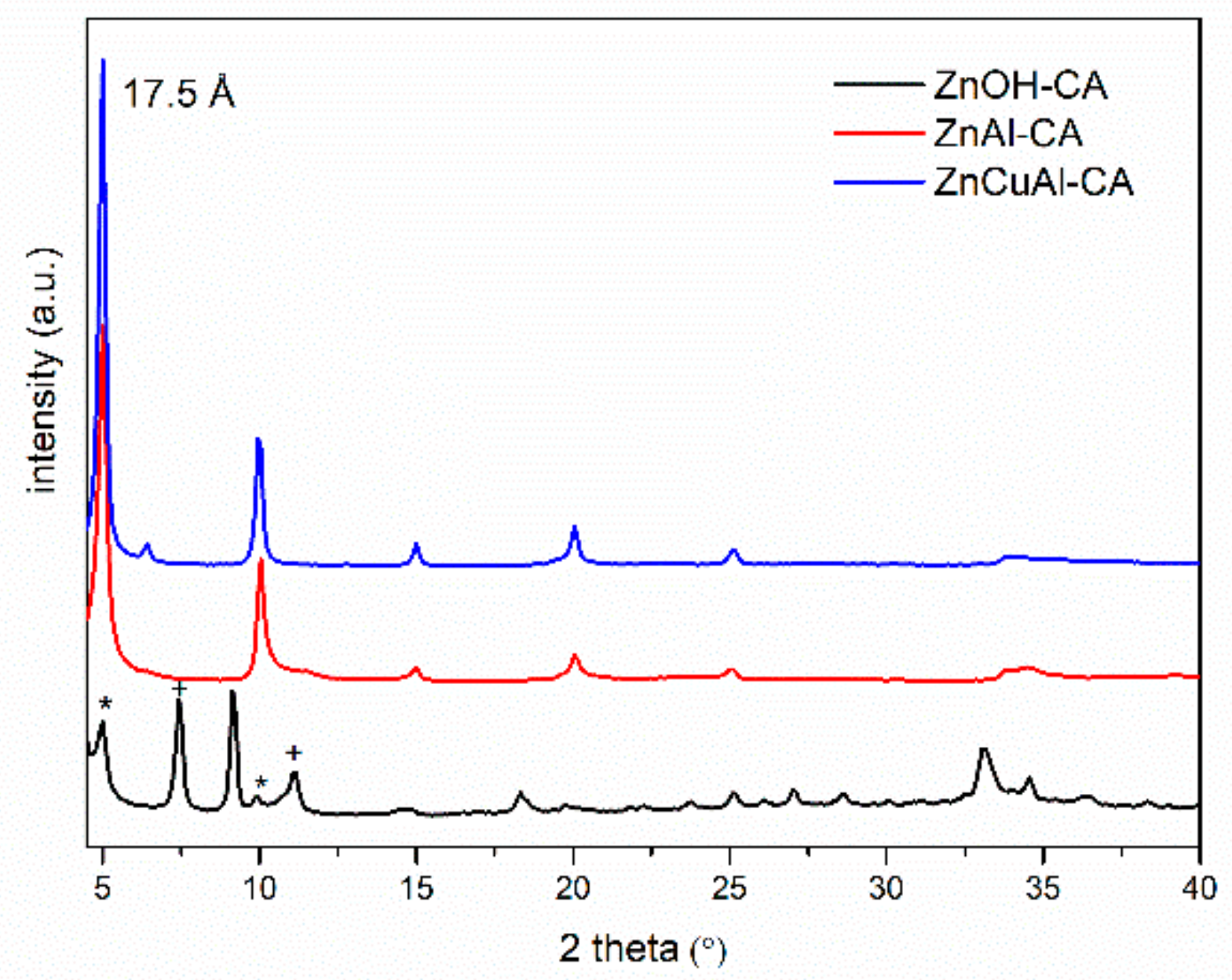
3.2. Characterization of the Intercalated Powders

The XRD spectra of the three cinnamate intercalation compounds are reported in Figure 3.
In each case, it is possible to observe an increase in the interlayer distance from the starting nitrate forms (8.9 Å for LDHs and 9.6 Å for LSHs) up to 17.5 Å, due to the effective intercalation of the organic anions. The ZnOH-CA spectra (black line) show a mixture of three different phases. The peaks at 11.8 Å and 7.9 Å that have been indicated using the symbol + are probably the second and third reflexes (004 and 006, respectively) of the intercalation compound, with an interlayer distance of 23.6 Å. This can be attributed to an intercalated phase of cinnamate, in which the LSH possesses a type IIb structure [24]. This is formed by one quarter of the octahedral zinc cations displaced from the main layer to the tetrahedral sites located above and below each empty tetrahedron [25]. A second phase underlined with the symbol * has a first reflex corresponding to an interlayer distance of 17.5 Å, as observed for ZnAl-CA and ZnCuAl LDHs. It is likely that this phase derives from the type I structures, where the metal centers are only coordinated in an octahedral arrangement, like in layered double hydroxide [25]. A partial residue of the nitrate phase was also observed at 9.6 Å. The ZnAl-CA and ZnCuAl-LDH phases both present an interlayer distance of 17.5 Å, which is compatible with cinnamate intercalation [21], and no residual nitrate was detected in either of the cases. According to Adam et al.’s model [21], the anions in this phase were considered to be orientated in a bilayer, where the molecules are partially tilted with respect to the plane. The FTIR spectra of the cinnamate intercalated powders are reported in Figure S2 of the SI, and they clearly confirm the XRD data, indicating the presence of cinnamate in the powders. The compounds exhibit most of the vibrations assigned to cinnamate, even if some of the vibrations are slightly shifted due to the electrostatic interaction between the organic anion and the inorganic layered structure [21,24]. The vibrations due to trans-C-H out-of-plane bending (976 cm−1) are evident in all three samples. Moreover, COO stretching of the intercalated cinnamate anion appears at 1531 and 1390 cm−1. Also, the C=C stretching signal is present in all the samples at 1636 cm−1. In the ZnOH-CA spectrum, the band at 1370 cm−1 confirms the presence of a certain amount of residual nitrate, as observed in the XRD spectrum.
The XRD spectra of the three compounds recovered after contact with salicylate solutions are reported in Figure 4. In this case, the intercalation of salicylate was successful only with ZnAl-LDH, obtaining a pure phase. The increase in interlayer distance from 8.9 Å to 16.5 Å confirms the presence of salicylate between the lamellae [6].
Both ZnOH-SAL and ZnCuAl-SAL are still in the pristine nitrate form, with an evident increase in the turbostratic disorder, especially for the ZnCuAl-SAL sample. In this last sample, we also observed a change in color, from light blue to light green; this change is probably due to a complex formation. The loss of effective intercalation can be attributed to many factors, but a detailed discussion falls outside the scope of this paper. Moreover, salicylate is detected in all three of the compounds, as demonstrated by the FT-IR (Figure S3 of SI). Regarding the ZnAl-SAL spectrum, clear evidence of the SAL presence is the asymmetric and symmetric stretching modes of the COO group observed at 1565 and 1360 cm−1. These bands occur at slightly lower wave-numbers in comparison to the free COO functional group in salicylate, according to the literature [26]. Also, the peak at 1252 cm−1 is due to the OH bending vibration mode of the phenolic group [13]. In the ZnOH-SAL spectrum, the bands due to the pristine solid are still present, but the typical signals of salicylate can be also detected. At 1570 cm−1, the asymmetric stretching mode of salicylate is evident, while the symmetric one is probably covered by the nitrate signal. The band at 1250 cm−1 due to the OH bending vibration mode of the phenolic group is also present. The ZnCuAl-SAL spectrum shows signals different from those observed for ZnAl-SAL. Shamraiz et al. [27] reported an FT-IR spectrum of [Cu(Hsal)2] with very similar band positions, giving evidence of a partial formation of a copper–salicylate complex. The presence of bands at 1564 and 1605 cm−1 are due to metal-bonded C–O, and the bands at 1407 and 1335 cm−1 are due to the C–C bond of the salicylate group. This result indicates that in ZnOH-SAL the organic molecules are probably adsorbed on the surface of the solid; while in ZnCuAl-SAL, a certain amount of [Cu(Hsal)2] has been formed. It can be concluded that even if the intercalation was not achieved, the samples are, nevertheless, both interesting candidates for testing the antimicrobial properties, since they clearly contain a suitable amount of antimicrobial species. All the samples were characterized by TGA in order to calculate the amount of conveyed molecules. The UV-Vis analysis carried out on the dissolved samples confirmed the observed results. In Table 2, the obtained results and the calculated amount of organic molecules in each compound are summarized. In the sample ZnOH-SAL, the organic molecule is probably absorbed on the surface.

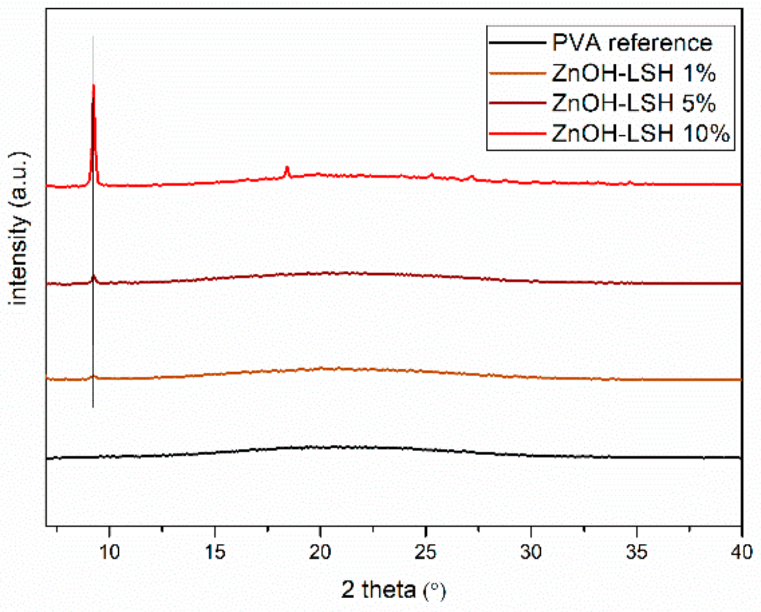
3.3. Chemical–Physical Characterization of the Coatings

The coatings were prepared by mixing PVA resin with a weighted amount of the prepared filler. Then, the as-prepared resins were coated on PET samples (7.5 × 3.6 cm). Five milliliters of the resin dispersion was poured on the substrate and filmed by rolling the product with a Mayer’s bar, as described previously (Figure 5). Two layers of the resins containing fillers were deposed on each substrate.
All the obtained films were characterized by XRD and FT-IR, but for simplicity, we reported only the characterizations of the following films: ZnOH-LSH 1%, ZnOH-LSH 5%, and ZnOH-LSH 10%. The same characterizations can be obtained by analyzing the XRD data of the other composite films.
In Figure 6, the XRD spectra are reported. It is evident that no change in the interlayer distance is detected, and the first reflex of the intercalate is always visible, meaning that no significant interactions are observed between the filler and the polymer (i.e., the formation of the intercalated composited or nanometric dispersion of the filler). The intensity of the first reflex increases linearly with the increase in filler content.
The FTIR analyses of the ZnOH-LSH films, compared with the spectra of the substrate and the pure powder, are reported in Figure S4. All the spectra present the same bands at similar positions due to the PVA-PET support and the most intense signal of ZnOH-LSH at 1370 cm−1. The spectrum of PVA-PET film exhibit bands in the range of 1412–1325 cm−1, which are ascribed to the bending of CH2 groups. A characteristic signal of the stretching vibration band of C=O is visible at 1733 cm−1. A C-O-C stretching vibration band is detectable at 1091 cm−1 and 1244 cm−1, suggesting the presence of an ester linkage between the hydroxyl group of PVA and the carboxyl groups of PET.
In Figure 7, the SEM micrographs of the sample ZnOH-LSH 10% are reported, in order to evaluate the morphological characteristics of the coating.
The sample clearly contains inorganic lamellar particles of ZnOH-NO3 with micrometric dimensions and irregular shape, ranging between 1 and 2 microns. The particles are uniformly dispersed on the coating, as also demonstrated by the EDX reported in Figure S5.

3.4. Antimicrobial Tests on the Coatings

The antimicrobial assays were carried out on ATCC strains and the clinical isolates S. aureus, E. faecalis, A. baumannii, K. pneumoniae, and C. albicans. Among them, it is important to underline that S. aureus, A. baumannii, and K. pneumoniae are considered as ESKAPE pathogens [3]; so, finding new strategies to reduce their spread is a really critical issue. The antibacterial properties of the films were determined using the Kirby–Bauer disk diffusion test according to the directions of the Clinical Laboratory Standards Institute [16]. Erythromycin and fluconazole were used as positive controls. The coatings containing ZnAl-LDH, ZnCuAl-LDH, and ZnOH-LSH were active against S. aureus, E. faecalis, and C. albicans, as reported in Table 3. In ZnAl-LDH and ZnOH-LSH, the antimicrobial performance increased with the increase in Zn content. This is in accordance with the study of Cheng et al. [28], which concludes that LDHs and LSHs [20] are also reservoirs of zinc ions and can slowly release this metal and reach an effective antimicrobial activity.
The coatings functionalized with ZnAl-CA, ZnCuAl-CA, and ZnOH-CA are active against E. faecalis and C. albicans (Table 4). It is worth underlining that ZnCuAl-CA is also active against K. pneumoniae [29], which is now considered one of the most dangerous pathogens for public health due to its antimicrobial resistance and the high human mortality rate. This effect is probably due to the combined release of Zn2+, Cu2+, and cinnamate.
In Table 5, the results of the coatings functionalized with ZnAl-SAL, ZnCuAl-SAL, and ZnOH-SAL are reported. The samples containing salicylate are the most interesting, confirming a synergistic activity of this antimicrobial molecule with Zn2+ and Cu2+. They are all active against S. aureus, E. faecalis, A. baumannii, K. pneumoniae, and C. albicans. The ZnCuAl-SAL ones are also active against K. pneumoniae. The wide activity of this sample can also be ascribed to the partial formation of [Cu(Hsal)2], which possesses an already documented antimicrobial activity [30].

4. Conclusions

In this work, innovative surface coating materials to prevent pathogen infections by simple contact were designed. A polyvinyl alcohol resin solution with increasing amounts of active fillers made up of intercalated cinnamate and salicylate in LDHs or LSHs was prepared and coated on a PET substrate. The materials were characterized using different techniques, including ICP-OES, XRD, FT-IR, and TGA. The antimicrobial susceptibility test was performed using the Kirby–Bauer disk diffusion method. The coatings containing ZnAl-LDH, ZnCuAl-LDH, and ZnOH-LSH were active against bacteria and yeast, and the antimicrobial performance increased with the increase in Zn content. The samples containing salicylate presented the synergistic activity of this antimicrobial molecule with Zn2+ and Cu2+ ions. ZnCuAl-CA and ZnCuAl-SAL were also active against the dangerous K. pneumoniae. Although this study is preliminary and requires further future investigation, the results clearly indicate that the use of these coatings will certainly be useful for preventing the spread of microbes that cause diseases, that can even be fatal and will lead to significant money savings for public health.

Supplementary Materials

The following supporting information can be downloaded at: https://www.mdpi.com/article/10.3390/physchem4030019/s1, Figure S1: FT-IR spectra of the powders, ZnOH-LSH black line, ZnAl-LDH red line, ZnCuAl-LDH blue line, Figure S2: FT-IR spectra of the powders, ZnOH-CA black line, ZnAl-CA red line, ZnCuAl-CA blue line, Figure S3: FT-IR spectra of the powders, ZnOH-SAL black line, ZnAl-SAL red line, ZnCuAl-SAL blue line, Figure S4: PVA black line, ZnOH-LSH 1% brown line, ZnOH-LSH 5% dark red line, ZnOH-LSH 10% red line, ZnOH-LSH, Figure S5: EDX analysis of the sample ZnOH-LSH 10%; Zn distribution is reported in red color.

Author Contributions

M.B., conceptualization, methodology, writing—original draft preparation, writing—review and editing, visualization; M.S., conceptualization, methodology, resources; R.E.G., investigation, writing—original draft preparation, visualization; I.D.G., validation, investigation; D.P., investigation, writing—original draft preparation; C.R., validation, investigation; R.N., conceptualization, methodology, writing—original draft preparation, writing—review and editing. All authors have read and agreed to the published version of the manuscript.

Funding

This research received no external funding.

Data Availability Statement

The original contributions presented in the study are included in the article/Supplementary Material, further inquiries can be directed to the corresponding author/s.

Acknowledgments

Raúl Escudero García is grateful for the Erasmus Mundus Chemical Nanoengineering (CNE) master program and the scholarship support. The authors thank Alessandro di Michele of the Physics Department of Perugia University for his valuable contribution to the SEM analyses.

Conflicts of Interest

Author Maria Bastianini, Michele Sisani, Raúl Escudero García, and Irene Di Guida were employed by the company Prolabin & Tefarm s.r.l. The remaining authors declare that the research was conducted in the absence of any commercial or financial relationships that could be construed as a potential conflict of interest.

References

  1. Zhang, Y.; Fan, W.; Sun, Y.; Chen, W.; Zhang, Y. Application of antiviral materials in textiles: A review. Nanotechnol. Rev. 2021, 10, 1092–1115. [Google Scholar] [CrossRef]
  2. Mingoia, M.; Conte, C.; Rienzo, D.A.; Dimmito, M.P.; Marinucci, L.; Magi, G.; Turkez, H.; Cufaro, M.C.; Del Boccio, P.; Di Stefano, A.; et al. Synthesis and Biological Evaluation of Novel Cinnamic Acid-Based Antimicrobials. Pharmaceuticals 2022, 15, 228. [Google Scholar] [CrossRef] [PubMed]
  3. Mulani, M.S.; Kamble, E.E.; Kumkar, S.N.; Tawre, M.S.; Pardesi, K.R. Emerging strategies to combat ESKAPE pathogens in the era of antimicrobial resistance: A review. Front. Microbiol. 2019, 10, 539. [Google Scholar] [CrossRef] [PubMed]
  4. Birkett, M.; Dover, L.; Cherian Lukose, C.; Wasy Zia, A.; Tambuwala, M.M.; Serrano-Aroca, Á. Recent Advances in Metal-Based Antimicrobial Coatings for High-Touch Surfaces. Int. J. Mol. Sci. 2022, 23, 1162. [Google Scholar] [CrossRef] [PubMed]
  5. Bastianini, M.; Scatto, M.; Sisani, M.; Scopece, P.; Patelli, A.; Petracci, A. Innovative Composites Based on Organic Modified Zirconium Phosphate and PEOT/PBT Copolymer. J. Compos. Sci. 2018, 2, 31. [Google Scholar] [CrossRef]
  6. Coiai, S.; Cicogna, F.; Pinna, S.; Spiniello, R.; Onor, M.; Oberhauser, W.; Coltelli, M.-B.; Passaglia, E. Antibacterial LDPE-based nanocomposites with salicylic and rosmarinic acid-modified layered double hydroxides. Appl. Clay Sci. 2021, 214, 106276. [Google Scholar] [CrossRef]
  7. Puccetti, M.; Donnadio, A.; Ricci, M.; Latterini, L.; Quaglia, G.; Pietrella, D.; Di Michele, A.; Ambrogi, V. Alginate Ag/AgCl Nanoparticles Composite Films for Wound Dressings with Antibiofilm and Antimicrobial Activities. J. Funct. Biomater. 2023, 14, 84. [Google Scholar] [CrossRef] [PubMed]
  8. Guzman, J.D. Natural cinnamic acids, synthetic derivatives and hybrids with antimicrobial activity. Molecules 2014, 19, 19292–19349. [Google Scholar] [CrossRef] [PubMed]
  9. Sykes, E.M.E.; White, D.; McLaughlin, S.; Kumar, A. Salicylic acids and pathogenic bacteria: New perspectives on an old compound. Can. J. Microbiol. 2023, 14, 1–14. [Google Scholar] [CrossRef]
  10. Moulay, S. Review: Poly(vinyl alcohol) Functionalizations and Applications. Polym. Plast. Technol. Eng. 2015, 54, 1289–1319. [Google Scholar] [CrossRef]
  11. Yeun, J.H.; Bang, G.S.; Park, B.J.; Ham, S.K.; Chang, J.H. Poly(vinyl alcohol) nanocomposite films: Thermooptical properties, morphology, and gas permeability. J. Appl. Polym. Sci. 2006, 101, 591–596. [Google Scholar] [CrossRef]
  12. Bastianini, M.; Sisani, M.; Naryyev, E.; Petracci, A.; Di Guida, I.; Narducci, R. Composite membranes based on polyvinyl alcohol and lamellar solids for water decontamination. New J. Chem. 2024, 48, 2128. [Google Scholar] [CrossRef]
  13. Ruiz, C.V.; López-González, M.; Giraldo, O. Composite films based on salicylic acid-layered zinc hydroxide and polyvinyl alcohol: Preparation, characterization, properties and potential applications. Polym. Test. 2021, 94, 107057. [Google Scholar] [CrossRef]
  14. Li, P.; Xu, Z.P.; Hampton, M.A.; Vu, D.T.; Huang, L.; Rudolph, V.; Nguyen, A.V. Control preparation of zinc hydroxide nitrate nanocrystals and examination of the chemical and structural stability. J. Phys. Chem. C 2012, 116, 10325–10332. [Google Scholar] [CrossRef]
  15. Di Michele, A.; Boccalon, E.; Costantino, F.; Bastianini, M.; Vivani, R.; Nocchetti, M. Insight into the synthesis of LDH using the urea method: Morphology and intercalated anion. Dalton Trans. 2024, accepted. [Google Scholar] [CrossRef] [PubMed]
  16. Clinical and Laboratory Standards Institute (CLSI). Clinical and Laboratory Standards Institute Methods for Dilution Antimicrobial Susceptibility Tests for Bacteria That Grow Aerobically Standard, Approval CDM-A. In M07-A9: Methods for Dilution Antimicrobial Susceptibility Tests Bacteria That Grow Aerobically; Clinical and Laboratory Standards Institute (CLSI): Wayne, PA, USA, 2018; Volume 91. [Google Scholar]
  17. Costantino, U.; Bugatti, V.; Gorrasi, G.; Montanari, F.; Nocchetti, M.; Tammaro, L.; Vittoria, V. New polymeric composites based on poly(ε-caprolactone) and layered double hydroxides containing antimicrobial species. ACS Appl. Mater. Interfaces 2009, 1, 668–677. [Google Scholar] [CrossRef] [PubMed]
  18. Bovio, B.; Locchi, S. Crystal structure of the orthorhombic basic copper nitrate, Cu2(OH)3NO3. J. Crystallogr. Spectrosc. Res. 1982, 12, 6. [Google Scholar] [CrossRef]
  19. Ruiz, C.V.; Rodríguez-Castellón, E.; Giraldo, O. Hybrid materials based on a layered zinc hydroxide solid and gallic acid: Structural characterization and evaluation of the controlled release behavior as a function of the gallic acid content. Appl. Clay Sci. 2019, 181, 105228. [Google Scholar] [CrossRef]
  20. Nabipour, H.; Sadr, M.H.; Thomas, N. Synthesis, controlled release and antibacterial studies of nalidixic acid-zinc hydroxide nitrate nanocomposites. New J. Chem. 2016, 40, 238–244. [Google Scholar] [CrossRef]
  21. Adam, N.; Mohd Ghazali, S.A.I.S.; Dzulkifli, N.N.; Hak, C.R.C.; Sarijo, S.H. Intercalations and characterization of zinc/aluminium layered double hydroxide-cinnamic acid. Bull. Chem. React. Eng. Catal. 2019, 14, 165–172. [Google Scholar] [CrossRef]
  22. Pérez, M.R.; Pavlovic, I.; Barriga, C.; Cornejo, J.; Hermosín, M.C.; Ulibarri, M.A. Uptake of Cu2+, Cd2+ and Pb2+ on Zn-Al layered double hydroxide intercalated with edta. Appl. Clay Sci. 2006, 32, 245–251. [Google Scholar] [CrossRef]
  23. Palacio, L.A.; Velásquez, J.; Echavarría, A.; Faro, A.; Ribeiro, F.R.; Ribeiro, M.F. Total oxidation of toluene over calcined trimetallic hydrotalcites type catalysts. J. Hazard. Mater. 2010, 177, 407–413. [Google Scholar] [CrossRef] [PubMed]
  24. Mohsin, S.M.N.; Hussein, M.Z.; Sarijo, S.H.; Fakurazi, S.; Arulselven, P.; Yun Hin, T.-Y. Synthesis of (cinnamate-zinc layered hydroxide) intercalation compound for sunscreen application. Chem. Cent. J. 2013, 7, 26. [Google Scholar] [CrossRef] [PubMed]
  25. Arizaga, G.G.C.; Satyanarayana, K.G.; Wypych, F. Layered hydroxide salts: Synthesis, properties and potential applications. Solid State Ionics 2007, 178, 1143–1162. [Google Scholar] [CrossRef]
  26. Silion, M.; Hritcu, D.; Popa, M.I. Intercalation of salicylic acid into ZnAl layered double hydroxides by ionic-exchange method. Optoelectron. Adv. Mater. Rapid Commun. 2009, 3, 817–820. [Google Scholar]
  27. Shamraiz, U.; Badshah, A.; Hussain, R.A.; Nadeem, M.A.; Saba, S. Surfactant-Free Fabrication of Copper Sulfides (CuS, Cu2S) via Hydrothermal Method. J. Clust. Sci. 2013, 24, 1181–1191. [Google Scholar] [CrossRef]
  28. Cheng, H.M.; Gao, X.W.; Zhang, K.; Wang, X.-R.; Zhou, W.; Li, S.-H.; Cao, X.-L.; Yan, D.-P. A novel antimicrobial composite: ZnAl-hydrotalcite with: P-hydroxybenzoic acid intercalation and its possible application as a food packaging material. New J. Chem. 2019, 43, 19408–19414. [Google Scholar] [CrossRef]
  29. Effah, C.Y.; Sun, T.; Liu, S.; Wu, Y. Klebsiella pneumoniae: An increasing threat to public health. Ann. Clin. Microbiol. Antimicrob. 2020, 19, 1. [Google Scholar] [CrossRef]
  30. Congradyova, A.; Jomová, K.; Kucková, L.; Kozísek, J.; Moncol, J.; Valko, M. Antimicrobial activity of copper (II) complexes. J. Microbiol. Biotechnol. Food Sci. 2014, 3, 67. [Google Scholar]
Figure 1. Main stages of the project.
Physchem 04 00019 g001
Figure 2. XRD spectra of ZnOH-LSH black line, ZnAl-LDH red line, and ZnCuAl-LDH blue line.
Physchem 04 00019 g002
Figure 3. XRD spectra of ZnOH-CA black line, ZnAl-CA red line, and ZnCuAl-CA blue line.
Physchem 04 00019 g003
Figure 4. XRD spectra of ZnOH-SAL black line, ZnAl-SAL red line, and ZnCuAl-SAL blue line.
Physchem 04 00019 g004
Figure 5. Image showing the step of pouring a layer of resin onto PET sheet (left), showing the Mayer’s bar spreading the resin along the whole sheet (middle), and showing the final product after it was dried in the oven (right).
Physchem 04 00019 g005
Figure 6. XRD spectra of PVA black line, ZnOH-LSH 1% brown line, ZnOH-LSH 5% dark red line, ZnOH-LSH 10% red line.
Physchem 04 00019 g006
Figure 7. SEM micrographs of the sample ZnOH-LSH 10%.
Physchem 04 00019 g007
Table 1. Amounts of PVA, water, and filler used for the preparation of the dispersion used for the coatings.
FilmPVA Amount (g)Water (mL)Filler (g)
1%9.91000.1
5%9.51000.5
10%9.01001.0
Table 2. Water loss and total loss% observed in the TGA of each sample; active content in weight% in the obtained compounds calculated by TGA and confirmed by UV-Vis.
SampleWater Loss%Total Loss%Active Weight
Content%
ZnOH-CA4.049.537
ZnAl-CA18.054.427
ZnCuAl-CA12.051.030
ZnOH-SAL5.053.035
ZnAl-SAL21.056.825
ZnCuAl-SAL5.053.130
Table 3. Zone of inhibition (ZOI) measured in mm as mean ± SD of three different experiments of the coatings containing ZnAl-LDH, ZnCuAl-LDH, and ZnOH-LSH. Results are expressed as zone of inhibition (ZOI) measured in mm. Data are the mean ± SD of three different experiments. NA: no activity; Nt: not tested.
S. aureusE. faecalisA. baumanniiK. pneumoniaeC. albicans
ZnAl-LDH 1%6.33 ± 1.538.00 ± 3.61-Nt7.00 ± 0.00
ZnAl-LDH 5%7.33 ± 2.087.33 ± 2.52-Nt7.00 ± 0.00
ZnAl-LDH 10%7.33 ± 2.088.67 ± 3.21-Nt8.33 ± 1.53
ZnCuAl-LDH 1%----6.33 ± 0.58
ZnCuAl-LDH 5%-5.08 ± 3.04--6.33 ± 1.15
ZnCuAl-LDH 10%----8.00 ± 1.00
ZnOH-LSH 1%-8.33 ± 1.53-Nt7.67 ± 3.79
ZnOH-LSH 5%-10.33 ± 0.58-Nt9.33 ± 4.93
ZnOH-LSH 10%8.33 ± 3.0611.33 ± 2.087.33 ± 2.08Nt11.00 ± 3.61
Erytromycin (15 µg)16.33 ± 1.1520.00 ± 2.6514.00 ± 2.65--
Fluconazole (25 µg)----5.67 ± 1.15
Table 4. Zone of inhibition (ZOI) measured in mm as mean ± SD of three different experiments of the coatings containing ZnAl-CA, ZnCuAl-CA, and ZnOH-CA. Results are expressed as zone of inhibition (ZOI) measured in mm. Data are the mean ± SD of three different experiments. NA: no activity; Nt: not tested.
S. aureusE. faecalisA. baumanniiK. pneumoniaeC. albicans
ZnAl-CA 1%-7.00 ± 0.00-Nt5.33 ± 0.58
ZnAl-CA 5%-8.00 ± 1.41-Nt8.67 ± 3.79
ZnAl-CA 10%-10.00 ± 1.73-Nt12.33 ± 0.58
ZnCuAl-CA 1%---5.33 ± 0.585.33 ± 0.58
ZnCuAl-CA 5%-5.67 ± 4.17-5.33 ± 0.587.00 ± 1.00
ZnCuAl-CA 10%-5.50 ± 3.73-6.00 ± 1.0011.33 ± 1.15
ZnOH-CA 1%-10.00 ± 5.00-Nt-
ZnOH-CA 5%-10.67 ± 5.13-Nt5.67 ± 1.15
ZnOH-CA 10%-10.00 ± 6.24-Nt8.00 ± 1.00
Erytromycin (15 µg)16.33 ± 1.1520.00 ± 2.6514.00 ± 2.65--
Fluconazole (25 µg)----5.67 ± 1.15
Table 5. Zone of inhibition (ZOI) measured in mm as mean ± SD of three different experiments of the coatings containing ZnAl-SAL, ZnCuAl-SAL, and ZnOH-SAL. Results are expressed as zone of inhibition (ZOI) measured in mm. Data are the mean ± SD of three different experiments. NA: no activity; Nt: not tested.
S. aureusE. faecalisA. baumanniiK. pneumoniaeC. albicans
ZnAl-SAL 1%6.33 ± 1.159.67 ± 5.03-Nt6.67 ± 2.89
ZnAl-SAL 5%7.00 ± 2.0010.00 ± 4.367.00 ± 2.00Nt7.33 ± 4.04
ZnAl-SAL 10%8.00 ± 2.6512.33 ± 2.315.67 ± 1.15Nt7.00 ± 3.46
ZnCuAl-SAL 1%-5.42 ± 3.307.33 ± 4.0410.00 ± 5.20-
ZnCuAl-SAL 5%8.33 ± 1.536.57 ± 3.499.00 ± 6.9310.00 ± 2.656.50 ± 2.12
ZnCuAl-SAL 10%9.33 ± 1.536.49 ± 3.4610.67 ± 7.3711.33 ± 2.31-
ZnOH-SAL 1%-7.33 ± 2.529.67 ± 3.79Nt5.33 ± 0.58
ZnOH-SAL 5%-6.67 ± 2.8910.00 ± 4.36Nt6.00 ± 1.73
ZnOH-SAL 10%-9.00 ± 5.2910.33 ± 4.16Nt10.33 ± 4.16
Erytromycin (15 µg)16.33 ± 1.1520.00 ± 2.6514.00 ± 2.65--
Fluconazole (25 µg)----5.67 ± 1.15
Disclaimer/Publisher’s Note: The statements, opinions and data contained in all publications are solely those of the individual author(s) and contributor(s) and not of MDPI and/or the editor(s). MDPI and/or the editor(s) disclaim responsibility for any injury to people or property resulting from any ideas, methods, instructions or products referred to in the content.

