Article

Riboflavin as a Coloring Agent of Tablets Affects the Photostability of Manidipine after the Change of Dosage Forms

1
Faculty of Pharmacy, Yasuda Women’s University, Yasuhigashi 6-13-1, Asaminami-ku, Hiroshima 731-0153, Japan
2
Akimoto Pharmacy, Akimoto Pharmacy Corporation, 7-17, Akama-cho, Shimonoseki 750-0007, Japan
3
School of Health and Social Services, Center for University-Wide Education, Saitama Prefectural University, 820 San-Nomiya, Koshigaya, Saitama 343-8540, Japan
*
Author to whom correspondence should be addressed.
Photochem 2024, 4(3), 377-387; https://doi.org/10.3390/photochem4030023
Submission received: 6 August 2024 / Revised: 4 September 2024 / Accepted: 6 September 2024 / Published: 10 September 2024

Abstract

:
Manidipine (MP) is widely used for reducing high blood pressure. Calslot® (CALS) tablets, which are the original MP medicines, and their generic medicines have been used for patients in clinical situations. The authors hypothesized that the photodegradability of MP drug substance in CALS tablets might be enhanced when the tablets were photo-exposed after the change of the dosage form by the presence of riboflavin (RF), which is utilized as a coloring agent and a well-known photosensitizer. The present study clarified that RF enhanced the photodegradation of MP when the powders and the suspensions of CALS tablets were ultraviolet light (UV) irradiated. The addition of RF to the suspension of MP standard substances also promoted MP photodegradation along with the increase of the generation rate of its main photoproduct, benzophenone. Finally, the authors performed the photostabilization of MP suspensions based on the addition of quercetin (QU), which is one of polyphenols and has both the antioxidative potency and the UV filtering potency. It is summarized that QU has a protective potency for MP’s own photodegradation, and it partially suppresses the photocatalytic effect of RF. Further studies focused on the photochemical behaviors of utilized additives for medicines are needed for their safe use.

1. Introduction

Manidipine (MP) is a member of the dihydropyridine drugs classification, and is widely used for reducing high blood pressure. Calslot® (CALS) tablets, which are the original MP medicines, and their generic medicines have been used for patients in clinical situations. Dihydropyridine drugs inhibit the calcium channels, resulting in the suppression of the transmembrane influx of calcium ions and the dilation of blood vessels [1]. Also, it is well known that dihydropyridine drugs are significantly photosensitive and an easily oxidized concomitant with the generation of pyridine derivatives [2,3,4]. The loss of dihydropyridine rings results in the loss of their pharmacological effects. In the case of nifedipine, photodegradation decreases the effect of the reduction in high blood pressure, but its main photoproduct (nitroso pyridine analogue) shows an antioxidative potency [5]. Several reports clarified that the photodegradation of pharmaceuticals induced the gain of adverse effects. For example, local venous pain caused by the injection of dacarbazine, which is an anticancer drug, is due to its photoproduct 5-diazo-imidazole-4-carboxamide [6]. One photoproduct of furosemide is its dimer [7], which shows that the mutagenicity is different from the parent compound [8]. Our previous studies also indicated that the photoproducts of several pharmaceuticals had more ecotoxicological potencies derived from their photoproducts [9,10,11]. Photo-exposure to pharmaceuticals might affect both the quantity and the quality, but the photostability of various pharmaceuticals, especially after the change of dosage form, is unclear because it was not evaluated by the maker. The change of the dosage forms is an efficient method to improve the medication compliance of patients, and it has been popular in clinical situations. It is reported that pulverizing or suspending several tablets of dihydropyridine drugs resulting in the more photo-sensitive [12,13].
In the present study, we focused on the photostability of CALS tablets. As shown in Figure 1, a previous study showed that MP was photodegraded with the generation of several photoproducts (P1, P2, and P3) including benzophenone (BP) by ultraviolet light (UV) irradiation [14]. BP seems to be generated by the elimination of a diphenyl methylene moiety from MP at the excited state. CALS tablets are composed of the active pharmaceutical ingredient (API) and several additives including riboflavin (RF), which is a well-known photosensitizer. It is hypothesized that the photodegradability of API in CALS tablets might be enhanced by the presence of RF. To prove this proposal, firstly, the photostability of CALS tablets was compared with that of API. In addition, the role of RF as a photocatalyst for MP photodegradation was clarified. Residual amounts of API were monitored using a high-performance liquid chromatography (HPLC) system. Secondly, the photostabilization of CALS tablets was performed based on the addition of quercetin (QU), which is one of the polyphenols. The authors previously investigated the photostabilization activity of QU for naproxen, which is one of the photosensitive pharmaceuticals, in an aqueous media and in the solid state [15,16]. The obtained results indicate that RF as a coloring agent enhances the photodegradation of MP in UV-irradiated powders and suspensions, and the addition of QU suppresses it partially.

2. Materials and Methods

2.1. Materials

Standard substances of MP, QU, and RF were purchased from the Tokyo Chemical Industry Corporation (Tokyo, Japan). CALS tablets (10 mg, batch number: HG011, Teva Takeda Pharm Ltd., Nagoya, Japan) were obtained from a commercial source. Methanol (MeOH) and formic acid were purchased from Fujifilm Wako Pure Chemical Corporation (Osaka, Japan). All reagents and organic solvents were of special and HPLC grade. Milli-Q (18.2 MΩ × cm) water was prepared by using a Milli-Q water purification system (Merck, Darmstadt, Germany).

2.2. Sample Preparation

CALS tablets were taken out of the press through package (PTP) sheets and pulverized or suspended for the preparation of test samples (powder sample and suspension sample). To prepare a suspension sample, one CALS tablet was added in 50 mL of Milli-Q water and sonicated for 10 min. Powder samples and suspension samples were exposed to a black light for 24 h. Control samples were also prepared following the same procedure, but they were covered with an aluminum foil to protect them from photo-exposure. UV-irradiated powder samples were added in 100 mL of a mixture of MeOH and water (1:1, v/v), and were sonicated for 15 min for the extraction of API. These solutions were filtered through a polyvinylidene difluoride (PVDF) membrane filter (Durapore® PVDF membrane, 0.45 μm, Merck), and the filtrate was subjected to HPLC analysis. UV-irradiated suspension samples were added into 50 mL of MeOH and a mixture of MeOH and water (1:1, v/v) to make up 100 mL exactly. After sonication for 15 min, the residue was removed as the same procedure of powder samples. The obtained filtrate was subjected to HPLC analysis.
MP standard samples of powder and suspension were prepared as follows: an aliquot of MP drug substance (10 mg) was put on the paper and utilized as a powder sample of API. To prepare the standard samples of suspension, the MP standard substance was initially dissolved in MeOH (10 mg/mL), and an aliquot of this solution (1 mL) was applied to a beaker. After the withdrawal of MeOH, 50 mL of Milli-Q water was added and sonicated for 10 min. This sample was utilized as a suspension sample of API. These standard samples were UV-irradiated and processed in the same way as the powder samples and suspension samples of CALS tablets. In the case of the experiments with RF and QU, standard solutions of RF and QU (10 mg/mL) dissolved in MeOH were added to the suspension samples of CALS tablets or MP drug substance to make their molar ratios (%) for MP between 0.01% and 2% for RF, and between 0.01% and 100% for QU. The prepared samples were subjected to the irradiation experiments after sonication for 10 min to make them uniform.
In addition, to evaluate the photostability of crushed CALS tablets in clinical situations, the powder samples were packaged by paper packages and cellophane packages (1 sample/package). Packaged samples were stored in a house (near a window and irradiated by sunlight during the daytime) or in the Akimoto Pharmacy (irradiated by fluorescent light (FL) or light-emitting diode (LED) light as the room light during the daytime) for up to 30 days. Also, packaged samples were UV-irradiated under the same conditions as the powder samples. After the irradiation, samples were taken out from the packages, and the same procedures as those used for the powder samples were performed for the preparation of the analytical samples. All experiments were performed in quadruplicate.

2.3. UV Irradiation Experiment

UV irradiation experiments were performed in a light cabinet with a black light lamp (20W FL20S BLB, Toshiba, Tokyo, Japan). The most abundant wavelength of this lamp is 365 nm, which is a component of sunlight. The UV irradiation intensity at 365 nm was 200 μW/cm2/s, as measured by a digital radiometer with a 365 nm sensor (UVX-36, UVP, Upland, CA, USA). Irradiation experiments were carried out under the following conditions: temperature: 20 °C, distance from the light source 20 cm for powder samples and 15 cm for suspension samples and water depth: 3 cm for suspension samples. Irradiation times were up to 24 h.

2.4. Evaluation of the Residual Amounts of MP in UV-Irradiated Samples

Monitoring of the residual amounts of MP and the generation rates of its main photoproduct was performed with an HPLC system, which was composed of an LC-20AB pump, a SIL-20AC autosampler, an SPD-M20A photodiode array (PDA) detector with LCsolution software, a CBM-20A system controller, a DGU-20A3 degasser, and a CTO-20A column oven (Shimadzu Corp., Kyoto, Japan). The analytical column used was the Shim-pack Arata C18 column (4.6 × 150 mm, particle size 5 μm, Shimadzu Corp.), which was kept at 40 °C during the analysis. The mobile phase included a mixture of MeOH and 0.1% (v/v) formic acid (45:55, v/v), and isocratic separations were performed. The flow rate was maintained at 1.0 mL/min, and the injection volume was 20 μL. The analytical time and the detection wavelength were set at 60 min and 254 nm, respectively. The retention time of MP was 20 min. The calculations for the amounts of MP and its photoproducts were carried out utilizing their detected peak areas obtained by an HPLC analysis, and they were shown as the residual rate (%) for the amount of MP without UV irradiation.

2.5. Statistical Analysis

The amounts of MP and its photoproducts in sample solutions were expressed as the mean ± standard deviation (SD). The statistical significance between the two groups was estimated by Student’s t-test. The thresholds for assessing significance were p < 0.05 (*), p < 0.01 (**), and p < 0.001 (***) versus the control samples (covered by an aluminum foil or without some additives).

3. Results

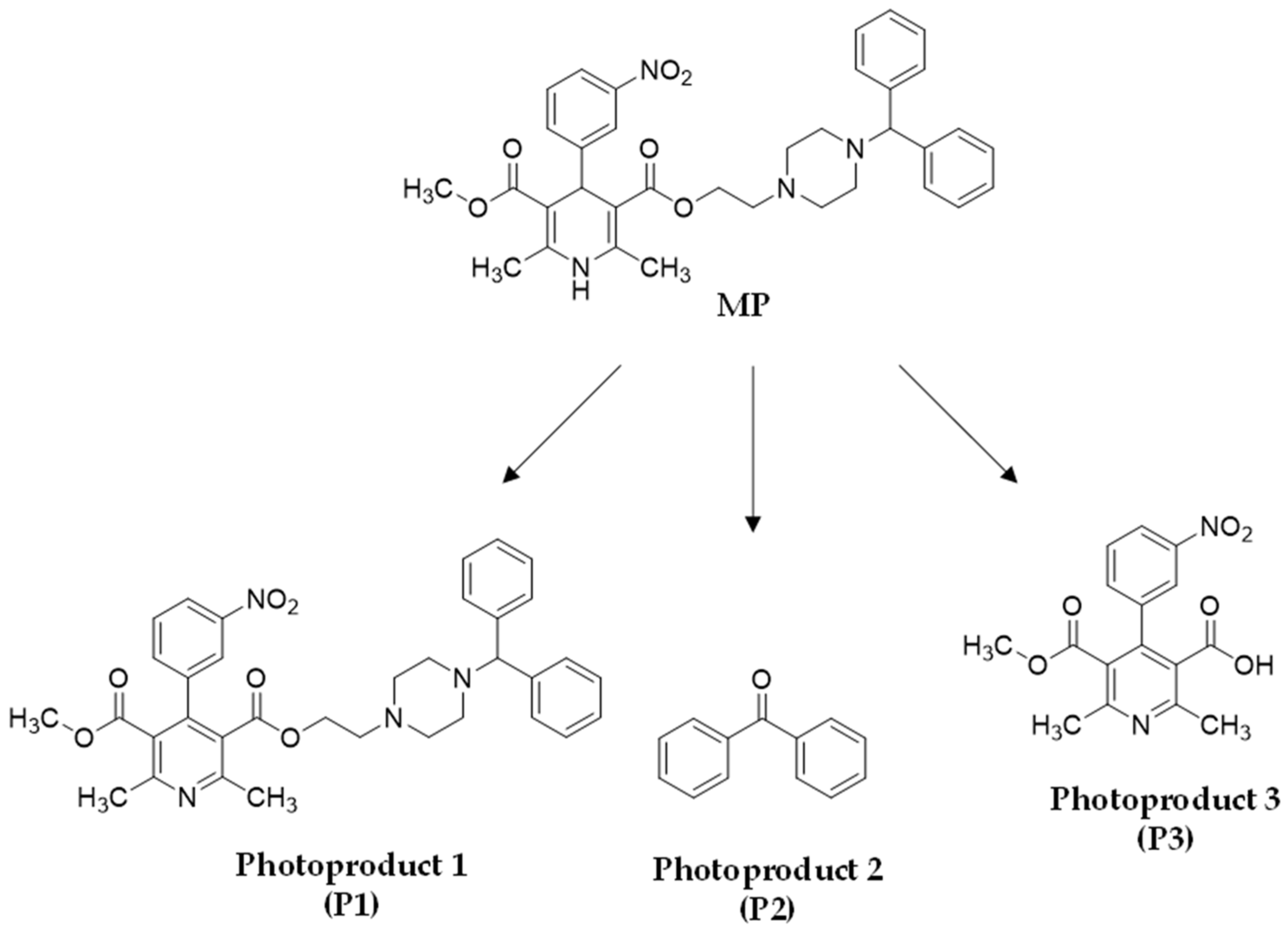
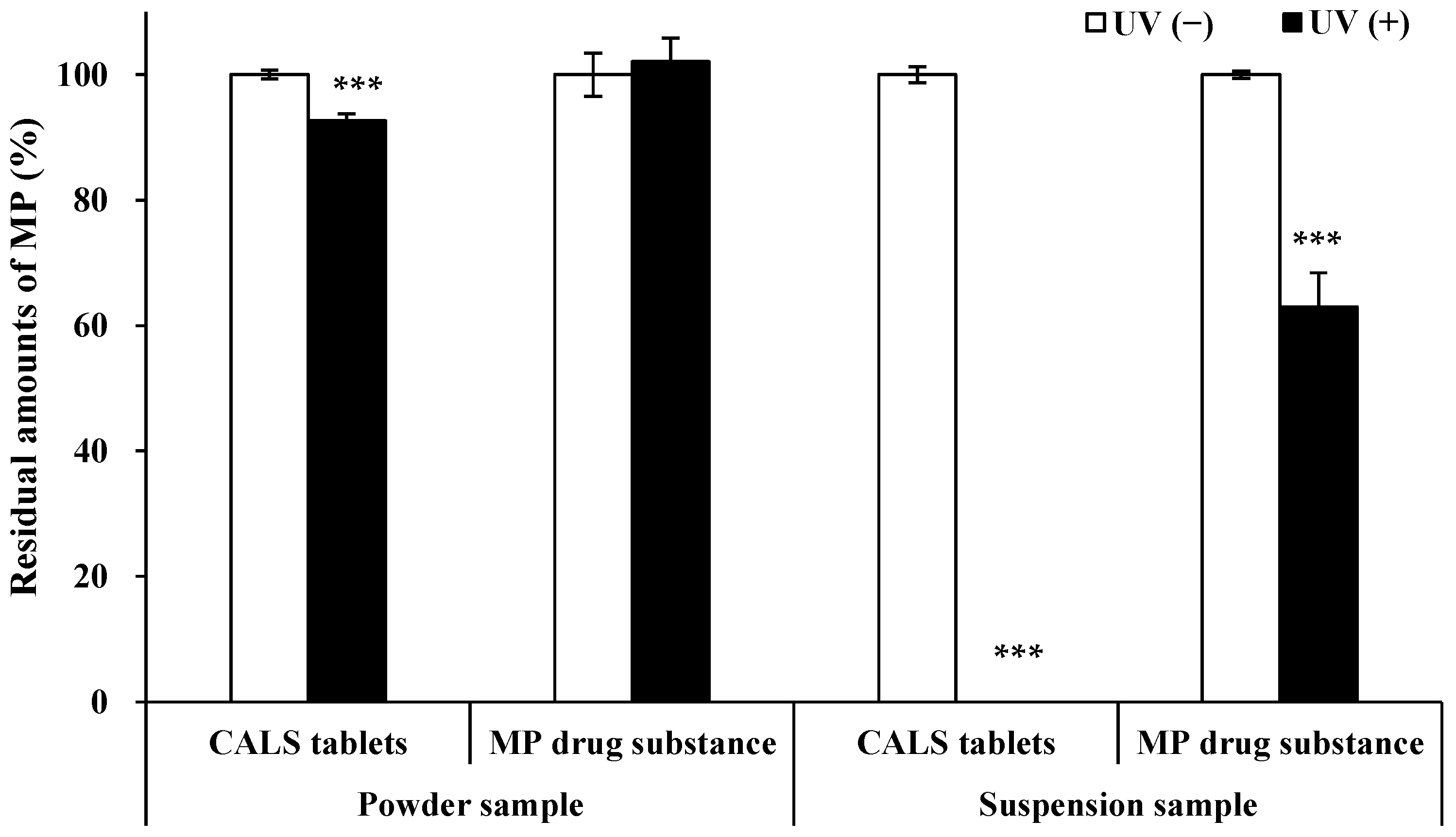
3.1. Photostability Evaluation of Altered Forms of CALS Tablets and MP Drug Substances

It is possible that photodegradation might change the quality of photo-exposed pharmaceuticals including their biological activities in addition to their quantity. The author carried out the photostability evaluation of the powder samples and suspension samples of CALS tablets by UV irradiation, in order to confirm their photosensitivity. As shown in Figure 2A,B, MP and P1, which was a potential impurity of CALS tablets, were degraded completely along with the generation of P2 and P3 in the suspension samples. In the case of UV-irradiated powder samples of CALS tablets, the peak of MP decreased after UV irradiation for 24 h with an increase in the peaks of P1 and P2 (P3 was not generated in the solid state). However, when the MP drug substance was suspended in an aqueous media, MP was not degraded completely by UV irradiation. The peaks of MP, P1, P2, and P3 were observed in the HPLC chromatogram (Figure 2D), but the peak areas of P2 and P3 were less than those recorded in the UV-irradiated suspension of MP tablets. Surprisingly, the powder of the MP drug substance was not affected by UV irradiation, and any MP photoproducts were not observed in the HPLC chromatogram.
The residual amounts of MP in powder samples and suspension samples of CALS tablets and MP drug substances with and without UV irradiation were summarized in Figure 3. In the case of altered forms of CALS tablets, the value of the residual amounts of MP in UV-irradiated powder samples was 92.6 ± 1.1%. However, MP in UV-irradiated powder samples of MP drug substances was not affected by UV irradiation, and the value of the residual amounts was comparable to the control samples (102.1 ± 3.7% and 100.0 ± 3.4%, respectively). The value of the residual amount of MP in UV-irradiated suspension samples of MP drug substances was 63.0 ± 5.4%, although MP in the suspension samples of CALS tablets was completely degraded after UV irradiation. The obtained results indicating an increase in the photodegradability of MP tablets after the change of dosage forms were the same as those obtained in our previous study [14].
Furthermore, in the case that CALS powders were packaged in paper packages and cellophane packages, UV irradiation induced a decrease in the contents of MP after 24 h irradiation, as shown in Figure S1. API, in these packages without UV irradiation, was not changed (residual amounts in paper packages and cellophane packages were 100.0 ± 2.6% and 100.0 ± 2.7%, respectively), but those values recorded after UV irradiation were significantly lower (96.1 ± 1.2% and 90.1 ± 0.7%, respectively). In addition, when these packages were stored near the window of the house for 1 month (samples were irradiated by sunlight during daytime), a 10% decrease in the API contents was observed, as shown in Figure S2. In the case that these packages were stored in the pharmacy (samples were irradiated by FL light or LED light during work time), several changes were not observed. These packages were utilized in clinical situations, so CALS powders might be affected when they are packaged and irradiated by sunlight.
Based on these results, it is suggested that additives in CALS tablets might promote the photodegradation of MP. Utilized additives in CALS tablets were as follows: corn starch, hydroxypropyl cellulose, low-substituted hydroxypropyl cellulose, magnesium stearate, riboflavin, and lactose monohydrate. Among them, the authors paid attention to riboflavin (RF), which is a well-known photocatalyst. There are several reports indicating the role of RF as a photocatalyst for pharmaceuticals [17,18,19]. The addition of RF promotes the photodegradation of sulindac, which is one of the non-steroidal anti-inflammatory drugs, resulting in the generation of its main photoproduct [20].

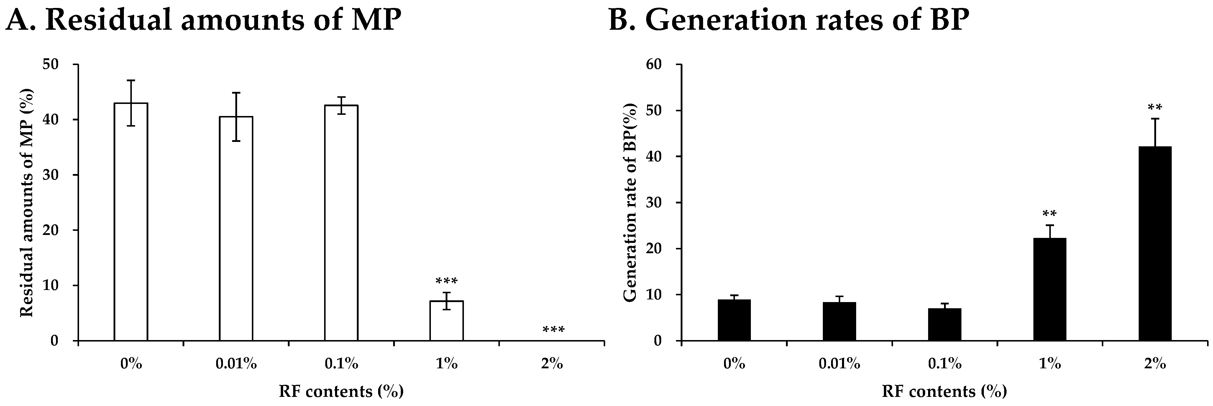
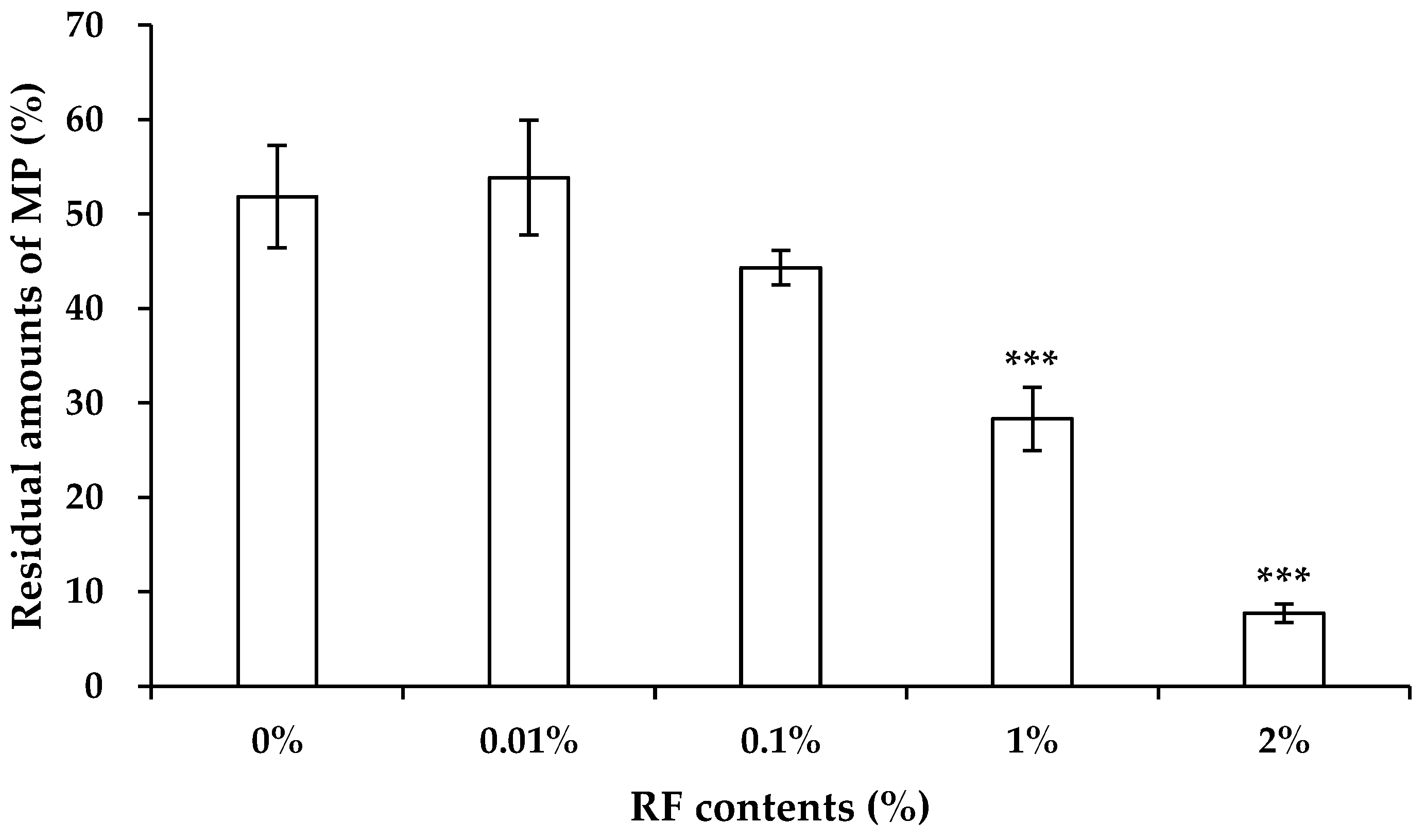
3.2. Evaluation of Photocatalysis Activity of RF

To prove the photocatalytic activity of RF for MP photodegradation, UV irradiation experiments were performed when the MP drug substance was irradiated, firstly, in the presence of RF. As shown in Figure 4, MP photodegradation was promoted when the RF contents were more than 1%. The residual amounts of MP after UV irradiation in the absence of RF were 43.0 ± 4.1%, but those in the presence of 1% RF were 7.2 ± 1.6%. In the case of the addition of 2% RF, MP was completely photodegraded after 24 h irradiation. Moreover, the generation rate of BP, which was designated as P2 and the main MP photoproduct, was increased concomitant with MP photodegradation in this condition; although, the generation rate did not increase with RF addition up to 0.1%. The generation rates of BP in the presence of 1% and 2% RF were 22.3 ± 2.8% and 42.2 ± 6.0%, respectively. These values were higher than in the absence of RF (8.9 ± 1.0%). There are several reports showing the toxicological effects of BP [21,22,23], so it is possible that RF might contribute to a decrease in the beneficial effect of MP and an increase in the adverse effect derived from BP.
Secondly, for the determination of the difference between the degradation rates of MP with and without RF, the time dependency of MP photodegradation in suspensions was compared, as shown in Figure 5. The photodegradation rate of MP with 1% RF (kinetic constant 22.7 h1) was higher than that without RF (kinetic constant 10.4 h1). On the other hand, MP photodegradation was not enhanced in the presence of RF when irradiated samples were covered by an aluminum foil. From these results, it is indicated that RF promotes MP photodegradation by photocatalytic activity. Several studies showed that RF acts as a photosensitizer and promotes the photodegradation of other chemical compounds [17,18,19]. RF promotes direct photodegradation of other compounds by the transfer of an excitation energy. Furthermore, indirect photodegradation was also enhanced because photo-exposed RF gives the energy to oxygen molecules and water molecules, resulting in the formation of a reactive oxygen species. In this study, MP photodegradation was promoted both in the powder samples and suspension samples, and the photodegradation in the suspension samples was significant. Because the enhancement of MP photodegradation was partial in the solid state, it seems to be that the transfer of an excitation energy between RF and MP might contribute to the promoted photodegradation. In the case of UV-irradiated suspensions, RF might transfer the energy to MP and other compounds, including water molecules, and promote MP photodegradation by the activation of direct and indirect photodegradation. Reactive oxygen species have a crucial role for the oxidation of a pyridine ring and the elimination of a diphenyl methylene moiety of MP, resulting in the generation of P1 and P2 [14]. MP photodegradation might be promoted by the generation of a reactive oxygen species, which is enhanced by the energy transfer from photo-sensitized RF. The details for the mechanism of the photocatalytic activity of RF are unclear, so additional studies are required.

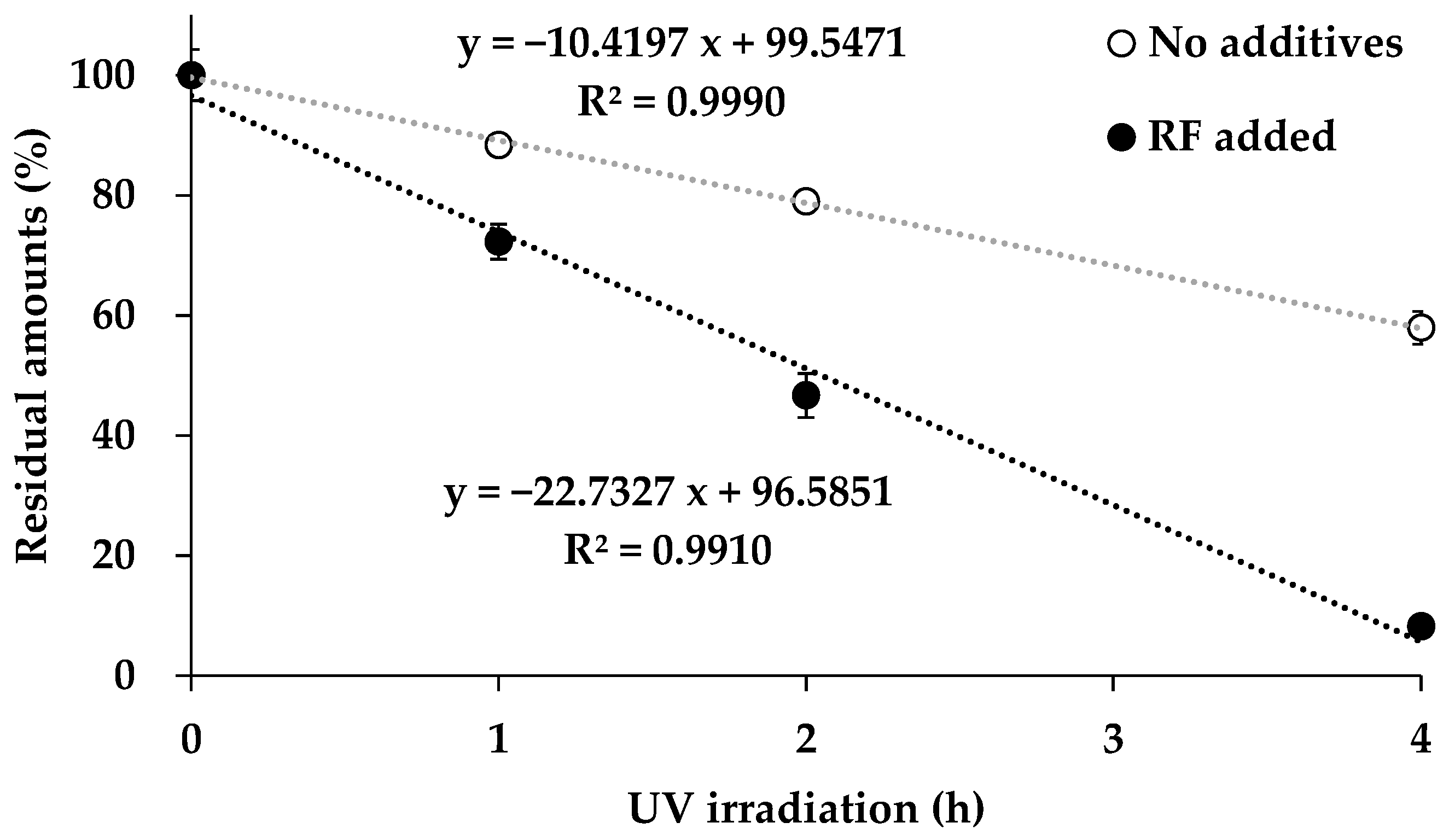
3.3. Study on the Photostabilization of CALS Suspensions

Our previous studies indicated that the additives having antioxidative potencies and UV filtering potencies suppressed the photodegradation of naproxen, which is well known as a photosensitive pharmaceutical [15,16]. In the next step, the authors performed the photostabilization of MP suspensions based on the addition of quercetin (QU), which is one of the polyphenols and has both the antioxidative potency and the UV filtering potency. It has already been clarified that the addition of QU suppresses the photodegradation of naproxen suspended in an aqueous media [15]. It was proposed that QU might quench naproxen at the excited state, in addition to the suppression of the generation of the reactive oxygen species. Herein, UV irradiation experiments were performed in the case that the MP drug substance was irradiated in the presence of QU. As shown in Figure 6, QU suppressed the MP photodegradation. The higher QU contents showed the higher photoprotective potency for MP. The value of residual amounts of MP without QU after 24 h irradiation was 46.0 ± 3.5%, but those with 0.01%, 0.1%, 1%, 10%, and 100% QU were 54.0 ± 1.1%, 56.5 ± 1.4%, 59.7 ± 0.8%, 60.9 ± 0.7%, and 80.9 ± 4.1%, respectively. In addition, QU suppressed the generation of BP derived from MP photodegradation in proportion to its contents (Figure S3). From these results, the dose-dependent protective effect of QU for MP photodegradation was indicated. It is suggested that QU might act as a photostabilizer for MP by several factors, including the protection from UV irradiation, the quenching of the excited state of MP, and the suppression of the generation of a reactive oxygen species. In the suspension of MP, the presence of QU could disrupt the photo-exposure to MP due to its significant UV absorption potency. In addition, QU has an antioxidative potency showing the higher reduction of Cu2+ to Cu+ compared with other tested polyphenols in the PAO test [15,16], so QU could suppress the behavior of reactive oxygen, otherwise resulting in the oxidation of a pyridine ring and the elimination of a diphenyl methylene moiety of MP. Moreover, QU might quench the excited state of UV-irradiated MP to form the basal state.
Furthermore, the authors compared the photocatalytic potency of RF with the photostabilizing potency of QU for MP photodegradation. The dose dependency of the protective effect of QU in the presence of 1% RF was evaluated, as shown in Figure 7. Residual amounts of MP after 24 h irradiation increased in proportion to the contents of QU. The values were as follows: 10.8 ± 5.1% for 0% QU, 21.4 ± 2.5% for 0.01% QU, 21.0 ± 4.2% for 0.1% QU, 23.4 ± 1.8% for 1% QU, 39.9 ± 5.8% for 10% QU, and 68.6 ± 2.2% for 100% QU. Overall, the residuals amounts were lower than those in the absence of RF (Figure 6). The generation rates of BP increased with RF similar to MP photodegradation, but QU also suppressed them dependent on its contents (Figure S4). In contrast to these experiments, the dose dependency of the photocatalytic effect of RF in the presence of 1% QU was evaluated, as shown in Figure 8. QU showed the protective effects on MP photodegradation, which was promoted by the presence of RF. MP was completely photodegraded by the addition of 2% RF (Figure 4), but the remain of MP after 24 h irradiation was observed in the presence of 2% RF and 1% QU (the residual amounts were 7.7 ± 1.0%). The generation rates of BP decreased by the addition of RF, and these values were less than those observed for the absence of QU, as shown in Figure S5. In the case of the addition of 2% RF in the presence of 1% QU, the generation rates of BP were 23.8 ± 3.0%, which were less than those recorded for the absence of QU (42.2 ± 6.0%). In addition, QU showed the photoprotective effect on MP photodegradation in the suspension samples of CALS tablets. Without QU, API was photodegraded completely after UV irradiation for 24 h, as shown in Figure 3, but API remained in the presence of 100% QU (the residual amounts of MP were 8.8 ± 3.1%). The residual amounts were less than those observed in the case that MP drug substance was suspended and UV-irradiated in the presence of 100% QU and 1% RF (Figure 7, 68.6 ± 2.2%). It is possible that the protective effect of QU might be attenuated by other additives or other additives might have a photocatalytic effect on MP photodegradation. The quenching activity of QU for MP at the excited state and the reactive oxygen species might be disrupted by the presence of other additives. Energy transfer and quenching between chemicals is complex, so additional research is needed to prove the interaction among MP and surrounding other additives. Based on the obtained results, it is summarized that QU has a protective potency for MP’s own photodegradation and partially suppresses the photocatalytic effect of RF.

4. Conclusions

This work revealed that RF promotes the photodegradation of MP tablets when they are crushed and suspended. RF is utilized as a coloring agent, but it has a potency to photo-catalyze the degradation of APIs after the change of the dosage form. In addition, the addition of QU, which has both an antioxidative potency and an UV-filtering potency, partially suppresses the photodegradation of MP. It might be an effective strategy to add several photostabilizers for crushed and suspended medicines, of which APIs are photodegradable, in clinical situations. Further studies focused on the photochemical behaviors of utilized additives for medicines are needed for their safe use.

Supplementary Materials

The following supporting information can be downloaded at: https://www.mdpi.com/article/10.3390/photochem4030023/s1. Figure S1: Residual amounts of MP in powders packaged by paper and cellophane after UV irradiation for 24 h. Values represent mean ± SD (n = 4). ** Difference compared with control samples (p < 0.01); Figure S2: Residual amounts of MP in powders packaged by paper and cellophane after the exposure by sunlight, FL light, and LED light. Values represent mean ± SD (n = 4). * Difference compared with control samples (p < 0.05) and *** difference compared with control samples (p < 0.001); Figure S3: Dose dependency of the effect of QU on the generation rates of BP in the UV-irradiated suspensions. Values represent mean ± SD (n = 4). * Difference compared with no additives (p < 0.05), ** difference compared with no additives (p < 0.01), and *** difference compared with no additives (p < 0.001); Figure S4: Dose dependency of the effect of QU on the generation rates of BP in the UV-irradiated suspensions in the presence of 1% RF. Values represent mean ± SD (n = 4). * Difference compared without QU (p < 0.05) and ** difference compared without QU (p < 0.01); Figure S5: Dose dependency of the effect of RF on the generation rates of BP in the UV-irradiated suspensions in the presence of 1% QU. Values represent mean ± SD (n = 4). *** Difference compared without RF (p < 0.001).

Author Contributions

Conceptualization, K.K. and H.N; Methodology, K.K., M.T. and K.H.; Validation, K.K.; Formal Analysis, K.K.; Investigation, K.K. and S.A.; Resources, H.N.; Data Curation, K.K.; Writing—Original Draft Preparation, K.K.; Writing—Review and Editing, S.A., N.U. and H.N.; Supervision, M.I. and H.N.; Project Administration, K.K. and H.N.; and Funding Acquisition, K.K. All authors have read and agreed to the published version of the manuscript.

Funding

This study was supported by JSPS KAKENHI grant number JP 20K15980.

Data Availability Statement

The datasets generated during the current study are available from the corresponding author on reasonable request.

Conflicts of Interest

The authors declare no conflicts of interest. Author Shiori Akimoto was employed by company Akimoto Pharmacy Corporation. The remaining authors declare that the research was conducted in the absence of any commercial or financial relationships that could be construed as a potential conflict of interest.

References

  1. Cheer, S.M.; McClellan, K. Manidipine: A review of its use in hypertension. Drugs 2001, 61, 1777–1799. [Google Scholar] [CrossRef] [PubMed]
  2. Jakimska, A.; Śliwka-Kaszyńska, M.; Nagórski, P.; Namieśnik, J.; Kot-Wasik, A. Phototransformation of amlodipine: Degradation kinetics and identification of its photoproducts. PLoS ONE 2014, 9, e109206. [Google Scholar] [CrossRef] [PubMed]
  3. Rango, G.; Garofalo, A.; Vetuschi, C. Photodegradation monitoring of amlodipine by derivative spectrophotometry. J. Pharm. Biomed. Anal. 2002, 27, 19–24. [Google Scholar]
  4. Chen, S.M.; Hsieh, M.C.; Chao, S.H.; Chang, E.E.; Wang, P.Y.; Wu, A.B. Separation and structure determination of nicardipine photoproducts by LC-ESI-MS. Biomed. Chromatogr. 2008, 22, 1008–1012. [Google Scholar] [CrossRef]
  5. Horinouchi, Y.; Tsuchiya, K.; Taoka, C.; Tajima, S.; Kihira, Y.; Matsuda, Y.; Shishido, K.; Yoshida, M.; Hamano, S.; Kawazoe, K.; et al. Antioxidant effects of photodegradation product of nifedipine. Chem. Pharm. Bull. 2011, 59, 208–214. [Google Scholar] [CrossRef]
  6. Asahi, M.; Matsushita, R.; Kawahara, M.; Ishida, T.; Emoto, C.; Suzuki, N.; Kataoka, O.; Mukai, C.; Hanaoka, M.; Ishizaki, J.; et al. Causative agent of vascular pain among photodegradation products of dacarbazine. J. Pharm. Pharmacol. 2002, 54, 1117–1122. [Google Scholar] [CrossRef]
  7. Della Greca, M.; Iesce, M.R.; Previtera, L.; Rubino, M.; Temussi, F. A new photoproduct of the drug furosemide in aqueous media. Environ. Chem. Lett. 2004, 2, 155–158. [Google Scholar] [CrossRef]
  8. Isidori, M.; Nardelli, A.; Parrella, A.; Pascarella, L.; Previtera, L. A multispecies study to assess the toxic and genotoxic effect of pharmaceuticals: Furosemide and its photoproduct. Chemosphere 2006, 63, 785–793. [Google Scholar] [CrossRef]
  9. Kawabata, K.; Sugihara, K.; Sanoh, S.; Kitamura, S.; Ohta, S. Ultraviolet-photoproduct of acetaminophen: Structure determination and evaluation of ecotoxicological effect. J. Photochem. Photobiol. A Chem. 2012, 249, 29–35. [Google Scholar] [CrossRef]
  10. Kawabata, K.; Sugihara, K.; Sanoh, S.; Kitamura, S.; Ohta, S. Photodegradation of pharmaceuticals in the aquatic environment by sunlight and UV-A, -B and -C irradiation. J. Toxicol. Sci. 2013, 38, 215–223. [Google Scholar] [CrossRef]
  11. Kawabata, K.; Akimoto, S.; Nishi, H. Photo-conversion of phenytoin to ecotoxicological substance benzophenone by ultraviolet light irradiation in aqueous media. Chromatography 2020, 41, 51–58. [Google Scholar] [CrossRef]
  12. Kawabe, Y.; Nakamura, H.; Hino, E.; Suzuki, S. Photochemical stabilities of some dihydropyridine calcium-channel blockers in powdered pharmaceutical tablets. J. Pharm. Biomed. Anal. 2008, 47, 618–624. [Google Scholar] [CrossRef] [PubMed]
  13. Kawabata, K.; Muraoka, H.; Miyara, M.; Kotake, Y.; Nishi, H. Photodegradation profiling of nitrendipine: Evaluation of active pharmaceutical ingredient, tablets and its altered forms. Anal. Sci. 2023, 39, 1791–1803. [Google Scholar] [CrossRef] [PubMed]
  14. Kawabata, K.; Hirai, K.; Akimoto, S.; Inagaki, M.; Nishi, H. Photostability evaluation of manidipine tablets and structural determination of its photoproducts. Anal Sci. 2024, 40, 1733–1747. [Google Scholar] [CrossRef]
  15. Kawabata, K.; Takato, A.; Oshima, S.; Akimoto, S.; Inagaki, M.; Nishi, H. Protective Effect of Selected Antioxidants on Naproxen Photodegradation in Aqueous Media. Antioxidants 2019, 8, 424. [Google Scholar] [CrossRef]
  16. Kawabata, K.; Miyoshi, A.; Nishi, H. Photoprotective effects of selected polyphenols and antioxidants on naproxen photodegradability in the solid-state. Photochem 2022, 2, 880–890. [Google Scholar] [CrossRef]
  17. Insińska-Rak, M.; Sikorski, M.; Wolnicka-Glubisz, A. Riboflavin and Its Derivates as Potential Photosensitizers in the Photodynamic Treatment of Skin Cancers. Cells 2023, 12, 2304. [Google Scholar] [CrossRef]
  18. Meng, J.; Xu, F.; Yuan, S.; Mu, Y.; Wang, W.; Hu, Z.H. Photocatalytic oxidation of roxarsone using riboflavin-derivative as a photosensitizer. Chem. Eng. J. 2019, 355, 130–136. [Google Scholar] [CrossRef]
  19. Castillo, C.; Criado, S.; Díaz, M.; García, N.A. Riboflavin as a sensitiser in the photodegradation of tetracyclines. Kinetics, mechanism and microbiological implications. Dye. Pigment. 2007, 72, 178–184. [Google Scholar] [CrossRef]
  20. Suga, M.; Makino, K.; Tabata, H.; Oshitari, T.; Natsugari, H.; Takahashi, H. Photoisomerization of sulindac and ozagrel hydrochloride by vitamin B2 catalyst under visible light irradiation. Pharm. Res. 2022, 39, 577–586. [Google Scholar] [CrossRef]
  21. Zhang, Q.; Ma, X.; Dzakpasu, M.; Wang, X.C. Evaluation of ecotoxicological effects of benzophenone UV filters: Luminescent bacteria toxicity, genotoxicity and hormonal activity. Ecotoxicol. Environ. Saf. 2017, 142, 338–347. [Google Scholar] [CrossRef] [PubMed]
  22. Kunz, P.Y.; Fent, K. Estrogenic activity of UV filter mixtures. Toxicol. Appl. Pharmacol. 2006, 217, 86–99. [Google Scholar] [CrossRef] [PubMed]
  23. Yamasaki, K.; Takeyoshi, M.; Yakabe, Y.; Sawaki, M.; Takatsuki, M. Comparison of the reporter gene assay for ER-alpha antagonists with the immature rat uterotrophic assay of 10 chemicals. Toxicol. Let. 2003, 142, 119–131. [Google Scholar] [CrossRef] [PubMed]
Figure 1. Photodegradation scheme of MP.
Photochem 04 00023 g001
Figure 2. HPLC chromatograms of suspension samples of CALS tablets and MP drug substances with and without UV irradiation for 24 h. (A) Suspension sample of CALS tablets, (B) UV-irradiated suspension sample of CALS tablets, (C) suspension sample of MP drug substance, and (D) UV-irradiated suspension sample of MP drug substance. Detection wavelength: 254 nm.
Photochem 04 00023 g002
Figure 3. Residual amounts of MP in powder samples and suspension samples after UV irradiation for 24 h. Values represent mean ± SD (n = 4). *** Difference compared with control samples (p < 0.001).
Photochem 04 00023 g003
Figure 4. The dose dependency of the effect of RF on the residual amounts of MP and the generation rates of BP in the UV-irradiated suspension samples for 24 h. (A) The residual amounts of MP and (B) generation rates of BP. Values represent mean ± SD (n = 4). ** Difference compared with no additives (p < 0.01) and *** difference compared with no additives (p < 0.001).
Photochem 04 00023 g004
Figure 5. Photodegradation rates of MP with and without RF after UV irradiation for up to 4 h.
Photochem 04 00023 g005
Figure 6. Dose dependency of the effect of QU on the residual amounts of MP in the UV-irradiated suspension samples for 24 h. Values represent mean ± SD (n = 4). * Difference compared with no additives (p < 0.05), ** difference compared with no additives (p < 0.01), and *** difference compared with no additives (p < 0.001).
Photochem 04 00023 g006
Figure 7. Dose dependency of the effect of QU on the residual amounts of MP in the UV-irradiated suspension samples in the presence of 1% RF for 24 h. Values represent mean ± SD (n = 4). * Difference compared without QU (p < 0.05) and *** difference compared without QU (p < 0.001).
Photochem 04 00023 g007
Figure 8. Dose dependency of the effect of RF on the residual amounts of MP in the UV-irradiated suspension samples in the presence of 1% QU for 24 h. Values represent mean ± SD (n = 4). *** Difference compared without RF (p < 0.001).
Photochem 04 00023 g008
Disclaimer/Publisher’s Note: The statements, opinions and data contained in all publications are solely those of the individual author(s) and contributor(s) and not of MDPI and/or the editor(s). MDPI and/or the editor(s) disclaim responsibility for any injury to people or property resulting from any ideas, methods, instructions or products referred to in the content.

