Article

Design and Development of a qPCR-Based Mitochondrial Analysis Workflow for Medical Laboratories

1
Faculty of Mathematics and Computer Science, University of Hagen, 58097 Hagen, Germany
2
ImmBioMed Business Consultants GmbH & Co. KG, 64319 Pfungstadt, Germany
3
Research Institute for Telecommunication and Cooperation (FTK), 44149 Dortmund, Germany
*
Author to whom correspondence should be addressed.
BioMedInformatics 2022, 2(4), 643-653; https://doi.org/10.3390/biomedinformatics2040042
Submission received: 23 October 2022 / Revised: 21 November 2022 / Accepted: 22 November 2022 / Published: 25 November 2022
(This article belongs to the Section Applied Biomedical Data Science)

Abstract

:
Mitochondrial DNA (mtDNA) damage is closely associated with typical diseases of aging, such as Alzheimer’s or Parkinson’s disease, and other health conditions, such as infertility. This damage manifests in reduced mitochondrial copy number and deletion mutations in mtDNA. Consequently, the analysis of mitochondrial damage by determining the parameters copy number and deletion ratio using quantitative real-time PCR (qPCR) is of interest for clinical diagnostics. To bring the findings from research into laboratory practice, a suitable and reliable process is needed, which must be thoroughly validated. This process includes the software used for the analysis, which must meet extensive regulatory and process requirements. Existing software does not adequately implement the requirements of laboratories and, in particular, does not provide direct support for the calculation of the aforementioned mtDNA parameters. The paper discusses the development of a new software-based analysis workflow that is designed specifically for laboratories to help with the calculation of mtDNA parameters. The software was developed using the User-Centered Design method and is based on the recently introduced prototype, “PlateFlow”. Initial user tests provide positive feedback. In the future, this workflow could form the basis for validations of mitochondrial tests in medical laboratories.

1. Introduction

Mitochondria are organelles found in eukaryotic cells. They have their own DNA, which is separate from the DNA in the cell’s nucleus. Mitochondria produce energy for the cell, help to regulate cell death [1], and are involved in the process of aging [2]. With aging, mitochondria become less efficient at producing energy. Mitochondrial DNA (mtDNA) is more susceptible to damage than the DNA in the nucleus [3]. This damage can accumulate over time and lead to the dysfunction of mitochondria. MtDNA damage has been associated with age-related diseases, such as Parkinson’s disease [4] and Alzheimer’s disease [5]. It has also been shown to be a factor in both female and male reproductive health conditions [6,7].
Laboratory diagnostics play a vital role in the management of patients with mitochondrial disorders. Laboratory testing can be used to confirm a diagnosis, guide treatment decisions, and monitor disease progression [8,9]. One way to measure mtDNA damage is by using quantitative Polymerase Chain Reaction (qPCR) [10]. With qPCR, the relative or absolute amount of a target nucleotide sequence in a sample can be measured. Using this method, two exemplary parameters related to mitochondrial function can be assessed: the mitochondrial DNA copy number and the deletion ratio [10]. Robust assays for assessing these two parameters have been established, together with formulas to calculate the specific ratios [6,7,10].
The focus of qPCR in the field of laboratory diagnostics lies on inter-experiment and inter-operator reproducibility and comparability [11,12]. This requires an analysis pipeline using a fixed and well-established protocol which must be followed so that samples yield the same results independent of the day of sample processing, the operator, or other changing conditions [13]. The determination of mtDNA damage or other qPCR-based parameters in medical laboratories is supported by software at all stages—from the entry of a sample order [14], through the evaluation of cycler raw data and quantification [15] to the calculation of relevant parameters, interpretation, documentation, and archiving of results [16]. These steps form a pipeline that can consist of different software components from different manufacturers [17]. Software used for medical diagnostics must be scientifically sound, legally compliant, and efficient [18]. The ideal medical diagnostic qPCR analysis pipeline does not only operate in a largely unsupervised and automated manner to reduce variation but is also modular in design so that the individual steps of the analysis pipeline can be interchanged [19]. The exchange of modules in the analysis pipeline may be necessary due to technical improvements or also due to changing regulations in the field of medical diagnostic software.
The requirements of medical laboratories are very different from those of research [20]. In research, project-specific work is usually conducted with self-contained experiments. The resulting data are analyzed in an open-ended manner, using multiple methods if necessary. Test parameters and methods can be adapted iteratively to obtain conclusive results and to lead the project in a promising direction. The focus of research is to investigate a particular hypothesis and its underlying mechanisms by performing numerous experiments with different methods and resulting parameters [21]. In contrast, medical laboratories have fixed, validated test procedures that must not be deviated from during productive operation and change rarely. The proper procedure must be ensured and documented as far as possible by the software involved. The challenge for software in medical laboratories is thus to be flexible enough for future developments like establishing new test procedures while remaining rigid enough during routine laboratory work to not allow out-of-specification deviations from the established protocol. Further challenges arise from a large number of regulations and standards that have to be observed by medical laboratories and their associated tools [16].
The transfer of research results into clinical practice is thus a complex problem area. This also applies to the case of determining mtDNA parameters for the diagnosis of mitochondrial disorders considered here. Thus, how can mtDNA data be efficiently analyzed in medical laboratories? To answer this question, the following sections first discuss the state of the art for laboratory mtDNA analysis before detailing the development and evaluation of a prototype software component that is designed to better meet the requirements of medical laboratories.

2. State of the Art

This section will provide an overview of the current state of the art for laboratory analysis of mtDNA parameters. First, we will discuss how mitochondrial damage can be analyzed using qPCR techniques. Next, we will review the regulatory requirements for the validation of software in laboratory practice. Finally, we will evaluate available qPCR software on their suitability for use in medical laboratories and mitochondrial damage analysis specifically.

2.1. Quantification of mtDNA Parameters Using qPCR

The circular mtDNA consists of different segments that are affected to different degrees by mutations or damage, with deletions playing a particularly important role. These deletions, which are copying errors in which certain sections of the mtDNA are completely removed, usually lead to a complete or partial loss of function of the mtDNA. To measure the rate of deletions, qPCR can be used to select and quantify two mitochondrial targets. The first target ( mtDNA min ) is located in a region where deletions are not known or very rare (minor arc). The second target ( mtDNA maj ) is located in a region where deletions are common (major arc). Hence, the target mtDNA min quantifies essentially all mtDNA copies while the target mtDNA maj quantifies intact mtDNA copies. Both targets can be determined by a multiplexing method in the same qPCR run. If the absolute concentrations c ( mtDNA maj ) and c ( mtDNA min ) of the targets can be determined, the deletion ratio mtDNA DR can be determined via Equation (1). Absolute concentrations can be determined, e.g., via a standard curve [22].
mtDNA DR = 1 c ( mtDNA maj ) c ( mtDNA min )
mtDNA DR = 1 2 Δ C q ( mtDNA maj ) 2 Δ C q ( mtDNA min )
= 1 2 Δ C q ( mtDNA min ) Δ C q ( mtDNA maj )
The deletion ratio can also be approximated by relative quantification using the Δ Δ C q method [22]. For this purpose, a reference sample (“calibrator”) with undamaged mitochondrial DNA is required for which the targets mtDNA maj and mtDNA min are likewise determined. By subtraction between the Cq-values of the sample and reference, Δ C q ( mtDNA maj ) and Δ C q ( mtDNA min ) can be determined. Normalization to a standard compensates for differences in qPCR response between the different targets, making Δ C q ( mtDNA maj ) and Δ C q ( mtDNA min ) more comparable than C q ( mtDNA maj ) and C q ( mtDNA min ) . After the normalization, the fold-change value [22] can be determined based on the difference between the two values (Equation (1)), which converts the logarithmic nature of the Cq-values into a linear ratio between mtDNA maj and mtDNA min .
Another important parameter is the number of mtDNA copies per cell. To determine this parameter mtDNA CN , a third target nDNA can be added for the determination of nuclear DNA. For example, the genes for β 2 microglobulin (B2M) [10] or RNase P (RPPH1) [6,7] are suitable for this purpose. Using known quantifications of c ( mtDNA min ) and c ( nDNA ) , the copy number mtDNA CN can then be determined via Equation (4):
mtDNA CN = 2 c ( mtDNA min ) c ( nDNA )
mtDNA CN = 2 Δ C q ( nDNA ) Δ C q ( mtDNA min )
Note that a factor of two is used as we are interested in the number of copies per cell and we expect two copies of the nDNA target per diploid cell. As for the deletion rate, it is possible to use a calibrator instead of determining the absolute concentration. To use the simple fold-change formula in Equation (5), the calibrator’s sample concentrations must be adjusted to result in a copy number of 1.

2.2. Legal Requirements for Software Used in Laboratory Diagnostics

Medical products and software used in laboratory diagnostics are regulated by various laws that vary by country and establish the requirements for their production, placing on the market and use. In the European Union, the In Vitro Diagnostics Regulation (IVDR) [18] regulates tools and devices used for medical diagnostics, including all software used for this purpose. Among other things, it requires that software development uses current technology standards, employs a software lifecycle process, takes into account aspects of usability and security, and is verified and validated prior to release [18].
To ensure quality control and performance, manufacturers must engage in Post-Marketing Surveillance (PMS) to actively collect experiences with their products. This process must be systematic so that Corrective and Preventative Actions (CAPA) can be taken if necessary [18]. This also means that the lifecycle of a software product used in laboratory diagnostics does not end with its development.
The IVDR states that only diagnostics approved for this purpose and which fulfill the conditions of the IVDR may be used in laboratories. Laboratories have the freedom to use so-called Lab-Developed Tests (LDTs) for which the laboratory assumes the entire regulatory responsibility. The laboratory must demonstrate and document conformity with Annex I of the IVDR including full technical validation. So-called Research-Use-Only (RUO) products provided by commercial suppliers are used by laboratories for medical diagnostics; these RUO products also count as Lab-developed Tests and consequently the laboratory is responsible for IVDR compliance.
The IVDR and related legislation create a regulatory framework and define certain objectives. Concrete guidelines for the implementation of these regulations and the validation of medical devices and software are generally not part of the legislation.

2.3. Specific Validation Requirements for Software Used in Laboratory Diagnostics

More concrete specifications for the validation of medical devices and software for diagnostics are part of numerous standards and guidelines that are constantly being developed in line with the state of the art. Due to their widespread use, some of these publications are considered binding and in some cases are required by regulatory authorities.
The most authoritative publications in this regard are those issued by standardization bodies such as the International Organization for Standardization (ISO). ISO 13485 [23] sets out detailed standards for quality management systems for medical device manufacturing. Additionally, ISO 15189 [24] establishes quality management guidelines specifically for medical laboratories. Furthermore, ISO 14971 [25] and ISO 22367 [26] provide principles and processes for risk management of medical devices and medical laboratories, respectively. Specifically for the development of medical software, IEC 62304 [27] discusses requirements for medical software development, maintenance, risk management, configuration management, and problem-solving [28]. IEC 82304 [29] outlines requirements for the validation of health software. ISO/IEC 27001 [30] addresses the increasingly important topic of information security for all types of organizations, including laboratories. Lastly, IEC 62366 [31] discusses requirements for the usability of medical devices.
In addition to these international standards, there are also various guidelines that in turn provide concrete suggestions for the implementation of the above standards and legislation. Examples are the guidelines of the Ad-hoc Commission “In Vitro Diagnostics” of the Association of the Scientific Medical Societies in Germany (AWMF; Arbeitsgemeinschaft der Wissenschaftlichen Medizinischen Fachgesellschaften), Subgroup Software [32], the MIQE guidelines [21] or guidelines of national interest groups like the German Medical Association [33]. For some areas of software development, publications that are not specific to medical software but have some generality are also relevant. As an example, ISO 22367 mentions “poorly designed user interface or processes” [26] (p. 8) as a risk factor for laboratories. Publications addressing common usability problems and solutions are thus also relevant for laboratory software. Nielsen [34] discusses some of the factors underlying usability heuristics, which he derives from the most common usability problems of software.

2.4. Existing qPCR Analysis Software

There are various providers offering software for qPCR analyses. In general, cyclers are already supplied with software that enables the determination of Cq-values from fluorescence values and, in some cases, also enables more advanced analyses. Such software is typically designed specifically for the manufacturer’s instruments and also allows the cycler to be programmed and operated. There is also manufacturer-independent software that supports devices from several suppliers. These include, for example, the software qbase+ [35,36], GenEx [37], and also PlateFlow [16], which we have developed.
qbase+ is a closed-source software of the vendor Biogazelle that includes various algorithms for qPCR analyses [35,38]. This includes support for the geNorm algorithm [39], which assists in the selection of suitable reference genes for gene activity experiments. In addition, more than one reference gene can be used for normalization, which contributes to more accurate measurements of gene activity.
Due to these algorithms and the project-based design of qbase+, it is particularly suitable for research or assay development. Particular functions for the diagnosis of specific pathological conditions, such as mitochondrial damage, are not available in the software [15,20,35]. The vendor recently announced that they will suspend all activity around qbase+ and will no longer update the software [36].
GenEx is a commercial software of the Swedish company MultiD Analyses AB. The software is available in different editions from Standard to Enterprise and offers a similarly large range of functions as qbase+. Particularly noteworthy in GenEx are the extensive statistical analysis options and visualizations. Predictive models based on neural networks or Support Vector Machines (SVM) are also supported by the software [37].
GenEx is also designed for general analyses and not for the diagnosis of specific pathological conditions. Direct determination of mitochondrial damage is thus not possible. Due to the project-based approach, GenEx is also more suitable for research or assay development within the laboratory rather than for productive regular operation in laboratory diagnostics. The software has also not been updated since 2019 [37].
We have developed PlateFlow as a proof-of-concept to create a modern software platform for qPCR-based analysis. The goal of the development was to create a modular design in which specific analysis workflows can be integrated into the software. Instead of a project-based approach, the software is designed to process cycler data in a more automated manner. Instead of maximizing flexibility during analysis, the focus is on the reproducibility and verifiability of results. During development, the focus was on implementing the technical and regulatory requirements of medical laboratories [16]. This stands in contrast to qBase+ and GenEx which are more suitable for research and assay development due to their design.
To the best of our knowledge, there is no cross-device qPCR software suitable for laboratory diagnostics that allows the detection of the mtDNA parameters mentioned above. Due to the modular extensibility of PlateFlow, it serves as a basis for the development of such a mitochondrial analysis workflow in the following sections.

3. Model and Implementation

The planned extension of PlateFlow for mtDNA analyses was discussed with a participating medical laboratory (ImmBioMed GmbH & Co. KG in Heidelberg, Germany) in several interviews and iteratively transferred into use cases, activity diagrams and requirements. The iterative methodology employed was based on the User-Centered Design (UCD) approach [40].
Figure 1 shows the identified use cases. The selection of the analysis strategy can be considered a basic functionality of PlateFlow, which must always be performed by the responsible employee before an analysis is initiated. The assessment of mtDNA parameters is a separate use case that can be divided into several sub-use cases like calculation of the relevant parameters, comparison of results against age-related reference intervals, or storing the analysis results.
Figure 2 shows an activity diagram of the same extension. The analysis starts by reading qPCR result data from which the relevant mtDNA parameters can be computed. The computation results can be visualized and stored for further processing and evaluation of medical findings.
Figure 3 shows the essential analysis parameters needed for the analysis. First, the three qPCR targets for mtDNA maj , mtDNA min , and nDNA can be chosen. Second, a calibrator can be selected to normalize the values as discussed in Section 2.1.
An example of a result table is shown in Figure 4. The table contains the sample name, the corresponding age if known, and the calculated mtDNA CN and mtDNA DR values. Age information is important because of the age dependency of the reference intervals. In this experiment, three biological samples, a non-template control (H2O), and three samples with known concentrations (STDMix_*) are shown. STDMix_03 was used as a calibrator and is thus fixed to a mtDNA CN of 1 and mtDNA DR of 0. As the displayed data are from early experiments and the laboratory test protocol has not been fully established yet, some artifacts like the mtDNA DR of 86.6% for the non-template control can be observed.
For the graphical representation of the values, a violin plot with a superimposed box plot was chosen (Figure 5), as this allows various statistical key figures to be displayed compactly. While the individual points are taken from the current run, the displayed distribution in the violin and box plots is based on a selectable reference file. The underlying data are the same as in Figure 4.
To show the age-dependency of the mtDNA parameters and compare the samples in the current analysis run to reference intervals, age (on the x-axis) can be plotted against mtDNA CN and mtDNA DR values (on the y-axis) (Figure 6). A regression line shows the age dependence of the data contained in the reference file. The gray background shows the 95% confidence interval of this reference data, while the data from the current analysis run are depicted in colored dots on top. The same example data were used as in Figure 4 and Figure 5.
All results, including the graphical representations, can be exported into a report to archive the results or to use them for subsequent processes such as reporting. The results including the visualizations can also be generated separately for each sample to simplify individual reporting. All analysis parameters and intermediate steps are also documented in the report, which makes possible operating errors recognizable even in retrospect.

4. Evaluation and Discussion

The development process for the mtDNA workflow was accompanied by the medical laboratory, and feedback was iteratively incorporated into the software. At two points in the middle and end of development, feedback was also collected and evaluated via a “think-aloud” protocol [41]. In this methodology, a prospective user (in this case, the laboratory staff) is guided step-by-step through the user interface using a real scenario, e.g., the execution of an analysis. The user is asked to verbalize his thinking process while performing the task. Missing functions or other deviations from the target state of the software can be detected reliably.
Overall, the evaluation resulted in positive feedback regarding the scope of functions for the diagnosis of mitochondrial disorders. Several missing functions were identified, some of which could be implemented directly. This involves, e.g., the possibility to define a standard sample (“calibrator”), which was not yet possible in the first development version. Another evaluation finding was the need for improving the integration of the software into other laboratory systems by extending the import/export possibilities. Finally, it was suggested that the accuracy of the calculated mtDNA parameters could be further improved by supporting the use of standard curves for determining the absolute concentration of the parameters.

5. Conclusions and Future Work

The determination of mtDNA parameters by qPCR in medical laboratories can make an important contribution to the diagnosis and treatment of patients with mitochondrial disorders. Existing software for the analysis of qPCR data is not suitable for the measurement of these disorders in a medical laboratory context. To fill this gap, we have extended the software “PlateFlow” with a mitochondrial workflow, specifically tailored to the needs of medical laboratories. By collaborating with a laboratory, feedback from potential users could be incorporated into the development and a first qualitative evaluation of the software could be performed.
One limitation of the current software is the focus on qPCR data exclusively, which does not allow the integration of other mitochondrial parameters. Another limitation is the limited import/export functionality, which makes integration with other laboratory software tools difficult. Finally, relying on a single calibrator sample requires very reliable and precise reactions between the different targets to get accurate measurements, even when technical replicates are used. Future work should thus improve the integration of PlateFlow with other laboratory systems and evaluate whether the clinical relevance of the calculated mtDNA parameters can be further improved by other analysis methods.

Author Contributions

Conceptualization, T.K. and M.H.; investigation, T.K., L.G., E.J. and F.L.-E.; writing—original draft preparation, T.K.; writing—review and editing, L.G., E.J., F.L.-E., P.M.K., M.K. and M.H.; visualization, T.K. and L.G.; supervision, M.K. and M.H.; project administration, T.K. All authors have read and agreed to the published version of the manuscript.

Funding

This research received no external funding.

Institutional Review Board Statement

Not applicable.

Informed Consent Statement

Not applicable.

Data Availability Statement

Not applicable.

Conflicts of Interest

Michael Kramer is the owner and managing director of ImmBioMed Business Consultants GmbH & Co. KG. Fernando Leonardi-Essmann is an employee at that company. His participation in the paper was however completely voluntary and there were no financial or other benefits besides academic recognition. Hence, the authors declare no conflict of interest.

References

  1. Bock, F.J.; Tait, S.W.G. Mitochondria as multifaceted regulators of cell death. Nat. Rev. Mol. Cell Biol. 2020, 21, 85–100. [Google Scholar] [CrossRef] [PubMed]
  2. Chocron, E.S.; Munkácsy, E.; Pickering, A.M. Cause or casualty: The role of mitochondrial DNA in aging and age-associated disease. Biochim. Biophys. Acta. Mol. Basis Dis. 2019, 1865, 285–297. [Google Scholar] [CrossRef] [PubMed]
  3. Zhao, L.; Sumberaz, P. Mitochondrial DNA Damage: Prevalence, Biological Consequence, and Emerging Pathways. Chem. Res. Toxicol. 2020, 33, 2491–2502. [Google Scholar] [CrossRef] [PubMed]
  4. Park, J.S.; Davis, R.L.; Sue, C.M. Mitochondrial Dysfunction in Parkinson’s Disease: New Mechanistic Insights and Therapeutic Perspectives. Curr. Neurol. Neurosci. Rep. 2018, 18, 21. [Google Scholar] [CrossRef] [Green Version]
  5. Misrani, A.; Tabassum, S.; Yang, L. Mitochondrial Dysfunction and Oxidative Stress in Alzheimer’s Disease. Front. Aging Neurosci. 2021, 13, 617588. [Google Scholar] [CrossRef]
  6. Ye, M.; Hu, B.; Shi, W.; Guo, F.; Xu, C.; Li, S. Mitochondrial DNA 4977 bp Deletion in Peripheral Blood Is Associated with Polycystic Ovary Syndrome. Front. Endocrinol. 2021, 12, 675581. [Google Scholar] [CrossRef]
  7. Wu, H.; Whitcomb, B.W.; Huffman, A.; Brandon, N.; Labrie, S.; Tougias, E.; Lynch, K.; Rahil, T.; Sites, C.K.; Pilsner, J.R. Associations of sperm mitochondrial DNA copy number and deletion rate with fertilization and embryo development in a clinical setting. Hum. Reprod. 2019, 34, 163–170. [Google Scholar] [CrossRef]
  8. Wong, L.J.C.; Boles, R.G. Mitochondrial DNA analysis in clinical laboratory diagnostics. Clin. Chim. Acta Int. J. Clin. Chem. 2005, 354, 1–20. [Google Scholar] [CrossRef] [PubMed]
  9. Rodenburg, R.J.T. Biochemical diagnosis of mitochondrial disorders. J. Inherit. Metab. Dis. 2011, 34, 283–292. [Google Scholar] [CrossRef] [PubMed] [Green Version]
  10. Phillips, N.R.; Sprouse, M.L.; Roby, R.K. Simultaneous quantification of mitochondrial DNA copy number and deletion ratio: A multiplex real-time PCR assay. Sci. Rep. 2014, 4, 3887. [Google Scholar] [CrossRef]
  11. de Gonzalo-Calvo, D.; Marchese, M.; Hellemans, J.; Betsou, F.; Skov Frisk, N.L.; Dalgaard, L.T.; Lakkisto, P.; Foy, C.; Scherer, A.; Garcia Bermejo, M.L.; et al. Consensus Guidelines for the Validation of qRT-PCR Assays in Clinical Research by the CardioRNA Consortium. Mol.-Ther.-Methods Clin. Dev. 2022, 24. [Google Scholar] [CrossRef] [PubMed]
  12. Sanders, R.; Bustin, S.; Huggett, J.; Mason, D. Improving the standardization of mRNA measurement by RT-qPCR. Biomol. Detect. Quantif. 2018, 15, 13–17. [Google Scholar] [CrossRef] [PubMed]
  13. Pan, Y.; Bodrossy, L.; Frenzel, P.; Hestnes, A.G.; Krause, S.; Lüke, C.; Meima-Franke, M.; Siljanen, H.; Svenning, M.M.; Bodelier, P.L.E. Impacts of inter- and intralaboratory variations on the reproducibility of microbial community analyses. Appl. Environ. Microbiol. 2010, 76, 7451–7458. [Google Scholar] [CrossRef] [PubMed] [Green Version]
  14. Sepulveda, J.L.; Young, D.S. The ideal laboratory information system. Arch. Pathol. Lab. Med. 2013, 137, 1129–1140. [Google Scholar] [CrossRef] [Green Version]
  15. Pabinger, S.; Rödiger, S.; Kriegner, A.; Vierlinger, K.; Weinhäusel, A. A survey of tools for the analysis of quantitative PCR (qPCR) data. Biomol. Detect. Quantif. 2014, 1, 23–33. [Google Scholar] [CrossRef] [Green Version]
  16. Krause, T.; Jolkver, E.; Bruchhaus, S.; Mc Kevitt, P.; Kramer, M.; Hemmje, M. A Preliminary Evaluation of “GenDAI”, an AI-Assisted Laboratory Diagnostics Solution for Genomic Applications. BioMedInformatics 2022, 2, 332–344. [Google Scholar] [CrossRef]
  17. Krause, T.; Jolkver, E.; Bruchhaus, S.; Kramer, M.; Hemmje, M. An RT-qPCR Data Analysis Platform. In Proceedings of the Collaborative European Research Conference (CERC 2021), Cork, Ireland, 9–10 September 2021. [Google Scholar]
  18. The European Parliament and the Council of the European Union. In Vitro Diagnostic Regulation; European Union: Maastricht, The Netherlands, 2017. [Google Scholar]
  19. Krause, T.; Jolkver, E.; Bruchhaus, S.; Kramer, M.; Hemmje, M. GenDAI—AI-Assisted Laboratory Diagnostics for Genomic Applications. In Proceedings of the 2021 IEEE International Conference on Bioinformatics and Biomedicine (BIBM), Houston, TX, USA, 9–12 December 2021. [Google Scholar] [CrossRef]
  20. Krause, T.; Jolkver, E.; Mc Kevitt, P.; Kramer, M.; Hemmje, M. A Systematic Approach to Diagnostic Laboratory Software Requirements Analysis. Bioengineering 2022, 9, 144. [Google Scholar] [CrossRef] [PubMed]
  21. Bustin, S.A.; Benes, V.; Garson, J.A.; Hellemans, J.; Huggett, J.; Kubista, M.; Mueller, R.; Nolan, T.; Pfaffl, M.W.; Shipley, G.L.; et al. The MIQE guidelines: Minimum information for publication of quantitative real-time PCR experiments. Clin. Chem. 2009, 55, 611–622. [Google Scholar] [CrossRef] [PubMed] [Green Version]
  22. Livak, K.J.; Schmittgen, T.D. Analysis of relative gene expression data using real-time quantitative PCR and the 2(-Delta Delta C(T)) Method. Methods 2001, 25, 402–408. [Google Scholar] [CrossRef] [PubMed]
  23. Standard ISO 13485:2016; Medical Devices—Quality Management Systems—Requirements for Regulatory Purposes. ISO International Organization for Standardization: Geneva, Switzerland, 2016.
  24. Standard ISO 15189:2012; Medical Laboratories—Requirements for Quality and Competence. ISO International Organization for Standardization: Geneva, Switzerland, 2012.
  25. Standard ISO 14971:2019; Medical Devices—Application of Risk Management to Medical Devices. ISO International Organization for Standardization: Geneva, Switzerland, 2019.
  26. Standard ISO 22367:2020; Medical Laboratories—Application of Risk Management to Medical Laboratories. ISO International Organization for Standardization: Geneva, Switzerland, 2020.
  27. Standard IEC 62304:2006; Medical Device Software—Software Life Cycle Processes. IEC International Electrotechnical Commission: Geneva, Switzerland, 2006.
  28. Johner, C. IEC 62304 2. Ausgabe: Alle Anwendungsbereiche und Änderungen. Available online: https://www.johner-institut.de/blog/iec-62304-medizinische-software/iec-62304-zweite-ausgabe/ (accessed on 13 October 2022).
  29. Standard IEC 82304-1:2016; Health Software—Part 1: General Requirements for Product Safety. IEC International Electrotechnical Commission: Geneva, Switzerland, 2016.
  30. Standard ISO/IEC 27001:2013; Information Technology—Security Techniques—Information Security Management Systems—Requirements. ISO International Organization for Standardization: Geneva, Switzerland, 2013.
  31. Standard IEC 62366-1:2015; Medical Devices—Part 1: Application of Usability Engineering to Medical Devices. IEC International Electrotechnical Commission: Geneva, Switzerland, 2015.
  32. Kraft, F.; Begemann, M.; Bietenbeck, A.; Ungelenk, M.; Kuhle, M.; Krawitz, P.; von Neuhoff, N.; Streichert, T. Hinweise der AWMF Subgruppe Software zur Umsetzung der Verordnung (EU) 2017/746 (IVDR) bei Software aus Eigenherstellung. Available online: https://www.awmf.org/fileadmin/user%5fupload/dateien/arbeitshilfen%5fund%5fmusterformblaetter/validierung%5fsoftware.docx (accessed on 21 November 2022).
  33. German Medical Association. Neufassung der Richtlinie der Bundesärztekammer zur Qualitätssicherung laboratoriumsmedizinischer Untersuchungen – Rili-BÄK. Dtsch. ÄRzteblatt Online 2019, 111. [Google Scholar] [CrossRef]
  34. Nielsen, J. Enhancing the explanatory power of usability heuristics. In Proceedings of the SIGCHI Conference on Human Factors in Computing Systems Celebrating Interdependence-CHI ’94, Toronto, ON, Canada, 26 April–1 May 2014; Adelson, B., Dumais, S., Olson, J., Eds.; ACM Press: New York, NY, USA, 1994; pp. 152–158. [Google Scholar] [CrossRef]
  35. What Makes Qbase+ Unique? Available online: https://www.qbaseplus.com/features (accessed on 14 October 2022).
  36. qbase+ | qPCR Analysis Software. Available online: https://www.qbaseplus.com/ (accessed on 14 October 2022).
  37. MultiD Analyses AB. GenEx. Available online: https://multid.se/genex/ (accessed on 23 October 2022).
  38. Hellemans, J.; Mortier, G.; de Paepe, A.; Speleman, F.; Vandesompele, J. qBase relative quantification framework and software for management and automated analysis of real-time quantitative PCR data. Genome Biol. 2007, 8, R19. [Google Scholar] [CrossRef] [PubMed] [Green Version]
  39. Vandesompele, J.; de Preter, K.; Pattyn, F.; Poppe, B.; van Roy, N.; de Paepe, A.; Speleman, F. Accurate normalization of real-time quantitative RT-PCR data by geometric averaging of multiple internal control genes. Genome Biol. 2002, 3. [Google Scholar] [CrossRef] [PubMed] [Green Version]
  40. Dopp, A.R.; Parisi, K.E.; Munson, S.A.; Lyon, A.R. A glossary of user-centered design strategies for implementation experts. Transl. Behav. Med. 2019, 9, 1057–1064. [Google Scholar] [CrossRef] [PubMed] [Green Version]
  41. Kushniruk, A.W.; Patel, V.L. Cognitive and usability engineering methods for the evaluation of clinical information systems. J. Biomed. Inform. 2004, 37, 56–76. [Google Scholar] [CrossRef] [PubMed]
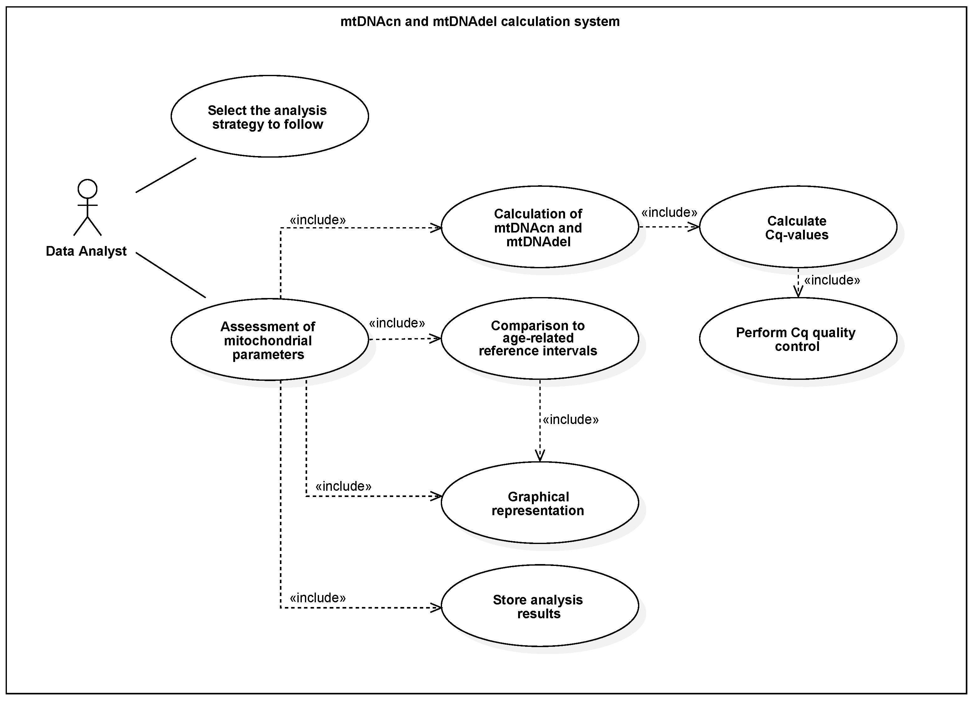
Figure 1. Use case for extension of PlateFlow for mtDNA analysis.
Biomedinformatics 02 00042 g001
Figure 2. Activity diagram of the mtDNA analysis workflow.
Biomedinformatics 02 00042 g002
Figure 3. Analysis settings for mtDNA assessment.
Biomedinformatics 02 00042 g003
Figure 4. Result table showing computed mtDNA CN and mtDNA DR values from example data.
Biomedinformatics 02 00042 g004
Figure 5. Violin plot showing non-production example results.
Biomedinformatics 02 00042 g005
Figure 6. Linear regression plot showing the age-dependency of mtDNA CN and mtDNA DR based on reference data and the currently analyzed dataset.
Biomedinformatics 02 00042 g006
Publisher’s Note: MDPI stays neutral with regard to jurisdictional claims in published maps and institutional affiliations.