Article

Utilizing Immunoinformatics for mRNA Vaccine Design against Influenza D Virus

by
Elijah Kolawole Oladipo
1,2,3,4,*,
Stephen Feranmi Adeyemo
1,
Modinat Wuraola Akinboade
2,
Temitope Michael Akinleye
1,5,
Kehinde Favour Siyanbola
1,
Precious Ayomide Adeogun
1,6,
Victor Michael Ogunfidodo
1,6,
Christiana Adewumi Adekunle
1,7,
Olubunmi Ayobami Elutade
1,8,
Esther Eghogho Omoathebu
1,9,
Blessing Oluwatunmise Taiwo
1,9,
Elizabeth Olawumi Akindiya
2,
Lucy Ochola
10 and
Helen Onyeaka
4,*
1
Division of Vaccine Design and Development, Helix Biogen Institute, Ogbomoso 212102, Oyo State, Nigeria
2
Division of Pharmacotherapies Design and Development, Helix Biogen Institute, Ogbomoso 212102, Oyo State, Nigeria
3
Department of Molecular Biology, Immunology and Bioinformatics, Adeleke University, Ede 232104, Osun State, Nigeria
4
Department of Chemical Engineering, University of Birmingham, Edgbaston, Birmingham B12 2TT, UK
5
Department of Anatomy, Advanced Research Center for Tumor Immunology, Inje University College of Medicine, 75 Bokji-ro, Busanjin-gu, Busan 47392, Republic of Korea
6
Department of Pure and Applied Biology, Ladoke Akintola University of Technology, Ogbomoso 212102, Oyo State, Nigeria
7
Department of Science Laboratory Technology, Ladoke Akintola University of Technology, Ogbomoso 212102, Oyo State, Nigeria
8
Department of Biochemistry, Ladoke Akintola University of Technology, Ogbomoso 212102, Oyo State, Nigeria
9
Department of Biological Sciences, Redeemer’s University, Ede PMB230, Osun State, Nigeria
10
Kenya Institute of Primate Research, End of Karen Road 25291, Nairobi 00502, Kenya
*
Authors to whom correspondence should be addressed.
BioMedInformatics 2024, 4(2), 1572-1588; https://doi.org/10.3390/biomedinformatics4020086
Submission received: 9 March 2024 / Revised: 22 May 2024 / Accepted: 3 June 2024 / Published: 12 June 2024
(This article belongs to the Special Issue Computational Biology and Artificial Intelligence in Medicine)

Abstract

:
Background: Influenza D Virus (IDV) presents a possible threat to animal and human health, necessitating the development of effective vaccines. Although no human illness linked to IDV has been reported, the possibility of human susceptibility to infection remains uncertain. Hence, there is a need for an animal vaccine to be designed. Such a vaccine will contribute to preventing and controlling IDV outbreaks and developing effective countermeasures against this emerging pathogen. This study, therefore, aimed to design an mRNA vaccine construct against IDV using immunoinformatic methods and evaluate its potential efficacy. Methods: A comprehensive methodology involving epitope prediction, vaccine construction, and structural analysis was employed. Viral sequences from six continents were collected and analyzed. A total of 88 Hemagglutinin Esterase Fusion (HEF) sequences from IDV isolates were obtained, of which 76 were identified as antigenic. Different bioinformatics tools were used to identify preferred CTL, HTL, and B-cell epitopes. The epitopes underwent thorough analysis, and those that can induce a lasting immunological response were selected for the construction. Results: The vaccine prototype comprised nine epitopes, an adjuvant, MHC I-targeting domain (MITD), Kozaq, 3′ UTR, 5′ UTR, and specific linkers. The mRNA vaccine construct exhibited antigenicity, non-toxicity, and non-allergenicity, with favourable physicochemical properties. The secondary and tertiary structure analyses revealed a stable and accurate vaccine construct. Molecular docking simulations also demonstrated strong binding affinity with toll-like receptors. Conclusions: The study provides a promising framework for developing an effective mRNA vaccine against IDV, highlighting its potential for mitigating the global impact of this viral infection. Further experimental studies are needed to confirm the vaccine’s efficacy and safety.

1. Introduction

In the taxonomic classification, influenza viruses belong to the Orthomyxoviridae family and have four types including A, B, C, and D [1]. The Influenza A virus (IAV), which belongs to the Alpha influenza genus, is the most severe, creating pandemics, whereas influenza B virus (IBV), of the genus Beta influenza virus, is less severe but can still generate epidemics. Influenza C virus (ICV), which is classified within the Gamma influenza virus genus, is typically linked with minimal symptoms, while Influenza D virus (IDV) is a new RNA virus that is less prevalent and has not yet been found to cause human illnesses [2,3]. It is a recently discovered zoonotic virus that developed in a pig with influenza-like illness (ILI) in Oklahoma, United States, in 2011 [4]. Since its discovery, IDV has been found in various species of animals, including pigs, cattle, goats, and sheep [5]. IDV’s global prevalence was proved by its isolation in Morocco, Togo, Benin, China, and Japan, highlighting its widespread dispersion [3]. Since 2012, it has been discovered that IDV is present among both cattle and small ruminants in Western and Northern Africa [6].
IDV is an enveloped virus that contains seven RNA segments, including Nucleoprotein (NP), Polymerase Acidic Protein (PA), Matrix Protein (M), Hemagglutinin Esterase Fusion (HEF), Non-Structural Proteins (NS), Polymerase Basic Protein 1 (PB1) and Polymerase Basic Protein 2 (PB2), which contain single-strand-negative-sense belonging to the genus Delta influenza virus [7]. The virions measure 80–120 nm in diameter. IDV is closely related to influenza C viruses, sharing about 50% amino acid similarity and a similar gene structure [8]. HEF is a viral surface protein that is responsible for the attachment, entry, and exit of the Influenza D virus (IDV) from host cells [9]. HEF has both hemagglutinin and esterase activities, which during virus release, binds to 9-O-acetylated sialic acids on the surface of the cell and cleaves them off [10]. The HEF protein is the principal target of neutralizing antibodies against Influenza D viruses, and it serves as the foundation for their classification into lineage. The IDV virus can be classified into five genetic lineages based on the coding sequences of HEF, which show considerable antigenic variation. The region of the HEF molecule that binds to receptors contains specific structures (epitopes) that are recognized by antibodies that can react with similar molecules. The five amino acids located at positions 210–214 in the HEF protein are responsible for determining the antigenic specificity of each lineage virus [11]. Understanding the structure and function of HEF may help to develop effective vaccines and therapeutics against IDV. The HEF of Influenza D virus (IDV) is vital in vaccine design as it is the most variable and immunogenic protein of IDV, and it mediates the key steps of viral entry and exit.
IDV is usually diagnosed utilizing molecular detection techniques such as the popular “real-time polymerase chain reaction (RT-PCR)” and genomic sequencing [12]. Serological tests can also be performed to determine the existence of IDV-specific antibodies in animal populations. Hemagglutination inhibition tests and other serological methods are also utilized to detect and characterize IDV in clinical and surveillance samples. The virus uses cattle as its natural reservoir and amplification host, with sporadic overflow to swine [13]. The highest occurrence of IDV has been recorded in young calves that exhibit indications of bovine respiratory disease (BRD) [14]. IDV has been identified as a contributing factor in bovine respiratory disease (BRD), causing mild to moderate respiratory illness in cattle. It has been shown to replicate efficiently at 37 °C in cell culture, which is the average lung temperature, and it can develop in more types of cells than the Type C virus. Despite the initial identification of IDV in pigs and cattle, the likelihood of zoonotic transmission to humans is unclear. While there have been no reports of any human sickness caused by the Influenza D virus, the possibility of human susceptibility to infection cannot be ruled out [15,16].
The virus’s pathogenic potential in humans is unclear; thus, vaccination and the continual monitoring of new infections in industrial animals are essential to protect both animal and human health. The potential risk of IDV transmission to humans is a major concern for public health, as it may accelerate viral genetic changes, leading to increased transmission and pathogenicity [3]. Vaccination is a potential method of preventing and controlling influenza, including IDV, but there are currently few commercially available vaccines for IDV as researchers have focused on developing effective IDV vaccines, including novel adjuvant trivalent inactivated vaccines [15]. This highlights the need to apply immunoinformatic methods to create an mRNA vaccine for IDV infections. The technological foundation of mRNA vaccines exhibits remarkable flexibility in production and application, enabling the encoding of any protein and the development of vaccines targeting various diseases [17]. mRNA vaccines present an attractive combination of immunological properties, safety, and flexibility, positioning them as a transformative vaccine technology platform [18]. By encoding the HEF sequences of different IDV lineages, an mRNA vaccine can potentially induce a broad and long-lasting immunity that can neutralize diverse strains of IDV. Moreover, an mRNA vaccine can also stimulate cellular immunity, which can help to clear the virus and prevent severe disease. HEF plays a critical role in the development of an effective and flexible mRNA vaccine for IDV. Therefore, this study’s goal is to develop a potent and universal mRNA vaccine against Influenza D Virus infections using immunoinformatic approaches to lessen the worldwide impact of this viral infection.

2. Methodology

2.1. Selection and Retrieval of Target Protein Sequences

HEF amino acid sequences of 88 Influenza D virus isolates were retrieved from the National Center for Biotechnology Information (NCBI) database [19]. The isolate’s selection was directed at all the continents. The sequences obtained consist of the pertinent genomic data necessary for the experiment and analysis. The criteria for retrieving the data included a complete coding sequence, host (all isolates were either bovine or swine), and high coverage level. The amino acid sequences obtained were cleaned, sorted, and prepared for antigenicity prediction, which was carried out using the VaxiJen v2.0 [20] bioinformatics tool.

2.2. Prediction of Cytotoxic T-Cell Lymphocyte (CTL) Epitope

Epitopes of the CTL were predicted by the use of NetMHCpan-4.1 [21]. In the analysis, the epitopes were generated for 6 cow alleles including BoLA-2:01201, BoLA-1:02301, BoLA-3:00101, BoLA-3:00201, BoLA-T2a, BoLA-6:04101, and BoLA-6:01301. CTL epitopes were also generated for 5 swine alleles including SLA-1:0401, SLA-2:0401, SLA-1:0801, SLA-3:0401, and SLA-2:0201 [22]. The antigenicity, toxicity, and allergenicity of the predicted epitopes were analyzed using VaxiJen v2.0 [20], ToxinPred v2 [23], and AllerTOP v.2.0 [24], respectively. The epitopes classified as “toxin” and “allergen”, and antigenicity values less than 0.45 were excluded from the study [25].

2.3. Prediction of Helper T-Lymphocytes (HTL) Epitope

HTL epitopes constitute a component of the immunoinformatic strategies employed in the development of vaccines. They play an important role in generating both cellular and humoral immune responses when they bind to specific MHC class II molecules. Potential HTL epitopes were predicted utilizing the IEDB server [26] and NetMHC-II server [27]. After this, the toxicity, antigenicity, and allergenicity of the epitopes were predicted. The epitopes that passed were further analyzed for their cytokine-inducing abilities using IFNepitope [28], IL-4pred [29], and IL-10pred [30] servers, respectively, for assessing interferon-(IFN-Y), interleukin-4 (IL-4) and interleukin-10 (IL-10) inducing capability.

2.4. Linear B-Lymphocyte (LBL) Epitope Prediction

The B-cells are integral to the adaptive immune system’s production of memory immune cells and direct activity against antigens and pathogens. The B-cell epitopes are antigenic regions recognized by antibodies for binding, leading to response immune actions against antigens [31]. Predicting the B-cell epitopes of the vaccine construct is essential to ensure long-term protection against the target organism [32]. Potential B-cell epitopes were predicted utilizing the ABCpred [33], and BEPIpred server [34].

2.5. Prediction of Conformational B-Cells Epitopes

The conformational B-cell epitopes of the 3-D refined model were predicted using the ElliPro [35] server from IEDB. One of the key elements in developing vaccines is understanding the conformational B-cell epitopes, which are specific sites on the antigens that play a role in immune function.

2.6. mRNA Primary Vaccine Construction

Following the procedure of [32], we designed the vaccine’s primary construct incorporating specific epitopes and co-translational residues. Initially, a 5′ cap, tailored to the host organism and obtained from NCBI [19], was incorporated to ensure stability and functionality within the vaccine formulation. Subsequently, the inclusion of the 5′ untranslated region (UTR), likewise obtained from the NCBI database, was employed to regulate the translation processes. Next, the KOZAK sequence, housing the start codon, was incorporated. Following this, tPA, sourced from the UniPROT server [36], was implemented, alongside a beta-defensin121 from Sus scrofa, utilized as an adjuvant. The HTL epitopes, associated with the adjuvant, utilized GPGPG and were interwoven with KK to connect the HTL epitopes with the B-cell epitopes. The B-cell epitopes were linked to CTL epitopes via EAAAK linkers. Also, MITD (MHC-I targeting domain) was integrated to facilitate the targeting of CTL epitopes to the compartment of MHC-I in the endoplasmic reticulum. Ultimately, the 3′ UTR and Poly-A tail were appended to enhance both mRNA stability and translational efficiency.

2.7. Prediction of Antigenicity, Toxicity and Allergenicity of Vaccine Candidate

The antigenicity, allergenicity and toxicity of the primary vaccine construct were evaluated. The VaxiJen server for antigenicity is based on an “alignment-independent prediction of protective antigens” categorizing viral, bacterial, and tumour antigens based on their physicochemical characteristics [20].

2.8. Prediction of Physicochemical Properties of the Vaccine Candidate

ExPASy ProtParam tool [37], an online server accessible for determining the physicochemical features of the vaccine, was utilized to predict the properties of the primarily designed construct, including the length of the construct, molecular weight, instability index, theoretical isoelectric point (pI), aliphatic index, estimated half-life, as well as the grand average of hydropathicity (GRAVY).

2.9. Prediction of the Solubility Properties of the Vaccine Construct

The solubility of the vaccine construct was predicted to determine the amino acid residue hydrophobicity and hydrophilicity using the Protein–Sol server [38].

2.10. Prediction of Secondary Vaccine Construct

The vaccine’s secondary structure shows information about the relationship of the amino acids present in the vaccine construct and helps to determine the orientation of the protein folding. SOPMA (Self Optimized Prediction Method) [39] was used in predicting the secondary construct of the vaccine.

2.11. Tertiary Vaccine Construct

The 3-D structure of the vaccine construct was predicted using ColabFold v1.5.5 [40], a tool created by DeepMind that creates a preliminary 3-D model of the structure from its amino acid sequence.

2.12. Refinement and Validation of the Tertiary Vaccine Construct

To enhance the accuracy of the anticipated tertiary structure of the mRNA vaccine, the initial model generated by ColabFold v1.5.5 underwent refinement utilizing the GalaxyRefine server [41]. This refinement tool employs a validated approach rooted in the community-wide CASP10 framework, which focuses on optimizing both the local and global aspects of the protein structure. The validation of the refined structure was executed via the PROCHECK [42] principle, by generating the plot of Ramachandran obtained from SAVES v6.0. A Ramachandran plot score exceeding 85% was deemed acceptable, indicating energetically favourable regions on the surface of the protein structure.

2.13. Molecular Docking

Molecular docking is a technique used to predict the interaction between a ligand and a receptor. This study verified the binding affinity between the receptors and the vaccine candidate. Specifically, the vaccine construct was docked with MHC molecules, namely toll-like receptor-2 (TLR-2) and toll-like receptor-4 (TLR-4), to test the vaccine’s affinity with humoral immune response cells. The RCSB protein data bank [43] was used to download the crystal structures of TLR-2 and TLR-4 while the HDOCK server [44] was used for the molecular docking, showing the binding affinity of the vaccine construct and the TLRs.
The methodology workflow for this study is presented in Figure 1.

3. Results

3.1. Selection and Retrieval of Target Protein Sequences

Of the 88 HEF sequences retrieved from NCBI, 76 passed the antigenicity threshold of 0.45 on VaxiJen v2.0. These were sorted to reveal 56 unique protein sequences which were subjected to further analysis. Table 1 shows the distribution of the retrieved isolates across each continent.

3.2. Evaluation of Cytotoxic T-Cell Lymphocyte Epitope

To select the most optimal and desirable epitopes for our vaccine development from a pool of CTL epitopes predicted across sequences from the six continents, we utilized an overlapping approach to prevent repetition and redundancy in the predicted epitopes. There were 256 unique epitopes with frequencies ranging from 9 to 1 predicted by the NetMHCpan-4.1 server. A total of 78 epitopes passed the “allergenicity” and “toxicity” tests. These were evaluated for antigenicity by utilizing the appropriate server. The results obtained showed that 30 out of the 66 epitopes scaled above the antigenicity threshold of 0.45. Further analysis revealed that 8 had immunogenicity scores above 1.2 as shown in Table 2. These were used for the construction of the vaccine.

3.3. Evaluation of Helper T-Lymphocytes (HTL) Epitope

NetMHC-II and IEDB servers were used in predicting the HTL epitopes. A total of 1314 and 362 epitopes were, respectively, predicted from the servers. Further analyses, such as toxicity and allergenicity, were conducted. Furthermore, the epitopes were screened for their IFN-γ, IL-4, and IL-10 inducibility. After undergoing immunogenicity testing, 10 epitopes (as shown in Table 3) were ultimately identified as successful HTL epitopes suitable for developing the primary mRNA vaccine construct.

3.4. Prediction and Evaluation of Linear B-Cell Epitope

The B-cell epitopes prediction of the antigenic sequences was carried out via the BEPIpred and ABCpred servers. A total of 17 promising epitopes were generated with sequence lengths ranging from 11–17 (Table 4). The epitopes were further screened down to 4 based on their exceptional non-allergenicity and anti-toxicity profile. A total of 4 epitopes were selected as part of the mRNA vaccine construct for IDV.

3.5. Prediction and Evaluation of Conformational B-Cells Epitopes

The results of the conformational or circular B-cell epitopes for the vaccine construct show its three-dimensional structure, including the various positions of the conformational B-cell, as demonstrated in Figure 2 below.

3.6. mRNA Primary Vaccine Construction

After conducting several analyses, eleven (11) epitopes comprising 3 HTL epitopes, 4 B-cell epitopes, and 4 CTL epitopes were selected for the primary vaccine construct. Additional components, such as the 5′ UTR, Kozak sequence, tPA, etc., were incorporated into the primary construct forming the mRNA vaccine, as illustrated in Figure 3 below.

3.7. Prediction of Antigenicity, Toxicity and Allergenicity of Vaccine Candidate

The antigenicity prediction of the construct via VaxiJen 2.0, with a threshold of 0.45, showed that it is antigenic. The AllerTOP 2.0 and ToxinPred v2, respectively, predicted the allergenicity and toxicity of the primary construct, indicating the non-allergenic and non-toxic nature of the construct.

3.8. Prediction of Physicochemical Properties of the Vaccine Candidate

The physical and chemical properties of the final construct are characterized by a molecular weight of 110.58 kDa with a length of 1058. The theoretical pI of the vaccine is 8.47, and the amino acid composition analysis reveals that it contains 101 negatively charged Aspartate and Glutamate, and 118 positively charged Argine and Lysine amino acids. The instability index was predicted to be 35.40, which classifies the vaccine construct as a stable molecule. The aliphatic index of the vaccine construct is 79.04, suggesting its potential thermostability. Additionally, the Grand Average of Hydropathicity (GRAVY) score is positive at 0.058, indicating that the vaccine is hydrophobic. The estimated half-life of the vaccine construct in Escherichia coli (in vivo) is >10 h, as reported in Table 5. The results suggest that the vaccine construct has a favourable physicochemical profile, indicating its potential for stability, solubility, and interaction with the immune system.

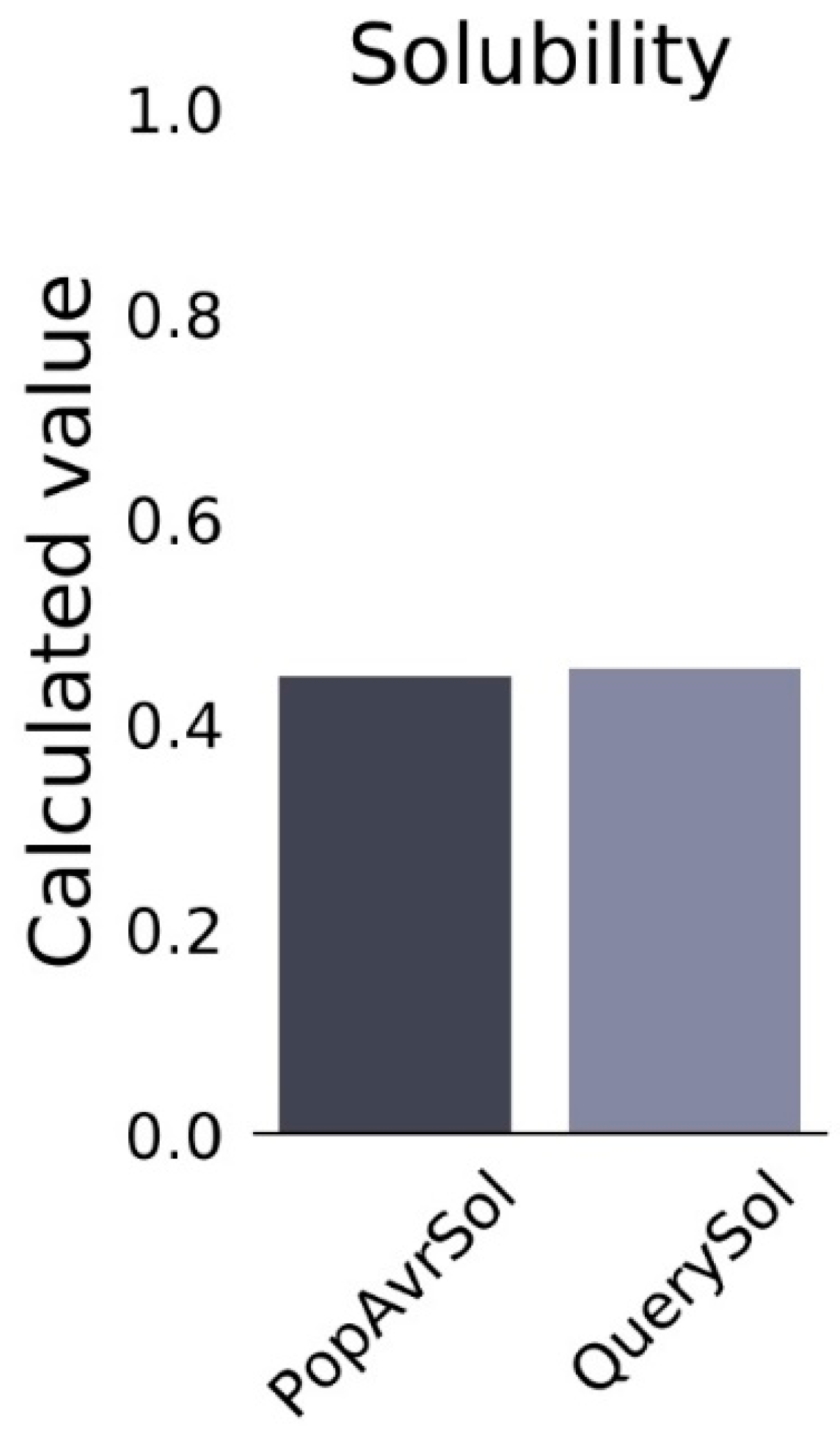
3.9. Prediction of the Solubility Properties of the Vaccine Construct

A subsequent ProteinSol assessment indicated the vaccine construct to be soluble, with a solubility score of 0.45 (Figure 4).

3.10. Secondary Vaccine Construct

The result of the analysis from the SOPMA server revealed that the construct is stable with 47.35% alpha-helix, 14.27% extended stranded, and 38.37% random coils as shown in Table 6. The outcome demonstrated that the secondary structure of the vaccine construct had high globular conformation and stability (Figure 5).

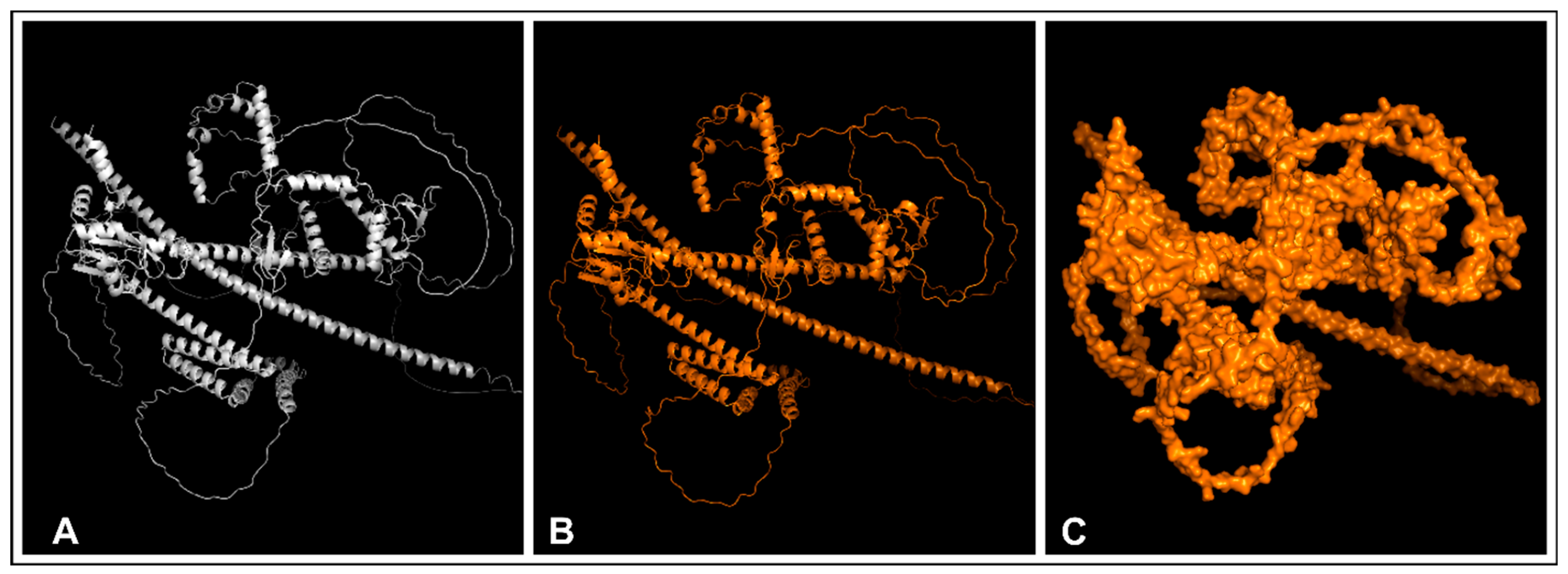
3.11. Tertiary Vaccine Construction, Refinement, and Validation of the Construct

The Colab Alphafold 2 server successfully predicted the 3-D structure, and the resulting structure was refined using GalaxyRefine server to ensure it maintained its native-like properties. This illustration, as shown in Figure 6, depicts the predicted 3-D structure, the refined structure and the surface structure of the refined vaccine construct. The protein’s quality was evaluated utilizing Ramachandran plots (Figure 7). The Ramachandran plot analysis confirmed that there are 1058 total residues in the amino acids of the IDV construct, of which approximately 94.2% are found in the region that is mostly favoured [A, B, L], and 5.1% are in the additional allowed regions. Notably, the amino acid residues located in disallowed regions constituted just 0.3% of the overall proportion, suggesting that the expected quality of the structure is relatively high.

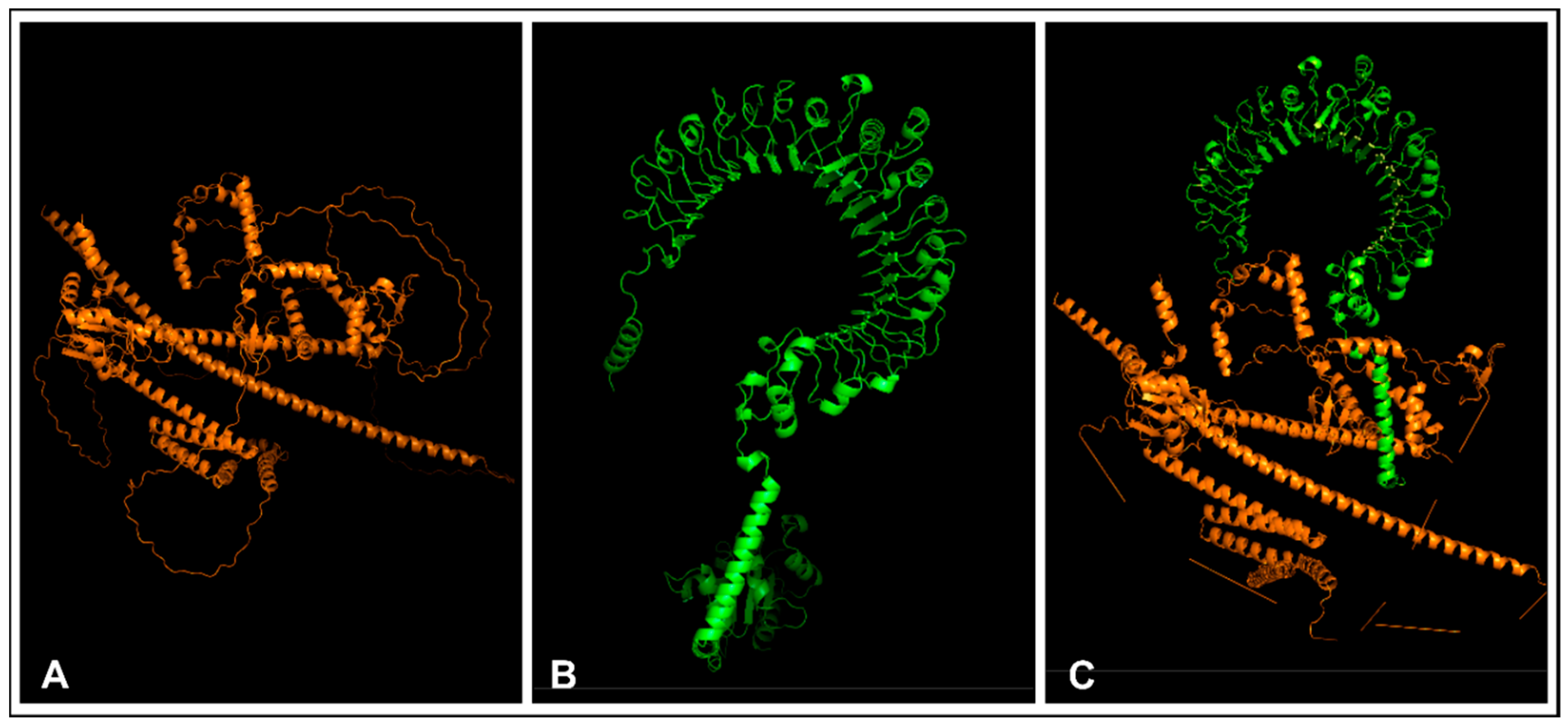
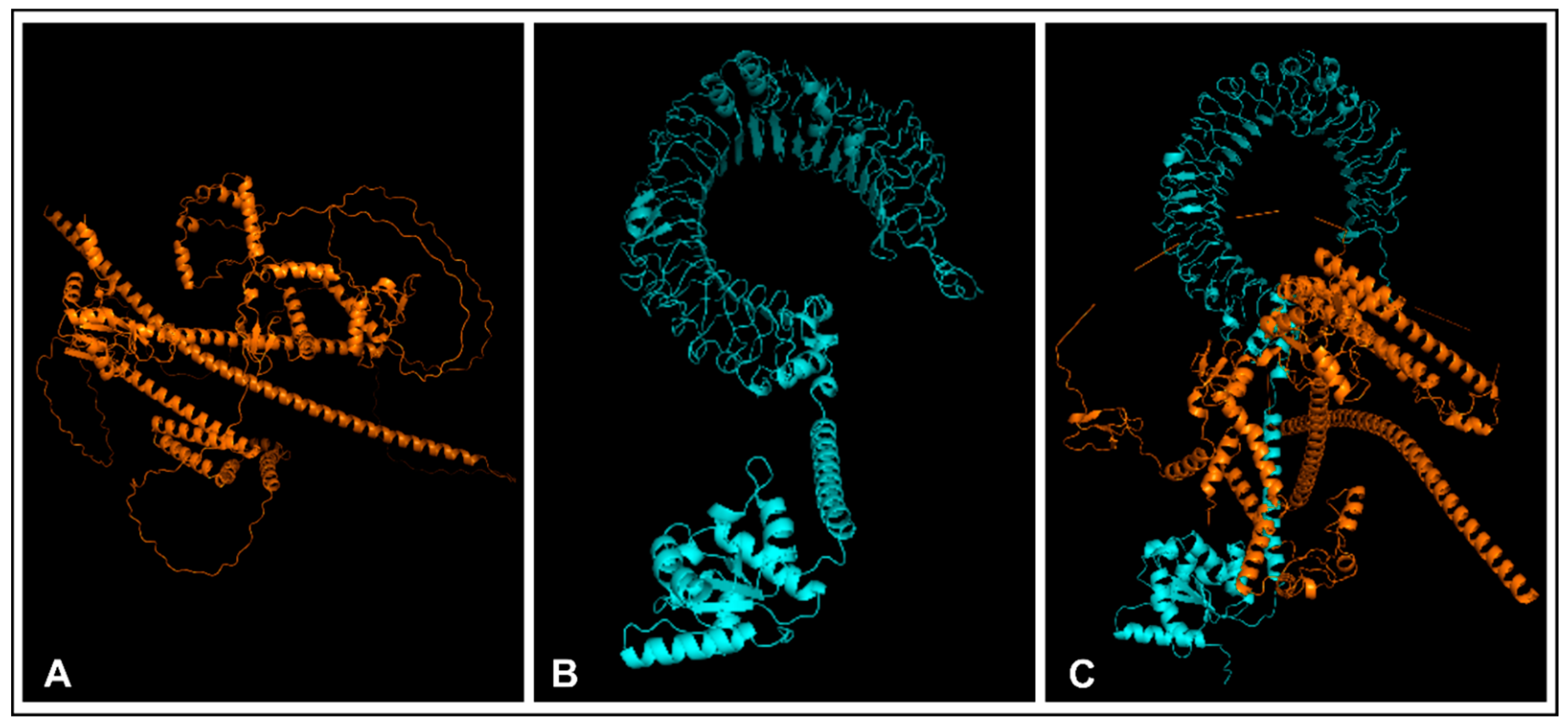
3.12. Molecular Docking

The 3-D refined structure was docked with TLR-2 and TLR-4, respectively. The docking results from the HDOCK server predicted the top 10 models of the structures. The ranking was determined by docking score, ligand RMSD(Å) and confidence score. Model 1 was chosen for both docked structures because it has the highest binding affinity (suggested by the lowest docking score). The results showed that the construct and the two TLRs have a strong affinity, as shown in Table 7, Figure 8 (construct and TLR-2) and Figure 9 (construct and TLR-4).

4. Discussion

Influenza D Virus (IDV) is a recently discovered enveloped RNA virus mostly found in swine and bovine [3]. It has also been found in other animal species exhibiting respiratory symptoms. Although there has been no record of human infection, it has been found that the IDV antibodies present in a sample of human serum through exposure or cross-reactivity with the Influenza C Virus have suggested a potential zoonotic risk, emphasizing the importance of surveillance and preventive measures to address this public health concern [45]. Hence, there is a need for an animal vaccine to be designed. This vaccine will contribute to the prevention and control of IDV outbreaks and the development of effective countermeasures against the emerging pathogen [46]. Our study presents a groundbreaking approach in the fight against IDV, as we focus on designing a universal mRNA vaccine utilizing the novel immunoinformatic approach, resulting in a safe, stable and soluble vaccine candidate.
HEF protein, which is the only surface glycoprotein of Influenza D Virus, was used as the target protein for this study. It has three main functions, including the ability to bind to the host cell receptors, the cleavage of the receptor as a result of its esterase activity, and the ability to mediate the fusion of the viral envelope. Also, HEF is vital for successful viral entry and replication in the host cell [47]. Eighty-eight (88) HEF sequences were retrieved and evaluated, and seventy-six (76) passed the antigenicity test.
After identifying the antigenic sequences, a further analysis was conducted to predict T-cell and B-cell epitopes. Identifying these epitopes is crucial for creating powerful antibodies that neutralize active proteins [48]. It is essential to accurately identify these epitopes in order to choose antibodies with a strong affinity, which contributes to the effectiveness of immunotherapy and immunodiagnostics. The role of T-cell epitopes in stimulating adaptive immunity by interacting with MHC molecules cannot be left out. According to [49,50,51], when choosing epitopes for an mRNA vaccine, it is vital to confirm that the epitopes are immunogenic and capable of inducing an immune response that can provide protection. This is why we selected epitopes pertinent to illicit necessary immune responses.
The vaccine was constructed using four CTL epitopes, three epitopes of HTL and four epitopes of B-cell after appropriate screening. The selected HTL epitopes excel due to their ability to stimulate interferon (IFN-γ), interleukin-4 (IL-4) and interleukin-10 (IL-10) production. The cytokines are crucial for stimulating the activation and maturation of T-helper 1 and T-helper 2 cells, respectively [52] suggesting that our vaccine construct can promote the release of essential cytokines. The prediction of each of the epitope’s antigenicity, toxicity, and allergenicity helped ensure that the vaccine candidate was safe, effective, and capable of stimulating an appropriate immune response.
The analysis of the physiochemical properties of the final vaccine construct was carried out immediately after the construction to know the molecular weight, aliphatic index, and instability index, and to ensure it was non-toxic or non-allergic. According to [53], the emergence of allergenicity and toxicity poses a substantial obstacle to the development of vaccines, manifesting after a vaccine has prompted an immune response that transitions to an allergic reaction. However, our vaccine construct indicated a non-allergenic and non-toxic nature, showcasing its likelihood of not triggering allergenic reactions when consumed, and minimizing the risk of harmful effects on cells or tissues, thereby rendering it safe for use. Also, the molecular weight is 110.58 kDa and the theoretical pI is 8.47, which suggests that the vaccine is alkaline in nature. The construct was considered stable, with an instability index of 35.40, which is below 40; this aligns with a similar study carried out by [54] on designing and evaluating a mRNA vaccine against BK virus using in silico studies. The GRAVY score and aliphatic index suggest that the final construct has a mild hydrophobic and thermostable character.
The vaccine candidate has a solubility score of 0.45 and can be effectively secreted in E. coli. According to [55], the solubility of mRNA vaccine protects mRNA from degradation, ensuring that mRNA can reach the target cells intact. Therefore, the solubility of vaccines is essential for their development and delivery. Glycosylation can affect the solubility of the vaccine by altering the function of the viral protein, stability, and folding, as well as their interactions with host receptors and immune cells [56]. HEF is glycosylated, meaning it has sugar chains attached to its surface. Therefore, enveloped viruses rely on glycosylation for immune escape and vaccine development employing adaptation and evolution for this purpose.
The orientation of protein folding can be determined by the results of the secondary structure [50,52]. The primary vaccine construct was inputted into an appropriate server to predict and analyze its secondary structure. The vaccine construct’s stabilized structure comprises 47.35% alpha-helix, 14.27% extended strands, and 38.37% random coils, as confirmed by the analysis. Thus, the result showed that the vaccine’s structure is highly globular, flexible, and stable in its secondary conformation.
The combination of AlphaFold 2 and GalaxyWEB has proved to be a highly effective approach in accurately predicting and refining the tertiary structure of the IDV vaccine. The Ramachandran plot results indicate a favourable distribution of amino acid residues, which is a promising sign of a structurally sound vaccine. Moreover, the use of multiple validation tools significantly increased the confidence in the accuracy and reliability of the predicted IDV vaccine structure.
The prediction of the binding affinity and position of a ligand and its corresponding receptor is a widely used bioinformatics approach, known as molecular docking. Ref. [57] suggest that molecular docking is a useful approach for simulating the binding affinity of epitopes to their corresponding MHC molecules. In this study, the vaccine construct’s ability to bind to TLRs on immune cells was determined by docking the tertiary construct, with TLR-2 and TLR-4, respectively. The HDOCK server provided 10 models of each of the docking structures. Model 1 was selected for both docked complexes using the confidence score, ligand RMSD(Å), and docking score. The vaccine construct has a higher binding affinity with TLR-2 and TLR-4, with docking scores of −359.19 and −379.08, respectively. This signifies the potential for triggering an immunological response because, according to [58] the strength of the binding between the receptor and its ligand increases as the energy decreases. Based on the analyses, it can be concluded that the IDV vaccine construct has the potential to be effective and safe. Therefore, the study’s overall findings suggest that this promising new vaccine candidate should be further developed and evaluated.

5. Conclusions

In light of the significant threat posed by the Influenza D virus, particularly its zoonotic potential, and impact on animal health, we have undertaken a comprehensive study leveraging immunoinformatic approaches to design an mRNA vaccine targeting this pathogen. The discoveries made in this study serve as a significant stepping stone toward the development of a stable and highly effective vaccine candidate for the Influenza D virus. The accurately selected epitopes and the favourable properties of the vaccine construct offer a solid foundation for further research and development. Therefore, this study provides a platform for rapid response and the proactive mitigation of future outbreaks of IDV or related infections.

Author Contributions

Conceptualization, E.K.O. and H.O.; Data curation, S.F.A. and T.M.A.; Formal analysis, E.K.O. and S.F.A.; Investigation, T.M.A. and L.O.; Project administration, S.F.A., M.W.A. and T.M.A.; Resources, E.K.O. and M.W.A.; Software, M.W.A., P.A.A., V.M.O. and C.A.A.; Supervision, E.K.O., L.O. and H.O.; Validation, K.F.S., V.M.O. and C.A.A.; Visualization, P.A.A., O.A.E., E.E.O., B.O.T. and E.O.A.; Writing—original draft, M.W.A., T.M.A., P.A.A., V.M.O., C.A.A., O.A.E., E.E.O., B.O.T. and E.O.A.; Writing—review and editing, E.K.O., S.F.A. and K.F.S. All authors have read and agreed to the published version of the manuscript.

Funding

This research received no external funding.

Institutional Review Board Statement

Not applicable.

Informed Consent Statement

Not applicable.

Data Availability Statement

No new data were created or analyzed in this study. Data sharing is not applicable to this article.

Acknowledgments

We would like to acknowledge Helix Biogen Institute in Ogbomoso, Oyo State, Nigeria, for their resources and technical assistance.

Conflicts of Interest

The authors declare no conflicts of interest.

References

  1. King, A.M.Q.; Adams, M.J.; Carstens, E.B.; Lefkowitz, E.J. (Eds.) Virus Taxonomy; Family—Orthomyxoviridae; Elsevier: San Diego, CA, USA, 2012; pp. 749–761. [Google Scholar]
  2. Hause, B.M.; Collin, E.A.; Liu, R.; Huang, B.; Sheng, Z.; Lu, W.; Wang, D.; Nelson, E.A.; Li, F. Characterization of a Novel Influenza Virus in Cattle and Swine: Proposal for a New Genus in the Orthomyxoviridae Family. mBio 2014, 5, e00031-14. [Google Scholar] [CrossRef] [PubMed]
  3. Asha, K.; Kumar, B. Emerging influenza D virus threat: What we know so far! J. Clin. Med. 2019, 8, 192. [Google Scholar] [CrossRef] [PubMed]
  4. Collin, E.A.; Sheng, Z.; Lang, Y.; Ma, W.; Hause, B.M.; Li, F. Cocirculation of two distinct genetic and antigenic lineages of proposed influenza D virus in cattle. J. Virol. 2015, 89, 1036–1042. [Google Scholar] [CrossRef] [PubMed]
  5. Nedland, H.; Wollman, J.; Sreenivasan, C.; Quast, M.; Singrey, A.; Fawcett, L.; Christopher-Hennings, J.; Nelson, E.; Kaushik, R.S.; Wang, D. Serological evidence for the co-circulation of two lineages of influenza D viruses in equine populations of the Midwest United States. Zoonoses Public Health 2018, 65, e148–e154. [Google Scholar] [CrossRef] [PubMed]
  6. Salem, E.; Cook EA, J.; Lbacha, H.A.; Oliva, J.; Awoume, F.; Aplogan, G.L.; Hymann, E.C.; Muloi, D.; Deem, S.L.; Alali, S.; et al. Serologic Evidence for Influenza C and D Virus among Ruminants and Camelids, Africa, 1991–2015. Emerg. Infect. Dis. 2017, 23, 1556–1559. [Google Scholar] [CrossRef]
  7. Kumar, B.; Asha, K.; Khanna, M.; Ronsard, L.; Meseko, C.A.; Sanicas, M. The emerging influenza virus threat: Status and new prospects for its therapy and control. Arch. Virol. 2018, 163, 831–844. [Google Scholar] [CrossRef] [PubMed]
  8. Hause, B.M.; Ducatez, M.; Collin, E.A.; Ran, Z.; Liu, R.; Sheng, Z.; Armien, A.; Kaplan, B.; Chakravarty, S.; Hoppe, A.D.; et al. Isolation of a novel swine influenza virus from Oklahoma in 2011 which is distantly related to human influenza C viruses. PLoS Pathog. 2013, 9, e1003176. [Google Scholar] [CrossRef] [PubMed]
  9. Molini, U.; Curini, V.; Jacobs, E.; Tongo, E.; Berjaoui, S.; Hemberger, M.Y.; Puglia, I.; Jago, M.; Khaiseb, S.; Cattoli, G.; et al. First influenza D virus full-genome sequence retrieved from livestock in Namibia, Africa. Acta Trop. 2022, 232, 106482. [Google Scholar] [CrossRef] [PubMed]
  10. Yu, J.; Hika, B.; Liu, R.; Sheng, Z.; Hause, B.M.; Li, F.; Wang, D. The Hemagglutinin-Esterase Fusion Glycoprotein Is a Primary Determinant of the Exceptional Thermal and Acid Stability of Influenza D Virus. MSphere 2017, 2, e00254-17. [Google Scholar] [CrossRef]
  11. Katayama, M.; Murakami, S.; Ishida, H.; Matsugo, H.; Sekine, W.; Ohira, K.; Takenaka-Uema, A.; Horimoto, T. Antigenic commonality and divergence of hemagglutinin-esterase-fusion protein among influenza D virus lineages revealed using epitope mapping. J. Virol. 2024, 98, e0190823. [Google Scholar] [CrossRef]
  12. Gray, G.C.; Robie, E.R.; Studstill, C.J.; Nunn, C.L. Mitigating future respiratory virus pandemics: New threats and approaches to consider. Viruses 2021, 13, 637. [Google Scholar] [CrossRef] [PubMed]
  13. Horimoto, T.; Hiono, T.; Mekata, H.; Odagiri, T.; Lei, Z.; Kobayashi, T.; Norimine, J.; Inoshima, Y.; Hikono, H.; Murakami, K.; et al. Nationwide Distribution of Bovine Influenza D Virus Infection in Japan. PLoS ONE 2016, 11, e0163828. [Google Scholar] [CrossRef] [PubMed]
  14. Mitra, N.; Cernicchiaro, N.; Torres, S.; Li, F.; Hause, B.M. Metagenomic characterization of the virome associated with bovine respiratory disease in feedlot cattle identified novel viruses and suggests an etiologic role for the influenza D virus. J. Gen. Virol. 2016, 97, 1771–1784. [Google Scholar] [CrossRef] [PubMed]
  15. Liu, R.; Sheng, Z.; Huang, C.; Wang, D.; Li, F. Influenza D virus. Curr. Opin. Virol. 2020, 44, 154–161. [Google Scholar] [CrossRef] [PubMed]
  16. Leibler, J.H.; Abdelgadir, A.; Seidel, J.; White, R.F.; Johnson, W.E.; Reynolds, S.J.; Gray, G.C.; Schaeffer, J.W. Influenza D virus exposure among US cattle workers: A call for surveillance. Zoonoses Public Health 2023, 70, 166–170. [Google Scholar] [CrossRef] [PubMed]
  17. Schlake, T.; Thess, A.; Fotin-Mleczek, M.; Kallen, J. Developing mRNA-vaccine technologies. RNA Biol. 2012, 9, 1319–1330. [Google Scholar] [CrossRef] [PubMed]
  18. Fang, E.; Liu, X.; Li, M.; Zhang, Z.; Song, L.; Zhu, B.; Wu, X.; Liu, J.; Zhao, D.; Li, Y. Advances in COVID-19 mRNA vaccine development. Signal Transduct. Target. Ther. 2022, 7, 94. [Google Scholar] [CrossRef]
  19. Sayers, E.W.; Bolton, E.E.; Brister, J.R.; Canese, K.; Chan, J.; Comeau, D.C.; Connor, R.; Funk, K.; Kelly, C.; Kim, S.; et al. Database resources of the National Center for Biotechnology Information. Nucleic Acids Res. 2022, 50, D20–D26. [Google Scholar] [CrossRef]
  20. Doytchinova, I.A.; Flower, D.R. VaxiJen: A server for prediction of protective antigens, tumour antigens and subunit vaccines. BMC Bioinform. 2007, 8, 4. [Google Scholar] [CrossRef]
  21. Reynisson, B.; Alvarez, B.; Paul, S.; Peters, B.; Nielsen, M. NetMHCpan-4.1 and NetMHCIIpan-4.0: Improved predictions of MHC antigen presentation by concurrent motif deconvolution and integration of MS MHC eluted ligand data. Nucleic Acids Res. 2020, 48, W449–W454. [Google Scholar] [CrossRef]
  22. Techakriengkrai, N.; Nedumpun, T.; Golde, W.T.; Suradhat, S. Diversity of the swine leukocyte antigen class I and II in commercial pig populations. Front. Vet. Sci. 2021, 8, 637682. [Google Scholar] [CrossRef] [PubMed]
  23. Gupta, S.; Kapoor, P.; Chaudhary, K.; Gautam, A.; Kumar, R.; Raghava, G.P.S. In silico approach for predicting toxicity of peptides and proteins. PLoS ONE 2013, 8, e73957. [Google Scholar] [CrossRef]
  24. Dimitrov, I.; Bangov, I.; Flower, D.R.; Doytchinova, I.A. AllerTOP v.2—A server for in silico prediction of allergens. J. Mol. Model. 2014, 20, 2278. [Google Scholar] [CrossRef] [PubMed]
  25. Oluwagbemi, O.O.; Oladipo, E.K.; Dairo, E.O.; Ayeni, A.E.; Irewolede, B.A.; Jimah, E.M.; Ogunleye, A.J. Computational construction of a glycoprotein multi-epitope subunit vaccine candidate for old and new South-African SARS-CoV-2 virus strains. Inform. Med. Unlocked 2022, 28, 100845. [Google Scholar] [CrossRef] [PubMed]
  26. Vita, R.; Mahajan, S.; Overton, J.A.; Dhanda, S.K.; Martini, S.; Cantrell, J.R.; Wheeler, D.K.; Sette, A.; Peters, B. The Immune Epitope Database (IEDB): 2018 update. Nucleic Acids Res. 2019, 47, D339–D343. [Google Scholar] [CrossRef]
  27. Andreatta, M.; Trolle, T.; Yan, Z.; Greenbaum, J.A.; Peters, B.; Nielsen, M. An automated benchmarking platform for MHC class II binding prediction methods. Bioinformatics 2018, 34, 1522–1528. [Google Scholar] [CrossRef] [PubMed]
  28. Dhanda, S.K.; Vir, P.; Raghava, G.P. Designing of interferon-gamma inducing MHC class-II binders. Biol. Direct 2013, 8, 30. [Google Scholar] [CrossRef] [PubMed]
  29. Dhanda, S.K.; Gupta, S.; Vir, P.; Raghava, G.P. Prediction of IL4-inducing peptides. Clin. Dev. Immunol. 2013, 2013, 263952. [Google Scholar] [CrossRef] [PubMed]
  30. Nagpal, G.; Usmani, S.S.; Dhanda, S.K.; Kaur, H.; Singh, S.; Sharma, M.; Raghava, G.P. Computer-aided designing of immunosuppressive peptides based on IL-10 inducing potential. Sci. Rep. 2017, 7, 42851. [Google Scholar] [CrossRef]
  31. Galanis, K.A.; Nastou, K.C.; Papandreou, N.C.; Petichakis, G.N.; Pigis, D.G.; Iconomidou, V.A. Linear B-Cell Epitope Prediction for In Silico Vaccine Design: A Performance Review of Methods Available via Command-Line Interface. Int. J. Mol. Sci. 2021, 22, 3210. [Google Scholar] [CrossRef]
  32. Oladipo, E.K.; Adeniyi, M.O.; Ogunlowo, M.T.; Irewolede, B.A.; Adekanola, V.O.; Oluseyi, G.S.; Omilola, J.A.; Udoh, A.F.; Olufemi, S.E.; Adediran, D.A.; et al. Bioinformatics Designing and Molecular Modelling of a Universal mRNA Vaccine for SARS-CoV-2 Infection. Vaccines 2022, 10, 2107. [Google Scholar] [CrossRef] [PubMed]
  33. Saha, S.; Raghava, G.P.S. Prediction of Continuous B-cell Epitopes in an Antigen Using Recurrent Neural Network. Proteins 2006, 65, 40–48. [Google Scholar] [CrossRef] [PubMed]
  34. Jespersen, M.C.; Peters, B.; Nielsen, M.; Marcatili, P. BepiPred-2.0: Improving sequence-based B-cell epitope prediction using conformational epitopes. Nucleic Acids Res. 2017, 45, W24–W29. [Google Scholar] [CrossRef] [PubMed]
  35. Ponomarenko, J.; Bui, H.H.; Li, W.; Fusseder, N.; Bourne, P.E.; Sette, A.; Peters, B. ElliPro: A new structure-based tool for the prediction of antibody epitopes. BMC Bioinform. 2008, 9, 514. [Google Scholar] [CrossRef] [PubMed]
  36. Coudert, E.; Gehant, S.; De Castro, E.; Pozzato, M.; Baratin, D.; Neto, T.; Sigrist, C.J.; Redaschi, N.; Bridge, A.; Consortium, T.U.; et al. Annotation of biologically relevant ligands in UniProtKB using ChEBI. Bioinformatics 2023, 39, btac793. [Google Scholar] [CrossRef] [PubMed]
  37. Gasteiger, E.; Hoogland, C.; Gattiker, A.; Duvaud, S.E.; Wilkins, M.R.; Appel, R.D.; Bairoch, A. Protein identification and analysis tools on the ExPASy server. In The Proteomics Protocols Handbook; Walker, J.M., Ed.; Humana Press: Totowa, NJ, USA, 2005; pp. 571–607. [Google Scholar] [CrossRef]
  38. Hebditch, M.; Carballo-Amador, M.A.; Charonis, S.; Curtis, R.; Warwicker, J. Protein–Sol: A web tool for predicting protein solubility from sequence. Bioinformatics 2017, 33, 3098–3100. [Google Scholar] [CrossRef]
  39. Deléage, G. ALIGNSEC: Viewing protein secondary structure predictions within large multiple sequence alignments. Bioinformatics 2017, 33, 3991–3992. [Google Scholar] [CrossRef] [PubMed]
  40. Mirdita, M.; Schütze, K.; Moriwaki, Y.; Heo, L.; Ovchinnikov, S.; Steinegger, M. ColabFold: Making protein folding accessible to all. Nat. Methods 2022, 19, 679–682. [Google Scholar] [CrossRef]
  41. Heo, L.; Park, H.; Seok, C. GalaxyRefine: Protein structure refinement is driven by side-chain repacking. Nucleic Acids Res. 2013, 41, W384. [Google Scholar] [CrossRef] [PubMed]
  42. Laskowski, R.A.; MacArthur, M.W.; Thornton, J.M. PROCHECK: Validation of protein structure coordinates. In International Tables of Crystallography, Volume, F: Crystallography of Biological Macromolecules; Rossmann, M.G., Arnold, E., Eds.; Kluwer Academic Publishers: Alphen aan den Rijn, The Netherlands, 2001; pp. 722–725. [Google Scholar]
  43. Berman, H.M.; Westbrook, J.; Feng, Z.; Gilliland, G.; Bhat, T.N.; Weissig, H.; Shindyalov, I.N.; Bourne, P.E. The Protein Data Bank. Nucleic Acids Res. 2000, 28, 235–242. [Google Scholar] [CrossRef]
  44. Yan, Y.; Tao, H.; He, J.; Huang, S. The HDOCK server for integrated protein–protein docking. Nat. Protoc. 2020, 15, 1829–1852. [Google Scholar] [CrossRef] [PubMed]
  45. Ruiz, M.; Puig, A.; Bassols, M.; Fraile, L.; Armengol, R. Influenza D virus: A review and update of its role in bovine respiratory syndrome. Viruses 2022, 14, 2717. [Google Scholar] [CrossRef] [PubMed]
  46. Brun, A. Vaccines and Vaccination for Veterinary Viral Diseases: A General Overview. Vaccine Technol. Vet. Viral Dis. 2016, 1349, 1–24. [Google Scholar] [CrossRef] [PubMed]
  47. Saegerman, C.; Gaudino, M.; Savard, C.; André Broes Ariel, O.; Meyer, G.; Ducatez, M. Influenza D virus in respiratory disease in Canadian, province of Québec, cattle: Relative importance and evidence of new reassortment between different clades. Transbound. Emerg. Dis. 2021, 69, 1227–1245. [Google Scholar] [CrossRef] [PubMed]
  48. Oladipo, E.K.; Akindiya, O.E.; Oluwasanya, G.J.; Akanbi, G.M.; Olufemi, S.E.; Adediran, D.A.; Babalola, M.O. Bioinformatics analysis of structural protein to approach a vaccine candidate against Vibrio cholerae infection. Immunogenetics 2023, 75, 99–114. [Google Scholar] [CrossRef] [PubMed]
  49. Oyarzun, P.; Kobe, B. Computer-aided design of T-cell epitope-based vaccines: Addressing population coverage. Int. J. Immunogenet. 2015, 42, 313–321. [Google Scholar] [CrossRef] [PubMed]
  50. Oluwagbemi, O.O.; Oladipo, E.K.; Kolawole, O.M.; Oloke, J.K.; Adelusi, T.I.; Irewolede, B.A.; Dairo, E.O.; Ayeni, A.E.; Kolapo, K.T.; Akindiya, O.E.; et al. Bioinformatics, Computational Informatics, and Modeling Approaches to the Design of mRNA COVID-19 Vaccine Candidates. Computation 2022, 10, 117. [Google Scholar] [CrossRef]
  51. Oladipo, E.K.; Ogunmolu, M.D.; Oyelakin, O.D.; Hammed, S.O.; Oluwasanya, G.J.; Hammed, S.O.; Onyeaka, H. Exploring the Potentials of Structural Proteins: Towards a Mrna Vaccine Candidate Against Marburg Virus Disease. Preprints 2023. [Google Scholar] [CrossRef]
  52. Oladipo, E.K.; Ajayi, A.F.; Ariyo, O.E.; Onile, S.O.; Jimah, E.M.; Ezediuno, L.O.; Adebayo, O.I.; Adebayo, E.T.; Odeyemi, A.N.; Oyeleke, M.O.; et al. Exploration of surface glycoprotein to design multi-epitope vaccine for the prevention of Covid-19. Inform. Med. Unlocked 2019, 21, 100438. [Google Scholar] [CrossRef]
  53. Dey, J.; Mahapatra, S.R.; Raj, T.K.; Kaur, T.; Jain, P.; Tiwari, A. Designing a novel multi-epitope vaccine to evoke a robust immune response against pathogenic multidrug-resistant Enterococcus faecium bacterium. Gut Pathog. 2022, 14, 21. [Google Scholar] [CrossRef]
  54. Mohammadi, Y.; Nezafat, N.; Negahdaripour, M.; Eskandari, S.; Zamani, M. In silico design and evaluation of a novel mRNA vaccine against BK virus: A reverse vaccinology approach. Immunol. Res. 2023, 71, 422–441. [Google Scholar] [CrossRef] [PubMed]
  55. Pardi, N.; Hogan, M.J.; Weissman, D. Recent advances in mRNA vaccine technology. Curr. Opin. Immunol. 2020, 65, 14–20. [Google Scholar] [CrossRef]
  56. Shental-Bechor, D.; Levy, Y. Effect of glycosylation on protein folding: A close look at thermodynamic stabilization. Proc. Natl. Acad. Sci. USA 2008, 105, 8256–8261. [Google Scholar] [CrossRef] [PubMed]
  57. Althurwi, H.N.; Alharthy, K.M.; Albaqami, F.F.; Altharawi, A.; Javed, M.R.; Muhseen, Z.T.; Tahir ul Qamar, M. mRNA-Based Vaccine Designing against Epstein-Barr Virus to Induce an Immune Response Using Immunoinformatic and Molecular Modelling Approaches. Int. J. Environ. Res. Public Health 2022, 19, 13054. [Google Scholar] [CrossRef]
  58. Fu, Y.; Zhao, J.; Chen, Z. Insights into the Molecular Mechanisms of Protein-Ligand Interactions by Molecular Docking and Molecular Dynamics Simulation: A Case of Oligopeptide Binding Protein. Comput. Math. Methods Med. 2018, 2018, 3502514. [Google Scholar] [CrossRef]
Figure 1. Workflow of the Methodology used in the study.
Biomedinformatics 04 00086 g001
Figure 2. Three-dimensional structures of eight predicted conformational B-cell epitopes: The yellow portion represents the B-cell epitope, while the grey portion represents the surrounding residues. The pI scores for each epitope are as follows: (A) 0.848 with eight residues, (B) 0.691 with 9 residues, (C) 0.831 with 10 residues, (D) 0.861 with 11 residues, (E) 0.885 with 11 residues, (F) 0.815 with 12 residues, (G) 0.923 with 13 residues, and (H) 0.799 with 20 residues.
Biomedinformatics 04 00086 g002
Figure 3. The schematic diagram showing the final mRNA vaccine construct. The 1058-amino acid-designed vaccine consists of 5′ Cap, 5′ UTR, Kozak sequence, tPA, adjuvant (purple) and three HTL (orange) epitopes linked by the GPGPG linker (black). The HTLs are joined together by GPGPG, the last HTL epitope and the first LBL (yellow) epitope are linked by the KK linker, including the other LBLs. The last LBL and the CTLs (green) are linked by EAAAK. MITD, 3′ UTR and Poly-A tail (121 alanine) are added at the C-terminal end of the vaccine construct for stability and purification.
Biomedinformatics 04 00086 g003
Figure 4. A graph showing the solubility prediction of the vaccine construct (QuerySol) and the average soluble E. coli protein (PopAvrSol).
Biomedinformatics 04 00086 g004
Figure 5. Secondary structure prediction of the vaccine construct.
Biomedinformatics 04 00086 g005
Figure 6. Three-dimensional structure of the vaccine construct. (A) Structure (Rank 1) of the vaccine from AlphaFold (B) Refined vaccine construct (in ribbon) from Galaxy Refine (C) Surface structure of the refined vaccine construct.
Biomedinformatics 04 00086 g006
Figure 7. Validation of the 3-D structure. (A) Results of the Ramachandran plot generated by the PROCHECK (B) Analysis of the Ramachandran plot.
Biomedinformatics 04 00086 g007
Figure 8. Molecular docking results. (A) Tertiary structure of the construct. (B) Toll-like receptor-2 (TLR-2). (C) Docked complex of TLR-2 (Green) and the construct (Orange).
Biomedinformatics 04 00086 g008
Figure 9. Molecular docking results. (A) Tertiary structure of the construct. (B) Toll-like receptor-4 (TLR-4). (C) Docked complex of TLR-4 (Blue) and the vaccine construct (Orange).
Biomedinformatics 04 00086 g009
Table 1. Total retrieved isolates across continents.
ContinentIsolates
Africa1
Europe25
North America43
South America4
Asia14
Oceania1
Table 2. Final CTL epitopes.
CTL EpitopesAntigenicityAllergenicityToxicityImmunogenicity
VANISMNLKAntigenNon-allergenNon-toxic1.6606
YSIKSTPRFAntigenNon-allergenNon-toxic1.4093
VNPRASPQVAntigenNon-allergenNon-toxic1.4867
RGIAGSRVKAntigenNon-allergenNon-toxic1.3654
AERELICIVAntigenNon-allergenNon-toxic1.3012
AEKELICIVAntigenNon-allergenNon-toxic1.2598
GDIEMRQLLAntigenNon-allergenNon-toxic1.2738
VNPRASSQVAntigenNon-allergenNon-toxic1.2661
Table 3. Final HTL epitopes.
EpitopesAntigenicityIL-4IL-10InterferonImmunogenicity
GLLFVGFVAGGVAGGAntigenInducerInducerPositive0.53738
AGGYFWGRSNERGGGAntigenInducerInducerPositive0.4476
VAGGYFWGRSNERGGAntigenInducerInducerPositive0.44493
LTTITAITACQAEREAntigenInducerInducerPositive0.41419
NRVAAYRGIASAEVKAntigenInducerInducerPositive0.32272
VAGGYFWGRSNERGGAntigenInducerInducerPositive0.44493
NRVAAYRGIASAEVKAntigenInducerInducerPositive0.32272
AGGYFWGRSSERGGGAntigenInducerInducerPositive0.2928
SYCFDTDGGYPIQVVAntigenInducerInducerPositive0.28454
RSYCFDTDGGYPIQVAntigenInducerInducerPositive0.23615
Table 4. Final B-cell epitopes.
B-Cell EpitopesAllergenicityToxicityImmunogenicity
FGLLFIGFVAGGVAGGYFNon-allergenNon-toxic0.65678
PEAGIDCFGLNWTQTKNon-allergenNon-toxic0.45256
GGIAQEAGPGCWYIDSNon-allergenNon-toxic0.44557
TAITACQAERELICIVQRNon-allergenNon-toxic0.3973
GGIAQEAGPGCWYVDSNon-allergenNon-toxic0.36809
LGSTIALCLLGLVAIAAFNon-allergenNon-toxic0.34982
KTTPYAGEADDNHGDINon-allergenNon-toxic0.31345
GYFWGRSNGRGGGASVNon-allergenNon-toxic0.29455
ASVINQKDWIGFGDSRNon-allergenNon-toxic0.28608
KRGGGIAQEAGPNon-allergenNon-toxic0.28007
TKRGGGIAQEANon-allergenNon-toxic0.24407
TKRGGGIAQENon-allergenNon-toxic0.20752
KRGGGIAQEANon-allergenNon-toxic0.20527
ASVINQKDWVGFGDSRNon-allergenNon-toxic0.19668
TKVVITSDPYYWGSTINon-allergenNon-toxic0.1955
GYRGIAPGTYSIRSTPNon-allergenNon-toxic0.17528
SPLWYAESSVNPGARPNon-allergenNon-toxic0.14735
Table 5. Physicochemical properties evaluation of the construct.
PropertyValue
Molecular weight110,576.04 Da
Number of Amino acids1058
Theoretical pI8.47
Total number of negatively charged residues (Asp and Glu)101
Total number of positively charged residues (Arg and Lys)118
FormulaC4822H2727N1347O1475S75
Total number of atoms15,446
Estimated half-life [The N-terminal of the sequence considered is A (Ala)]30 h (mammalian reticulocytes, in vitro)
>20 h (yeast, in vivo)
>10 h (Escherichia coli, in vivo)
Instability index35.40 (<40)
Aliphatic index79.04 (>50)
Grand average of hydropathicity (GRAVY)0.058
Predicted Scaled Solubility0.454
Table 6. Study of the secondary structure of the designed vaccine.
Name of the Examined UnitNumber of ResiduesPercentages
Alpha-helix50147.35%
310 helixes00.00%
Beta bridge00.00%
Extended strand15114.27%
Beta turn00.00%
Random coil40638.37%
Other states00.00%
Table 7. Summary of the docking complexes.
Docking ComplexesDocking ScoreConfidence ScoreLigand RMSD (Å)
Construct and TLR-2−359.190.9850115.12
Construct and TLR-4−379.080.989967.81
Disclaimer/Publisher’s Note: The statements, opinions and data contained in all publications are solely those of the individual author(s) and contributor(s) and not of MDPI and/or the editor(s). MDPI and/or the editor(s) disclaim responsibility for any injury to people or property resulting from any ideas, methods, instructions or products referred to in the content.

