Simple Summary
The implications of PD-L1 expression in oral squamous cell carcinoma (OSCC) remain unclear. Most published series and systematic reviews suffer from cohort heterogeneity and the use of different immunohistochemical tools, leading to controversial and non-conclusive results without significant clinical relevance. A retrospective monocentric study, including all patients with OSCC diagnosed and treated between January 2020 and May 2022, was performed. The method used to measure PD-L1 was reliable and accurate, with a correlation coefficient between PD-L1 expression in a biopsy and a surgical piece of 0.83 (p < 0.0001). PD-L1 expression was observed in large tumors (p < 0.001) and was correlated with the presence of lymph node metastases (p < 0.004). This study aimed to assess the immunohistochemical expression of PD-L1 in squamous cell carcinomas of the oral cavity with a reliable and accurate method and its possible associations with different histopathological, clinical, and prognostic variables in a patient cohort with OSCC from a single institution.
Abstract
Background: PD1 and its ligand PD-L1 are related to prognosis in many solid tumors; however, their role in oral squamous cell carcinoma (OSCC) remains unclear. Methods: A retrospective monocentric study including all patients with OSCC diagnosed and treated between January 2020 and May 2022 was performed. PD-L1 expression was assessed per a combined positive score (CPS), considering a CPS of > or equal to 1 as positive (1–20 indicating “low expression” and ≥20 indicating “high”). A descriptive analysis of the patient cohort and tumors was performed, including tumor size, stage, lymph node involvement, recurrence, and survival. Results: In total, 65 patients (65 tumors) were analyzed. A total of 66.15% of the tumors were in advanced stages (III-IV), of which 97.67% expressed PD-L1+, compared with 71.42% in the early stages (I–II). T4 tumors expressed PD-L1 in 100% of cases, compared with 54% in T1 tumors. A total of 50.79% of the tumors showed lymph node involvement (pN+), with 100% of the pN+ showing PD-L1+. The prevalence of pN+ was 59.38% vs. 40.63% for high vs. low PD-L1 expression, respectively. Patients’ follow-ups ranged from 2 to 34.5 months. No significant difference was seen between overall survival (OS) and PD-L1 +/− (CPS ≥ 1 vs. CPS < 1) or high (CPS ≥ 20) and low (CPS < 20) PD-L1 expression (p < 0.97 and 0.64, respectively). Conclusions: The method used to measure PD-L1 (a laboratory test with Dako 22C3 anti-PD-L1 primary antibodies) was reliable and accurate, with a correlation coefficient between PD-L1 expression in the biopsy and the surgical piece of 0.83 (p < 0.0001). A CPS of ≥1 was observed in large tumors (p < 0.001) and was correlated with that of lymph node metastases (p < 0.004). Further analysis of PD-L1 expression in OSCC and studies to determine its relevance in tumor biology and prognosis is needed.
1. Introduction
Oral squamous cell carcinoma (OSCC) is the most common type of cancer in the head and neck area. Every year, approximately 300,000 new cases are diagnosed worldwide [1]. It is linked to higher morbidity and a poor prognosis when diagnosed at advanced stages. Although adjuvant radiation therapy or combined chemoradiotherapy is frequently used for treatment, present treatment is generally based on the surgical resection of the primary tumor, which may be followed by radio-, chemo-, and immunotherapy. The selection of adjuvant treatments relies on factors such as the tumor location, histological grading, histopathological features (such as extracapsular extension in N+ disease and/or the presence of positive surgical margins), radiographic findings, and the TNM system grading [2].
In recent years, immunotherapy has been added to the therapeutic arsenal against many different tumors, supported by the results of different clinical trials [3,4,5,6,7,8]. In addition, some early-stage clinical trials have begun to administer neoadjuvant immunotherapy before surgery [9,10]. This immunotherapy is based on the different survival strategies of tumor cells and their relationship with the microenvironment. One of these potential routes of tumor escape from the action of the immune system is the expression of the PD1 ligand protein (PD-L1) by tumor cells, which induces anergy in cytotoxic CD8 lymphocytes and prevents them from attacking the tumor cells by interacting with their PD1 protein. Blocking the binding of PD-L1 to PD-1 via an immune checkpoint inhibitor (anti-PD-L1 or anti-PD-1) allows T cells to destroy tumor cells [11,12].
PD-L1 expression has been studied via immunohistochemistry in many tumors, including head and neck squamous cell carcinomas; however, such expression levels are characterized by high inter- and intratumoral heterogeneity due to factors that are not completely understood. Even in squamous cell carcinomas of the head and neck, variable expression is observed depending on the location of the primary tumor [13]. The immune checkpoint PD1 and its ligand PD-L1 are related to the prognosis of many solid tumors. However, their role in oral squamous cell carcinomas (OSCCs) remains unclear. Most published series and systematic reviews have included cohort heterogeneity (e.g., in terms of the geographical region, tumor site, sex distribution, and habits such as alcohol and tobacco use), used different immunohistochemistry (IHC) tools, and even applied different treatment protocols, leading to controversial and non-conclusive results without significant clinical relevance [14].
This study aimed to assess the immunohistochemical expression of PD-L1 in squamous cell carcinomas of the oral cavity with a reliable and accurate method, as well as its possible association with different histopathological, clinical, and prognostic variables, in a patient cohort with OSCC from a single institution with a peculiar distribution, including a female majority and 50% regular smokers. In addition, we aimed to determine an accurate and reliable method for measuring PD-L1 in OSCC.
2. Materials and Methods
A retrospective observational study of 65 OSCCs from 65 patients was performed. All patients were collected from Hospital Universitario 12 de Octubre, Madrid, between January 2020 and May 2022. Tumors were stained for PD-L1 using the combined positive score (CPS), considering PD-L1 as positive when CPS ≥ 1. The ESTROBE guidelines were followed. Ethical approval was granted by the CEIM/CEI of Hospital Universitario 12 de Octubre. The inclusion criteria were as follows: OSCC diagnosed with at least one PD-L1 measurement performed (either on a biopsy, surgical specimen, or both) and an imaging test for staging (in case no pathological staging was performed). Oropharyngeal and HPV+ SCCs were excluded, as well as carcinoma in situ tumors. Patients who did not receive surgery as the first treatment and those who had not had at least two months of follow-up were excluded from the survival analysis.
Clinicopathologic features, including age at diagnosis, sex, smoking history (e.g., never smoked or current/former smoker), tumor sites (e.g., the tongue, cervical metastasis from a previous resected OSCC, gums, buccal mucosa, mouth floor, or retromolar trigone), the pathologic TNM stage (the American Joint Committee on Cancer 8th Edition of the Oral Cancers Staging System), and comorbidity that can alter the immune system (e.g., diabetes mellitus (DM), chronic corticosteroids, transplant recipients, and chronic obstructive pulmonary disease (COPD)) were analyzed in this study. Histologic differentiation was designated as either well, moderately, or poorly differentiated. The extranodal extension (ENE) of the metastatic lymph nodes was classified as positive or negative.
The patients were followed up until death or 1 November 2022. Overall survival (OS) was defined as the interval from the day of surgery to the day of death for any cause; disease-free survival (DFS) from the day of surgery to the day of disease progression in a CT scan/other imaging test or death by any cause; and disease-specific survival (DSS) from the day of surgery to the day of death from OSCC.
The immunohistochemical study was performed in the Pathology Department of our hospital using 4 µm thick sections of tissue embedded in paraffin and fixed in formalin. We used a commercially available laboratory test with Dako 22C3 anti-PD-L1 primary antibodies (Agilent Dako, CA, USA, catalog number K8005) on a Ventana Benchmark ULTRA platform. Antigen retrieval was performed using the Cell Conditioning 1 antigen retrieval solution (Ventana Medical Systems) for 64 min at 95 °C. The specimens were incubated with primary anti-PD-L1 monoclonal antibodies at a concentration of 1:25 for 64 min at 37 °C and with the Optic View DAB IHQ Detection Kit. An internal control with normal tonsils was performed to assess the validity of the PD-L1 immunoreaction. Cases were considered adequate for assessment if at least 100 tumor cells were observed. We considered positive partial or complete membrane staining of infiltrative tumor cells and membranes and/or cytoplasmic staining of immune cells (lymphocytes and macrophages) near the tumor. The CPS was used to calculate the PD-L1 expression level. A CPS of less than 1 was considered negative. Our trained pathologist assessed the PD-L1 expression with a CPS evaluation (the number of positive (immune plus tumor) cells divided by the number of viable tumor cells and multiplied by 100) in the stained slides of HNSCCs in a blinded fashion without knowledge of any clinical data.
The CPS classification as high (20), low (1–20), or negative (<1) was based on the KEYNOTE-048 study [3]. In that study, pembrolizumab with chemotherapy improved OS vs. cetuximab with chemotherapy in patients with a CPS of 20 or more and a CPS of 1 or more. Hence, the clinically relevant CPS cutoffs of ≥1 and ≥20 were used (Figure 1).
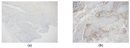
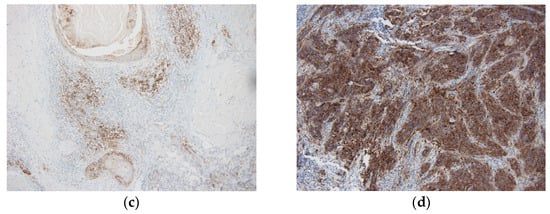
Figure 1.
All figures are at 10× magnification. (a) Immunohistochemical staining of tongue tumor with CPS < 1 (negative expression); (b) immunohistochemical staining of oral cavity tumor with CPS of 5 (low expression); (c) immunohistochemical staining of oral cavity tumor with CPS of 40 (high expression); (d) immunohistochemical staining of oral cavity tumor with CPS of 100 (high expression).
IBM SPSS 24.0 (IBM Corp., Armonk, NY, USA) was used for the statistical analysis. The chi-squared test was used for clinicopathological variables with categorical data unless any category had a value of < 5, wherein Fisher’s exact test was used. Continuous data were analyzed using either Mann–Whitney’s U or Kruskal–Wallis’s H tests. Survival outcomes were calculated using Kaplan–Meier curves and the log-rank test. The reliability of the CPS in biopsy and surgical specimens was determined by calculating the correlation coefficient (IC). We defined the IC as poor (95% CI < 0.5), moderate (0.5 ≥ 95% CI < 0.75), good (0.75 ≥ 95% CI < 0.9), or excellent (95% CI ≥ 0.9).
3. Results
3.1. CPS Measured in Biopsy and Surgical Piece
Sixty-five tumors from sixty-five patients were analyzed. PD-L1 was measured on two occasions, first, in the tumor biopsy and later, in the surgical resection piece. The CPS was determined in the biopsy and the surgical piece sample in 23 cases. A PD-L1 measurement in the biopsy was available in 52 tumors: 6 (11.53%) tumors were PD-L1−, while 46 (88.46%) tumors were PD-L1+, with a mean CPS of 34.83. A PD-L1 measurement in the surgical piece was available in 38 tumors: 7 (18.42%) were PD-L1-negative, and 31 (81.58%) were PD-L1-positive, with a mean CPS of 48.23. The correlation coefficient between the PD-L1 expression in the biopsy and the surgical piece was 0.83 (p < 0.0001). Sixty tumors (89.55%) were PD-L1-positive in the biopsy or the surgical resection piece on at least one occasion. Thirty-seven (55%) tumors had a high CPS (>20). The CPS was higher in the biopsy than in the surgical piece in five tumors, while six tumors had a higher CPS in the surgical piece. Simplifying these measurements for statistical analysis, we considered a tumor PD-L1-positive when a positive value was obtained in one of the two PD-L1 measurements. In the case of being positive in both measurements, we considered the highest value for statistical analysis.
3.2. Clinicopathological Correlation
The PD-L1 status (as a categorized variable) and clinicopathological variables were compared (Table 1). The patients’ mean age was 67.92 (Ds 14.24). Using 60 years as the cutoff point, no statistically significant differences were observed in PD-L1 status and age (CPS ≥ 1 vs. <1, p = 0.89; CPS ≥ 20 vs. <20, p = 0.47). Our sample included 29 (44.61%) men and 36 women (55.38%). No significant difference was seen in sex for PD-L1 expression (p = 0.92). In total, 32 (49.23%) patients were non-smokers, and 33 (50.76%) patients were smokers or ex-smokers. No significant differences were seen in smoking status for PD-L1 expression (p = 0.65). There was no significant correlation between being a smoker or non-smoker and sex (p = 0.47); therefore, the patient’s sex was not a confounding factor between smoking and PD-L1 expression in our sample.

Table 1.
Comparison between PD-L1 status and clinicopathological variables.
In total, 18 (27.69%) patients were immunosuppressed (i.e., DM, chronic corticosteroids, transplant recipients, and COPD), and 47 (72.30%) were not immunosuppressed. No statistically significant differences were seen between patients’ PD-L1 expression and immune status (p = 0.96), and no significant difference was seen between the immune status and the degree of high, low, or negative PD-L1 expression (p = 0.43). The most common OSCC location was the tongue, in 22 (33.84%) patients. No significant differences were seen between the tumor location and PD-L1 expression (p = 0.074), nor between the location, smoking status, and PD-L1 expression (p = 0.3). Twenty-three (35.38%) tumors were T4, the most common tumor size. In total, 86.95% of T4 tumors expressed PD-L1 in the surgical piece (tumor resection), compared with 54.54% in T1 tumors. However, all T4 tumors expressed PD-L1 at least once (there were three T4 tumors that were PD-L1-positive in the biopsy and PD-L1-negative in the surgical piece). Significant differences were seen between the expression status of PD-L1 and the tumor size when the CPS cutoff value was set at ≥1 and ≥20 (p < 0.001 and p = 0.009, respectively). This significant difference is explained because most of the PD-L1-negative tumors were T1. Thirty-two (50.79%) of the tumors showed lymph node involvement (pN+). In total, 100% of the N+ tumors were PD-L1+ in at least one measurement (the biopsy or surgical piece). Three N+ tumors were PD-L1-positive in the biopsy and PD-L1-negative in the surgical piece. The prevalence of pN+ was 59.38% vs. 40.63% for high vs. low PD-L1 expression, respectively. The cervical node extension significantly correlated with the PD-L1 expression (CPS ≥ 1) (p = 0.004). Similar to the tumor size, this significant difference is explained because most of the PD-L1-negative tumors were N0. No significant differences were seen when we compared the cervical node extension (N0 vs. N+) between high (CPS ≥ 20) and low expressions (CPS < 20) of PD-L1 (p = 0.7). In total, 43 (66.15%) tumors were in advanced stages (III and IV), of which 36 (83.72%) were stage IV, and all were PD-L1+. In total, 97.67% of the advanced tumors expressed PD-L1+, while 71.42% of the early tumors expressed PD-L1. A significant difference was seen between the tumor stage and PD-L1 expression (p < 0.001) when the CPS cutoff value was set at one. Neither the tumor differentiation grade nor extranodal extension were significantly associated with PD-L1 expression (p = 0.12 and p = 0.13, respectively).
This study included 59 (90.76%) primary tumors and 6 (9.23%) recurrences. In total, 52 (80%) of the primary tumors were PD-L1-positive, of which 30 (57.7%) exhibited high expression (CPS > 20) and 22 (42.30%) low expression (CPS < 20). In total, 100% of the recurrences (of six tumors) expressed PD-L1, three of them with high expression and three with low expression. No significant differences were seen between primary and recurring tumors for PD-L1 expression (p = 0.38).
3.3. Survival
A total of 57 patients underwent surgery as the first treatment. Three patients were initially treated with chemoradiotherapy, four patients just with radiotherapy, and one with best supportive care. Due to the heterogeneity of the treatments, we decided to restrict the survival analysis to only the patients’ data at the time of the first surgically treated tumor; thus, only 57 patients were included in the survival analysis.
Patients’ follow-ups ranged from 2 to 34.5 months, with 11 cancer-specific deaths. The median follow-up of the patients was 13.68 months [6.36, 14.17]. The median OS was reached at 11.9 months, and the interquartile range was [6.36, 19.11]. A significantly worse OS was found for N+ status (N0 vs. N+; p = 0.0006), advanced stages (I and II vs. III and IV; p = 0.0067), tumor size (p = 0.003), and extranodal extension (ENE− vs. ENE+; p = 0.0033). No significant difference was seen between PD-L1 +/− (CPS ≥ 1 vs. CPS < 1) or high (CPS ≥ 20) vs. low (CPS < 20) PD-L1 expression and OS (p = 0.97 and 0.64, respectively) (Figure 2).
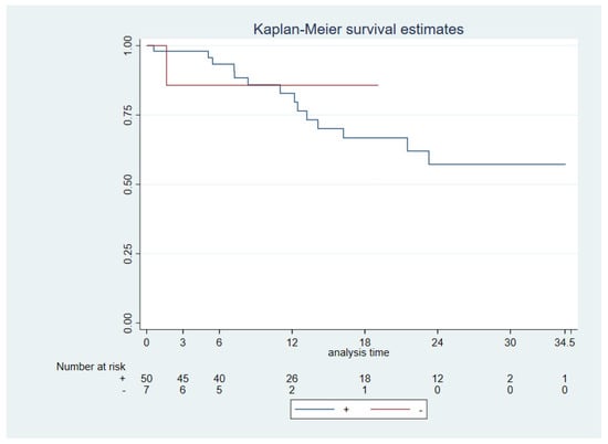
Figure 2.
Overall survival. PD-L1 + (CPS ≥ 1) vs. PD-L1 − (CPS < 1) (log−rank p = 0.97).
Disease-free survival (DFS) was measured from the months of surgery to the day of disease progression in a CT scan/other imaging test or death by any cause. The ENE was close to achieving a significant difference in DFS (p = 0.061). No significant difference was seen between PD-L1 +/− (CPS ≥ 1 vs. CPS < 1) or high (CPS ≥ 20) vs. low (CPS < 20) PD-L1 expressions and DSF (p = 0.11 and p = 0.88, respectively) (Figure 3). In this cohort, 33.89% had tumor recurrences (20 tumors). Twelve of these tumors were PD-L1-high, and seven were PD-L1-low. Just one tumor was PD-L1-negative in the biopsy of the recurrence; previously, this tumor was PD-L1-low (with a CPS of 10).
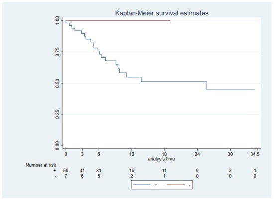
Figure 3.
Disease−free survival. PD-L1 + (CPS ≥ 1) vs. PD-L1 − (CPS < 1) (log−rank p = 0.11).
All patients who died due to the OSCC were PD-L1-positive; nevertheless, no significant difference was found between PD-L1 +/− (CPS ≥ 1 vs. CPS < 1) or high (CPS ≥ 20) vs. low (CPS < 20) PD-L1 expression and DSS (p = 0.43 and p = 0.99, respectively) (Figure 4). A significantly worse DSS was seen for ENE+ (ENE+ vs. ENE−; p = 0.003), cervical node extension (N+ vs. N−; p = 0.0005), advanced stages (I and II vs. III and IV; p = 0.005), and tumor size (p = 0.0014).
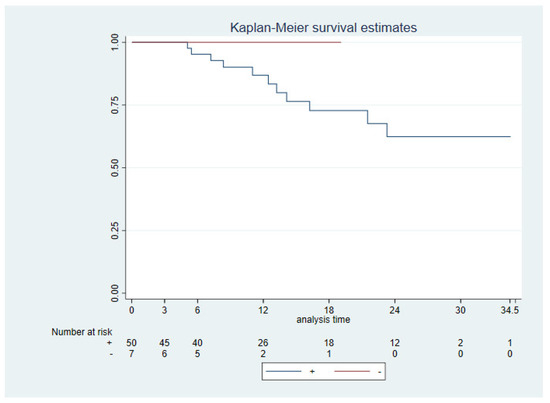
Figure 4.
Disease−specific survival. PD-L1 + (CPS ≥ 1) vs. PD-L1 − (CPS < 1) (log−rank p = 0.43).
4. Discussion
In this study, PD-L1 expression was evaluated with the Combined Positive Score (CPS) scoring system, which measured the PD-L1 expression in tumor and immune cells. The presence of immune cells in tumors impacts the capacity for an adequate clinical response to immunotherapy [13]. A CPS above 1% predicts a response to anti-PD-L1 therapies [3,15,16]. The TPS can also measure PD-L1, but this score does not consider the presence of immune cells in the tumor, so it should be complemented with IC (tumor-infiltrating immune cells) to predict the response to immune checkpoint inhibitors (ICIs) [17].
In our study, 58 (89.23%) of the 65 tumors were PD-L1-positive, and 36 (55.38%) tumors had a high CPS (>20). Other studies in the literature have found similar PD-L1 expression frequencies in head and neck epidermoid tumors. Dilinaer Wusiman et al. [18] and the KETNOTE-048 study [3] reported values of 89.9% and 85% with a CPS of ≥1, and 43.7% and 43% with a CPS of >20, respectively. Like us, both studies used the 22C3 assay for immunohistochemical studies. In contrast, a prevalence of 48% (CPS > 1) was found in Miranda et al.’s cohort [19]. Other studies used the TPS for PD-L1 measurements, such as Levounel et al. [20], reporting a 58% prevalence of PD-L1+ OSCC in their cohort, with a cutoff TPS of >5%.
In our cohort, the CPS of five tumors was higher in the biopsy than in the surgical piece, while six tumors had a higher CPS in the surgical piece. Three N+ and T4 tumors were PD-L1-positive in the biopsy and PD-L1-negative in the surgical piece. Gaetano Paolino et al. [21] reported that PD-L1 expression was usually higher in lymph node metastases than in surgical specimens, and lower expression was found in biopsies. Jacob H. Rasmussen [22] considers tumor heterogenicity the most important factor in PD-L1 measurement variations in HNSCCs.
Despite this, we obtained a good correlation coefficient between the CPS scores of the biopsy and surgical specimens (0.83; p < 0.0001). In addition, we conducted internal tests with tonsil tissue, which provided internal validity to the results and allowed us to consider our measurements and method to be valid and reliable. Simona Crosta et al. [14] compared five PD-L1 measurement protocols with PD-L1 detection with microarrays and found that the interobserver reliability was moderate, with an ICC of 0.774 (95% CI (0.651; 0.871)). Considering this, specific training is needed for PD-L1 measurements as they influence patients’ therapeutic decisions. Similar results were described in Andrea Ambrosini-Spaltro et al.’s study, which observed a concordance between tumor resections of head and neck epidermoid tumors and tumor biopsies of 86.7% (k = 0.688) [11].
Positive expression of PD-L1 (CPS ≥ 1) was significantly more frequent in advanced T stages. In our cohort, most PD-L1-negative tumors were T1 (71.42%). These results follow those of other studies reported in the literature. One explanation for this observed phenomenon could be that the tumor undergoes faster growth when it begins to express PD-L1 due to the inhibition of the immune system. However, other studies have related PD-L1 expression (CPS ≥ 1) to smaller tumor sizes [18,23,24]. It has been suggested this may be due to complex molecular mechanisms that stop tumor mitosis [25]. Further study of the relationship between PD-L1 and tumor size is needed.
No significant correlation was found between PD-L1 (CPS ≥ 1) and sex, smoking status, or tumor location. Some studies [20,26] have suggested that PD-L1 could be involved in the oncogenesis of tongue tumors, and PD-L1 may be overexpressed in the early stages of tongue cancer. Levounel et al. [20] found a significantly higher TPS in tongue tumors and suggested that PD-L1 could be involved in the oncogenesis of tongue tumors in non-smoking female patients. A recent pilot study [27] showed a significant increase in PD-L1 expression in progressing dysplasia of OSCC compared with non-progressive dysplasia; therefore, PD-L1 could be related to tumor development and oncogenesis. Concordantly, one meta-analysis [28] showed that PD-L1 expression was significantly higher in non-smoking patients. One explanation in the literature for the lower PD-L1 expression in smoking patients is the lower INF gamma signaling, which is a potent PD-L1 inducer [29]. In our study, we included patients who were smokers and ex-smokers in the same group, without having access to a record of the pack-year index, so this measurement may have been artifactual. The effect of smoking can be objectified by measuring its mutational signature, as performed by Mandal et al. in 2016 [30], who, thus, associated smoking with lower T-cell infiltrate and IFN-γ signaling.
When using the cutoff point of 60 years old, no statistically significant differences were observed in PD-L1 status (CPS ≥ 1 vs. < 1, p = 0.611; CPS ≥ 20 vs. < 20, p = 0.17). In line with this, Levounel et al. [28] described no significant association between PD-L1 status and age (>56, >60, and >65). Age plays an important role in PD-L1 regulation and individual responses to immunotherapy, reducing the response in elderly patients. A meta-analysis conducted by Yi Ming et al. suggested that patients older than 75 might not receive OS benefits from anti-PD-L1 treatment in solid tumors [31].
No differences in PD-L1 expression were observed in OSCCs in immunosuppressed vs. non-immunosuppressed patients. Dilinaer Wusiman et al. [18] found a significantly lower incidence of PD-L1 HNSCC tumors in diabetic type II patients.
PD-L1 was not associated with a worse prognosis in terms of overall and disease-specific survival in our study. The number of survival-related events in our study was not statistically powered to draw any conclusions about how PD-L1 affects survival. In addition, our study could have had biases due to being conducted retrospectively at a single institution. Therefore, even if we found a correlation between PD-L1 and survival, we would have to consider these results with caution. Reviewing the literature, Lin Y. M. et al. [32] described high PD-L1 expression as an independent risk factor in men and smokers, and PD-L1 could be an independent prognostic marker for OSCC patients who are men and/or smokers. Concordantly, two meta-analyses suggested that PD-L1 may harm patient survival in HNSCC. Levounel et al. [28] found that overexpression of PD-L1 in OSCC was linked to worse survival, measured as the DSS and DFS. The other meta-analysis performed by Wu L. et al. [33] suggested that PD-L1 may be an effective marker of a poor prognosis in salivary gland carcinomas. Contrarily, X. Sun et al. [25] found a better prognosis in gastrointestinal stromal tumors that expressed PD-L1 and had tumor-infiltrating CD8+ cells. Wei-Fa Yang et al. [34] reviewed 23 studies of PD-L1’s association with prognosis in HNSCC and found no significant difference between PD-L1-positive and -negative HNSCC patients in OS or DSS. Despite the meta-analyses, the lack of consensus on how we measure PD-L1 makes us unsure of its implications for the survival of patients with head and neck tumors; hence, another meta-analysis should be carried out once the method is standardized and further survival studies of PD-L1 in head and neck tumors are published. Further studies with more OSCCs are needed to elucidate PD-L1’s influence on the prognosis of OSCC.
Another implication of PD-L1 in the prognosis of oral cavity tumors would be its effect on selecting neoadjuvant treatment. Ju W. T. et al. [9] conducted a pilot neoadjuvant study in oral cavity tumors with camrelizumab and apatinib in 20 patients with locally advanced tumors. Tumors with a CPS of greater than 10 achieved a greater pathological response, and neoadjuvant treatment was safe for subsequent surgery. More studies will be necessary to determine the benefits of neoadjuvant therapy with anti-PD-L1 drugs; however, the results seem promising.
5. Conclusions
This study proposed the measurement of PD-L1 with a valid (with internal tonsil controls) and reliable method (CI 0.83; p < 0.0001) and related PD-L1 expression to the main clinicopathological variables and prognostic values. We obtained an OSCC PD-L1+ expression of 89.23% (CPS > 1), similar to that found in other studies. PD-L1+ tumors were associated with a larger tumor size, lymph node metastasis, and advanced stages. We found no correlation between PD-L1 and any other clinical variables. PD-L1+ tumors were not associated with a worse prognosis in our sample. A larger sample size and longer follow-up are necessary to determine the prognostic role of PD-L1 in OSCC.
Author Contributions
Conceptualization, G.S.A.; methodology, J.J.-A.; software, I.P.-H.; validation, G.S.A. and I.Z.R.; formal analysis, F.L.-J.; investigation, I.P.-H. and F.L.-J.; resources, F.L.-J.; data curation, I.P.-H., F.L.-J. and P.C.P.; writing—original draft preparation, F.L.-J.; writing—review and editing, F.L.-J.; visualization, I.Z.R. and M.M.-N.; supervision, G.S.A.; project administration, G.S.A.; funding acquisition, G.S.A. All authors have read and agreed to the published version of the manuscript.
Funding
This research received no external funding.
Institutional Review Board Statement
This study was conducted per the Declaration of Helsinki and approved by the Ethics Committee of “Comité de Ética de Investigación del Hospital Universitario 12 de Octubre” (CPMP/ICH/135/95 date 14 March 2023).
Informed Consent Statement
This study was conducted on leftover specimens from surgical samples. Cases were completely anonymized and used blindly.
Data Availability Statement
The original contributions presented in the study are included in the article, further inquiries can be directed to the corresponding author.
Acknowledgments
The authors thank Gregorio Sánchez Aniceto for his patience.
Conflicts of Interest
The authors declare no conflicts of interest.
References
- Sung, H.; Ferlay, J.; Siegel, R.L.; Laversanne, M.; Soerjomataram, I.; Jemal, A.; Bray, F. Global Cancer Statistics 2020: GLOBOCAN Estimates of Incidence and Mortality Worldwide for 36 Cancers in 185 Countries. CA Cancer J. Clin. 2021, 71, 209–249. [Google Scholar] [CrossRef] [PubMed]
- Ettinger, K.S.; Ganry, L.; Fernandes, R.P. Oral Cavity Cancer. Oral Maxillofac. Surg. Clin. N. Am. 2019, 31, 13–29. [Google Scholar] [CrossRef] [PubMed]
- Burtness, B.; Harrington, K.J.; Greil, R.; Soulières, D.C.D.; KEYNOTE-048 Investigators. Pembrolizumab alone or with chemotherapy versus cetuximab with chemotherapy for recurrent or metastatic squamous cell carcinoma of the head and neck (KEYNOTE-048): A randomised, open-label, phase 3 study. Lancet 2019, 394, 1915–1928, Erratum in Lancet 2020, 395, 272; Erratum in Lancet 2020, 395, 564; Erratum in Lancet 2021, 397, 2252. [Google Scholar] [CrossRef]
- Kiyota, N.; Hasegawa, Y.; Takahashi, S.C.D. A randomized, open-label, phase III clinical trial of nivolumab vs. therapy of investigator’s choice in recurrent squamous cell carcinoma of the head and neck: A subanalysis of Asian patients versus the global population in CheckMate 141. Oral Oncol. 2017, 73, 138–146. [Google Scholar] [CrossRef]
- Cortes, J.; Cescon, D.W.; Rugo, H.S.; Nowecki, Z.; Im, S.A.; Yusof, M.M.; Gallardo, C.; Lipatov, O.; Barrios, C.H.; Holgado, E.; et al. Pembrolizumab plus chemotherapy versus placebo plus chemotherapy for previously untreated locally recurrent inoperable or metastatic triple-negative breast cancer (KEYNOTE-355): A randomised, placebo-controlled, double-blind, phase 3 clinical trial. Lancet 2020, 396, 1817–1828. [Google Scholar] [CrossRef] [PubMed]
- Sun, J.M.; Shen, L.; Shah, M.A.; Enzinger, P.; Adenis, A.; Doi, T.; Kojima, T.; Metges, J.P.; Li, Z.; Kim, S.B.; et al. Pembrolizumab plus chemotherapy versus chemotherapy alone for first-line treatment of advanced oesophageal cancer (KEYNOTE-590): A randomised, placebo-controlled, phase 3 study. Lancet 2021, 398, 759–771. [Google Scholar] [CrossRef] [PubMed]
- Powles, T.; Csőszi, T.; Özgüroğlu, M.; Matsubara, N.; Géczi, L.; Cheng, S.Y.; Fradet, Y.; Oudard, S.; Vulsteke, C.; Morales Barrera, R.; et al. Pembrolizumab alone or combined with chemotherapy versus chemotherapy as first-line therapy for advanced urothelial carcinoma (KEYNOTE-361): A randomised, open-label, phase 3 trial. Lancet Oncol. 2021, 22, 931–945. [Google Scholar] [CrossRef]
- O’Brien, M.; Paz-Ares, L.; Marreaud, S.; Dafni, U.; Oselin, K.; Havel, L.; Esteban, E.; Isla, D.; Martinez-Marti, A.; Faehling, M.; et al. Pembrolizumab versus placebo as adjuvant therapy for completely resected stage IB-IIIA non-small-cell lung cancer (PEARLS/KEYNOTE-091): An interim analysis of a randomised, triple-blind, phase 3 trial. Lancet Oncol. 2022, 23, 1274–1286. [Google Scholar] [CrossRef]
- Ju, W.T.; Xia, R.H.; Zhu, D.W.; Dou, S.J.; Zhu, G.P.; Dong, M.J.; Wang, L.Z.; Sun, Q.; Zhao, T.C.; Zhou, Z.H.; et al. A pilot study of neoadjuvant combination of anti-PD-1 camrelizumab and VEGFR2 inhibitor apatinib for locally advanced resectable oral squamous cell carcinoma. Nat. Commun. 2022, 13, 5378. [Google Scholar] [CrossRef]
- Schoenfeld, J.D.; Hanna, G.J.; Jo, V.Y.; Rawal, B.; Chen, Y.H.; Catalano, P.S.; Lako, A.; Ciantra, Z.; Weirather, J.L.; Criscitiello, S.; et al. Neoadjuvant Nivolumab or Nivolumab Plus Ipilimumab in Untreated Oral Cavity Squamous Cell Carcinoma: A Phase 2 Open-Label Randomized Clinical Trial. JAMA Oncol. 2020, 6, 1563–1570. [Google Scholar] [CrossRef]
- Wu, T.; Tang, C.; Tao, R.; Yong, X.; Jiang, Q.; Feng, C. PD-L1-Mediated Immunosuppression in Oral Squamous Cell Carcinoma: Relationship With Macrophage Infiltration and Epithelial to Mesenchymal Transition Markers. Front. Immunol. 2021, 12, 693881. [Google Scholar] [CrossRef]
- Qiao, X.-W.; Jiang, J.; Pang, X.; Huang, M.-C.; Tang, Y.-J.; Liang, X.; Tang, Y. The Evolving Landscape of PD-1/PD-L1 Pathway in Head and Neck Cancer. Front. Immunol. 2020, 11, 1721. [Google Scholar] [CrossRef] [PubMed]
- Ambrosini-Spaltro, A.; Limarzi, F.; Gaudio, M.; Calpona, S.; Meccariello, G. PD-L1 expression in head and neck carcinoma by combined positive score: A comparison among preoperative biopsy, tumor resection, and lymph node metastasis. Virchows Arch. 2022, 481, 93–99. [Google Scholar] [CrossRef]
- Crosta, S.; Boldorini, R.; Bono, F.; Brambilla, V.; Dainese, E.; Fusco, N.; Gianatti, A.; L’Imperio, V.; Morbini, P.; Pagni, F. PD-L1 Testing and Squamous Cell Carcinoma of the Head and Neck: A Multicenter Study on the Diagnostic Reproducibility of Different Protocols. Cancers 2021, 13, 292. [Google Scholar] [CrossRef] [PubMed]
- Tumeh, P.C.; Harview, C.L.; Yearley, J.H.; Shintaku, I.P.; Taylor, E.J.; Robert, L.; Chmielowski, B.; Spasic, M.; Henry, G.; Ciobanu, V.; et al. PD-1 blockade induces responses by inhibiting adaptive immune resistance. Nature 2014, 515, 568–571. [Google Scholar] [CrossRef]
- Cohen, E.E.W.; Bell, R.B.; Bifulco, C.B.; Burtness, B.; Gillison, M.L.; Harrington, K.J.; Le, Q.T.; Lee, N.Y.; Leidner, R.; Lewis, R.L.; et al. The Society for Immunotherapy of Cancer consensus statement on immunotherapy for the treatment of squamous cell carcinoma of the head and neck (HNSCC). J. Immunother. Cancer 2019, 7, 184. [Google Scholar] [CrossRef]
- Paver, E.C.; Cooper, W.A.; Colebatch, A.J.; Ferguson, P.M.; Hill, S.K.; Lum, T.; Shin, J.S.; O’Toole, S.; Anderson, L.; Scolyer, R.A.; et al. Programmed death ligand-1 (PD-L1) as a predictive marker for immunotherapy in solid tumours: A guide to immunohistochemistry implementation and interpretation. Pathology 2021, 53, 141–156. [Google Scholar] [CrossRef]
- Wusiman, D.; Guo, L.; Huang, Z.; Li, Z.; Liu, S.; Ying, J.; Li, W.; An, C. The clinicopathological significance of PD-L1 expression assessed by the combined positive score (CPS) in head and neck squamous cell carcinoma. Pathol. Res. Pract. 2022, 236, 153934. [Google Scholar] [CrossRef]
- Miranda-Galvis, M.; Rumayor Piña, A.; Sales de Sá, R.; Almeida Leite, A.; Agustin Vargas, P.; Calsavara, V.F.; Lópes Pinto, C.A.; Teng, Y.; Kowalski, L.P. PD-L1 expression patterns in oral cancer as an integrated approach for further prognostic classification. Oral Dis. 2021, 27, 1699–1710. [Google Scholar] [CrossRef] [PubMed]
- Lenouvel, D.; González-Moles, M.Á.; Ruiz-Ávila, I.; Chamorro-Santos, C.; González-Ruiz, L.; González-Ruiz, I.; Ramos-García, P. Clinicopathological and prognostic significance of PD-L1 in oral cancer: A preliminary retrospective immunohistochemistry study. Oral Dis. 2021, 27, 173–182. [Google Scholar] [CrossRef]
- Paolino, G.; Pantanowitz, L.; Barresi, V.; Pagni, F.; Munari, E.; Moretta, L.; Brunelli, M.; Bariani, E.; Vigliar, E.; Pisapia, P.; et al. PD-L1 evaluation in head and neck squamous cell carcinoma: Insights regarding specimens, heterogeneity and therapy. Pathol. Res. Pract. 2021, 226, 153605. [Google Scholar] [CrossRef] [PubMed]
- Rasmussen, J.H.; Olin, A.B.; Lelkaitis, G.; Hansen, A.E.; Andersen, F.L.; Johannesen, H.H.; Kjaer, A.; Fischer, B.M.; Specht, L.; Bentzen, S.M.; et al. Intratumor heterogeneity is biomarker specific and challenges the association with heterogeneity in multimodal functional imaging in head and neck squamous cell carcinoma. Eur. J. Radiol. 2021, 139, 109668. [Google Scholar] [CrossRef] [PubMed]
- Sanchez-Canteli, M.; Granda-Díaz, R.; Del Rio-Ibisate, N.; Allonca, E.; López-Alvarez, F.; Agorreta, J.; Garmendia, I.; Montuenga, L.M.; García-Pedrero, J.M.; Rodrigo, J.P. PD-L1 expression correlates with tumor-infiltrating lymphocytes and better prognosis in patients with HPV-negative head and neck squamous cell carcinomas. Cancer Immunol. Immunother. 2020, 69, 2089–2100. [Google Scholar] [CrossRef]
- Chen, J.; Gu, P.; Wu, H. Uncovering PD-L1 and CD8+ TILS Expression and Clinical Implication in Cervical Squamous Cell Carcinoma. Biomed. Res. Int. 2020, 2020, 8164365. [Google Scholar] [CrossRef] [PubMed]
- Sun, X.; Shu, P.; Fang, Y.; Yuan, W.; Zhang, Q.; Sun, J.; Fu, M.; Xue, A.; Gao, X.; Shen, K.; et al. Clinical and Prognostic Significance of Tumor-Infiltrating CD8+ T Cells and PD-L1 Expression in Primary Gastrointestinal Stromal Tumors. Front. Oncol. 2021, 11, 789915. [Google Scholar] [CrossRef] [PubMed]
- Yoshida, S.; Nagatsuka, H.; Nakano, K.; Kogashiwa, Y.; Ebihara, Y.; Yano, M.; Yasuda, M. Significance of PD-L1 Expression in Tongue Cancer Development. Int. J. Med. Sci. 2018, 15, 1723–1730. [Google Scholar] [CrossRef] [PubMed]
- Dave, K.; Ali, A.; Magalhaes, M. Increased expression of PD-1 and PD-L1 in oral lesions progressing to oral squamous cell carcinoma: A pilot study. Sci. Rep. 2020, 10, 9705. [Google Scholar] [CrossRef] [PubMed]
- Lenouvel, D.; González-Moles, M.Á.; Ruiz-Ávila, I.; Gonzalez-Ruiz, L.; Gonzalez-Ruiz, I.; Ramos-García, P. Prognostic and clinicopathological significance of PD-L1 overexpression in oral squamous cell carcinoma: A systematic review and comprehensive meta-analysis. Oral Oncol. 2020, 106, 104722. [Google Scholar] [CrossRef] [PubMed]
- de la Iglesia, J.V.; Slebos, R.J.C.; Martin-Gomez, L.; Wang, X.; Teer, J.K.; Tan, A.C.; Gerke, T.A.; Aden-Buie, G.; van Veen, T.; Masannat, J.; et al. Effects of Tobacco Smoking on the Tumor Immune Microenvironment in Head and Neck Squamous Cell Carcinoma. Clin. Cancer Res. 2020, 26, 1474–1485. [Google Scholar] [CrossRef]
- Mandal, R.; Şenbabaoğlu, Y.; Desrichard, A.; Havel, J.J.; Dalin, M.G.; Riaz, N.; Lee, K.W.; Ganly, I.; Hakimi, A.A.; Chan, T.A.; et al. The head and neck cancer immune landscape and its immunotherapeutic implications. JCI Insight 2016, 1, e89829. [Google Scholar] [CrossRef]
- Weng, Y.M.; Peng, M.; Hu, M.X.; Yao, Y.; Song, Q.B. Clinical and molecular characteristics associated with the efficacy of PD-1/PD-L1 inhibitors for solid tumors: A meta-analysis. OncoTargets Ther. 2018, 11, 7529–7542. [Google Scholar] [CrossRef]
- Lin, Y.M.; Sung, W.W.; Hsieh, M.J.; Tsai, S.C.; Lai, H.W.; Yang, S.M.; Shen, K.H.; Chen, M.K.; Lee, H.; Yeh, K.T.; et al. High PD-L1 Expression Correlates with Metastasis and Poor Prognosis in Oral Squamous Cell Carcinoma. PLoS ONE 2015, 10, e0142656. [Google Scholar] [CrossRef]
- Wu, L.; Jiang, C.; Zhu, Z.; Sun, Y.; Zhang, T. Prognostic role of PD-L1 expression in patients with salivary gland carcinoma: A systematic review and meta-analysis. PLoS ONE 2022, 17, e0272080. [Google Scholar] [CrossRef]
- Yang, W.F.; Wong, M.C.M.; Thomson, P.J.; Li, K.Y.; Su, Y.X. The prognostic role of PD-L1 expression for survival in head and neck squamous cell carcinoma: A systematic review and meta-analysis. Oral Oncol. 2018, 86, 81–90. [Google Scholar] [CrossRef]
Disclaimer/Publisher’s Note: The statements, opinions and data contained in all publications are solely those of the individual author(s) and contributor(s) and not of MDPI and/or the editor(s). MDPI and/or the editor(s) disclaim responsibility for any injury to people or property resulting from any ideas, methods, instructions or products referred to in the content. |
© 2024 by the authors. Licensee MDPI, Basel, Switzerland. This article is an open access article distributed under the terms and conditions of the Creative Commons Attribution (CC BY) license (https://creativecommons.org/licenses/by/4.0/).