Review

Advancing Bladder Cancer Biomarker Discovery: Integrating Mass Spectrometry and Molecular Imaging

by
Vadanasundari Vedarethinam
Department of Biomedical Engineering, Stony Brook University, New York, NY 11794, USA
Submission received: 14 January 2025 / Revised: 15 March 2025 / Accepted: 21 March 2025 / Published: 24 March 2025

Simple Summary

Bladder cancer, a heterogeneous disease, demands precise diagnostic and therapeutic methods. This review highlights the integration of metabolomics and molecular imaging as transformative tools in bladder cancer management. Metabolomics analyzes small molecules across serum, urine, and tissue, identifying metabolic signatures linked to tumor progression and therapeutic responses. Techniques like LC-MS and NMR detect alterations in glycolysis, amino acid metabolism, and lipid biosynthesis. Concurrently, molecular imaging methods such as PET and MRI provide the real-time visualization of tumor biology and spatial–functional dynamics. The synergy of these technologies bridges diagnostic gaps, overcoming the limitations of invasive methods like cystoscopy. Despite challenges such as high costs and data complexity, integrating metabolomics and imaging improves staging accuracy, biomarker discovery, and treatment monitoring. Future research should emphasize multi-omics integration, AI-driven analysis, and clinical validation to enhance accessibility. This multidisciplinary approach holds promise for advancing precision oncology and personalized treatment strategies.

Abstract

Bladder cancer, a highly heterogeneous disease, necessitates precise diagnostic and therapeutic strategies to enhance patient outcomes. Metabolomics, through comprehensive small-molecule analysis, provides valuable insights into cancer-associated metabolic alterations at the cellular, tissue, and systemic levels. Concurrently, molecular imaging modalities like PET, MRI, and CT enable the non-invasive, real-time visualization of tumor biology, facilitating the spatial and functional assessment of biomarkers. Key findings highlight the identification of metabolomic profiles correlated with cancer progression, recurrence, and treatment responses across serum, urine, and tissue samples. Advanced analytical platforms, such as LC-MS and NMR, uncover distinct metabolic signatures and pathway alterations in glycolysis, amino acid metabolism, and lipid biosynthesis. Molecular imaging further enhances staging accuracy and treatment monitoring by visualizing metabolic activity and receptor expression. The integration of these technologies addresses the limitations of invasive diagnostic methods and paves the way for precision oncology. Future advancements should focus on multi-omics integration, AI-driven analysis, and large-scale clinical validation to ensure broad accessibility and transformative impacts on bladder cancer management.

1. Introduction

The evaluation of metabolic changes has primarily relied on measuring individual hormones and metabolites using imaging techniques and standard clinical laboratory tests. In contrast, metabolomics provides a comprehensive approach by systematically analyzing a wide range of metabolites, including nutrients, drugs, signaling molecules, and their metabolic byproducts, in biological samples such as blood, urine, and tissue extracts [1]. This emerging field offers a powerful platform for identifying cancer biomarkers and elucidating the drivers of tumorigenesis.
Historically, the study of human disease has followed a reductionist approach, isolating and identifying individual factors contributing to pathological states. In contrast, systems biology aims to understand complex biological systems by analyzing the collective interactions of molecular constituents that define a phenotype. Central to systems biology is the capability to measure the diverse aspects of cell states, enabled by advancements in genomics, transcriptomics, proteomics, and metabolomics [2]. Among these disciplines, metabolomics is distinct in that it provides a functional readout of metabolic processes, offering a direct assessment of metabolic phenotypes. Unlike other omics approaches, metabolomics reflects the cumulative effects of alterations at the DNA, RNA, and protein levels. In many cases, metabolomics can be the most sensitive method for detecting pathological variants, as even subtle changes in protein expression or structure can lead to significant shifts in protein activity and metabolite levels. Additionally, metabolites themselves can influence protein activity, thereby impacting nearly all biological processes, including DNA replication, RNA transcription, and protein translation [3].
Advancements in functional and molecular imaging biomarkers are enhancing the evaluation of targeted therapy outcomes. Molecular imaging integrates biomedical imaging and molecular biology to non-invasively visualize and quantify the spatial and temporal dynamics of biological processes in living organisms, serving biochemical, biological, diagnostic, and therapeutic purposes. Representative molecular imaging techniques include molecular magnetic resonance imaging (mMRI), single-photon emission computed tomography (SPECT), radionuclide imaging (PET), optical imaging (bioluminescence and fluorescence),magnetic resonance spectroscopy (MRS), photoacoustic imaging, and multimodal imaging. While techniques like radionuclide and optical imaging often rely on the injection of molecular probes to generate imaging signals, others, such as mMRI and photoacoustic imaging, can monitor drug efficacy using endogenous molecules or exogenous molecular probes [4].
This review highlights the current and future potential of metabolomics to enhance cancer diagnosis, monitoring, and treatment. It begins by exploring the intricate interactions between metabolism and cancer at cellular, tissue, and systemic levels. Next, we discuss the fundamental technical aspects of metabolomics, including instrumentation, sample sources, and the advantages and limitations of various methodologies. Finally, we present examples of how metabolomics has been applied in clinical and translational research, illustrating its potential for future applications. For a deeper exploration of the role of metabolism in cancer, readers are encouraged to consult the extensive reviews cited throughout this manuscript.

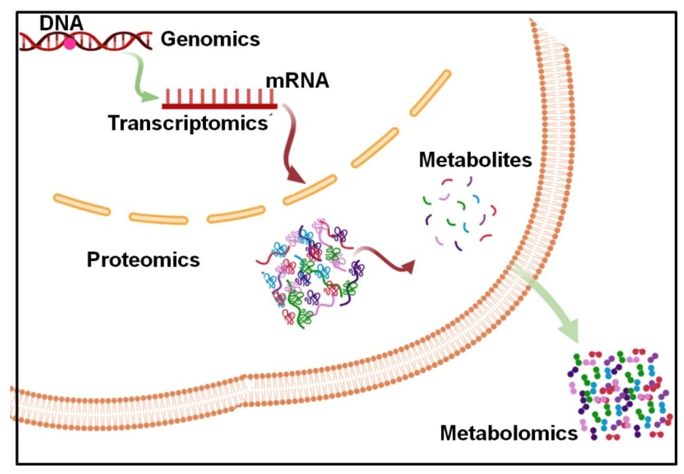
Cancer Metabolism and Metabolites

Cancer cell metabolism is driven by changes in intracellular signaling pathways, which are disrupted by mutations in oncogenes and tumor-suppressor genes. Mutated oncogenes can directly activate cancer cell metabolism, while altered metabolic enzymes can contribute to malignant transformation. Metabolism is an essential energy-producing process that helps maintain cell homeostasis and supports growth and proliferation [5]. Normal cells have intricate signaling networks managed by key regulatory enzymes, which sense environmental signals and control metabolic activities to produce the energy needed for survival in a highly regulated way. When cells proliferate, they activate metabolic pathways to meet the increased demand for adenosine triphosphateneeded for cell division. However, this metabolic increase also produces byproducts, such as reactive oxygen species, which can damage cells and lead to DNA mutations [6]. Consequently, changes in cell metabolism can initiate tumorigenesis. Mutations in oncogenes and tumor-suppressor genes can alter multiple signaling pathways, thereby reshaping cell metabolism to support the tumor formation process. These changes enable cells to adapt to the metabolic demands of cancer, with some metabolic alterations playing an essential role in malignant transformation [7].
In recent years, numerous studies on mitochondrial function in cancer cells have highlighted that the Warburg effect is more closely tied to changes in signaling pathways regulating glucose uptake and utilization than to mitochondrial defects. This renewed focus on the Warburg effect in cancer research underscores the importance of understanding aerobic glycolysis in cancer cells. The combination of oncogene and tumor suppressor mutations, a hypoxic microenvironment, mtDNA mutations, and other factors contribute to the metabolic shifts seen in tumors [8]. Gaining deeper insights into cancer cell energy metabolism can pave the way for innovative diagnostic and therapeutic strategies. This review will explore the metabolic characteristics of tumor cells, examining altered enzymatic activities involved in aerobic glycolysis and current approaches targeting metabolic pathways for cancer treatment

2. Literature Search Strategy

This narrative review was conducted to comprehensively evaluate the role of metabolomics and molecular imaging in bladder cancer biomarker discovery and diagnostics. A structured literature search was performed using the PubMed, Scopus, and Web of Science databases to identify relevant peer-reviewed studies. The search covered publications from different time sets to ensure the inclusion of both foundational and recent advancements in the field. Peer-reviewed original research articles, systematic reviews, and meta-analyses focusing on metabolomics and imaging-based biomarker discovery in bladder cancer included. Studies conducted on human subjects, in vitro models, and preclinical animal models relevant to bladder cancer diagnostics were included. Articles published in English to maintain consistency and accessibility of data were included. The selected literature was critically analyzed to extract insights into biomarker identification, metabolic pathway alterations, and the integration of imaging with metabolomics for improved diagnostic accuracy. This comprehensive approach ensures that the review provides a balanced synthesis of existing knowledge while highlighting future directions in precision oncology for bladder cancer.

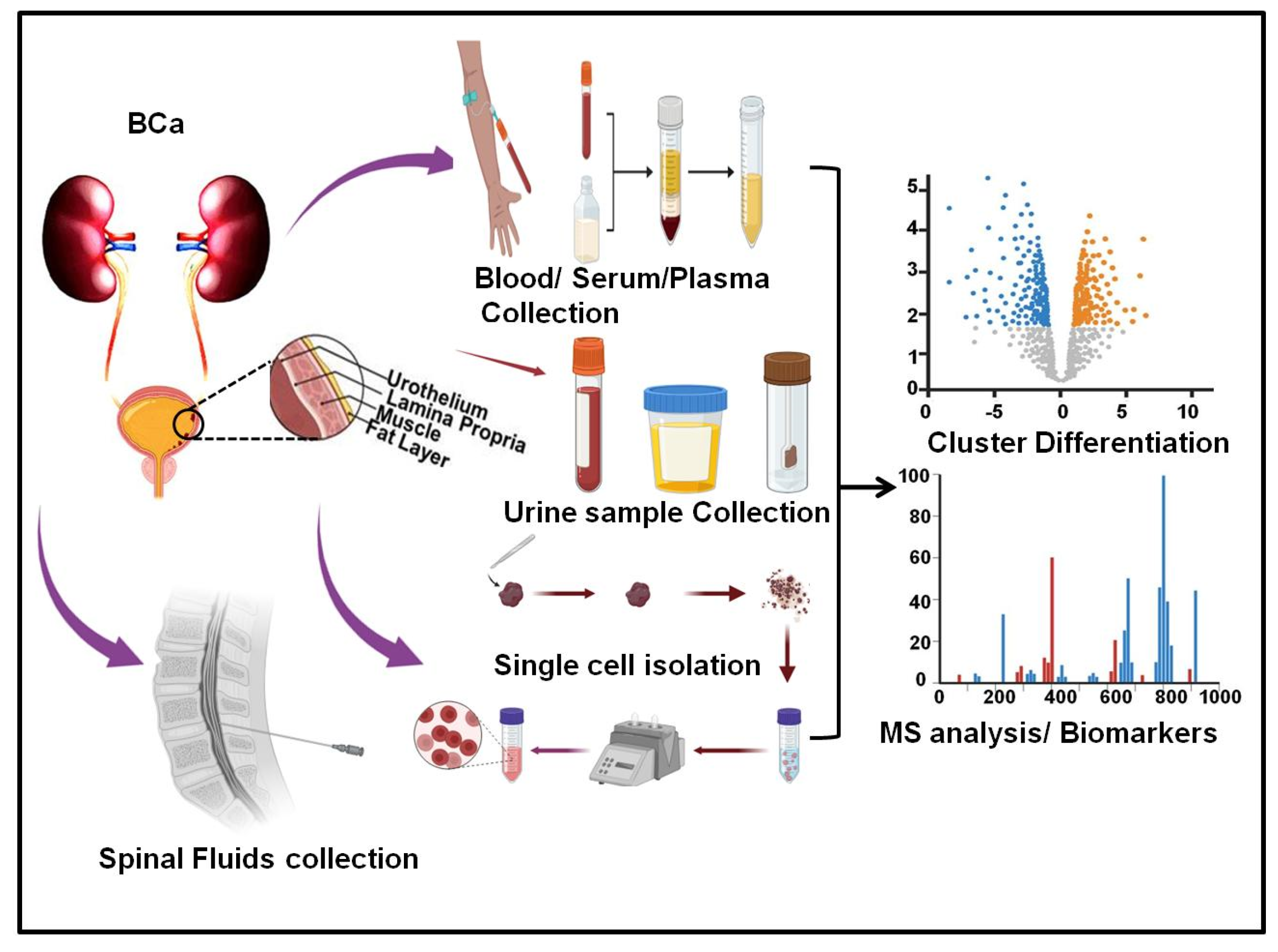
2.1. Serum Metabolic Analysis in Bladder Cancer

To diagnose primary or recurrent bladder cancer (BC), cystoscopy and urine cytology are the standard diagnostic procedures. However, these methods have notable limitations, necessitating the development of a non-invasive, highly sensitive, specific, and convenient diagnostic approach. Mass spectrometry and nuclear magnetic resonance (NMR) have become essential tools for analyzing metabolic profiles, enabling the quantitative and qualitative measurement of small-molecule metabolites. These techniques facilitate a comprehensive and rapid analysis, making metabolomics a powerful approach in cancer research. Metabolomics not only aids in identifying potential biomarkers but also helps uncover the mechanisms underlying cancer pathogenesis [9]. Promising results have been observed in various cancers, including breast, prostate, lung, and liver cancer. A study identified metabolic signatures related to glucose, the tricarboxylic acid cycle, lipids, amino acids, and nucleotide pathways by profiling the global metabolome using GC-MS and LC-MS. This study revealed significant pathway alterations between normal urothelium and high-grade urothelial carcinoma at various stages [10].
Advancements in analytical methods, such as reverse-phase high-performance liquid chromatography (RP-HPLC) coupled with triple quadrupole MS, have further enhanced the quantitative determination and validation of BC metabolites [11]. Clinical validation using biosamples from BC patients can be match-controlled to achieve recovery and precision values within regulatory guideline ranges. Multivariate regression analysis confirmed an association between these metabolic profiles and patient survival, highlighting the potential of metabolomics for advancing BC diagnostics and prognostics (Figure 1).
Numerous studies have been conducted to identify serum metabolites as potential biomarkers for early cancer detection and monitoring. A principal component analysis of the metabolome data revealed distinct metabolic profiles between African American and European American BLCA sera [12]. In AA BLCA sera, metabolites such as taurine, glutamine, glutamic acid, and several amino acids were upregulated, while orotate, ketoglutarate, and glyceraldehyde 3-phosphate were downregulated compared to EA BLCA.
These differences were disease-specific, as similar patterns were not observed in healthy AA and EA controls. Pathway analysis linked these metabolic changes to alterations in aspartate, alanine, glutamate, and lysine degradation pathways, as well as purine catabolism and tryptophan, serine, and fatty acid metabolism [13]. Another study investigated this, >300 metabolites were targeted, and 190 metabolites spanning various classes (amino acids, free fatty acids, tricarboxylic acid cycle intermediates, glycolysis/PPP metabolites, and nucleic acids/nucleotides) were detected in serum samples [14].
A serum metabolic profiling study identified 40 differential metabolites in smoker BC patients, including elevated levels of amino acids such as tyrosine, phenylalanine, proline, serine, valine, isoleucine, glycine, and asparagine, as well as taurine. An integromic analysis combining metabolomic and transcriptomic data revealed 17 intersecting genes significantly associated with survival in smoker BC patients, with catechol-O-methyltransferase, iodotyrosine deiodinase, and tubulin tyrosine ligase emerging as key contributors [15].
In a serum metabolic profiling study using GC-MS and LC-MS, 328 and 9536 peaks were detected in positive and negative ionization modes, respectively, identifying 305 and 3324 metabolites across 120 serum samples. The OPLS-DA was employed, and highlighting significant metabolic differences between groups, used to create volcano plots. Results indicated notable variations between the pathological complete response(pCR) and non-pCR groups, which were further confirmed by the hierarchical clustering analysis of significantly altered metabolites.
A total of 100 differential metabolites was analyzed using the KEGG database, revealing 40 significantly enriched metabolic pathways from another study. The primary pathways linked to treatment response included central carbon metabolism involving glycine, alanine, asparagine, and glutamine, protein digestion and absorption, aminoacyl-tRNA biosynthesis, and D-amino acid metabolism, suggesting their role in the efficacy of the TCbHP regimen. The top 20 enriched pathways for upregulated metabolites were dominated by central carbon metabolism and amino acid biosynthesis, while downregulated metabolites were linked to pathways such as lysosome function, apoptosis, and ascorbate metabolism. These findings underscore the complex metabolic changes associated with a response to TCbHP treatment [16].
Notably, 1H NMR-based metabolomic analysis helps to identify distinct serum metabolic profiles in BC patients compared to urinary tract calculi patients and healthy subjects. Significant differences were observed between low-grade (LBC) and high-grade (HBC) BC patients and between pre- and post-transurethral resection of bladder tumor (TURBT) patients. BC patients exhibited decreased levels of metabolites such as isoleucine/leucine, tyrosine, phenylalanine, lactate, glycine, and citrate, alongside increased levels of lipids (V/LDL), glucose, and acetoacetate. Notably, alterations in metabolites such as glucose, tyrosine, and phenylalanine correlated with the metastatic status of BC. These metabolite changes were specific to BC and not observed in calculi patients, highlighting their disease relevance. Post-TURBT metabolic profiles showed partial recovery toward those of healthy subjects, reflecting the treatment response. The study also revealed disrupted metabolic pathways in BC, including aromatic amino acid metabolism, glycolysis, the citrate cycle, and lipogenesis. Elevated lipogenesis, characterized by increased lipid and ketone body levels, was linked to tumor growth and development. These findings suggest that specific metabolite alterations, particularly in amino acids and lipid metabolism, could serve as potential biomarkers for non-invasive BC diagnosis and monitoring. Overall, the results underscore the utility of metabolomic profiling in understanding BC pathogenesis and highlight the potential of serum metabolites as biomarkers to improve detection, surveillance, and therapeutic strategies [17]. From the observed literature, blood serum samples from patients with low-grade and high-grade bladder cancer (BC) and healthy controls, identifying six significantly altered metabolites: dimethylamine, malonate, lactate, glutamine, histidine, and valine. Notably, a double-blind study involving 106 patients with suspected BC validated the diagnostic utility of these metabolites for early BC detection [18].

2.2. Urine Metabolic Analysis in Bladder Cancer

Detecting the presence, recurrence, and progression of diseases, as well as identifying optimal treatments for specific cancer types, has been a central focus of research in recent years. Biomarkers found in liquid biopsies play a pivotal role in this context, offering real-time monitoring of disease states through non-invasive methods. Liquid biopsy samples, including urine, serum, plasma, saliva, cerebrospinal fluid, and pleural fluid, enable the effective prognosis of advanced diseases. In bladder cancer, serum and urine are the most commonly utilized liquid biopsy specimens. Given the altered metabolism observed in cancers, analyzing metabolites in these specimens provides valuable insights into the disease state [19].
A Venn diagram (to identify two or more sets of samples) provides selected results from the OPLS-DA model and univariate analysis. In the overlapping region, metabolites met the predefined criteria of VIP with >1 and FC > 0, qualifying them as potential metabolic biomarkers (Figure 2). Based on the relative abundance of differential metabolites, pathway enrichment analysis using the Small-Molecule Pathway Database (SMPDB) identified several metabolic pathways. Among those, seven pathways—phenylacetate metabolism, propanoate metabolism, fatty acid metabolism, pyruvate metabolism, arginine and proline metabolism, glycine and serine metabolism, and bile acid biosynthesis—were significantly enriched, each containing at least two annotated metabolites [20].
Lin et al. conducted a comparative proteomic analysis of urinary exosomes in 10 healthy controls and 10 age-matched bladder cancer patients, comparing the exosome-derived proteome with the overall urine proteome. Using MS-based label-free proteomics, they identified 1222 proteins, of which 56 showed significantly elevated levels in the urinary exosomes of bladder cancer patients. This study highlights the potential of urinary exosomes as a promising source for enriching and identifying bladder cancer protein biomarkers [21]. Chen et al. conducted comparative and targeted proteomic analyses of urinary microparticles from nine hernia patients (control) and patients with bladder cancer. Their research identified 107 differentially expressed proteins, and they achieved the precise quantification of 29 proteins including peptides using LC−MRM/MS in 48 urine samples from bladder cancer, hernia, and urinary tract infection/hematuria cases. Significant changes in the concentrations of 24 proteins were observed between bladder cancer and hernia, with area-under-the-curve values ranging from 0.702 to 0.896. The study also quantified tumor-associated calciumsignal transducer 2 (TACSTD2) in raw urine samples using a commercial ELISA, confirming its potential utility in diagnosing bladder cancer. This study underscores the strong association between TACSTD2 and bladder cancer and emphasizes the potential of urinary microparticles as a non-invasive diagnostic tool [22]. Proteomic analysis of urinary exosomes (EVs) and tumor-derived exosomes (Te-EVs) revealed that the majority of proteins identified in Te-EVs were also present in urinary EVs. A novel approach combining the proteomic analysis of both types of exosomes led to the identification of reliable urinary EV biomarker proteins (HSP90, SDC1, and MARCKS) for bladder cancer (BCa) detection. These BCa-specific EV proteins have the potential to serve as both biomarkers and therapeutic targets. Future research will focus on developing a high-throughput detection system for these BC biomarker EV proteins to facilitate clinical applications and further investigate their functional roles [23].
A selection of metabolites with high accuracy, sensitivity, and specificity was identified, including from urine sample with high sensitivity and specificity include benzenepropanoic acid, Desaminotyrosine, 3,8-dioxa-2,9-disiladecane, and d-ribose. Other notable metabolites included ribitol, d-fructose, and d-mannose, all showing good predictive characteristics with anaccuracy > 0.80 and sensitivity and specificity > 0.80. The data suggest that these identified compounds could be valuable in differentiating bladder cancer from control conditions [24]. Leucine was found to be upregulated in both urine and tissue but downregulated in serum. In contrast, lysine exhibited inconsistent regulation, being downregulated in urine in two studies while upregulated in one [25]. For non-essential amino acids, serine displayed inconsistent regulation in urine, with one study reporting downregulation and another indicating upregulation [26]. Cysteine was consistently downregulated in urine. Aspartic acid showed upregulation in both urine and tissue [27]. Proline exhibited variable regulation without a clear trend in urine and tissue [25].

2.3. Tissue Metabolic Analysis in Bladder Cancer

Proteomic profiling was conducted using a super-SILAC strategy, which used a common heavy-labeled internal reference for accurate protein comparisons across samples. A total of 15,714 proteins were quantified, and 3781 were filtered based on strict criteria. These proteins showed a broad range of characteristics, including varied isoelectric points, molecular weights, solubility, and diverse subcellular localizations [28]. This finding was supported by pathway analysis and provided an insight into how FGFR3 mutations could make tumors more susceptible to proapoptotic triggers, which could be enhanced by agents like birinapant. This research contributes significant proteomic, genomic, and transcriptomic insights that refine bladder cancer classifications and highlight potential protein markers for subtype identification. This study also suggests that FGFR3 alterations could be targeted for therapeutic strategies involving TRAIL sensitization, complementing existing anti-FGFR treatments and addressing the challenge of treatment resistance in clinical settings [29].In metabolite detection, one study reported the upregulation of lactic acid in tissue, highlighting the need for further research into its tissue-specific regulation [30]. Valine, as one of the metabolites, demonstrated more consistent regulation in tissue [31]. Aspartic acid exhibited upregulation in tissue [32]. Proline showed variable regulation without a distinct trend in urine and tissue [25].
The primary variation across data sets was due to the preservation method, affecting protein intensities observed in MS analysis. This suggests that data from Formalin-Fixed, Paraffin-Embedded (FFPE) and Optimal Cutting Temperature compound (OCT) samples should not be combined for accurate comparisons. The OCT-preserved samples allowed for the analysis of more proteins compared to FFPE, but the OCT data were more dispersed. FFPE samples, on the other hand, exhibited better clustering of data, particularly when comparing different tumor stages. Nine cancer-related proteins showed similar deregulation across different tumor stages in both FFPE and OCT methods, with Galectin-1 (LGALS1) being notably upregulated in T2/T3 tumors [33]. This finding was consistent with independent validation, where higher cytoplasmic IHC staining was observed in high-stage tumors compared to low-stage tumors. The analysis identified proteins such as TGFBI, ANXA5, TPT1, FN1, CORO1C, LGALS1, PGM1, MAOA, ETFB, ACTB, FTH1, ANXA1, ACTN1, SERPINH1, CAT, CAP1, ACTN4, and AKR1C1, underscoring the potential for discovering biomarkers linked to different cancer stages [34].
Metabolomics not only holds promise for cancer diagnosis but also plays a crucial role in guiding oncologic surgeries. Rapid ionization mass spectrometry with electrosurgical tools can be used to assess tumor margins in during treatemnt. This innovative approach enables simultaneous metabolomic analysis and surgical intervention, providing live feedback to enhance precision (Table 1). However, its widespread adoption is limited by high costs. Beyond its diagnostic applications in oncology, the correlation between pre-treatment serum phenylalanine levels and survival outcomes in metastatic breast cancer highlights the potential of metabolomics for prognostic evaluation [35].

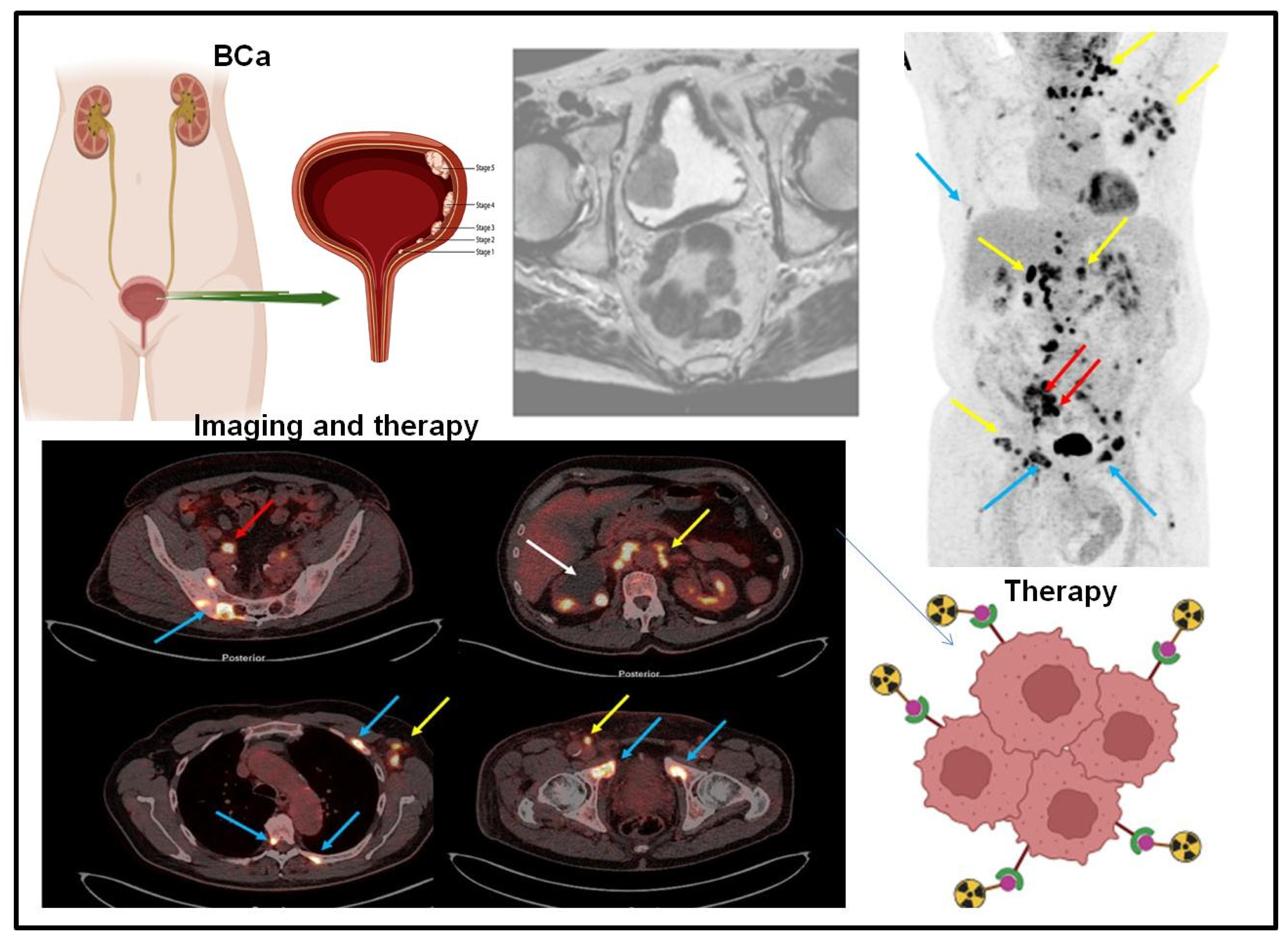
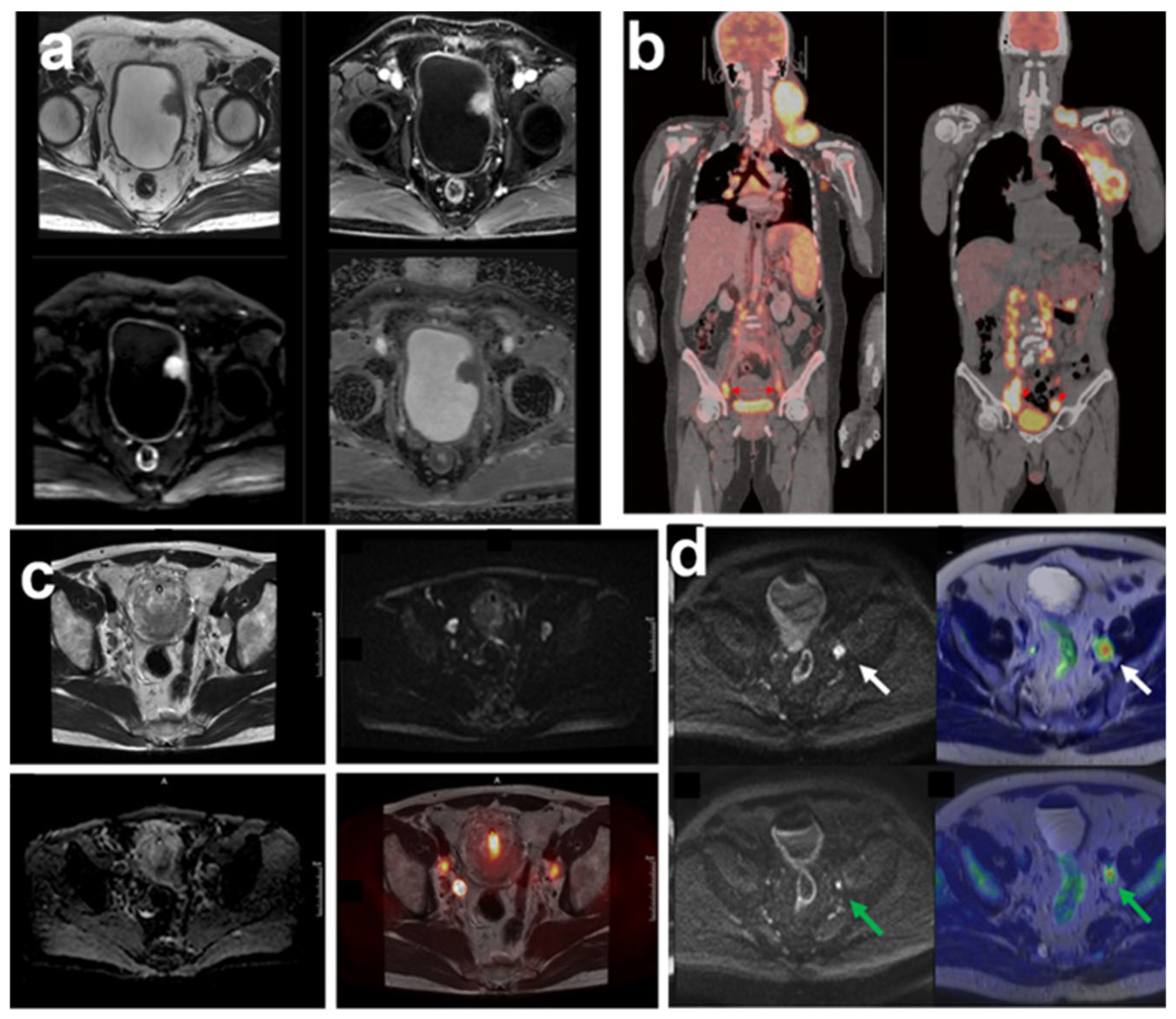
3. Targeted Molecular Imaging of Bladder Cancer

The primary method for evaluating T-staging of urinary bladder tumors is the transurethral resection of the bladder (TURB). The diagnostic accuracy for T-staging is reported to be 62% to 85% for MRI and 35% to 55% for CT, with MRI demonstrating superior performance [42]. Urothelial tumors are typically FDGavid with high FDG uptake, and in some cases, primary tumors in the urinary tract and bladder can be detected, particularly if FDG activity in the urine is diluted. To address this challenge, several methods have been investigated to reduce urine FDG activity, including bladder irrigation, catheterization, FDG wash-out, dual-phase imaging, early PET imaging, late PET imaging after voiding, oral hydration, and forced diuresis [43]. A retrospective review was conducted on 62 consecutive patients with MIBC who underwent a first FDG-PET/CT between five years. After NAIC, these patients underwent a second FDG-PET/CT, followed by radical cystectomy. Patients with no hypermetabolism in the bladder and lymph nodes on the second FDG-PET/CT were classified as metabolic complete responders, while those with no evidence of residual disease on histopathology were considered pathologic complete responders. The accuracy of the second FDG-PET/CT in distinguishing complete responders from patients with residual disease was evaluated using histopathology as the gold standard [44].
Molecular imaging in clinical practice primarily relies on PET, SPECT, and MRS. PET and SPECT utilize various radiotracers for medical imaging, particularly in oncology, to visualize metabolic and functional processes in tumors. MRS, a specialized MRI technique, detects changes in proton/nuclei excitation and relaxation, enabling the assessment of metabolite levels, including choline, pyruvate, lactate, lipids, and polyamines. Advanced MRS techniques, such as 1H, 19F, 31P, and 13C MRS, have been developed and extensively reviewed in the literature. Clinically, MRS plays a significant role in oncology, offering insights into tumor metabolism, progression, and treatment response (Figure 3).
Imaging with PET/CT provides greater accuracy in staging bladder cancer by identifying localized, nodal, and metastatic disease. Approximately 26.3% of patients experience a change in management decisions based on PET/CT results.
Sensitivity and specificity for detecting pelvic lymph nodes were 54.3% and 98.9%, while for metastasis, they were 76.8% and 96.9%, respectively [46,47]. Assessing the accuracy and prognostic significance of CT-based lymph node (LN) staging in variant histologies of bladder cancer, the overall accuracy of CT for detecting LN metastases in variant histologies was 62%, with a sensitivity of 46% and a specificity of 70%.For urothelial and non-urothelial variants, accuracies were 67% and 57%, respectively. A loss of fatty hilum and an increased regional LN number were significant prognostic factors for poor cancer-specific survival (CSS) and overall survival (OS). Patients with LN metastases showed significantly worse survival compared to LN-negative patients, highlighting LN status as a critical prognostic marker [48].
The diagnostic accuracy of multiparametric MRI (mp-MRI) was 88%, outperforming DW-MRI (82%), DCE-MRI (74%), and high-resolution T2W-MRI (52%) in differentiating non-muscle-invasive from muscle-invasive tumors and organ-confined from non-organ-confined tumors (Figure 4). Agreement between mp-MRI and histopathological staging (κ = 0.679) was higher than for DW-MRI (κ = 0.566), DCE-MRI (κ = 0.566), or T2W-MRI (κ = 0.274). Overstaging rates were reduced from 48% with T2W-MRI, 26% with DCE-MRI, and 18% with DW-MRI to 12% with mp-MRI [49]. 18F-FDG PET/MRI shows promise in diagnosing bladder cancer (BLCA), but its role in staging and evaluating neoadjuvant therapy (NAT) response remains unclear. This retrospective study of 40 BLCA patients assessed 18F-FDG PET/MRI and its parameters for tumor staging and NAT response prediction. Results indicate good performance in both areas, with ΔSUVmean emerging as a key parameter for predicting NAT response. Overall, 18F-FDG PET/MRI demonstrated potential as a valuable tool in BLCA diagnosis and treatment planning [46]. FDG-PET/CT demonstrated high sensitivity with 95% for overall disease, 100% for the primary tumor, and 97% for lymph nodes, though specificity ranged from 30% to 42%. These findings highlight FDG-PET/CT as a valuable tool for distinguishing complete responders from those with residual disease, enabling the early identification of patients who may need alternative treatment pathways following NAIC [44]. Hybrid SPECT/CT enabled the detection of all sentinel nodes, outperforming planar lymphoscintigraphy and gamma-probe techniques. A total of 21 sentinel nodes were identified across six patients, including metastatic and non-metastatic nodes. SPECT/CT detected SLNs missed by conventional methods, particularly smaller nodes and anatomically challenging locations. Hybrid SPECT/CT enhanced SLN detection accuracy, offering valuable insights into metastatic spread [50].
The majority of bladder SUCs were located in the trigone and displayed large tumor size, irregular shape, low ADC values, VI-RADS scores ≥ 4, necrosis, and invasive characteristics. Univariate analysis identified significant differences between SUCs and CUCs in tumor location, shape, LAD, SAD, ADC value, VI-RADS score, necrosis, extravesical extension, pelvic peritoneal spread, and hydronephrosis/ureteral effusion. Multinomial logistic regression highlighted SAD and necrosis as independent predictors, with the combined model achieving an AUC of 0.849 in ROC analysis [51]. Kim and colleagues conducted a meta-analysis to assess the diagnostic accuracy of 11C-choline and 11C-acetate in lymph node staging for BCa, reporting pooled sensitivity and specificity values of 66% and 89%, respectively [52]. Notably, 11C-choline and 11C-acetate PET/CT demonstrated low sensitivity but moderate specificity in detecting metastatic lymph nodes in patients with bladder cancer (BCa). The utility of 11C-acetate for staging is particularly limited by a high rate of false-positive uptake, often caused by inflammation or infection, especially in patients who have undergone prior intravesical Bacillus Calmette–Guérin (BCG) therapy [53]. [68Ga]FAPI-PET/CT can show promise as a diagnostic radioligand in bladder cancer, with an initial analysis of FAP-ligand with superiority over [18F]FDG in a small cohort of patients [54]. 68Ga-FAP-2286 PET can detect metastatic lymph nodes smaller than 1 cm and may offer greater specificity than 18F-FDG PET. However, these findings require validation through prospective studies involving larger patient cohorts [55]. Dual-isotope studies using 177Lu or 177Lu/125Ifor tumor targeting of radiolabeled MMP-2/9 ACPPs suggest that activation primarily occurs in the vascular compartment rather than being tumorspecifics [56].VEGF exhibits moderate to high expression levels in both primary tumors and lymph node metastases, making it a promising candidate for imaging modalities targeting urothelial carcinoma [57]. In bladder cancer, CK20 and BCL2 show positivity rates of 53.3% and 10.0%, respectively. Both markers serve as prognostic indicators for tumor grading, with lower BCL2 expression and higher CK20 expression correlating with higher tumor grades [58].
These biomarkers serve as non-invasive diagnostic tools for detecting and monitoring bladder cancer. However, despite their clinical utility, they may exhibit limitations in sensitivity and specificity. As a result, they are commonly used alongside other diagnostic approaches, such as cystoscopy, to ensure a more comprehensive assessment. Furthermore, the FDA has granted Breakthrough Therapy Designation to the BCa Assay, a quantitative urine-based test designed to detect bladder cancer and upper tract urothelial carcinoma. This designation is intended to accelerate the assay’s development and review, recognizing its potential to enhance diagnostic accuracy. While FDA-approved urinary biomarkers play a crucial role in bladder cancer management, their optimal effectiveness is achieved when integrated with conventional diagnostic methods (Table 2).

4. Integration of Mass Spectrometry and Molecular Imaging for Bladder Cancer Biomarker Detection as Advancing Precision Oncology

Bladder cancer is a significant clinical challenge with varying degrees of aggressiveness and outcomes. The early detection and precise characterization of bladder cancer are crucial for guiding personalized treatment strategies. Over the years, the integration of advanced analytical techniques, such as mass spectrometry and molecular imaging, has significantly enhanced the ability to detect and monitor bladder cancer biomarkers. Mass spectrometry is an analytical technique used to measure the mass and composition of molecules. It is particularly valuable in proteomics, metabolomics, and lipidomics. In bladder cancer research, MS can identify and quantify specific peptides, proteins, and other biomolecules that are indicative of cancer progression. MS can detect differential protein expression profiles associated with bladder cancer subtypes. For example, specific proteins like CK20, BCL2, and MMP-2/9 have been studied for their roles in cancer progression. MS facilitates the identification of modifications such as phosphorylation, glycosylation, and ubiquitination, which are critical in tumor signaling pathways. Molecular imaging focuses on visualizing the molecular and cellular aspects of cancer through imaging techniques. This includes modalities like PET, MRI, CT, optical imaging, and SPECT. In bladder cancer, molecular imaging aims to capture real-time data on tumor metabolism, receptor binding, and the expression of specific markers. PET and MRI are used to visualize cancer cell activity and tumor heterogeneity. For instance, imaging techniques are applied for detecting metabolic activity and specific cellular markers. Longitudinal imaging allows for monitoring biomarker expression over time and assessing treatment responses. The combination of MS and molecular imaging offers a comprehensive approach to cancer biomarker detection. While MS excels in the molecular characterization of biomarkers, molecular imaging provides spatial and functional insights into cancerous tissues. Together, they complement each other by combining high-resolution molecular analysis with in vivo visualization of disease processes. MS identifies novel protein markers, while molecular imaging verifies their localization and activity within tumors. Combining both techniques allows for a thorough understanding of how tumors respond to therapies, leading to better therapeutic strategies.
Limitations: MS requires high-quality samples and stringent protocols for accurate biomarker detection. Both MS and molecular imaging technologies are expensive and may limit their use in resource-limited settings. High-dimensional data generated from both techniques require advanced bioinformatics tools for analysis. Consistency in sample processing and imaging protocols is critical for reproducibility of results. Molecular imaging often requires advanced interpretation that may vary between different centers. Although promising, findings need validation in larger patient cohorts and longitudinal studies.
Several commercially available platforms facilitate high-throughput multi-omics analyses, enabling integrated investigations of metabolomics, proteomics, and genomics. For instance, Metabolon® and Biocrates® offer standardized mass spectrometry-based metabolomics kits for biomarker discovery. Olink® and SomaScan® utilize proteomic profiling technologies to assess protein biomarkers at a large scale. These platforms enhance reproducibility and allow for comprehensive molecular profiling in cancer research. Their application in bladder cancer has the potential to improve early diagnosis, treatment stratification, and therapy monitoring.

5. Future Directions in the Integration of Mass Spectrometry and Molecular Imaging for Bladder Cancer Biomarker Detection

The integration of mass spectrometry and molecular imaging is at the forefront of cancer research, offering new insights into tumor biology and personalized treatment strategies. While substantial progress has been made, several key areas remain to be explored to fully realize the potential of these technologies. One of the most promising future directions is the integration of multi-omics data. By combining genomics, transcriptomics, proteomics, metabolomics, and imaging, a more comprehensive understanding of bladder cancer can be achieved. This holistic approach provides detailed molecular information while molecular imaging visualizes these biomarkers in vivo. Systems-level integration can identify complex interactions between various biomarkers and tumor microenvironments, revealing the underlying mechanisms of cancer progression and resistance. The integration of MS and molecular imaging can significantly advance personalized medicine in bladder cancer treatment. The ability to tailor diagnostic and therapeutic approaches based on specific biomarker profiles is key to improving patient outcomes. Future efforts should focus on utilizing MS to detect unique biomarker signatures and leveraging molecular imaging to assess tumor responses in real time, ensuring tailored treatment strategies. Additionally, molecular imaging should be used for continuous biomarker assessment throughout treatment, while MS can analyze metabolic changes and therapeutic targets.
Current bladder cancer biomarkers present challenges in early detection due to the limited sensitivity and specificity of mass spectrometry and cystoscopy in molecular imaging. Future research aims to integrate mass spectrometry with molecular imaging to enhance sensitivity, specificity, and non-invasive diagnostic approaches. Techniques such as REIMS-associated endoscopy and spatial metabolomics hold significant potential in this regard. Additionally, gas chromatography–mass spectrometry (GC-MS) and electronic signal noise technologies show promise in detecting bladder cancer through urinary volatile organic compounds. While molecular imaging techniques like PET/CT are valuable for staging, restaging, and assessing responses to neoadjuvant chemotherapy, further advancements are needed to address technical limitations. Real-time, non-destructive tissue analysis during endoscopy could improve diagnostic accuracy, and combining multiple imaging modalities with mass spectrometry data may offer a more comprehensive understanding of the disease.

6. Conclusions

The integration of metabolomics and molecular imaging represents a significant advancement in bladder cancer research, enabling precise diagnostics, prognostic assessments, and tailored therapies. Metabolomics detects subtle metabolic alterations, while molecular imaging provides spatial and functional insights into tumor biology. Leveraging advanced platforms like LC-MS and PET/MRI enhances biomarker discovery across serum, urine, and tissue, offering a deeper understanding of cancer progression and treatment response. Despite challenges such as standardization, high costs, and the need for clinical validation, these technologies hold great potential for clinical translation. Future efforts should focus on multi-omics integration, large-scale validation, and cost-effective diagnostic solutions. The application of AI-driven analysis will further refine biomarker identification and clinical decision-making. By addressing these challenges, metabolomics and imaging can drive precision oncology, improving bladder cancer detection and treatment.

Funding

This research received no external funding.

Conflicts of Interest

The author declare no competing interests.

References

  1. Wang, X.; Dai, C.; Wu, Y.; Liu, Y.; Wei, D. Molecular-electromechanical system for unamplified detection of trace analytes in biofluids. Nat. Protoc. 2023, 18, 2313–2348. [Google Scholar] [CrossRef] [PubMed]
  2. Diaz, P.M.; Leehans, A.; Ravishankar, P.; Daily, A. Multiomic Approaches for Cancer Biomarker Discovery in Liquid Biopsies: Advances and Challenges. Biomark. Insights 2023, 18, 11772719231204508. [Google Scholar] [CrossRef]
  3. Bai, J.W.; Qiu, S.Q.; Zhang, G.J. Molecular and functional imaging in cancer-targeted therapy: Current applications and future directions. Signal Transduct. Target. Ther. 2023, 8, 89. [Google Scholar] [CrossRef]
  4. Rowe, S.P.; Pomper, M.G. Molecular imaging in oncology: Current impact and future directions. CA Cancer J. Clin. 2022, 72, 333–352. [Google Scholar] [CrossRef] [PubMed]
  5. Yang, J.; Shay, C.; Saba, N.F.; Teng, Y. Cancer metabolism and carcinogenesis. Exp. Hematol. Oncol. 2024, 13, 10. [Google Scholar]
  6. Pavlova, N.N.; Zhu, J.; Thompson, C.B. The hallmarks of cancer metabolism: Still emerging. Cell Metab. 2022, 34, 355–377. [Google Scholar] [PubMed]
  7. Zhao, J.; Jin, D.; Huang, M.; Ji, J.; Xu, X.; Wang, F.; Zhou, L.; Bao, B.; Jiang, F.; Xu, W.; et al. Glycolysis in the tumor microenvironment: A driver of cancer progression and a promising therapeutic target. Front. Cell Dev. Biol. 2024, 12, 1416472. [Google Scholar]
  8. Zhong, X.; He, X.; Wang, Y.; Hu, Z.; Huang, H.; Zhao, S.; Wei, P.; Li, D. Warburg effect in colorectal cancer: The emerging roles in tumor microenvironment and therapeutic implications. J. Hematol. Oncol. 2022, 15, 160. [Google Scholar] [CrossRef]
  9. Jia, K.; Wang, Y.; Jiang, L.; Lai, M.; Liu, W.; Wang, L.; Liu, H.; Cao, X.; Li, Y.; Nie, Z. Urine Metabolic Profiling for Rapid Lung Cancer Screening: A Strategy Combining Rh-Doped SrTiO3-Assisted Laser Desorption/Ionization Mass Spectrometry and Machine Learning. ACS Appl. Mater. Interfaces 2024, 16, 12302–12309. [Google Scholar] [CrossRef]
  10. Khmelevskaya, E.A.P.E.S.; Buiko, E.E.; Ufandeev, A.A.; Kaydash, O.A.; Ivanov, V.V.; Baykov, A.N.; Parochkina, E.V.; Udut, E.V. Precision Medicine in Oncology: The Role and Prospects of Mass Spectrometry. Bull. Sib. Med. 2024, 23, 162–182. [Google Scholar] [CrossRef]
  11. Zhao, Y.; Sun, W.; Ji, Z.; Liu, X.; Qiao, Y. Serum metabolites as early detection markers of non-muscle invasive bladder cancer in Chinese patients. Front. Oncol. 2023, 13, 1061083. [Google Scholar] [CrossRef] [PubMed]
  12. Vieira de Sousa, T.; Guedes de Pinho, P.; Pinto, J. Metabolomic Signatures of Treatment Response in Bladder Cancer. Int. J. Mol. Sci. 2023, 24, 17543. [Google Scholar] [CrossRef]
  13. Vantaku, V.; Donepudi, S.R.; Piyarathna, D.W.B.; Amara, C.S.; Ambati, C.R.; Tang, W.; Putluri, V.; Chandrashekar, D.S.; Varambally, S.; Terris, M.K.; et al. Large-scale profiling of serum metabolites in African American and European American patients with bladder cancer reveals metabolic pathways associated with patient survival. Cancer 2019, 125, 921–932. [Google Scholar] [CrossRef]
  14. Amara, C.S.; Ambati, C.R.; Vantaku, V.; Badrajee Piyarathna, D.W.; Donepudi, S.R.; Ravi, S.S.; Arnold, J.M.; Putluri, V.; Chatta, G.; Guru, K.A.; et al. Serum Metabolic Profiling Identified a Distinct Metabolic Signature in Bladder Cancer Smokers: A Key Metabolic Enzyme Associated with Patient Survival. Cancer Epidemiol. Biomark. Prev. 2019, 28, 770–781. [Google Scholar] [CrossRef]
  15. Akand, M.; Jatsenko, T.; Muilwijk, T.; Gevaert, T.; Joniau, S.; Van der Aa, F. Deciphering the molecular heterogeneity of intermediate- and (very-)high-risk non-muscle-invasive bladder cancer using multi-layered -omics studies. Front. Oncol. 2024, 14, 1424293. [Google Scholar] [CrossRef] [PubMed]
  16. Zhang, N.; Huang, Y.; Wang, G.; Xiang, Y.; Jing, Z.; Zeng, J.; Yu, F.; Pan, X.; Zhou, W.; Zeng, X. Metabolomics assisted by transcriptomics analysis to reveal metabolic characteristics and potential biomarkers associated with treatment response of neoadjuvant therapy with TCbHP regimen in HER2 + breast cancer. Breast Cancer Res. 2024, 26, 64. [Google Scholar] [CrossRef] [PubMed]
  17. Cao, M.; Zhao, L.; Chen, H.; Xue, W.; Lin, D. NMR-based Metabolomic Analysis of Human Bladder Cancer. Anal. Sci. Int. J. Jpn. Soc. Anal. Chem. 2012, 28, 451–456. [Google Scholar] [CrossRef]
  18. Bansal, N.; Gupta, A.; Mitash, N.; Shakya, P.S.; Mandhani, A.; Mahdi, A.A.; Sankhwar, S.N.; Mandal, S.K. Low- and high-grade bladder cancer determination via human serum-based metabolomics approach. J. Proteome Res. 2013, 12, 5839–5850. [Google Scholar]
  19. Lodewijk, I.; Dueñas, M.; Rubio, C.; Munera-Maravilla, E.; Segovia, C.; Bernardini, A.; Teijeira, A.; Paramio, J.M.; Suárez-Cabrera, C. Liquid Biopsy Biomarkers in Bladder Cancer: A Current Need for Patient Diagnosis and Monitoring. Int. J. Mol. Sci. 2018, 19, 2514. [Google Scholar] [CrossRef]
  20. Wang, R.; Kang, H.; Zhang, X.; Nie, Q.; Wang, H.; Wang, C.; Zhou, S. Urinary metabolomics for discovering metabolic biomarkers of bladder cancer by UPLC-MS. BMC Cancer 2022, 22, 214. [Google Scholar] [CrossRef]
  21. Lee, J.; McKinney, K.Q.; Pavlopoulos, A.J.; Niu, M.; Kang, J.W.; Oh, J.W.; Kim, K.P.; Hwang, S. Altered Proteome of Extracellular Vesicles Derived from Bladder Cancer Patients Urine. Mol. Cells 2018, 41, 179–187. [Google Scholar] [PubMed]
  22. Chen, C.L.; Lai, Y.F.; Tang, P.; Chien, K.Y.; Yu, J.S.; Tsai, C.H.; Chen, H.W.; Wu, C.C.; Chung, T.; Hsu, C.W.; et al. Comparative and targeted proteomic analyses of urinary microparticles from bladder cancer and hernia patients. J. Proteome Res. 2012, 11, 5611–5629. [Google Scholar] [CrossRef] [PubMed]
  23. Tomiyama, E.; Matsuzaki, K.; Fujita, K.; Shiromizu, T.; Narumi, R.; Jingushi, K.; Koh, Y.; Matsushita, M.; Nakano, K.; Hayashi, Y.; et al. Proteomic analysis of urinary and tissue-exudative extracellular vesicles to discover novel bladder cancer biomarkers. Cancer Sci. 2021, 112, 2033–2045. [Google Scholar] [CrossRef]
  24. Lin, J.Y.; Juo, B.R.; Yeh, Y.H.; Fu, S.H.; Chen, Y.T.; Chen, C.L.; Wu, K.P. Putative markers for the detection of early-stage bladder cancer selected by urine metabolomics. BMC Bioinform. 2021, 22, 305. [Google Scholar] [CrossRef]
  25. Loras, A.; Suárez-Cabrera, C.; Martínez-Bisbal, M.C.; Quintás, G.; Paramio, J.M.; Martínez-Máñez, R.; Gil, S.; Ruiz-Cerdá, J.L. Integrative Metabolomic and Transcriptomic Analysis for the Study of Bladder Cancer. Cancers 2019, 11, 686. [Google Scholar] [CrossRef] [PubMed]
  26. Putluri, N.; Shojaie, A.; Vasu, V.T.; Vareed, S.K.; Nalluri, S.; Putluri, V.; Thangjam, G.S.; Panzitt, K.; Tallman, C.T.; Butler, C.; et al. Metabolomic profiling reveals potential markers and bioprocesses altered in bladder cancer progression. Cancer Res. 2011, 71, 7376–7386. [Google Scholar] [CrossRef]
  27. Shen, C.; Sun, Z.; Chen, D.; Su, X.; Jiang, J.; Li, G.; Lin, B.; Yan, J. Developing urinary metabolomic signatures as early bladder cancer diagnostic markers. Omics 2015, 19, 1–11. [Google Scholar] [CrossRef]
  28. Mertens, L.S.; Fioole-Bruining, A.; Vegt, E.; Vogel, W.V.; van Rhijn, B.W.; Horenblas, S. Impact of (18)F-fluorodeoxyglucose (FDG)-positron-emission tomography/computed tomography (PET/CT) on management of patients with carcinoma invading bladder muscle. BJU Int. 2013, 112, 729–734. [Google Scholar]
  29. Groeneveld, C.S.; Sanchez-Quiles, V.; Dufour, F.; Shi, M.; Dingli, F.; Nicolle, R.; Chapeaublanc, E.; Poullet, P.; Jeffery, D.; Krucker, C.; et al. Proteogenomic Characterization of Bladder Cancer Reveals Sensitivity to Apoptosis Induced by Tumor Necrosis Factor-related Apoptosis-inducing Ligand in FGFR3-mutated Tumors. Eur. Urol. 2024, 85, 483–494. [Google Scholar]
  30. Nizioł, J.; Ossoliński, K.; Płaza-Altamer, A.; Kołodziej, A.; Ossolińska, A.; Ossoliński, T.; Krupa, Z.; Ruman, T. Untargeted metabolomics of bladder tissue using liquid chromatography and quadrupole time-of-flight mass spectrometry for cancer biomarker detection. J. Pharm. Biomed. Anal. 2024, 240, 115966. [Google Scholar]
  31. Wittmann, B.M.; Stirdivant, S.M.; Mitchell, M.W.; Wulff, J.E.; McDunn, J.E.; Li, Z.; Dennis-Barrie, A.; Neri, B.P.; Milburn, M.V.; Lotan, Y.; et al. Bladder cancer biomarker discovery using global metabolomic profiling of urine. PLoS ONE 2014, 9, e115870. [Google Scholar] [CrossRef]
  32. Shen, C.; Khonsari, M.M. Numerical optimization of texture shape for parallel surfaces under unidirectional and bidirectional sliding. Tribol. Int. 2015, 82, 1–11. [Google Scholar] [CrossRef]
  33. Schoeb, D.S.; Wollensak, C.; Kretschmer, S.; González-Cerdas, G.; Ataman, C.; Kayser, G.; Dressler, F.F.; Gratzke, C.; Zappe, H.; Miernik, A. Ex-vivo evaluation of miniaturized probes for endoscopic optical coherence tomography in urothelial cancer diagnostics. Ann. Med. Surg. 2022, 77, 103597. [Google Scholar]
  34. Valdés, A.; Bitzios, A.; Kassa, E.; Shevchenko, G.; Falk, A.; Malmström, P.U.; Dragomir, A.; Segersten, U.; Lind, S.B. Proteomic comparison between different tissue preservation methods for identification of promising biomarkers of urothelial bladder cancer. Sci. Rep. 2021, 11, 7595. [Google Scholar]
  35. Tenori, L.; Oakman, C.; Claudino, W.M.; Bernini, P.; Cappadona, S.; Nepi, S.; Biganzoli, L.; Arbushites, M.C.; Luchinat, C.; Bertini, I.; et al. Exploration of serum metabolomic profiles and outcomes in women with metastatic breast cancer: A pilot study. Mol. Oncol. 2012, 6, 437–444. [Google Scholar] [CrossRef] [PubMed]
  36. McGee, E.E.; Zeleznik, O.A.; Balasubramanian, R.; Hu, J.; Rosner, B.A.; Wactawski-Wende, J.; Clish, C.B.; Avila-Pacheco, J.; Willett, W.C.; Rexrode, K.M.; et al. Differences in metabolomic profiles between Black and White women in the U.S.: Analyses from two prospective cohorts. Eur. J. Epidemiol. 2024, 39, 653–665. [Google Scholar] [CrossRef]
  37. Jacyna, J.; Kordalewska, M.; Artymowicz, M.; Markuszewski, M.; Matuszewski, M.; Markuszewski, M.J. Pre- and Post-Resection Urine Metabolic Profiles of Bladder Cancer Patients: Results of Preliminary Studies on Time Series Metabolomics Analysis. Cancers 2022, 14, 1210. [Google Scholar] [CrossRef]
  38. Pinto, J.; Amaro, F.; Lima, A.R.; Carvalho-Maia, C.; Jerónimo, C.; Henrique, R.; Bastos, M.L.; Carvalho, M.; Guedes de Pinho, P. Urinary Volatilomics Unveils a Candidate Biomarker Panel for Noninvasive Detection of Clear Cell Renal Cell Carcinoma. J. Proteome Res. 2021, 20, 3068–3077. [Google Scholar]
  39. Jiang, X.; Gao, F.; Chen, X.; Yu, Y.; Ding, G.; Wu, J. Ultrasensitive and High Reproducible Detection of Urinary Metabolites Using the Tip-Contact Extraction Method Coupled with Negative LDI-MS. J. Proteome Res. 2021, 20, 4022–4030. [Google Scholar]
  40. Zhu, S.; Corsetti, S.; Wang, Q.; Li, C.; Huang, Z.; Nabi, G. Optical sensory arrays for the detection of urinary bladder cancer-related volatile organic compounds. J. Biophotonics 2019, 12, e201800165. [Google Scholar] [CrossRef]
  41. Zhu, S.; Huang, Z.; Nabi, G. Fluorometric optical sensor arrays for the detection of urinary bladder cancer specific volatile organic compounds in the urine of patients with frank hematuria: A prospective case-control study. Biomed. Opt. Express 2020, 11, 1175–1185. [Google Scholar] [CrossRef] [PubMed]
  42. Einerhand, S.M.H.; van Gennep, E.J.; Mertens, L.S.; Hendricksen, K.; Donswijk, M.L.; van der Poel, H.G.; van Rhijn, B.W.G. 18F-fluoro-2-deoxy-D-glucose positron emission tomography/computed tomography in muscle-invasive bladder cancer. Curr. Opin. Urol. 2020, 30, 654–664. [Google Scholar] [CrossRef] [PubMed]
  43. Wondergem, M.; van der Zant, F.M.; Rafimanesh-Sadr, L.; Knol, R.J.J. Effect of forced diuresis during 18F-DCFPyL PET/CT in patients with prostate cancer: Activity in ureters, kidneys and bladder and occurrence of halo artefacts around kidneys and bladder. Nucl. Med. Commun. 2019, 40, 652–656. [Google Scholar] [CrossRef]
  44. Fitoussi, O.; Roche, J.B.; Riviere, J.; Wallerand, H.; Poulain, J.E.; Gordien, P.; Galland, S.; Henriques, B.; Dupin, C.; Vincent, M.; et al. Accuracy of FDG-PET/CT for Response Evaluation of Muscle-Invasive Bladder Cancer following Neoadjuvant or Induction Chemotherapy. Urol. Int. 2023, 107, 239–245. [Google Scholar] [CrossRef]
  45. Bouchelouche, K. PET/CT in Bladder Cancer: An Update. Semin. Nucl. Med. 2022, 52, 475–485. [Google Scholar] [CrossRef] [PubMed]
  46. Li, T.; You, Q.; Zhang, S.; Li, R.; Xie, S.; Li, D.; Ai, S.; Yang, R.; Guo, H. Performance of (18)F-FDG PET/MRI and its parameters in staging and neoadjuvant therapy response evaluation in bladder cancer. iScience 2024, 27, 109657. [Google Scholar] [CrossRef]
  47. Shahait, M.; Abu-Hijlih, R.; Farkouh, A.; Obeidat, S.; Salah, S.; Abdlkadir, A.S.; Al-Ibraheem, A. Fluorodeoxyglucose positron emission tomography (18F-FDG PET)-computed tomography (CT) in the initial staging of bladder cancer: A single institution experience. J. Egypt. Natl. Canc Inst. 2023, 35, 21. [Google Scholar] [CrossRef]
  48. Rodler, S.; Solyanik, O.; Ingenerf, M.; Fabritius, M.; Schulz, G.B.; Jokisch, F.; Volz, Y.; Westhofen, T.; Ebner, B.; Casuscelli, J.; et al. Accuracy and prognostic value of radiological lymph node features in variant histologies of bladder cancer. World J. Urol. 2022, 40, 1707–1714. [Google Scholar] [CrossRef]
  49. Afifi, A.H.; Abdel Maksoud, T.A.S.; El-Noueam, K.I.; Ataa, M.A.; Abdallah, D.M. Multiparametric-MRI as a comprehensive study in evaluation, characterization & local staging of urinary bladder carcinomas. Egypt. J. Radiol. Nucl. Med. 2017, 48, 493–507. [Google Scholar]
  50. Sherif, A.; Garske, U.; Torre, M.d.L.; Thörn, M. Hybrid SPECT-CT: An Additional Technique for Sentinel Node Detection of Patients with Invasive Bladder Cancer. Eur. Urol. 2006, 50, 83–91. [Google Scholar] [CrossRef]
  51. Zhuo, J.; Han, J.; Yang, L.; Wang, Y.; Shi, G.; Yan, Z.; Yang, L.; Han, R.; Huang, F.; Ban, X.; et al. CT and MRI features of sarcomatoid urothelial carcinoma of the bladder and its differential diagnosis with conventional urothelial carcinoma. Cancer Imaging 2024, 24, 102. [Google Scholar] [CrossRef] [PubMed]
  52. Kim, S.J.; Koo, P.J.; Pak, K.; Kim, I.J.; Kim, K. Diagnostic accuracy of C-11 choline and C-11 acetate for lymph node staging in patients with bladder cancer: A systematic review and meta-analysis. World J. Urol. 2018, 36, 331–340. [Google Scholar] [CrossRef]
  53. Schöder, H.; Ong, S.C.; Reuter, V.E.; Cai, S.; Burnazi, E.; Dalbagni, G.; Larson, S.M.; Bochner, B.H. Initial results with (11)C-acetate positron emission tomography/computed tomography (PET/CT) in the staging of urinary bladder cancer. Mol. Imaging Biol. 2012, 14, 245–251. [Google Scholar] [CrossRef]
  54. Novruzov, E.; Dendl, K.; Ndlovu, H.; Choyke, P.L.; Dabir, M.; Beu, M.; Novruzov, F.; Mehdi, E.; Guliyev, F.; Koerber, S.A.; et al. Head-to-head Intra-individual Comparison of [(68)Ga]-FAPI and [(18)F]-FDG PET/CT in Patients with Bladder Cancer. Mol. Imaging Biol. 2022, 24, 651–658. [Google Scholar] [CrossRef] [PubMed]
  55. Koshkin, V.S.; Kumar, V.; Kline, B.; Escobar, D.; Aslam, M.; Cooperberg, M.R.; Aggarwal, R.R.; de Kouchkovsky, I.; Chou, J.; Meng, M.V.; et al. Initial Experience with (68)Ga-FAP-2286 PET Imaging in Patients with Urothelial Cancer. J. Nucl. Med. 2024, 65, 199–205. [Google Scholar] [CrossRef]
  56. van Duijnhoven, S.M.; Robillard, M.S.; Nicolay, K.; Grüll, H. Tumor targeting of MMP-2/9 activatable cell-penetrating imaging probes is caused by tumor-independent activation. J. Nucl. Med. 2011, 52, 279–286. [Google Scholar] [CrossRef] [PubMed]
  57. van der Fels, C.A.M.; Leliveld, A.; Buikema, H.; van den Heuvel, M.C.; de Jong, I.J. VEGF, EGFR and PSMA as possible imaging targets of lymph node metastases of urothelial carcinoma of the bladder. BMC Urol. 2022, 22, 213. [Google Scholar] [CrossRef]
  58. Pourebrahimi, E.; Moradi Tabriz, H.; Miratashi Yazdi, S.A.; Nazar, E.; Batmani, S. The value of BCL2 and CK20 expression in predicting behavioral patterns of bladder cancer, a cross sectional study. Ann. Med. Surg. 2022, 81, 104372. [Google Scholar] [CrossRef]
  59. Nitta, Y.; Konishi, H.; Makino, T.; Tanaka, T.; Kawashima, H.; Iovanna, J.L.; Nakatani, T.; Kiyama, H. Urinary levels of hepatocarcinoma-intestine-pancreas/pancreatitis-associated protein as a diagnostic biomarker in patients with bladder cancer. BMC Urol. 2012, 12, 24. [Google Scholar] [CrossRef]
  60. Bruch, P.G.; Plage, H.; Hofbauer, S.; Kornienko, K.; Weinberger, S.; Roßner, F.; Schallenberg, S.; Kluth, M.; Lennartz, M.; Blessin, N.C.; et al. Cytokeratin 20 expression is linked to stage progression and to poor prognosis in advanced (pT4) urothelial carcinoma of the bladder. Exp. Mol. Pathol. 2023, 131, 104860. [Google Scholar] [CrossRef]
  61. Bejrananda, T.; Kanjanapradit, K.; Saetang, J.; Sangkhathat, S. Impact of immunohistochemistry-based subtyping of GATA3, CK20, CK5/6, and CK14 expression on survival after radical cystectomy for muscle-invasive bladder cancer. Sci. Rep. 2021, 11, 21186. [Google Scholar]
  62. Desai, S.; Dug Lim, S.; Jimenez, R.E.; Chun, T.; Keane, T.E.; McKenney, J.K.; Zavala-Pompa, A.; Cohen, C.; Young, R.H.; Amin, M.B. Relationship of Cytokeratin 20 and CD44 Protein Expression with WHO/ISUP Grade in pTa and pT1 Papillary Urothelial Neoplasia. Mod. Pathol. 2000, 13, 1315–1323. [Google Scholar]
  63. Dum, D.; Menz, A.; Völkel, C.; De Wispelaere, N.; Hinsch, A.; Gorbokon, N.; Lennartz, M.; Luebke, A.M.; Hube-Magg, C.; Kluth, M.; et al. Cytokeratin 7 and cytokeratin 20 expression in cancer: A tissue microarray study on 15,424 cancers. Exp. Mol. Pathol. 2022, 126, 104762. [Google Scholar]
  64. Jangir, H.; Nambirajan, A.; Seth, A.; Sahoo, R.K.; Dinda, A.K.; Nayak, B.; Kaushal, S. Prognostic stratification of muscle invasive urothelial carcinomas using limited immunohistochemical panel of Gata3 and cytokeratins 5/6, 14 and 20. Ann. Diagn. Pathol. 2019, 43, 151397. [Google Scholar] [PubMed]
  65. Jung, M.; Kim, B.; Moon, K.C. Immunohistochemistry of cytokeratin (CK) 5/6, CD44 and CK20 as prognostic biomarkers of non-muscle-invasive papillary upper tract urothelial carcinoma. Histopathology 2019, 74, 483–493. [Google Scholar] [PubMed]
  66. Sánchez-Carbayo, M.; Urrutia, M.; González de Buitrago, J.M.; Navajo, J.A. Evaluation of two new urinary tumor markers: Bladder tumor fibronectin and cytokeratin 18 for the diagnosis of bladder cancer. Clin. Cancer Res. 2000, 6, 3585–3594. [Google Scholar]
  67. Fouad, H.; Salem, H.; Ellakwa, D.E.; Abdel-Hamid, M. MMP-2 and MMP-9 as prognostic markers for the early detection of urinary bladder cancer. J. Biochem. Mol. Toxicol. 2019, 33, e22275. [Google Scholar]
  68. Schulz, A.; Loloi, J.; Pina Martina, L.; Sankin, A. The Development of Non-Invasive Diagnostic Tools in Bladder Cancer. Onco Targets Ther. 2022, 15, 497–507. [Google Scholar]
  69. Ng, K.; Stenzl, A.; Sharma, A.; Vasdev, N. Urinary biomarkers in bladder cancer: A review of the current landscape and future directions. Urol. Oncol. 2021, 39, 41–51. [Google Scholar]
  70. Alavi, A.; Izadpanahi, M.H.; Haghshenas, L.; Faridizad, R.; Eslami, M.J.; Ghadimi, K. Comparing urine levels of BLCA-4 nuclear matrix protein in patients with bladder cancer and non-bladder cancer. Int. J. Physiol. Pathophysiol. Pharmacol. 2019, 11, 289–292. [Google Scholar]
  71. Mi, Y.; Zhao, Y.; Shi, F.; Zhang, M.; Wang, C.; Liu, X. Diagnostic accuracy of urine cytokeratin 20 for bladder cancer: A meta-analysis. Asia Pac. J. Clin. Oncol. 2019, 15, e11–e19. [Google Scholar] [CrossRef]
  72. Roupret, M.; Gontero, P.; McCracken, S.R.C.; Dudderidge, T.; Stockley, J.; Kennedy, A.; Rodriguez, O.; Sieverink, C.; Vanié, F.; Allasia, M.; et al. Diagnostic Accuracy of MCM5 for the Detection of Recurrence in Nonmuscle Invasive Bladder Cancer Followup: A Blinded, Prospective Cohort, Multicenter European Study. J. Urol. 2020, 204, 685–690. [Google Scholar] [CrossRef]
  73. Furuya, H.; Pagano, I.; Chee, K.; Kobayashi, T.; Wong, R.S.; Lee, R.; Rosser, C.J. Comparison of Commercial ELISA Kits, a Prototype Multiplex Electrochemoluminescent Assay, and a Multiplex Bead-Based Immunoassay for Detecting a Urine-Based Bladder-Cancer-Associated Diagnostic Signature. Diagnostics 2019, 9, 166. [Google Scholar] [CrossRef]
  74. Yang, H.-J.; Liu, T.; Xiong, Y. Anti-cancer effect of LINC00478 in bladder cancer correlates with KDM1A-dependent MMP9 demethylation. Cell Death Discov. 2022, 8, 242. [Google Scholar] [PubMed]
  75. Hirasawa, Y.; Pagano, I.; Chen, R.; Sun, Y.; Dai, Y.; Gupta, A.; Tikhonenkov, S.; Goodison, S.; Rosser, C.J.; Furuya, H. Diagnostic performance of Oncuria™, a urinalysis test for bladder cancer. J. Transl. Med. 2021, 19, 141. [Google Scholar] [CrossRef] [PubMed]
  76. Karashima, T.; Umemoto, S.; Kishida, T.; Osaka, K.; Nakagawa, M.; Yoshida, E.; Yoshimura, T.; Sakaguchi, M.; Nishimoto, H.; Tai, M.; et al. Clinical evaluation of urine laminin-γ2 monomer as a potent biomarker for non-muscle invasive bladder cancer. Cancer Med. 2023, 12, 2453–2462. [Google Scholar] [CrossRef] [PubMed]
  77. Li, F.; Wang, P.; Ye, J.; Xie, G.; Yang, J.; Liu, W. Serum EZH2 is a novel biomarker for bladder cancer diagnosis and prognosis. Front. Oncol. 2024, 14, 1303918. [Google Scholar] [CrossRef]
  78. Rouprêt, M.; Gontero, P.; McCracken, S.R.C.; Dudderidge, T.; Stockley, J.; Kennedy, A.; Rodriguez, O.; Sieverink, C.; Vanié, F.; Allasia, M.; et al. Reducing the Frequency of Follow-up Cystoscopy in Low-grade pTa Non–muscle-invasive Bladder Cancer Using the ADXBLADDER Biomarker. Eur. Urol. Focus 2022, 8, 1643–1649. [Google Scholar] [CrossRef]
  79. Ecke, T.H.; Meisl, C.J.; Schlomm, T.; Rabien, A.; Labonté, F.; Rong, D.; Hofbauer, S.; Friedersdorff, F.; Sommerfeldt, L.; Gagel, N.; et al. BTA stat®, NMP22® BladderChek®, UBC® Rapid Test, and CancerCheck® UBC® rapid VISUAL as urinary marker for bladder cancer: Final results of a German multicenter study. Urol. Oncol. Semin. Orig. Investig. 2023, 41, 484.e417–484.e426. [Google Scholar] [CrossRef]
  80. Plage, H.; Furlano, K.; Neymeyer, J.; Weinberger, S.; Gerdes, B.; Hubatsch, M.; Ralla, B.; Franz, A.; Fendler, A.; de Martino, M.; et al. CEA (CEACAM5) expression is common in muscle-invasive urothelial carcinoma of the bladder but unrelated to the disease course. BJUI Compass 2024, 5, 585–592. [Google Scholar] [CrossRef]
  81. VandenBussche, C.J.; Heaney, C.D.; Kates, M.; Hooks, J.J.; Baloga, K.; Sokoll, L.; Rosenthal, D.; Detrick, B. Urinary IL-6 and IL-8 as predictive markers in bladder urothelial carcinoma: A pilot study. Cancer Cytopathol. 2024, 132, 50–59. [Google Scholar] [PubMed]
  82. Boström, P.J.; Aaltonen, V.; Söderström, K.-O.; Uotila, P.; Laato, M. Expression of cyclooxygenase-1 and -2 in urinary bladder carcinomas in vivo and in vitro and prostaglandin E2 synthesis in cultured bladder cancer cells. Pathology 2001, 33, 469–474. [Google Scholar] [PubMed]
  83. Kong, T.; Qu, Y.; Zhao, T.; Niu, Z.; Lv, X.; Wang, Y.; Ding, Q.; Wei, P.; Fu, J.; Wang, L.; et al. Identification of novel protein biomarkers from the blood and urine for the early diagnosis of bladder cancer via proximity extension analysis. J. Transl. Med. 2024, 22, 314. [Google Scholar] [CrossRef] [PubMed]
  84. Furuya, H.; Tabula, L.; Lee, R.; Kralovec, P.; Ramsden, M.; Wong, R.; Rosser, C.J. Analytical validation of ONCURIA™ a multiplex bead-based immunoassay for the non-invasive bladder cancer detection. Pract. Lab. Med. 2020, 22, e00189. [Google Scholar]
  85. Gómez, B.B.; López-Cortés, R.; Casas-Nebra, F.J.; Vázquez-Estévez, S.; Pérez-Fentes, D.; Chantada-Vázquez, M.D.P.; Bravo, S.B.; Núñez, C. Detection of Circulating Serum Protein Biomarkers of Non-Muscle Invasive Bladder Cancer after Protein Corona-Silver Nanoparticles Analysis by SWATH-MS. Nanomaterials 2021, 11, 2384. [Google Scholar] [CrossRef]
  86. Tang, X.; Cao, Y.; Liu, J.; Wang, S.; Yang, Y.; Du, P. The diagnostic and prognostic value of nuclear matrix protein 22 in bladder cancer. Transl. Cancer Res. 2020, 9, 7174–7182. [Google Scholar]
  87. Nerli, R.B.; Ghagane, S.C.; Rangrez, S.; Chandra, S.; Thakur, M.L.; Gomella, L. Detection of bladder cancer using voided urine sample and by targeting genomic VPAC receptors. Indian. J. Urol. 2021, 37, 345–349. [Google Scholar] [CrossRef]
  88. Ghagane, S.C.; Shadab, R.; Nerli, R.B.; Rai, S.; Thakur, M.; Gomella, L. Voided Urine Sample in the Diagnosis of Prostate Cancer in Patients with Serum PSA Ranging between 2.6 to 10 ng/mL. Asian Pac. J. Cancer Biol. 2023, 8, 339–343. [Google Scholar]
  89. Thakur, M.L.; Tripathi, S.K.; Gomella, L.G.; Salmanoglu, E.; Kim, S.; Kelly, W.K.; Keith, S.W.; Intenzo, C.; McCue, P.; Hoffman-Censits, J.; et al. Imaging urothelial bladder cancer: A VPAC PET targeted approach. Can. J. Urol. 2021, 28, 10596–10602. [Google Scholar]
  90. Muilwijk, T.; Baekelandt, L.; Akand, M.; Daelemans, S.; Marien, K.; Waumans, Y.; van Dam, P.-J.; Kockx, M.; Van den Broeck, T.; Van Cleynenbreugel, B.; et al. Fibroblast Activation Protein-α and the Immune Landscape: Unraveling T1 Non–muscle-invasive Bladder Cancer Progression. Eur. Urol. Open Sci. 2024, 66, 67–74. [Google Scholar] [CrossRef]
Figure 1. Omics revolution: exploring genomics, proteomics, transcriptomics, and metabolomics in modern science.
Onco 05 00013 g001
Figure 2. From sample to insight: comprehensive workflows in mass spectrometry for sample collection, processing, and data analysis.
Onco 05 00013 g002
Figure 3. The PET/CT ofhigh FDG uptake in tumor located in the right ureter and lymph node metastaseswith high FDG uptake. Corresponding axial CT tumor in the right ureter and the small lymph node metastasis [45].
Onco 05 00013 g003
Figure 4. Molecular imaging of urothelial carcinoma tumor is in close proximity tothe bladder. (a) MRI imaging of suspicious lymph nodes that could indicate metastasis. (b) contrast-enhanced MRI, arrows shows areas of increased signal intensity, suggesting active tumor growth. (c) Uptake of radiotracer in pre-transurethral resection 18F-FDG PET/MRI images with significant and heterogeneous tumor mass. (d) Uptake of radiotracer inpre-transurethral resection and post-chemotherapy11C-acetate PET/MRI image with the uptake of 11C-acetate. Arrows indicates irregular or thickened area of the bladder wall, suggesting the presence of a tumor.
Onco 05 00013 g004
Table 1. Micro/macro molecule detection by mass spectrometry with the platform of multi-omics.
Sample TypeAnalytical PlatformTool of AnalysisSpecified BiomarkersRef.
SerumLC MSPCA16-hydroxy-10-oxohexadecanoic acid, PGF2a ethanolamide, sulfoglycolithocholate, and threoninyl-alanine[11]
SerumLC MSMetaboAnalyst 3.0Taurine, glutamine, glutamate, aspartate, and serine[13]
PlasmaLC MSPCAN6, N6-dimethlylysine, riglycerides, phosphatidylcholines, lysophosphatidylethanolamines, phosphatidylethanolamines, and organoheterocyclic compounds, but higher levels of phosphatidylethanolamine plasmalogens, phosphatidylcholine plasmalogens, cholesteryl esters, and carnitines[36]
UrineHPLC-TOF/MS and GC-QqQ/MSData Acquisition Reprocessor(DA Reprocessor)Propanoic acid, creatinine l, 2-deoxy-ribonic acid, and benzenediol[37]
SerumHPLC and LC-MS/MSTCGA and KEGG/HMDBUrea, glycine, Aminobutyraldehyde, pyruvate, Putrescine, sarcosine, alanine, lactic acid, and 2-Hydroxypyridine betaine aldehyde.[14]
PlasmaLC-MSPCA and OPLS-DA3-Hydroxynonanoic acid, DOPA sulfate, Glutamyl-Threonine, and N-lactoyl-Leucine
UrineHS–SPME-GC-MSVOC and VCC PLS-DA models b3-methylbutanal, benzaldehyde, 2-furaldehyde, 4-heptanone, and p-cresol[38]
UrineLDI-MSPCA and OPLS-DAMelamine, cyromazine and imazalil, serine, creatinine, proline, valine, Cysteine, Nicotinic acid, taurine, Citraconic acid, Allysine, N-acetylvaline (L) N-acetylthreonine, and Lauric acid[39]
UrineLC-MSPCA and OPLS-DAEthylbenzene, Hexanal, Laurie aldehyde, and Nonanoyl chloride[40]
UrineLC-MS and GC MSPLSDAOther organic compounds[41]
Table 2. Notable FDA-approved bladder cancer biomarker identifications.
Type of CancerSample TypeBiomarkersRef.
BCaUrineHepatocarcinoma-intestine-pancreas/pancreatitis-associated protein [59]
UrineCytokeratin 20[60]
UrineCK20[61]
TissueCK 8[62]
TissueCK7 and CK20[63]
TissueCK 5/6[64]
TissueCK 5/6 &20[65]
Bladder tumor fibronectin and CK 20[66]
UrineMatrix Metalloproteinase 9 (MMP-9) and Metalloproteinase 2 (MMP-2)[67]
UrineBladder tumor antigen[68]
NMP22[69]
UrineBLCA-4 nuclear matrix protein[70]
UrineCytokeratin 20[71]
UrineMCM5[72]
UrineMMP-9[73]
TissueMMP-9[74]
UrineVEGFA[75]
UrineLaminin-γ2 monomer[76]
BloodEZH2[77]
UrineMCM5 protein[78]
UrineBladder tumor antigen[79]
TissueCarcinoembryonic antigen[80]
UrineInterleukin-8[81]
TissueCyclooxygenase-2[82]
SerumAREG, RET, WFDC2, FGFBP1, ESM-1, and PVRL4[83]
UrineIL8, MMP10, MMP9, SDC1, VEGF, and CA9[84]
SerumBeta-2-glycoprotein 1[85]
Urine NMP22[86]
TissueVPAC[87]
SerumNMP22[88]
BloodVPAC receptor[89]
TissueFAP[90]
Disclaimer/Publisher’s Note: The statements, opinions and data contained in all publications are solely those of the individual author(s) and contributor(s) and not of MDPI and/or the editor(s). MDPI and/or the editor(s) disclaim responsibility for any injury to people or property resulting from any ideas, methods, instructions or products referred to in the content.

