Article

Growth-Promoting and Protective Effect of Trichoderma atrobrunneum and T. simmonsii on Tomato against Soil-Borne Fungal Pathogens

by
Dimitrios Natsiopoulos
1,
Apostolos Tziolias
1,
Ioannis Lagogiannis
2,
Spyridon Mantzoukas
3 and
Panagiotis A. Eliopoulos
1,*
1
Laboratory of Plant Health Management, Department of Agrotechnology, University of Thessaly, Gaiopolis, 41500 Larissa, Greece
2
Plant Protection Division of Patras, ELGO-Demeter, 26442 Patras, Greece
3
Department of Agriculture, University of Ioannina, 47100 Arta, Greece
*
Author to whom correspondence should be addressed.
Crops 2022, 2(3), 202-217; https://doi.org/10.3390/crops2030015
Submission received: 12 June 2022 / Revised: 21 June 2022 / Accepted: 27 June 2022 / Published: 29 June 2022
(This article belongs to the Special Issue Molecular Variability of Crop Pathogens)

Abstract

:
Trichoderma fungi are promising candidates for biocontrol agents and plant growth promoters. Trichoderma atrobrunneum and T. simmonsii were evaluated for the control of soil-borne phytopathogenic fungi, in the present study. Dual culture tests with Rhizoctonia solani and Fusarium oxysporum f. sp. lycopersici were used to conduct in vitro evaluation. In the presence of Trichoderma, phytopathogen’s growth rate was inhibited up to 59.70% for R. solani and 42.57% for F. oxysporum. Greenhouse trials with potted tomato plants demonstrated that Trichoderma caused a significant increase of stem height and fresh stem weight in pathogen-inoculated plants, compared with the negative control (plants artificially inoculated with the phytopathogen only). Except for T. simmonsii, plant growth was not significantly enhanced by a Trichoderma presence in the positive control (healthy plants). The overall performance of the two Trichoderma species studied was equivalent to that of the T. harzianum T22 commercial strain. All the tested species were found to be effective in suppressing colony growth and disease development of the soil borne pathogens in dual cultures and potted plants, indicating that they could be used as biocontrol agents. Our findings are discussed in the context of enhancing endophytic microorganisms’ application in crop production systems.

1. Introduction

Control of crop fungal diseases has been achieved mainly by chemical fungicides, for centuries. These chemicals help farmers to preserve and increase crop quality and quantity all over the world [1,2]. However, their extensive and often irresponsible use over a very long period has caused very serious problems, such as pathogen resistance development, recurrence of secondary plant pathogens, environmental pollution, human health risks, etc. These side-effects have generated the drive to develop alternative crop protection tools and strategies, that are not only cost-effective and reliable but also more ecologically and human-friendly [1].
Biological control, the application of beneficial microorganisms to minimize the activities and population of plant pathogens is regarded as a reliable, cost-effective, environmental and human-friendly non-chemical approach to control plant pathogens [3,4,5]. Among biocontrol agents (BCAs), fungal antagonists play the most important role in controlling plant diseases, comprising a total of 300 species belonging to 13 classes and 113 genera [6].
Today, the genus Trichoderma is considered the most important fungal BCA [6,7]. Isolates of more than 25 Trichoderma species have been thoroughly studied and have shown great potential for significantly inhibiting phytopathogen growth [8]. Trichoderma harzianum is regarded the most widely used BCA for a variety of phytopathogens [9]. Moreover, many strains of Trichoderma have often demonstrated excellent ability not only to produce several metabolites that play a major role in biocontrol efficiency [10] but also to adopt various mechanisms mainly competition, antibiotism and mycoparasitism [7,11].
Fungicidal is not the only action that have been recorded on Trichoderma, given that it may also act as plant growth promoter [12,13,14,15,16,17,18]. This dual beneficial effect of Thrichoderma (disease control and plant growth promotion) has been reported in many important crops, such as cucumber [19], melon [17], maize [20,21], soybean [20,22], wheat [20], lentil [20], tea [23], chickpea [24,25], common bean [26,27], muskmelon [28], potato [29] and tomato [30,31].
In the present study we tested newly discovered strains of T. atrobrunneum and T. simmonsii, two Trichoderma species that have not yet been very thoroughly studied. The first one has been evaluated as control agent against Fusarium wilt in cucumber and demonstrated both efficient protective effect and growth stimulation [32]. The same effect has been verified against Rhizoctonia root rot in cowpea seedlings [33]. The second species, T. simmonsii, has been studied for its plant growth promotion/and biocontrol activity against Phytopthtora capsici on bell pepper with promising results [34]. It also promoted soybean seed germination and seedling growth [35]. Moreover, several strains of both tested species showed signs of good antagonism against Armillaria pathogens in in vitro assays [36,37].
Our study aimed to evaluate biological control and plant growth promotion abilities of two Trichoderma isolates on tomato infected by soil borne fungal pathogens. The isolates were first assessed in vitro for their antagonism against Fusarium oxysporum f. sp. lycopersici and Rhizoctonia solani in dual culture tests. Further evaluation tests in plant growth took place in greenhouse trials on potted plants. Our findings are discussed in the context of enhancing endophytic microorganisms’ application in crop production systems.

2. Materials and Methods

2.1. Isolation and Identification of Fungal Pathogens

Fusarium oxysporum f. sp. lycopersici was isolated from infected tomato plants in the glasshouse of the Department of Agrotechnology, Larissa, Central Greece. Stems of the infected tomato plant were chopped into small pieces, their surface sterilized in a 1% sodium hypochloride for 3 min, then rinsed in sterile distilled water. After the plant tissues were dried in a sterile filter, they were incubated in a PDA growth medium for 8 days in temperature of 25 °C under dark conditions (in incubators). After six days of incubation, small colonies of fungus appeared and were transferred to fresh PDA plates to obtain petri dishes with pure cultures of the pathogen.
Species identification was carried out by the senior author through microscopic examination based on certain morphological characters like colony texture and pigmentation, conidial appearance and shape, the number of septa in macro-conidia, etc. [38,39].
Rhizoctonia inoculum was isolated from potted tomato seedlings, which were sown in soil infected with Rhizoctonia. The fungus was isolated from diseased tomato plants with visible symptoms 10 days post sowing. The fungus was isolated as above. Species identification was carried out by the senior author through the microscopic examination of the sclerotia, the shape of the cells, the hyphal branching and septa formation near it, the colony color, etc., from the fungal culture three weeks after incubation [40].

2.2. Isolation of Trichoderma Species

Trichoderma atrobrunneum was isolated in Serres (Northern Greece) from corn cobs, where green mold had developed between the kernels after a period of extreme rainfall with low temperatures and after severe insect damage. The fungus was transferred to a Petri dish, incubated for 7 days at 25 °C and kept as a pure culture.
Trichoderma simmonsii was isolated from soil sample collected from Devon Great Consols, a closed copper–arsenic mine near Tavistock in Devon, UK. The soil was heavily contaminated with copper and arsenic. Soil was air dried at 30 °C for three days, ground and sieved. Fungi were isolated following the soil–plate method [41]. About 0.05 g of the dry soil was spread in Petri dishes with PDA and then incubated at 25 °C. Fungal colonies developed after 3 days and a small sample of the mycelium from each colony was transferred in a new dish to keep a pure culture for each fungus and incubated. From all the transferred fungal colonies, in one Petri dish the fungus was identified as Trichoderma spp. through microscopic examination based on colony growth and color, the pigmentation of hyphae and the arrangement of conidiophores and conidia [42].
A commercial strain of Trichoderma harzianum T22 (Trianum-P, Koppert B.V., Berkel en Rodenrijs, The Netherlands) was used as a positive control in our experiments. It was developed by spreading the granules in a Petri dish with PDA and incubated at 25 °C.

2.3. Molecular Characterisation of Trichoderma Species

Trichoderma isolates were subcultured several times on plates with Sabouraud dextrose agar (SDA) to ensure purity and monosporic cultures. Following the method outlined by Rogers and Bendich [43], the genomic DNA (gDNA) was extracted. Applying universal primer sets ITS4 (5′-TCCTCCGCTTATTGATATGC-3′) and ITS5 (5′-GGAAGTAAAAGTCGTAAC AAGG-3′), a fragment of the ITS I/5.8s/ITS II region of the ribosomal DNA (rDNA) was expanded. PCR reactions (30 µL) included 50 ng of template gDNA, 1.25 µL of each 10 pM oligonucleotide, 1 µL of 10 mM dNTPs, 1 µL of 2 U/µL Taq DNA polymerase (Minotech, Guangzhou, China), 1.5 μL of MgCl2, and 2.5 µL of 10× PCR buffer. The PCR protocol for the amplification of ITS regions includes 31 cycles at 94 °C for 60 s, 55 °C for 60 s and 72 °C for 90 s, followed by a final elongation at 72 °C for 5 min. PCR products were kept at 4 °C. The quantity and quality of PCR products were determined by gel electrophoresis using 2% agarose gel, which was stained with SYBR Safe DNA Gel Stain (Invitrogen, Waltham, MA, USA) and visualized under UV light (BIO-RAD, Molecular Imager Gel Doc XR System, Hercules, CA, USA).
The purification and the sequencing of the amplified products took place at Eurofins Genomics, Ebersberg, Germany. The similarity of the fungal DNA sequences in the present work with homologous sequences was matched using the Basic Local Alignment Search Tool (NCBI BLAST) [44]. The guidelines of Cai and Druzhinina [45] were adopted for the molecular identification of Trichoderma species. According to this protocol, a species of Trichoderma can be identified if its ITS sequence reaches at least one similarity value ≥76% to the sequences in the dataset and the two other DNA barcoding markers are highly similar to the corresponding sequences of the reference strain of one species as rpb2 ≥ 99% and tef1 ≥ 97%.

2.4. Dual Culture Tests

The dual culture method [46] was performed to determine the antagonistic effect of Trichoderma isolates against the pathogens F. oxysporum and R. solani. A small piece of mycelial disc (5 mm diameter) from the growing edge of the 7-day old fungal cultures (Trichoderma and phytopathogens) was placed on the opposite of the PDA Petri dish (size 90 × 15 mm), 10 mm from the edge of the plate and equal distances apart. All dishes were incubated at 25 °C. Control dishes were monocultures of F. oxysporum and R. solani, identical to treatment dishes but without the presence of Trichoderma.
The inhibition of the phytopathogen growth rate was recorded daily by measuring the radial growth of the pathogens for 6 days. To measure the radial growth over the PDA surface, the open-source Java freeware ImageJ 1.8v (http://rsbweb.nih.gov/ij/, accessed on 15 April 2021) was used after photography [47]. Photographs of examined dishes were taken daily with a stereo microscope (MOTIC SFC-11C N2GG, Motic Europe S.L.U., Barcelona, Spain), equipped with digital camera and connected with PC. Each treatment was replicated 6 times.
The inhibition growth rate (IGR) was calculated using the following Formula (1):
IGR = [(CRG − TRG)/CRG] × 100%,
where CRG is the mean control radial growth, meaning the mean radial of the pathogenic fungal colony in the monoculture (in absence of Trichoderma), and TRG is the mean treatment radial growth, meaning the mean radial of the pathogenic fungal colony in the dual culture (in presence of Trichoderma).

2.5. Inoculum Preparation

PDA dishes with 7-day old pure monocultures of F. oxysporum and Trichoderma were used. After incubation, sterile water was poured into the plates and the mycelium was scraped with a sterile glass rod. The spore suspension was filtered and put into tubes, and the spore concentration was determined using a Neubauer hemocytometer (TIEFE 0.100 mm 1/400 9 mm). The concentration of the F. oxysporum, T. harzianum, T. atrobrunneum and T. simmonsii conidial suspensions were 1.08 × 10⁸, 0.53 × 10⁸, 4.04 × 10⁸ and 2.33 × 10⁸ conidia/mL, respectively.
For R. solani treatments, mycelium discs ~2 cm from Rhizoctonia 7-day old monocultures were used to inoculate plants.

2.6. Greenhouse Tests on Potted Tomato Plants

2.6.1. Fusarium

Tomato seeds (variety Campbell 33) were placed in small pots. The seedlings were individually transferred to 5 lt pots [23 cm diameter (top) × 17.4 cm deep × 17.5 cm diameter (base)] after 15 days. Two days after transplantation, each pot was inoculated with 4ml of the desired water conidial suspension (F. oxysporum and/or Trichoderma) according to experimental design. Tomato plants inoculated only with Trichoderma were used as positive control (Trichoderma-only plants), whereas plants inoculated only with F. oxysporum (Fusarium-only plants) and healthy plants without any fungus, were used as negative controls. There were 6 replications for each treatment.
Experimental plants were kept for 50 days under greenhouse conditions. After that time interval, stem height, stem and root fresh weight for each plant took place stem height was measured with a ruler and weights were recorded with a lab analytical balance (BT 2000 PCE Instruments UK Ltd., Southampton, UK) to assess the protective effect of Trichoderma species against F. oxysporum in potted tomato plants.
All pots were watered properly (1–2 times per week according to weather conditions) without water run-off.

2.6.2. Rhizoctonia

Two mycelium discs ~2 cm from 7-day old monocultures were used as the inoculum of R. solani. They were placed in the pots the same time the tomato seeds were sown. After 24 h, each pot was inoculated with 4 mL of the desired water conidial suspension of the Trichoderma strain according to experimental design. For each Trichoderma species, the concentration of the conidial suspensions was identical with above (in the case of F. oxysporum) and was measured 8.65 × 10⁸, 6.09 × 10⁸ and 3.9 × 10⁸ conidia/mL for T. harzianum T22, T. atrobrunneum and T. simmonsii, respectively.
Tomato plants inoculated only with Trichoderma were used as positive control (Trichoderma-only plants), whereas plants only inoculated with R. solani (Rhizoctonia-only plants) and healthy plants without any fungus were used as negative controls. There were 6 replications for each treatment.
Experimental plants were kept for 30 days under greenhouse conditions. After that time interval, stem height and stem fresh weight for each plant took place (similarly with above) to assess the effect of the Trichoderma species against R. solani in potted tomato plants. Root fresh weight could not be measured for R. solani treated plants given that root development was minor due to the presence of R. solani. All pots were watered properly (1–2 times per week according to weather conditions) without water run-off.

2.7. Endophytic Re Isolation from Plant Tissues

Eight samples of tomato leaves, stems and roots were cut into 1-cm2 diameter and 0.5-cm2 thick discs in a laminar flow chamber. Measurements for endophytic colonization were made in the end of the experiment. The samples were surface sterilized by immersion in 96% ethanol solution for one minute, in 6% sodium hypochlorite solution for five minutes and, finally, in 96% ethanol solution for thirty seconds [48]. Sterile leaves, stems and roots samples were then inoculated SDA substrate using a sterile metal hook and incubated in the dark at 25 °C ± 2 and 80% humidity. The conidial growth sequence lasted 14 days at sealed Petri with parafilm.
The germination of Trichoderma conidia on the tomato leaves, stems and roots was evaluated using an optical microscope (40×) (type of microscope) and the number of samples which displayed fungal growth was measured. The above-mentioned process was completed inside a laminar flow chamber (Equip Vertical Air Laminar Flow Cabinet Clean Bench, Mechanical Application LTD, Athens, Greece).

2.8. Statistical Analysis

Our data (radial growth, stem height, fresh stem and root weight) were subjected to analysis of variance in order to evaluate the significance of the differences among various treatments. Specifically, simple one-way ANOVAs were performed on data pooled from all treatments to test the significance of the effect of Trichoderma presence (a) in phytopathogen growth in Petri dishes and (b) in the development of diseased and healthy tomato plants. All statistical tests were performed by using SPSS v.27.0. [49].

3. Results

3.1. Molecular Characterization of Trichoderma Species

All Trichoderma isolates were molecularly characterized to species. The sequence of our strain, collected from the soil of an abandoned mine, had a 99.16% matched identity with T. simmonsii, while the other one recorded a 99.49% matched identity with T. atrobrunneum (Table 1).

3.2. Dual Culture Tests

The in vitro results showed a significant inhibition of mycelial growth of F. oxysporum after 4 days, caused by the presence of Trichoderma. As presented in Table 2, all Trichoderma isolates inhibited significantly the radial mycelial growth of F. oxysporum after 96 h (df = 3, 23; F = 66.78; p < 0.0001) and 120 h (df = 3, 23; F = 315.00; p < 0.0001) (Table 2). It should be noted that T. atrobrunneum caused significantly higher growth reduction on the phytopathogen even compared with the commercial T. harzianum strain (Table 2).
Trichoderma isolates showed an even stronger inhibitory effect on R. solani, causing a significant reduction in the phytopathogen’s mean radial growth from the first day (df = 3, 23; F = 15.40; p < 0.0001) (Table 3). No significant differences in the growth inhibition of R. solani existed among the three tested Trichoderma species in almost all time intervals (Table 3).
The inhibition growth rates (IGR) calculated during the present study are depicted in Figure 1. The highest IGR values for both F. oxysporum (42.57%) and R. solani (59.70%) were obtained by T. atrobrunneum after 120 h. Overall, for treatments with exposure time >96 h, R. solani proved to be much more vulnerable in the presence of Trichoderma, recording higher IGR values (50.00–59.70%) than F. oxysporum (18.88–42.57%) (Figure 1).

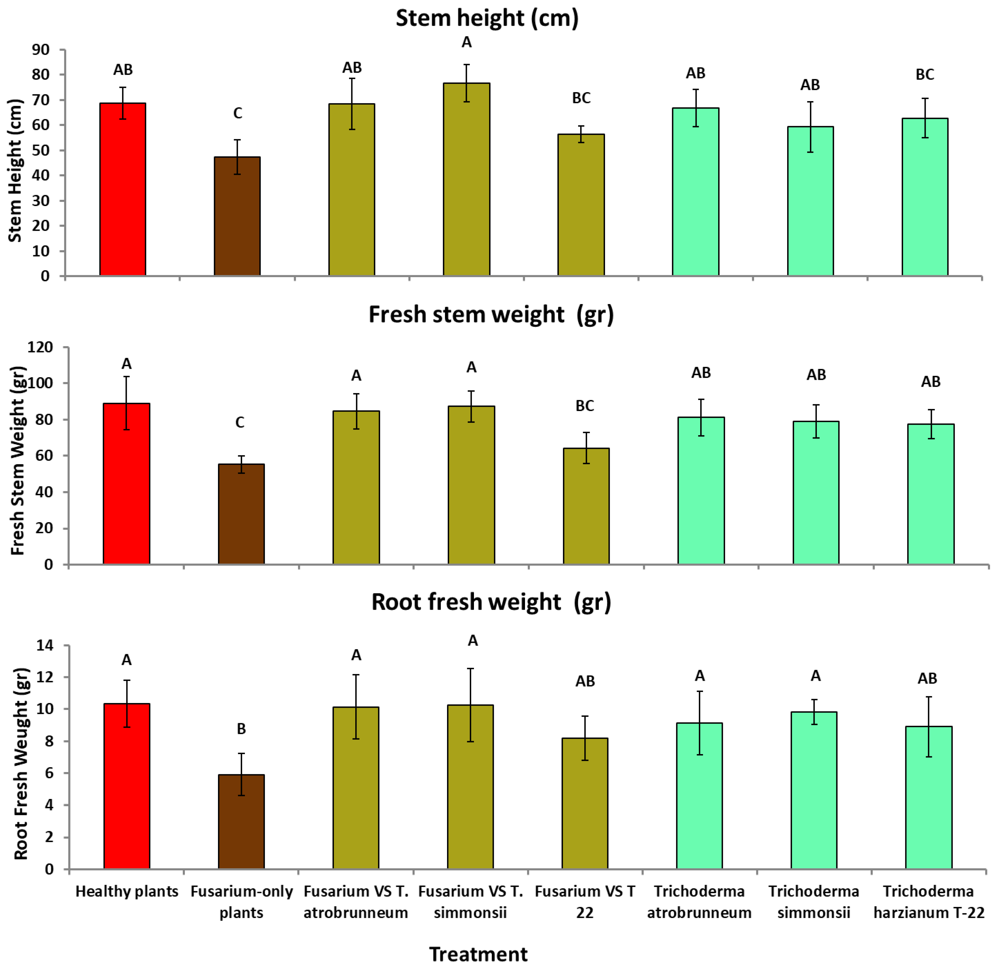
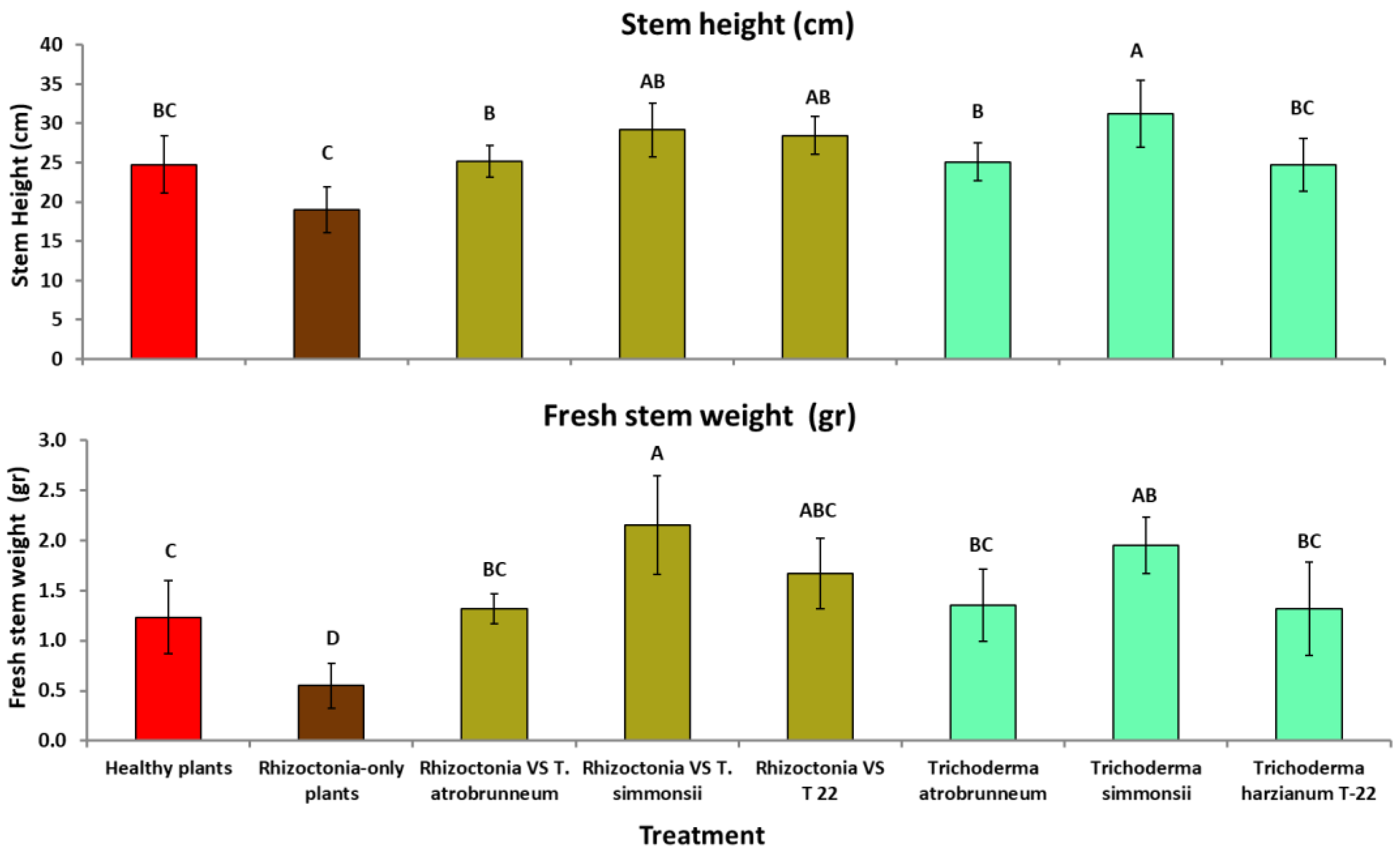
3.3. Greenhouse Tests

The effects of Trichoderma on tomato growth are presented in Figure 2 and Figure 3. As expected, the growth of diseased tomato plants inoculated with phytopathogen only (brown bars) was dramatically reduced compared with the control and with Trichoderma treated plants in almost all cases (Figure 2 and Figure 3).
In the Fusarium experiment, the differences in growth parameters among control and treated plants were significant in all cases (stem height: df = 7, 47; F = 8.34; p < 0.0001; fresh stem weight: df = 7, 47; F = 9.14; p < 0.0001; root fresh weight: df = 7, 47; F = 4.61; p = 0.0007). Generally, the presence of Trichoderma in F. oxysporum diseased plants (dark yellow bars) managed to stop the growth reduction in most cases, given that there were not any significant differences with the healthy control plants. Another interesting conclusion is that our Trichogramma strains (T. atrobrunneum and T. simmonsii) performed better than the commercial Trichoderma strain (T-22), reaching higher growth parameters values. However, differences were not always significant (Figure 2).
As far as the R. solani experiment is concerned, differences among various control and treated plants were significant (stem height: df = 7, 47; F = 8.48; p < 0.0001; fresh stem weight: df = 7, 47; F = 11.61; p < 0.0001). Plants inoculated with T. simmonsii (healthy or diseased) demonstrated the highest growth values, exceeding even the healthy plants’ growth (red bars) (Figure 3). Similarly to F. oxysporum plants, the inoculation with Trichoderma in R. solani diseased plants managed to stop the plant growth reduction in most cases.
Both tested Trichoderma strains stimulated plant growth mainly in the diseased plants (Fusarium- and Rhizoctonia-only plants) but not always in the healthy ones, considering that, in most treatments (except for T. simmonsii in the R. solani experiment, Figure 3), growth parameters of Trichoderma-only plants (green bars) were either lower or not significantly increased compared with those of healthy plants (red bars) (Figure 2 and Figure 3).

3.4. Re-Isolation of Trichoderma from Leaves, Stems and Roots on SDA Substrate

The establishment of Trichoderma endophytes in tomato plants was evaluated at the end of the greenhouse trials. Successful re-isolations of the fungus were obtained from all treated plants (at least one sample form the plant was colonized). On the other hand, no Trichoderma was detected in control plants. In total, about 90% of leaf, 70% of stem and 85% of roots samples from treated plants were successfully colonized by Trichoderma.

4. Discussion

Actions and methods for promoting sustainability in agricultural production systems, such as integrated pest management (IPM) and organic farming, have been greatly boosted by the urgent need to preserve the environment and human health from the harmful effects of agrochemicals [50]. The use of BCAs, which are based on living microbes or their metabolites and suppress plant diseases, is a key component of these techniques [5,50,51]. The efficiency of a wide variety of non-pathogenic fungi against soil-borne plant diseases has been the subject of significant research [52]. These control measures are in line with the European Green Deal (EGD) that aims to increase organic farming from 8% to 25% and reduce chemical pesticide application by 50% by 2030 [53].
Endophytic beneficial fungi that are naturally present in the plant ecosystem have been utilized for plant disease control for decades [54,55]. They manage to eliminate fungal phytopathogens in the plant mainly by competing for food sources, producing metabolites that inhibit the pathogen growth and by occupying specific sites in the plant first [55,56,57]. Among those antagonistic microorganisms, Trichoderma is by far the most promising and well-studied genus [58], having been investigated for more than 100 years for its beneficial effects. A plethora of scientific publications and reviews, including dozens of strains, have studied its mechanisms and interactions with plants ([11,59,60] and others), its antibiotics and metabolites ([59,61] and others), its action as biocontrol agent against various pathogens ([6,25,62] and others) and as biostimulant ([12,13,14,15,25] and others).
Trichoderma has demonstrated great effectiveness against soil-borne plant diseases. This has been demonstrated for R. solani, not only in dual culture tests [63,64,65] but also in field experiments, including crops like sugar beet [63], potato [66], cucumber [67], bean [68], tobacco [69], maize [70], cowpea [33] and tomato [33]. It has been well documented that Trichoderma suppresses R. solani through mycoparasitism and the production of antifungal compounds [66,71,72]. Moreover, various Trichoderma species have shown great potential in suppressing tomato wilt caused by F. oxysporum f. sp. lycopersici both in vitro [73,74,75,76,77,78,79] and in vivo [75,76,77,78,80,81].
We examined new strains from two species that have not been thoroughly investigated, especially against soil-borne pathogens. In a very recent study, a strain of T. atrobrunneum showed an inhibition rate 39.1 and 47.5% in dual culture tests with two F. oxysporum isolates after 5 days [32]. Despite the different strain, this conclusion is very close with our results (42.57% IGR after 5 days). Another strain of the same species inhibited the growth of R. solani by 58.9% [33] and the growth of Colletotrichum lagenarium by 95% [82] after 9 days.
As far as T. simmonsii is concerned, three strains caused 65.7, 68.6 and 81.9% inhibition in the growth of R. solani in dual culture tests after 9 days [33]. Our relevant value (57.78% after 5 days) does not differ notably, given that the exposure time was much shorter. In the same study, one strain stimulated cowpea plant growth significantly, increasing root and aerial part weight compared with R. solani-only control plants. Similar growth promotion by T. simmonsii in tomatoes was observed in the present study.
Tested Trichoderma strains showed differentiated potential for plant growth promotion given that their presence in healthy plants did not result in a significant increase in the development of tomato aerial parts and root system in most of the treatments. Generally, the ability of Trichoderma to promote plant growth under various biotic and abiotic stress conditions has been well established in numerous recent and older studies [12,13,14,15,16,17,18,19,25,30]. Plant growth mechanisms are stimulated by the presence of Trichoderma, include phosphorous mobilization, by extracellular phosphatases and the production of indole-3-acetic acid derivatives [83]. Although we did not verify that T. atrobrunneum may act effectively as a plant growth promoter, other strains of this species caused very high K, P and Zn solubilization [82], like other Trichoderma species [84,85,86,87].
The only verified case of biostimulant action in the present study was that of T. simmonsii in the R. solani experiments. The fact that the same Trichoderma strain caused significant growth increase in tomato plants only in the second experiment may be attributed to the early inoculation (one day after sow) compared with late inoculation (15 days after sow) in the first experiment. Similar growth promoting effects for this T. simmonsii have been reported in the shoot and total biomass of rice seedlings [88], ball peppers [34] and soybeans [35].
Our Trichoderma isolates enhanced tomato growth in the presence of F. oxysporum or R. solani, but in the absence of the pathogen they did not promote plant growth, in most cases. This finding supports the theory that many antagonistic microbes help the plant to grow indirectly by preventing the pathogen’s harmful effects and lowering disease intensity [89,90,91].
Even though a plethora of Trichoderma-based products are commercially available, most of them are concentrated on a very limited number of well-known species [9,92,93,94]. Many Trichoderma species remain undiscovered or uninvestigated. In the present study, two new Trichoderma strains of not so well-studied species were investigated for the first time as promising biocontrol agents against Fusarium wilt and foot rot in tomato. Although our results clearly showed their biocontrol potential, the exact mechanism should be elucidated and studied in depth to avoid the occurrence where strains that perform well in lab conditions fail to perform effectively in the field [95,96,97,98,99].

5. Conclusions

In conclusion, the evaluation of our endophytic Trichoderma strains revealed their notable biocontrol capability and their usage as biocontrol agents against fungal infections appears to be a promising approach. More research into the practical issues of large-scale production and formulation, on the other hand, is needed to develop a biocontrol product that is stable, effective, secure and increasingly successful. Endophytic fungal antagonists are a viable non-chemical alternative technique for long-term disease management on a wide range of plants and crops.

Author Contributions

Conceptualization, D.N., A.T. and P.A.E.; methodology, D.N. and P.A.E.; validation, P.A.E. and S.M.; formal analysis, D.N. and I.L.; experimentation, D.N. and A.T.; resources, P.A.E. and S.M.; data curation, D.N. and A.T.; writing—original draft preparation, P.A.E.; writing—review and editing, D.N., I.L. and S.M.; supervision, P.A.E. All authors have read and agreed to the published version of the manuscript.

Funding

This research received no external funding.

Institutional Review Board Statement

Not applicable.

Informed Consent Statement

Not applicable.

Data Availability Statement

Not applicable.

Conflicts of Interest

The authors declare no conflict of interest.

References

  1. Leadbeater, A. Recent Developments and Challenges in Chemical Disease Control—A Review. Plant Protect. Sci. 2016, 51, 163–169. [Google Scholar] [CrossRef] [Green Version]
  2. Zubrod, J.P.; Bundschuh, M.; Arts, G.; Brühl, C.A.; Imfeld, G.; Knäbel, A.; Payraudeau, S.; Rasmussen, J.J.; Rohr, J.; Scharmüller, A.; et al. Fungicides: An Overlooked Pesticide Class? Environ. Sci. Technol. 2019, 53, 3347–3365. [Google Scholar] [CrossRef] [PubMed]
  3. Pal, K.K.; McSpadden Gardener, B. Biological Control of Plant Pathogens. Plant Health Instructor; The American Phytopathological Society: St. Paul, MN, USA, 2006; pp. 1–25. [Google Scholar] [CrossRef] [Green Version]
  4. Tariq, M.; Khan, A.; Asif, M.; Khan, F.; Ansari, T.; Shariq, M.; Siddiqui, M.A. Biological Control: A Sustainable and Practical Approach for Plant Disease Management. Acta Agric. Scand. B Soil Plant Sci. 2020, 70, 507–524. [Google Scholar] [CrossRef]
  5. Thambugala, K.M.; Daranagama, D.A.; Phillips, A.J.L.; Kannangara, S.D.; Promputtha, I. Fungi vs. Fungi in Biocontrol: An Overview of Fungal Antagonists Applied Against Fungal Plant Pathogens. Front. Cell. Infect. Microbiol. 2020, 10, 604923. [Google Scholar] [CrossRef] [PubMed]
  6. Sood, M.; Kapoor, D.; Kumar, V.; Sheteiwy, M.S.; Ramakrishnan, M.; Landi, M.; Araniti, F.; Sharma, A. Trichoderma: The “Secrets” of a Multitalented Biocontrol Agent. Plants 2020, 9, 762. [Google Scholar] [CrossRef] [PubMed]
  7. Asad, S.A. Mechanisms of Action and Biocontrol Potential of Trichoderma against Fungal Plant Diseases—A Review. Ecol. Complex. 2022, 49, 100978. [Google Scholar] [CrossRef]
  8. Poveda, J. Trichoderma as biocontrol agent against pests: New uses for a mycoparasite. Biol. Control 2021, 159, 104634. [Google Scholar] [CrossRef]
  9. Fraceto, L.F.; Maruyama, C.R.; Guilger, M.; Mishra, S.; Keswani, C.; Singh, H.B.; de Lima, R. Trichoderma harzianum-Based Novel Formulations: Potential Applications for Management of Next-Gen Agricultural Challenges. J. Chem. Technol. Biotechnol. 2018, 93, 2056–2063. [Google Scholar] [CrossRef]
  10. Vinale, F.; Sivasithamparam, K. Beneficial Effects of Trichoderma Secondary Metabolites on Crops. Phytother. Res. 2020, 34, 2835–2842. [Google Scholar] [CrossRef]
  11. Harman, G.E. Overview of Mechanisms and Uses of Trichoderma spp. Phytopathology 2006, 96, 190–194. [Google Scholar] [CrossRef] [Green Version]
  12. Ousley, M.A.; Lynch, J.M.; Whipps, J.M. Effect of Trichoderma on Plant Growth: A Balance between Inhibition and Growth Promotion. Microb. Ecol. 1993, 26, 277–285. [Google Scholar] [CrossRef] [PubMed]
  13. Ousley, M.A.; Lynch, J.M.; Whipps, J.M. Potential of Trichoderma spp. as consistent plant growth stimulators. Biol. Fertil. Soils 1994, 17, 85–90. [Google Scholar] [CrossRef]
  14. Saba, H. Trichoderma—A Promising Plant Growth Stimulator and Biocontrol Agent. Mycosphere 2012, 3, 524–531. [Google Scholar] [CrossRef]
  15. Stewart, A.; Hill, R. Applications of Trichoderma in plant growth promotion. In Biotechnology and Biology of Trichoderma; Gupta, V., Schmoll, M., Herrera-Estrella, A., Upadhyay, R., Druzhinina, I., Tuohy, M., Eds.; Elsevier: Amsterdam, The Netherlands, 2014; pp. 415–428. [Google Scholar]
  16. Bora, L.C.; Bora, P.; Gogoi, M. Potential of Trichoderma spp. for Pest Management and Plant Growth Promotion in NE India. In Trichoderma: Host Pathogen Interactions and Applications; Sharma, A.K., Sharma, P., Eds.; Springer: Singapore, 2020; pp. 205–220. [Google Scholar] [CrossRef]
  17. Sánchez-Montesinos, B.; Diánez, F.; Moreno-Gavira, A.; Gea, F.J.; Santos, M. Plant Growth Promotion and Biocontrol of Pythium ultimum by Saline Tolerant Trichoderma Isolates under Salinity Stress. Int. J. Environ. Res. Public Health 2019, 16, 2053. [Google Scholar] [CrossRef] [Green Version]
  18. Sánchez-Montesinos, B.; Diánez, F.; Moreno-Gavíra, A.; Gea, F.J.; Santos, M. Role of Trichoderma aggressivum f. europaeum as Plant-Growth Promoter in Horticulture. Agronomy 2020, 10, 1004. [Google Scholar] [CrossRef]
  19. Inbar, J.; Abramsky, M.; Cohen, D.; Chet, I. Plant Growth Enhancement and Disease Control By Trichoderma harzianum in Vegetable Seedlings Grown under Commercial Conditions. Eur. J. Plant. Pathol. 1994, 100, 337–346. [Google Scholar] [CrossRef]
  20. Rinu, K.; Sati, P.; Pandey, A. Trichoderma gamsii (NFCCI 2177): A Newly Isolated Endophytic, Psychrotolerant, Plant Growth Promoting, and Antagonistic Fungal Strain. J. Basic Microbiol. 2014, 54, 408–417. [Google Scholar] [CrossRef]
  21. Degani, O.; Rabinovitz, O.; Becher, P.; Gordani, A.; Chen, A. Trichoderma longibrachiatum and Trichoderma asperellum Confer Growth Promotion and Protection against Late Wilt Disease in the Field. J. Fungi 2021, 7, 444. [Google Scholar] [CrossRef]
  22. Macena, A.M.F.; Kobori, N.N.; Mascarin, G.M.; Vida, J.B.; Hartman, G.L. Antagonism of Trichoderma-Based Biofungicides against Brazilian and North American Isolates of Sclerotinia sclerotiorum and Growth Promotion of Soybean. BioControl 2020, 65, 235–246. [Google Scholar] [CrossRef]
  23. Shang, J.; Liu, B.; Xu, Z. Efficacy of Trichoderma asperellum TC01 against Anthracnose and Growth Promotion of Camellia sinensis Seedlings. Biol. Control 2020, 143, 104205. [Google Scholar] [CrossRef]
  24. Srivastava, A.; Singh, R.P.; Srivastava, A.K.; Saxena, A.K.; Arora, D.K. Growth promotion and charcoal rot management in chickpea by Trichoderma harzianum. J. Plant Prot. Res. 2008, 48, 81–92. [Google Scholar] [CrossRef] [Green Version]
  25. Poveda, J. Biological Control of Fusarium oxysporum f. sp. ciceri and Ascochyta rabiei Infecting Protected Geographical Indication Fuentesaúco-Chickpea by Trichoderma Species. Eur. J. Plant Pathol. 2021, 160, 825–840. [Google Scholar] [CrossRef]
  26. Carvalho, D.D.C.; de Mello, S.C.M.; Lobo Júnior, M.; Geraldine, A.M. Biocontrol of Seed Pathogens and Growth Promotion of Common Bean Seedlings by Trichoderma harzianum. Pesqui. Agropecu. Bras. 2011, 46, 822–828. [Google Scholar] [CrossRef] [Green Version]
  27. de Souza Pedro, E.A.; Harakava, R.; Lucon, C.M.M.; Guzzo, S.D. Promoção do crescimento do feijoeiro e controle da antracnose por Trichoderma spp. Pesqui. Agropecu. Bras. 2012, 47, 1589–1595. [Google Scholar] [CrossRef] [Green Version]
  28. Nuangmek, W.; Aiduang, W.; Kumla, J.; Lumyong, S.; Suwannarach, N. Evaluation of a Newly Identified Endophytic Fungus, Trichoderma phayaoense for Plant Growth Promotion and Biological Control of Gummy Stem Blight and Wilt of Muskmelon. Front. Microbiol. 2021, 12, 410. [Google Scholar] [CrossRef]
  29. Hicks, E.; Bienkowski, D.; Braithwaite, M.; McLean, K.; Falloon, R.; Stewart, A. Trichoderma strains suppress Rhizoctonia diseases and promote growth of potato. Phytopathol. Mediterr. 2014, 53, 502–514. [Google Scholar]
  30. Fontenelle, A.D.B.; Guzzo, S.D.; Lucon, C.M.M.; Harakava, R. Growth promotion and induction of resistance in tomato plant against Xanthomonas euvesicatoria and Alternaria solani by Trichoderma spp. Crop Prot. 2011, 30, 1492–1500. [Google Scholar] [CrossRef]
  31. Zaw, M.; Matsumotom, M. Plant growth promotion of Trichoderma virens, Tv911 on some vegetables and its antagonistic effect on Fusarium wilt of tomato. Environ. Control Biol. 2020, 58, 7–14. [Google Scholar] [CrossRef] [Green Version]
  32. Pavlovskaya, N.; Gneusheva, I.; Solokhina, I.; Ageeva, N. The Biological Activity of Subspecies Trichoderma harzianum against Fusarium oxysporum, the Causative Agent of Fusarium Wilt Cucumber in Vitro. BIO Web Conf. 2020, 21, 00021. [Google Scholar] [CrossRef]
  33. Wang, C.; Zhuang, W. Evaluating Effective Trichoderma Isolates for Biocontrol of Rhizoctonia solani Causing Root Rot of Vigna unguiculata. J. Integr. Agric. 2019, 18, 2072–2079. [Google Scholar] [CrossRef]
  34. Rokni, N.; Shams Alizadeh, H.; Bazgir, E.; Darvishnia, M.; Mirzaei-Najafgholi, H. The Tripartite Consortium of Serendipita indica, Trichoderma simmonsii, and Bell Pepper (Capsicum annum). Biol. Control 2021, 158, 104608. [Google Scholar] [CrossRef]
  35. Bakhshandeh, E.; Gholamhosseini, M.; Yaghoubian, Y.; Pirdashti, H. Plant Growth Promoting Microorganisms Can Improve Germination, Seedling Growth and Potassium Uptake of Soybean under Drought and Salt Stress. Plant Growth Regul. 2020, 90, 123–136. [Google Scholar] [CrossRef]
  36. Chen, L.; Bóka, B.; Kedves, O.; Nagy, V.D.; Szűcs, A.; Champramary, S.; Roszik, R.; Patocskai, Z.; Münsterkötter, M.; Huynh, T.; et al. Towards the Biological Control of Devastating Forest Pathogens from the Genus Armillaria. Forests 2019, 10, 1013. [Google Scholar] [CrossRef] [Green Version]
  37. Rees, H.J.; Bashir, N.; Drakulic, J.; Cromey, M.G.; Bailey, A.M.; Foster, G.D. Identification of Native Endophytic Trichoderma spp. for Investigation of In Vitro Antagonism towards Armillaria mellea Using Synthetic- and Plant-Based Substrates. J. Appl. Microbiol. 2021, 131, 392–403. [Google Scholar] [CrossRef]
  38. Booth, C. The Genus Fusarium; Commonwealth Mycological Institute: Kew, UK, 1971; 237p. [Google Scholar]
  39. Nelson, P.E.; Tousson, T.A.; Marasas, W.F.O. Fusarium Species: An Illustrated Manual for Identification; Pennsylvania State University Press: University Park, PA, USA, 1983; 206p. [Google Scholar]
  40. Sneh, B.; Burpee, L.; Ogoshi, A. Identification of Rhizoctonia Species; American Phytopathological Society: Saint Paul, MN, USA, 1991; 133p. [Google Scholar]
  41. Warcup, J.H. The Soil-Plate Method for Isolation of Fungi from Soil. Nature 1950, 166, 117–118. [Google Scholar] [CrossRef]
  42. Gams, W.; Bissett, J. Morphology and Identification of Trichoderma. In Trichoderma and Gliocladium: Basic Biology, Taxonomy and Genetics Vol. 1; Harman, G.E., Kubicek, C.P., Eds.; Taylor and Francis: London, UK, 1998; pp. 3–34. [Google Scholar]
  43. Rogers, S.O.; Bendich, A.J. Extraction of DNA from Milligram Amounts of Fresh, Herbarium and Mummified Plant Tissues. Plant Mol. Biol. 1985, 5, 69–76. [Google Scholar] [CrossRef]
  44. Destéfano, R.H.R.; Destéfano, S.A.L.; Messias, C.L. Detection of Metarhizium anisopliae var. anisopliae within Infected Sugarcane Borer Diatraea saccharalis (Lepidoptera, Pyralidae) Using Specific Primers. Genet. Mol. Biol. 2004, 27, 245–252. [Google Scholar] [CrossRef] [Green Version]
  45. Cai, F.; Druzhinina, I.S. In Honor of John Bissett: Authoritative Guidelines on Molecular Identification of Trichoderma. Fungal Divers. 2021, 107, 1–69. [Google Scholar] [CrossRef]
  46. Morton, D.T.; Stroube, N.H. Antagonistic and stimulatory effects of microorganisms upon Sclerotium rolfsii. Phytopathology 1955, 45, 419–420. [Google Scholar]
  47. Schneider, C.A.; Rasband, W.S.; Eliceiri, K.W. NIH Image to ImageJ: 25 Years of Image Analysis. Nat. Methods 2012, 9, 671–675. [Google Scholar] [CrossRef]
  48. Meyling, N.V.; Eilenberg, J. Occurrence and distribution of soil borne entomopathogenic fungi within a single organic agroecosystem. Agric. Ecosyst. Environ. 2006, 113, 336–341. [Google Scholar] [CrossRef]
  49. IBM Corp. IBM SPSS Statistics for Windows; Version 27.0; IBM Corp: Armonk, NY, USA, 2020. [Google Scholar]
  50. Ab Rahman, S.S.F.; Singh, E.; Pieterse, C.M.J.; Schenk, P.M. Emerging Microbial Biocontrol Strategies for Plant Pathogens. Plant Sci. 2018, 267, 102–111. [Google Scholar] [CrossRef] [PubMed] [Green Version]
  51. He, D.C.; He, M.H.; Amalin, D.M.; Liu, W.; Alvindia, D.G.; Zhan, J. Biological control of plant diseases: An evolutionary and eco-economic consideration. Pathogens 2021, 10, 1311. [Google Scholar] [CrossRef] [PubMed]
  52. Niu, B.; Wang, W.; Yuan, Z.; Sederoff, R.R.; Sederoff, H.; Chiang, V.L.; Borriss, R. Microbial Interactions Within Multiple-Strain Biological Control Agents Impact Soil-Borne Plant Disease. Front. Microbiol. 2020, 11, 585404. [Google Scholar] [CrossRef] [PubMed]
  53. Hulot, J.F.; Hiller, N. Exploring the Benefits of Biocontrol for Sustainable Agriculture—A Literature Review on Biocontrol in Light of the European Green Deal; Institute for European Environmental Policy: Brussels, Belgium, 2021; pp. 1–42. [Google Scholar]
  54. Wilson, D. Fungal Endophytes: Out of Sight but Should Not Be out of Mind. Oikos 1993, 68, 379–384. [Google Scholar] [CrossRef]
  55. Heydari, A.; Pessarakli, M. A review on biological control of fungal plant pathogens using microbial antagonists. J. Biol. Sci. 2010, 10, 273–290. [Google Scholar] [CrossRef] [Green Version]
  56. Ghorbanpour, M.; Omidvari, M.; Abbaszadeh-Dahaji, P.; Omidvar, R.; Kariman, K. Mechanisms underlying the protective effects of beneficial fungi against plant diseases. Biol. Control 2018, 117, 147–157. [Google Scholar] [CrossRef]
  57. Latz, M.A.; Jensen, B.; Collinge, D.B.; Jørgensen, H.J. Endophytic fungi as biocontrol agents: Elucidating mechanisms in disease suppression. Plant Ecol. Divers. 2018, 11, 555–567. [Google Scholar] [CrossRef] [Green Version]
  58. Ferreira, F.V.; Musumeci, M.A. Trichoderma as Biological Control Agent: Scope and Prospects to Improve Efficacy. World J. Microbiol. Biotechnol. 2021, 37, 90. [Google Scholar] [CrossRef]
  59. Vinale, F.; Sivasithamparam, K.; Ghisalberti, E.L.; Marra, R.; Woo, S.L.; Lorito, M. Trichoderma–Plant–Pathogen Interactions. Soil Biol. Biochem. 2008, 40, 1–10. [Google Scholar] [CrossRef]
  60. Mukhopadhyay, R.; Kumar, D. Trichoderma: A Beneficial Antifungal Agent and Insights into Its Mechanism of Biocontrol Potential. Egypt J. Biol. Pest Control 2020, 30, 133. [Google Scholar] [CrossRef]
  61. Ghisalberti, E.L.; Sivasithamparam, K. Antifungal antibiotics produced by Trichoderma spp. Soil Biol. Biochem. 1991, 23, 1011–1020. [Google Scholar] [CrossRef]
  62. Ghazanfar, M.U.; Raza, M.; Raza, W.; Qamar, M.I. Trichoderma as Potential Biocontrol Agent, Its Exploitation in Agriculture: A Review. Plant Prot. 2018, 2, 109–135. [Google Scholar]
  63. Anees, M.; Tronsmo, A.; Edel-Hermann, V.; Hjeljord, L.G.; Héraud, C.; Steinberg, C. Characterization of Field Isolates of Trichoderma Antagonistic against Rhizoctonia solani. Fungal Biol. 2010, 114, 691–701. [Google Scholar] [CrossRef] [PubMed]
  64. de Melo, I.S.; Faull, J.L. Parasitism of Rhizoctonia solani by Strains of Trichoderma spp. Sci. agric. 2000, 57, 55–59. [Google Scholar] [CrossRef]
  65. Halifu, S.; Deng, X.; Song, X.; Song, R.; Liang, X. Inhibitory Mechanism of Trichoderma virens ZT05 on Rhizoctonia solani. Plants 2020, 9, 912. [Google Scholar] [CrossRef] [PubMed]
  66. Abbas, A.; Jiang, D.; Fu, Y. Trichoderma spp. as antagonist of Rhizoctonia solani. J. Plant Pathol. Microbiol. 2017, 8, 1–9. [Google Scholar]
  67. Trillas, M.I.; Casanova, E.; Cotxarrera, L.; Ordovás, J.; Borrero, C.; Avilés, M. Composts from Agricultural Waste and the Trichoderma asperellum Strain T-34 Suppress Rhizoctonia solani in Cucumber Seedlings. Biol. Control 2006, 39, 32–38. [Google Scholar] [CrossRef]
  68. Mayo, S.; Gutierrez, S.; Malmierca, M.G.; Lorenzana, A.; Campelo, M.P.; Hermosa, R.; Casquero, P.A. Influence of Rhizoctonia solani and Trichoderma spp. in growth of bean (Phaseolus vulgaris L.) and in the induction of plant defense-related genes. Front. Plant Sci. 2015, 6, 685. [Google Scholar] [CrossRef] [Green Version]
  69. Cole, J.S.; Zvenyika, Z. Integrated control of Rhizoctonia solani and Fusarium solani in tobacco transplants with Trichoderma hurziunum and triadimenol. Plant Pathol. 1988, 37, 271–277. [Google Scholar] [CrossRef]
  70. Herrera, W.; Valbuena, O.; Pavone-Maniscalco, D. Formulation of Trichoderma asperellum TV190 for biological control of Rhizoctonia solani on corn seedlings. Egypt J. Biol. Pest Control 2020, 30, 44. [Google Scholar] [CrossRef]
  71. Heflish, A.A.; Abdelkhalek, A.; Al-Askar, A.A.; Behiry, S.I. Protective and Curative Effects of Trichoderma asperelloides Ta41 on Tomato Root Rot Caused by Rhizoctonia solani Rs33. Agronomy 2021, 11, 1162. [Google Scholar] [CrossRef]
  72. Inayati, A.; Sulistyowati, L.; Aini, L.Q.; Yusnawan, E. Mycoparasitic Activity of Indigenous Trichoderma virens Strains Against Mungbean Soil Borne Pathogen Rhizoctonia solani: Hyperparasite and Hydrolytic Enzyme Production. AGRIVITA J. Agric. Sci. 2020, 42, 229–242. [Google Scholar] [CrossRef]
  73. Barari, H. Biocontrol of Tomato Fusarium Wilt by Trichoderma Species under in Vitro and in Vivo Conditions. Cercet. Agron. Mold. 2016, 49, 91–98. [Google Scholar] [CrossRef] [Green Version]
  74. Prasad, L.; Chaudhary, S.; Sagar, S.; Tomar, A. Mycoparasitic Capabilities of Diverse Native Strain of Trichoderma spp. against Fusarium oxysporum f. sp. lycopersici. J. Appl. Nat. Sci. 2016, 8, 769–776. [Google Scholar] [CrossRef] [Green Version]
  75. Moosa, A.; Sahi, S.T.; Haq, I.-U.; Farzand, A.; Khan, S.A.; Javaid, K. Antagonistic Potential of Trichoderma Isolates and Manures Against Fusarium Wilt of Tomato. Int. J. Veg. Sci. 2017, 23, 207–218. [Google Scholar] [CrossRef]
  76. Babychan, M.; Simon, S. Efficacy of Trichoderma spp. against Fusarium oxysporum f. Sp. lycopersici. (FOL) Infecting Pre-and Post-Seedling of Tomato. J. Pharmacogn. Phytochem. 2017, 6, 616–619. [Google Scholar]
  77. Patel, S.; Saraf, M. Biocontrol efficacy of Trichoderma asperellum MSST against tomato wilting by Fusarium oxysporum f. sp. lycopersici. Arch. Phytopathol. Plant Prot. 2017, 50, 228–238. [Google Scholar] [CrossRef]
  78. Sallam, N.M.A.; Eraky, A.M.I.; Sallam, A. Effect of Trichoderma spp. on Fusarium Wilt Disease of Tomato. Mol. Biol. Rep. 2019, 46, 4463–4470. [Google Scholar] [CrossRef]
  79. Jagraj, S.; Vipul, K.; Seweta, S.; Adesh, K.; Vinit, P.S. In vitro evaluation of Trichoderma species against Fusarium oxysporum f. sp. lycopersici causing tomato wilt. Plant Pathol. J. 2018, 17, 59–64. [Google Scholar] [CrossRef] [Green Version]
  80. Ghazalibiglar, H.; Kandula, D.R.W.; Hampton, J.G. Biological Control of Fusarium Wilt of Tomato by Trichoderma Isolates. N. Z. Plant Prot. 2016, 69, 57–63. [Google Scholar] [CrossRef] [Green Version]
  81. Jamil, A. Antifungal and plant growth promoting activity of Trichoderma spp. against Fusarium oxysporum f. sp. lycopersici colonizing tomato. J. Plant Prot. Res. 2021, 61, 243–253. [Google Scholar]
  82. Hewedy, O.A.; Mansour, A.; Ali, M.G.; Lateif, K.S.A.; Ismaiel, M.H.; El-Meihy, R.M. Comprehensive Characterization and Screening of Different Trichoderma Isolates as Plant Growth Promoters: Insight to Metal Solubilization, Enzymatic Activity, and Antagonistic Effect. Rev. Res. Sq. 2022; Preprint. [Google Scholar] [CrossRef]
  83. Monfil, V.O.; Casas-Flores, S. Chapter 32—Molecular Mechanisms of Biocontrol in Trichoderma spp. and Their Applications in Agriculture. In Biotechnology and Biology of Trichoderma; Gupta, V.K., Schmoll, M., Herrera-Estrella, A., Upadhyay, R.S., Druzhinina, I., Tuohy, M.G., Eds.; Elsevier: Amsterdam, The Netherlands, 2014; pp. 429–453. [Google Scholar] [CrossRef]
  84. Anil, K.; Lakshmi, T. Phosphate Solubilization Potential and Phosphatase Activity Of Rhizospheric Trichoderma spp. Braz. J. Microbiol. 2010, 41, 787–795. [Google Scholar] [CrossRef] [PubMed]
  85. Saravanakumar, K.; Shanmuga Arasu, V.; Kathiresan, K. Effect of Trichoderma on Soil Phosphate Solubilization and Growth Improvement of Avicennia marina. Aquat. Bot. 2013, 104, 101–105. [Google Scholar] [CrossRef]
  86. França, D.V.C.; Kupper, K.C.; Magri, M.M.R.; Gomes, T.M.; Rossi, F. Trichoderma spp. isolates with potential of phosphate solubilization and growth promotion in cherry tomato. Pesqui. Agropecuária Trop. 2017, 47, 360–368. [Google Scholar] [CrossRef] [Green Version]
  87. Aishwarya, S.; Viswanath, H.S.; Singh, A.; Singh, R. Biosolubilization of different nutrients by Trichoderma spp. and their mechanisms involved: A review. Int. J. Adv. Agric. Sci. Tech. 2020, 7, 34–39. [Google Scholar]
  88. Aban, J.L.; Barcelo, R.C.; Oda, E.E.; Reyes, G.A.; Balangcod, T.D.; Gutierrez, R.M.; Hipol, R.M. Dominant Root Associated Fungi (RAF) from Drynaria quercifolia L. Either Induce or Retard Growth of PSB Rc10 Rice (Oryza sativa L.) in Gibberellic Acid-Inhibited Medium. App. Environ. Res. 2017, 39, 89–98. [Google Scholar] [CrossRef]
  89. Berg, G. Plant-Microbe Interactions Promoting Plant Growth and Health: Perspectives for Controlled Use of Microorganisms in Agriculture. Appl. Microbiol. Biotechnol. 2009, 84, 11–18. [Google Scholar] [CrossRef]
  90. Hayat, R.; Ali, S.; Amara, U.; Khalid, R.; Ahmed, I. Soil Beneficial Bacteria and Their Role in Plant Growth Promotion: A Review. Ann. Microbiol. 2010, 60, 579–598. [Google Scholar] [CrossRef]
  91. Beneduzi, A.; Ambrosini, A.; Passaglia, L.M.P. Plant Growth-Promoting Rhizobacteria (PGPR): Their Potential as Antagonists and Biocontrol Agents. Genet. Mol. Biol. 2012, 35, 1044–1051. [Google Scholar] [CrossRef] [Green Version]
  92. Woo, S.L.; Ruocco, M.; Vinale, F.; Nigro, M.; Marra, R.; Lombardi, N.; Pascale, A.; Lanzuise, S.; Manganiello, G.; Lorito, M. Trichoderma-Based Products and Their Widespread Use in Agriculture. Open Mycol. J. 2014, 8, 71–126. [Google Scholar] [CrossRef] [Green Version]
  93. Samuels, G.; Hebbar, P. Trichoderma Identification and Agricultural Applications; American Phytopathological Society Press: St. Paul, NY, USA, 2015; 204p. [Google Scholar]
  94. Manoharachary, C.; Singh, H.B.; Varma, A. Trichoderma: Agricultural Applications and Beyond; Springer: Cham, Switzerland, 2020; 367p. [Google Scholar] [CrossRef]
  95. Sid Ahmed, A.; Ezziyyani, M.; Pérez Sánchez, C.; Candela, M.E. Effect of Chitin on Biological Control Activity of Bacillus spp. and Trichoderma harzianum against Root Rot Disease in Pepper (Capsicum annuum) Plants. Eur. J. Plant Pathol. 2003, 109, 633–637. [Google Scholar] [CrossRef]
  96. Rabeendran, N.; Jones, E.E.; Moot, D.J.; Stewart, A. Biocontrol of Sclerotinia Lettuce Drop by Coniothyrium minitans and Trichoderma hamatum. Biol. Control 2006, 39, 352–362. [Google Scholar] [CrossRef]
  97. Villalta, O.N.; Wite, D.; Hunt, J.; Stewart, A.; Porter, I.J. Biological control of Sclerotinia minor on lettuce using Trichoderma and Coniothyrium species. Acta Hortic. 2012, 944, 51–58. [Google Scholar] [CrossRef]
  98. Blaya, J.; López-Mondéjar, R.; Lloret, E.; Pascual, J.A.; Ros, M. Changes Induced by Trichoderma harzianum in Suppressive Compost Controlling Fusarium Wilt. Pestic. Biochem. Physiol. 2013, 107, 112–119. [Google Scholar] [CrossRef]
  99. Martínez-Medina, A.; Del Mar Alguacil, M.; Pascual, J.A.; Van Wees, S.C.M. Phytohormone Profiles Induced by Trichoderma Isolates Correspond with Their Biocontrol and Plant Growth-Promoting Activity on Melon Plants. J. Chem. Ecol. 2014, 40, 804–815. [Google Scholar] [CrossRef]
Figure 1. Inhibition growth rate (IGR) values of F. oxysporum (up) and R. solani (down) in dual culture tests with Trichoderma species (IGR = ((CRG − TRG)/CRG) × 100%, CRG: mean radial growth of the phytopathogen in monoculture, TRG: mean radial growth of the phytopathogen in treatment (dual culture with Trichoderma).
Crops 02 00015 g001
Figure 2. Effects of Trichoderma treatments on the growth of healthy and F. oxysporum-inoculated tomato plants. Columns of the same chart followed by the same letter are not significantly different (Tukey–Kramer HSD Test, p < 0.05), all tests were performed in potted greenhouse plants, all treatments included six replications, DAS 50 days.
Crops 02 00015 g002
Figure 3. Effects of Trichoderma treatments on the growth of healthy and R. solani-inoculated tomato plants. Columns of the same chart followed by the same letter are not significantly different (Tukey–Kramer HSD Test, p < 0.05), all tests were performed in potted greenhouse plants, all treatments included six replications, DAS 30 days.
Crops 02 00015 g003
Table 1. Molecular characterization of Trichoderma strains tested during present study.
SpeciesCollection SiteBlast ID NumberDNA SequenceIdentity (%)GenBank Closest Hit
Trichoderma simmonsiiDevon, United Kingdom from soil of abandoned mineGMMBSMTJ01RGGAGGGCATTACCGAGTTTACAACTCCCAAACCCAATGTGAACGTTACCAAACTGTTGCCTCGGCGGGATCTCTGCCCCGGGTGCGTCGCAGCCCCGGACCAAGGCGCCCGCCGGAGGACCAACCTAAAACTCTTATTGTATACCCCCTCGCGGGTTTTTTTATAATCTGAGCCTTTCTCGGCGCCTCTCGTAGGCGTTTCGAAAATGAATCAAAACTTTCAACAACGGATCTCTTGGTTCTGGCATCGATGAAGAACGCAGCGAAATGCGATAAGTAATGTGAATTGCAGAATTCAGTGAATCATCGAATCTTTGAACGCACATTGCGCCCGCCAGTATTCTGGCGGGCATGCCTGTCCGAGCGTCATTTCAACCCTCGAACCCCTCCGGGGGGTCGGCGTTGGGGATCGGCCCTCCCTTAGCGGGTGGCCGTCTCCGAAATACAGTGGCGGTCTCGCCGCAGCCTCTCCTGCGCAGTAGTTTGCACACTCGCATCGGGAGCGCGGCGCGTCCACAGCCGTTAAACACCCAACTTCTGAAATGTTGACCTCGGATCAGGTAGGAATACCCGCTGAACTTAAGCATATCA99.16NR_137297.1
Trichoderma atrobrunneumSerres, Greece from corn combsGMMU60DF013GGAGGGCATTACCGAGTTTACAACTCCCAAACCCAATGTGAACGTTACCAAACTGTTGCCTCGGCGGGATCTCTGCCCCGGGTGCGTCGCAGCCCCGGACCAAGGCGCCCGCCGGAGGACCAACCAAAACTCTTATTGTATACCCCCTCGCGGGTTTTTTTTATAATCTGAGCCTTCTCGGCGCCTCTCGTAGGCGTTTCGAAAATGAATCAAAACTTTCAACAACGGATCTCTTGGTTCTGGCATCGATGAAGAACGCAGCGAAATGCGATAAGTAATGTGAATTGCAGAATTCAGTGAATCATCGAATCTTTGAACGCACATTGCGCCCGCCAGTATTCTGGCGGGCATGCCTGTCCGAGCGTCATTTCAACCCTCGAACCCCTCCGGGGGGTCGGCGTTGGGGATCGGCCCTGCCTTGGCGGTGGCCGTCTCCGAAATACAGTGGCGGTCTCGCCGCAGCCTCTCCTGCGCAGTAGTTTGCACACTCGCATCGGGAGCGCGGCGCGTCCACAGCCGTTAAACACCCAACTTCTGAGTTTGCACACTCGCATCGGGAGCGCGGCGCGTCCACAGCCGTTAAACACCCAACTTCTGA99.49NR_137298.1
Table 2. Mean radial growth (in cm) of Fusarium oxysporum f. sp. lycopersici in the presence or absence of Trichoderma sp. in dual culture tests.
FungusTime
24 h48 h72 h96 h120 h
F. oxysporum only a0.57 ± 0.10 aE *1.08 ± 0.12 aD1.77 ± 0.15 aC2.38 ± 0.15 aB2.92 ± 0.10 aA
F. oxysporum and T. harzianum b0.55 ± 0.10 aD1.07 ± 0.12 aC1.70 ± 0.17 aB1.87 ± 0.08 bAB1.90 ± 0.09 bA
F. oxysporum and T. atrobrunneum0.55 ± 0.05 aC1.05 ± 0.05 aB1.60 ± 0.09 aA1.65 ± 0.05 cA1.68 ± 0.06 cA
F. oxysporum and T. simmonsii0.55 ± 0.05 aD1.05 ± 0.08 aC1.75 ± 0.10 aB1.93 ± 0.05 bA1.95 ± 0.05 bA
a: negative control (phytopathogen monoculture), b: positive control, * means of the same column followed by the same small letter are not significantly different, means of the same row followed by the same capital letter are not significantly different (Tukey–Kramer HSD Test, p < 0.05), all tests were performed in Petri dishes with PDA at 25 °C, all treatments included six replications.
Table 3. Mean radial growth (in cm) of Rhizoctonia solani in the presence or absence of Trichoderma sp. in dual culture tests.
FungusTime
24 h48 h72 h96 h120 h
R. solani only a1.10 ± 0.09 aE *3.03 ± 0.08 aD5.17 ± 0.14 aC6.57 ± 0.08 aB7.82 ± 0.08 aA
R. solani and T. harzianum b0.97 ± 0.05 bC2.03 ± 0.16 bB3.17 ± 0.08 bA3.20 ± 0.09 bA3.22 ± 0.10 bA
R. solani and T.atrobrunneum0.88 ± 0.08 bcC2.05 ± 0.05 bB3.08 ± 0.08 bA3.12 ± 0.12 bA3.15 ± 0.10 bA
R. solani and T. simmonsii0.85 ± 0.05 cC1.95 ± 0.10 bB3.15 ± 0.12 bA3.28 ± 0.15 bA3.30 ± 0.13 bA
a: negative control (phytopathogen monoculture), b: positive control, * means of the same column followed by the same small letter are not significantly different, means of the same row followed by the same capital letter are not significantly different (Tukey–Kramer HSD Test, p < 0.05), all tests were performed in Petri dishes with PDA at 25 °C, all treatments included six replications.
Publisher’s Note: MDPI stays neutral with regard to jurisdictional claims in published maps and institutional affiliations.

