Article

Isolation and Optimization of Phages Infecting Members of the Streptococcus bovis/Streptococcus equinus Complex

by
Jenny Laverde Gomez
1,
Cory Schwarz
1,2,
Marina Tikhonova
1,
Clark Hamor
3,
Yizhi J. Tao
3,
Pedro J. J. Alvarez
1,2,* and
Jacques Mathieu
1,2
1
Sentinel Environmental, 11231 Richmond Avenue D105, Houston, TX 77082, USA
2
Department of Civil and Environmental Engineering, Rice University, 6100 Main Street, Houston, TX 77005, USA
3
Department of Biosciences, Rice University, 6100 Main Street, Houston, TX 77005, USA
*
Author to whom correspondence should be addressed.
Appl. Microbiol. 2025, 5(1), 28; https://doi.org/10.3390/applmicrobiol5010028
Submission received: 1 January 2025 / Revised: 20 February 2025 / Accepted: 1 March 2025 / Published: 4 March 2025

Abstract

:
Background: Cattle production is a cornerstone of U.S. agriculture but faces increasing pressure to balance profitability with environmental sustainability. Optimizing the ruminal microbiome to enhance feed efficiency could help address both challenges. Members of the Streptococcus bovis/Streptococcus equinus complex (SBSEC) are key contributors to ruminal acidosis and related digestive disorders due to their role in carbohydrate fermentation and lactic acid production. Bacteriophages targeting this bacterial group present a promising approach to mitigate this problem with high precision and without promoting the spread of antibiotic resistance. Methods: A collection of SBSEC-targeting bacteriophages were isolated from cattle rumen fluid and feces and further characterized. Characterization included host-range evaluation, whole genome sequencing, and growth inhibition assessment via optical density measurements. Selected bacteriophages underwent training to enhance infectivity. Results: Eleven lytic and one lysogenic phage were isolated. Several phages demonstrated sustained bacterial growth suppression, showing efficacy against SBSEC bacteria from diverse sources despite narrow host ranges. Co-evolutionary training was done in a subset of phages to improve bacteriolytic activity but had an inconsistent effect on the ability of phages to inhibit the growth of their naïve host. Genomic sequencing and phylogenetic analysis revealed uniqueness and clustering into three distinct groups that matched phenotypic characteristics. Conclusions: This study demonstrates the potential of bacteriophages as precise biological control agents, with successful isolation and enhancement of phages targeting SBSEC bacteria. Eleven lytic genome-sequenced phages show promise for development as cattle feed additives, though further research is needed to optimize their application in agricultural settings.

1. Introduction

Cattle production is a cornerstone of US agricultural activity, representing 88.4 billion in total cash receipts for agricultural commodities in 2023 [1]. However, the industry faces dual growing pressures: the need for increased production to meet rising demand while improving long-term sustainability and reducing impacts to the environment [2,3,4,5]. Beef production faces intense scrutiny due to the resource intensiveness of cattle production practices [6,7] thus technologies that can efficiently increase beef production while maintaining or improving economic and environmental sustainability are critically needed.
Enhancing feed efficiency in cattle is widely regarded as a crucial strategy for improving the overall sustainability of the beef industry [8,9,10]. It has been estimated that a 10% increase in feed efficiency can lead to a substantial 43% increase in profits. Accordingly, there has been substantial research effort into improving cattle feed efficiency [11,12]. Though the factors associated with variation in feed efficiency are still far from being completely understood, many contributing processes have been identified, including feeding behavior and physical activity, digestibility, energy metabolism, protein turnover, body composition, the endocrine system, and lastly, rumen microbiota [13,14,15,16,17,18].
Rumen microbiota both influences and is influenced by diet, shaping cattle feed efficiency [19,20,21]. Despite significant advancements in identifying specific rumen microorganisms and understanding their functions, our knowledge of rumen microbiology and the ecological networks required to degrade diverse substrates remains incomplete [22,23]. Therefore, new technologies are urgently needed to enable microbiome engineering and capitalize on recent discoveries.
Top-down microbiome engineering strategies, widely used in the cattle industry, involve modifying pre-existing microbial communities. These include dietary adjustments, antibiotics, prebiotics, essential oils, rumen transfaunation, probiotics (direct-fed microbials), and vaccines, all aimed at improving animal performance through microbiome modulation [24,25,26]. Generally, these strategies lack specificity, yield inconsistent results or produce temporary improvements that are challenging to maintain [27,28,29,30,31,32]. While the need for precise microbiome manipulation tools is widely acknowledged, few cost-effective solutions have been developed for animal agriculture [22]. Bacteriophages (phages), viruses that selectively and exclusively infect and lyse bacteria, offer a highly specific, self-amplifying, and environmentally friendly alternative to traditional antimicrobials [33]. Unlike broad-spectrum antibiotics, phages precisely target bacterial hosts without disrupting beneficial microbial communities, making them an attractive tool for microbiome modulation. First described by Félix d’Hérelle in the early 20th century [34], phage research has undergone a renaissance in recent years, with renewed interest in their applications across medicine, agriculture and biotechnology [35].
As such, we have isolated and characterized phages infecting several problematic ruminal bacterial species [36,37], including phages targeting a group of facultatively anaerobic gastrointestinal streptococci, the Streptococcus bovis/Streptococcus equinus complex (SBSEC). Streptococcus is a genus of diverse spherical Gram-positive lactic acid bacteria, containing commensal species native to the microbial communities of humans and animals but also pathogenic species responsible for numerous diseases. In ruminants, SBSEC have been observed to outgrow other ruminal bacterial flora under optimal growth conditions, producing a large amount of lactate and capsular polysaccharide causing acute ruminal acidosis and bloat [38,39]. Feedlot cattle are commonly fed ionophores (such as monensin and other antibiotics) to increase feed efficiency [40], stop SBSEC overgrowth and prevent the subsequent drop in ruminal pH [41]. However, this practice raises serious industrial and public health concerns highlighting the need for other methods of microbial control and ruminal community modulation. Given the importance of SBSEC, the first suggestion of phage therapy for microbial control in the rumen was for “S. bovis” [42] and substantial progress was made previously to isolate and characterize SBSEC phages and ruminal phages [42,43,44,45,46]. However, many lysogenic phages infecting SBSEC were identified, and only a few were genetically characterized. Recently, whole genome sequences of lytic ruminal phages have been reported and new SBSEC phages have been isolated [47,48,49]. Further experimental in vitro and in vivo studies with viral isolates are expected to remain crucial for the elucidation of biological properties required for the development of effective phage therapies and answer outstanding questions in rumen virus research [50].
In this publication, we describe 12 phages isolated from ruminal contents and cattle feces infecting SBSEC isolates and evaluate their suitability as antimicrobial agents, possibly for future use as feed additives to reduce or prevent SBSEC mediated acidosis.

2. Materials and Methods

2.1. Sampling and Storage of Ruminal Contents and Cattle Fecal Matter

Over 100 rumen fluid samples were collected from a beef processing plant in Amarillo, Texas, the Beef Cattle Research Center (Kansas State University) and the USDA Livestock Issues Research Unit (Lubbock, TX, USA) over the course of several years. Some rumen fluid samples were from cattle exposed to acidotic diets [51], while others were sourced from tylosin-fed feedlot cattle, and from cannulated cattle not fed tylosin. Samples shipped overnight on ice were processed within 24 h and stored at −80 °C in 10% dimethyl sulfoxide. Fresh fecal samples were sourced from grazing land in central Texas and stored at 4 °C or frozen at −80 °C in 10% dimethyl sulfoxide until use.

2.2. Bacterial Host and Bacteriophage Isolation

Samples were either streaked directly or enriched for bacterial isolation. Enrichments were performed to improve the isolation rate of both SBSEC strains and bacteriophages. Briefly, 200 µL of bovine rumen fluid or stool were added to a minimal medium consisting of 20 g/L pancreatic digest of casein, 10 g/L yeast extract, 0.1 mg/L resazurin, 1 g/L L-cysteine, 4 mg/L CaCl2, 8 mg/L MgSO4, 20 mg/L KH2PO4, 20 mg/L K2HPO4, 40 mg/L NaCl, 160 mg/L NaHCO3. The medium was sparged with an anaerobic gas mix consisting of 10% CO2, 5% H2 and balancing N2 and was supplemented with 1% maltose or 1% starch [52,53,54]. After overnight incubation at 37 °C, bacterial strains were isolated by streaking enrichments on Modified Edwards Medium (Oxoid) in anaerobic conditions. The species of SBSEC bacteria was confirmed by sequencing of their 16S rRNA gene [55]. After isolation, all SBSEC isolates were grown and maintained in brain heart infusion (BHI) broth in aerobic conditions. After enrichment and SBSEC isolation, all subsequent work was done under aerobic conditions. A list of the bacterial isolates used in this study can be found in the Supplementary Information (Table S1). For bacteriophage isolation, lysates from the enrichment culture were produced by centrifugation (10,000× g for 10 min) and filter sterilization using 25-mm diameter, 0.22-µm pore size PVDF hydrophilic syringe filter (Choice™, Thermo Scientific™, Waltham, MA, USA). The resulting lysates were spotted on lawns of the bacterial hosts in double layer assays (DLA), further detailed below [56].

2.3. Double Layer Assay and Bacteriophage Propagation

DLA were prepared with Brain Heart Infusion (BHI) (RPI Corp., Mt. Prospect, IL, USA) agar that was overlaid with BHI top agar (0.4% agarose) supplemented with 5% glycerol, 1 mM MgCl2, 1 mM CaCl2 and 5% of host bacteria and incubated aerobically. Clear plaques or zones of clearance were serially diluted and spotted again until single plaques could be picked using a sterile 1-mL micropipette tip. Agar plugs containing single plaques were resuspended in SM buffer (50 mM Tris-HCl (pH 7.5), 0.1 M NaCl, 8 mM MgSO4, 0.01% w/v gelatin) and amplified in the corresponding host. Phages were amplified using DLA or using liquid cultures infected with phages under aerobic conditions. Phage titers were assayed via serial dilution and spotting on DLA. Clarified phage stocks were concentrated by the addition of 1:2 volume of sterile polyethylene glycol (PEG) 3× solution (30% PEG 8000, 3 M NaCl) and centrifugation at 12,000× g for 30 min after overnight incubation at 4 °C. The resulting pellet was suspended in SM buffer. Phage stocks were stored at 4 °C under aerobic conditions.
The host range of the isolated phages was evaluated using a library of SBSEC bacterial isolates. Phages stocks at high titer (1 to 5 × 10⁷ PFU/mL) were spotted on DLA lawns of SBSEC isolates and their ability to cause lysis was recorded.

2.4. Phage Genetic and Phenotypic Characterization

2.4.1. Whole Genome Sequencing

High molecular weight DNA of each bacteriophage was extracted using a traditional phenol chloroform DNA extraction method [57] with the following differences: after PEG precipitation phages were resuspended in SMB, phenol chloroform extraction was done 3 times, and nucleic acids were resuspended in water instead of TE buffer in the final step. The DNA quantity and quality were assessed using Take 3 microvolume plate and BioTek Synergy H1 microplate spectrophotometer (Agilent Technologies, Inc., Santa Clara, CA, USA). DNA concentrations were also determined using the fluorescent dsDNA quantification kit Broad Range BioDynami (BioDynami, Huntsville, AL, USA), and molecular weight of the samples was verified by electrophoresis of 200 ng DNA on a 1% agarose gel using a Quick-Load® Purple 1 kb Plus DNA Ladder (New England Biolabs, Ipswich, MA, USA). If required, size selection was done with magnetic beads using 0.5 or 0.45 volumes of beads, according to the manufacturer’s instruction (Omega Bio-Tek, Inc., Norcross, GA, USA).
Libraries for genome sequencing were prepared from high molecular weight DNA using a Rapid barcoding sequencing kit 24 V14 (SQK-RBK114.24, Oxford Nanopore Technologies, Oxford, UK) and sequenced on a MinION sequencer (Oxford Nanopore Technologies, Oxford, UK) using a Flongle adaptor and a Flongle flow cell (R.10.4.1 chemistry) according to the manufacturer’s instructions. Reads were base called (super-accurate basecalling) and demultiplexed using MinKNOW and filtered by size with SeqKit v 2.9.0 [58,59]. Assemblies were generated using Flye v 2.9.5 [60] and also compared to assemblies generated by Unicycler v 0.5.0 [61,62] and Canu v 2.2 [63,64] to ensure the same assembly was produced by at least two methods. A subset of phage genome sequencing was performed by Plasmidsaurus using the same technique: Oxford Nanopore Technology for linear PCR/sequencing (25 to 125 kbp) with custom assembly (Plasmidsaurus Inc., South San Francisco, CA, USA).

2.4.2. Genome Annotation and Phylogenetic Analysis

Assembled viral genomes were annotated using Pharokka v1.4.0 [65] and manually refined using BLASTx (v 2.16.0) [66]. BLASTx was done using the NCBI web browser BLAST services, using translated nucleotide queries against the standard non-redundant protein sequences (nr) database and the default threshold of 0.05. Genomes were submitted to NCBI GenBank under the following accession numbers: PY1, PQ621712; PY2, PQ621713; PY3, PQ621714; PY4, PQ621715; PY7, PQ621716; PY9, PQ621717; 36L, PQ621707; Bsingle, PQ621708; Pika, PQ621709; PYS40, PQ621710; CSJC, PQ621704; Taco, PQ621705; Mushu, PQ621706; and Vroast, PV146553. Genomes and associated annotations were visualized with Proksee [67]. Alignments of similarity of the genomes were generated using clinker in the online CompArative GEne Cluster Analysis Toolbox (CAGECAT) [68] and OrthoANI [69]. Phylogenetic analysis including other published Streptococcus phage genome sequences was carried out using the Virus Classification and Tree Building Online Resource (VICTOR) web service https://victor.dsmz.de (accessed on 13 February 2025), a method for genome-based phylogeny and classification of prokaryotic viruses [70]. VICTOR analysis conducts all pairwise comparisons of the nucleotide sequences using the Genome-BLAST Distance Phylogeny (GBDP) method [71] under settings recommended for prokaryotic viruses [70]. Taxon boundaries at the species, genus and family level are estimated with the OPTSIL program [72], the recommended clustering thresholds and an F value (fraction of links required for cluster fusion) of 0.5 [70,73]. Phylogenetic analysis was done including full Streptococcus phage genomes. They consist of all complete “Streptococcus phage” sequences found in the NCBI nucleotide database that had SBSEC Streptococcus as a host. It also included phage genomes that had greater than 50% identity over more than 45% query cover to the genome of the phages from this study as found by NCBI BLAST. We also included 1 Mitis group and 2 Pyogenic group Streptococcus phages for comparison to dissimilar Streptococcus species phages. The phage genomes included in the phylogenetic analysis [47,48,49,74,75,76,77,78] are listed in Supplementary Table S2.

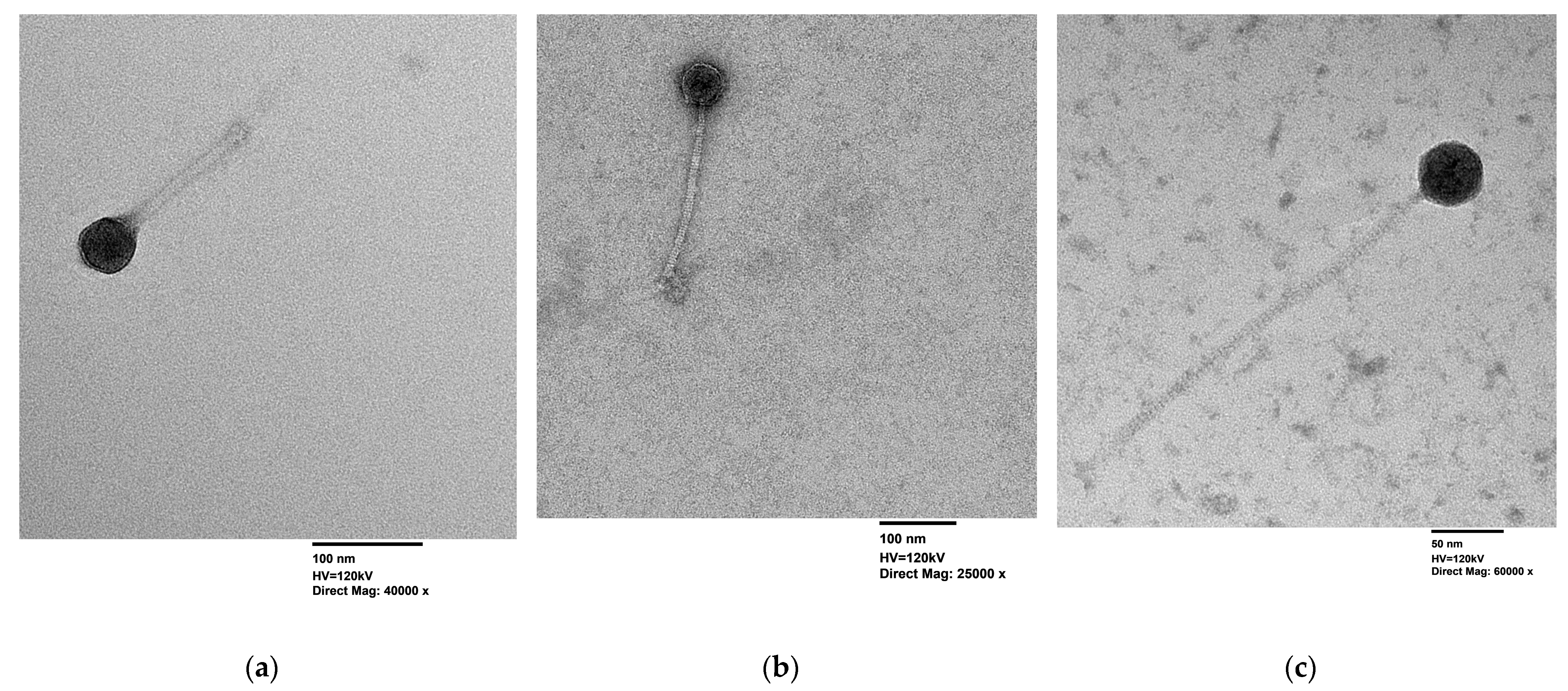
2.4.3. Transmission Electron Microscopy (TEM)

Phage particles were imaged by transmission electron microscopy (TEM) as described in [36]. Briefly, glow discharged formvar Carbon 400 mesh grids (Ted Pella Inc., 01754-F, Redding, CA, USA) were prepared using 7.5 mg/mL uranyl formate (Polysciences Inc., 24762, Warrington, PA, USA) as the staining solution. Three µL of concentrated phage stock (which was filtered to remove cellular debris) was applied to each grid for 1 min before removal using a filter paper. After drying overnight, grids were imaged using a JEOL JEM-1400Flash transmission electron microscope (JEOL Ltd., Tokyo, Japan) equipped with a tungsten filament and 15-megapixel AMT NanoSprint15 sCMOS camera (Advanced Microscopy Techniques, Woburn, MA, USA).

2.4.4. Assessing Inhibition of Bacterial Growth

Bacterial isolates were grown in BHI broth until mid to late log phase of growth was reached (approximately 5 h until OD600 = 0.5 to 0.7). Cell density was adjusted to 2 × 10⁷ CFU/mL in double-strength BHI broth. Phage lysates of known titer were adjusted to 2 × 10⁷ PFU/mL and serially diluted ten-fold in SMB to assay phage activity at various multiplicity of infections (MOI). Then equal volumes of 100 µL of cells and 100 µL phage lysates were mixed in duplicate wells of a 96-well culture plate. Each experiment was done in triplicate. Bacterial growth at 37 °C was monitored by measuring absorbance at 600 nm using a BioTek synergy H1 plate reader at 10-min intervals with discontinuous shaking for 20 h (10 s double orbital shaking 807 cm before each read). Data from the BioTek Synergy H1 microplate reader was acquired with the Gen5 software version 3.10.06 (Biotek Instruments, Agilent Technologies, Inc., Santa Clara, CA, USA). Growth curve experiments were performed with a minimum of two biological replicates. Statistical analysis and data visualizations were performed either in Microsoft Excel (Microsoft Office 16) or Python 3.10, using Matplotlib version 3.10. or Seaborn 0.13.2 plotting software. The graphical abstract was made using Biorender. Area under the curve (AUC) was calculated using Simpson’s rule [79,80]. This metric evaluates phage performance over time; a higher AUC indicates more bacterial host growth and a lower AUC indicates robust host growth inhibition. Results were visualized using bar plots that displayed the mean normalized responses with propagated error bars. Significance markers (***, **, *, ns) were added above bars based on the p-values (p < 0.001, p < 0.01, p < 0.05, p ≥ 0.05). Error bars represent propagated standard errors to account for variability in replicates and normalization. Independent two-sample t-tests assuming unequal variances (Welch’s t-test [81]) were performed to compare the normalized responses of untrained and trained phages for each condition.

2.4.5. Lifestyle Characterization

Lifestyle prediction analysis was performed on all sequenced phage genomes using the several algorithmic tools: PhageTerm (Galaxy version 1.0.12) [82,83], PhaTYP 0.3.0 [83] within PhaBox2 2.1.10 [84] and PhageAI (version 1.0.2) [85]. For 7 selected phages with ambiguous lifestyle predictions, representing both phage types under investigation, experimental validation of lysogenization capacity was conducted [86]. The temperate phage Vroast, though only partially characterized, was also included as a positive control for lysogenization. To detect lysogenized phages in bacterial survivors of infection, [86] two approaches were employed. First, spot tests were incubated extensively up to four days to promote mesa formation. Mesas are zones in the center of lysis spots which, after a prolonged incubation, show confluent bacterial growth due to the bacteria becoming resistant to the phage, possibly (but not exclusively) due to homoimmunity [86]. Bacteria from these mesas were isolated on BHI agar, and five single colonies per mesa were selected for analysis. When mesas yielded no bacterial growth, liquid infections were performed using initial bacterial concentrations of 1 × 10⁸ CFU/mL at MOI = 1, conditions known to enhance lysogeny rates [87,88]. All surviving clones underwent three successive streak purifications to eliminate residual free phage. Survivors were tested for phage immunity through double-layer agar assays, where the original phage was spotted onto bacterial lawns of the surviving isolates [86].
The presence or absence of phage DNA in bacterial genomes was then verified by PCR using phage-specific primers (Supplemental Material Table S3). Furthermore, surviving bacterial isolates were also subject to mitomycin C induction [89], using a concentration of 1.49 µM and exposure time of 14 h. The resulting lysates were spotted onto double-layer agar plates containing the naïve host to check for plaque formation.

2.4.6. Stability of Phages in Rumen Fluid

Stability of SBSEC phages in rumen fluid was evaluated in a subset of 8 phages presented in this study. Phage suspensions were diluted in 5 mL sterile rumen fluid (Bar Diamond Inc., Parma, ID, USA) at a concentration of 1 × 10⁷ PFU/mL and incubated at 37 °C under anaerobic atmosphere. The fraction of surviving phages was quantified by spotting serially diluted phages in rumen fluid on DLA. All experiments were done in duplicate and spots on DLA were done in triplicate.

2.5. Phage Training to Increase Killing Efficiency

Three of the isolated bacteriophages were subject to 30 rounds of serial passaging with their host adapting methods used by others [90,91,92,93]. Briefly, mid log host bacterial cultures were used to inoculate 5 mL BHI broth at 108 CFU/mL and infected with the corresponding phage at MOI = 0.01, followed by incubation at 37 °C statically, overnight. Serial passaging was done by transferring 100 µL of the infected culture to 5 mL of fresh BHI medium and incubating again for 8 h or overnight until 30 rounds were completed or phage loss was observed. For every 10 cycles, lysates were made by centrifugation (10,000× g for 10 min) and filter sterilization using a 25 mm diameter, 0.22 µm pore size PVDF hydrophilic syringe filter (Choice™, Thermo Scientific™, Waltham, MA, USA). The viability of the phages was confirmed by spotting lysates on a DLA of the corresponding naïve host.

3. Results

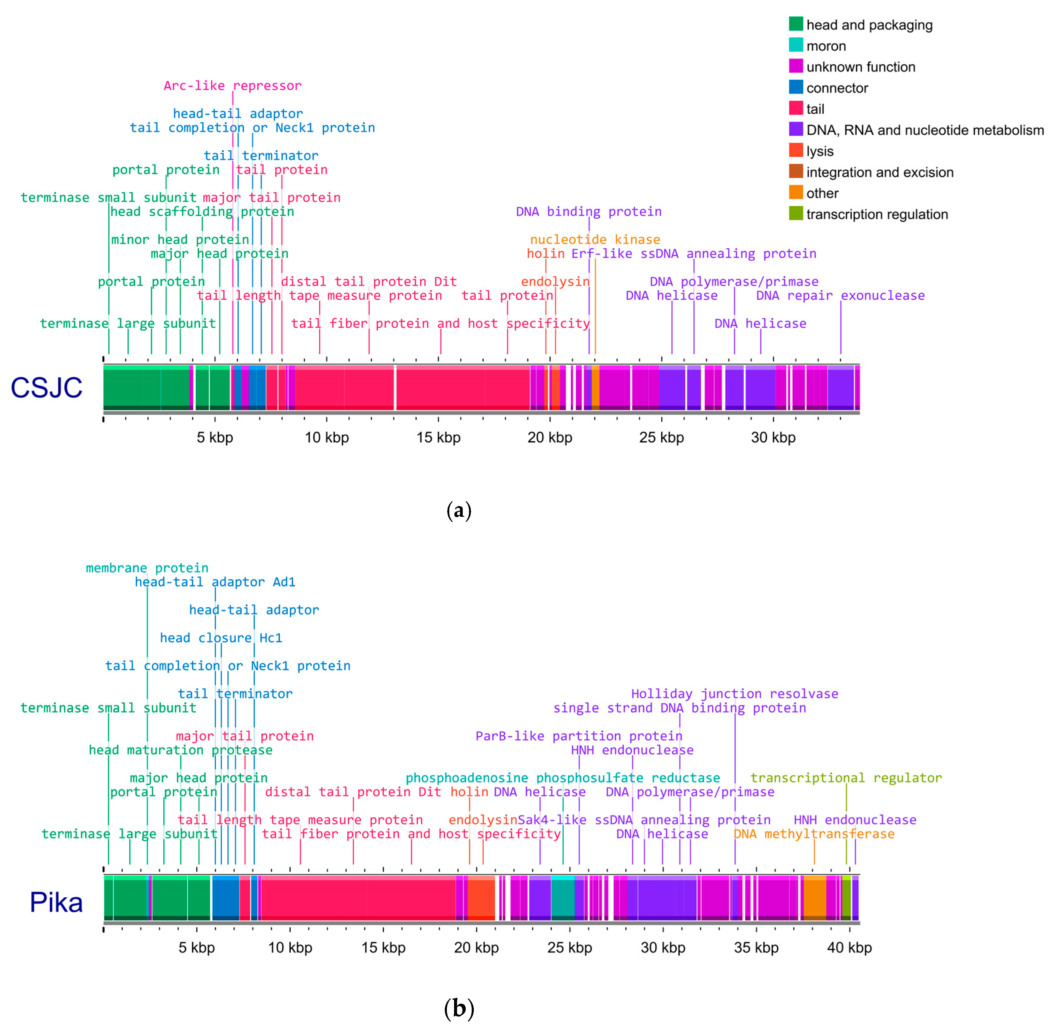
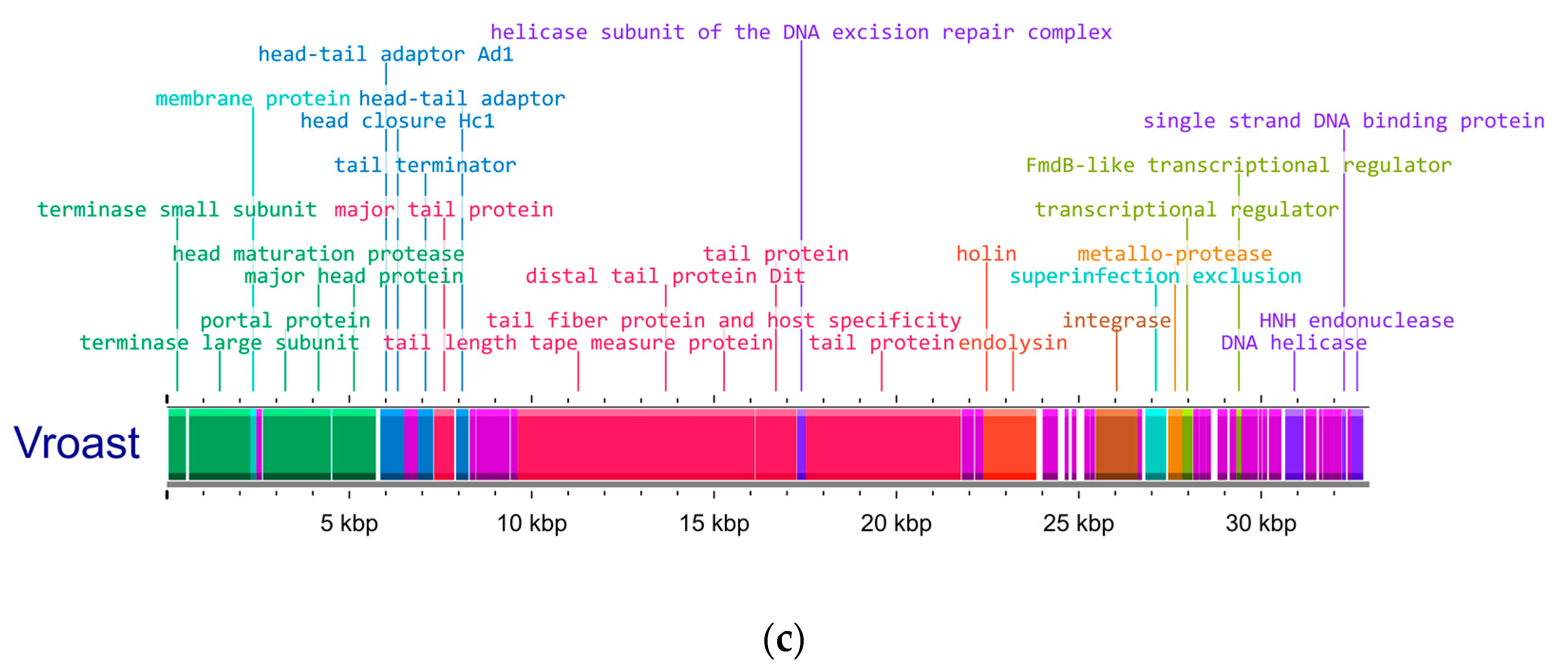
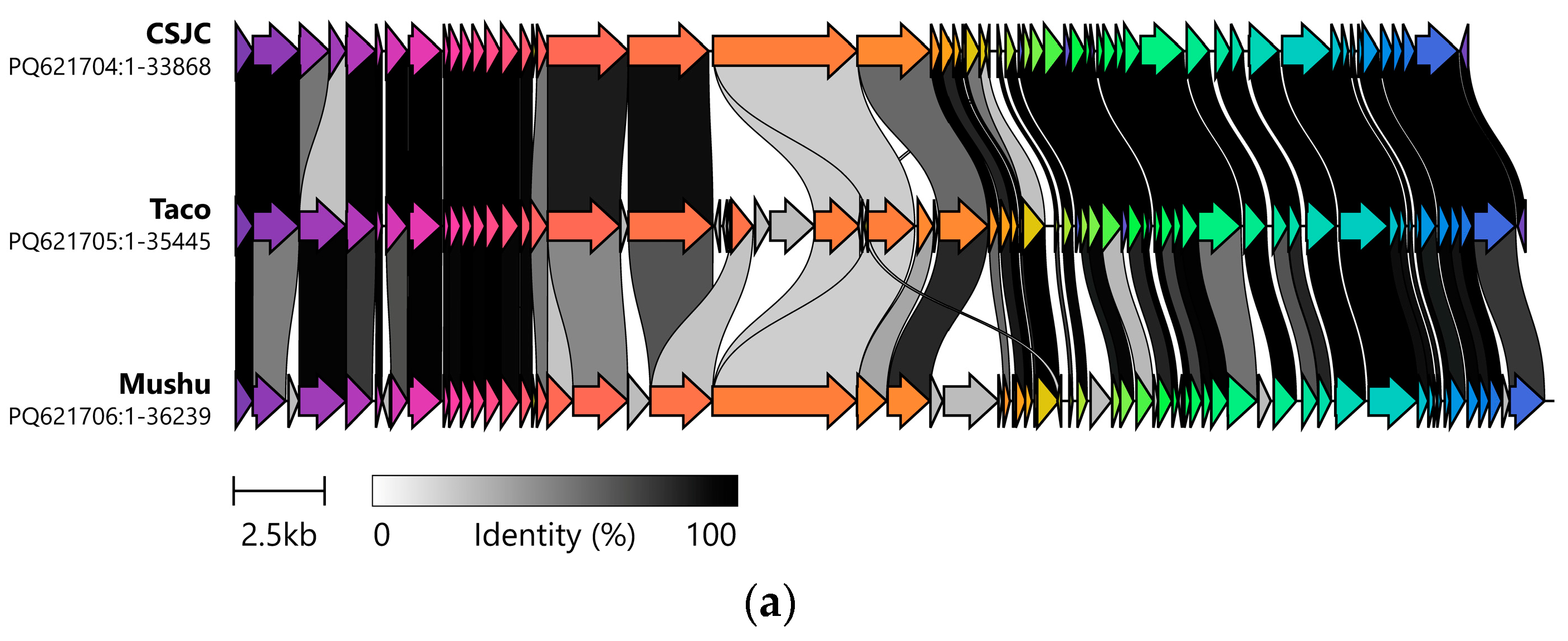
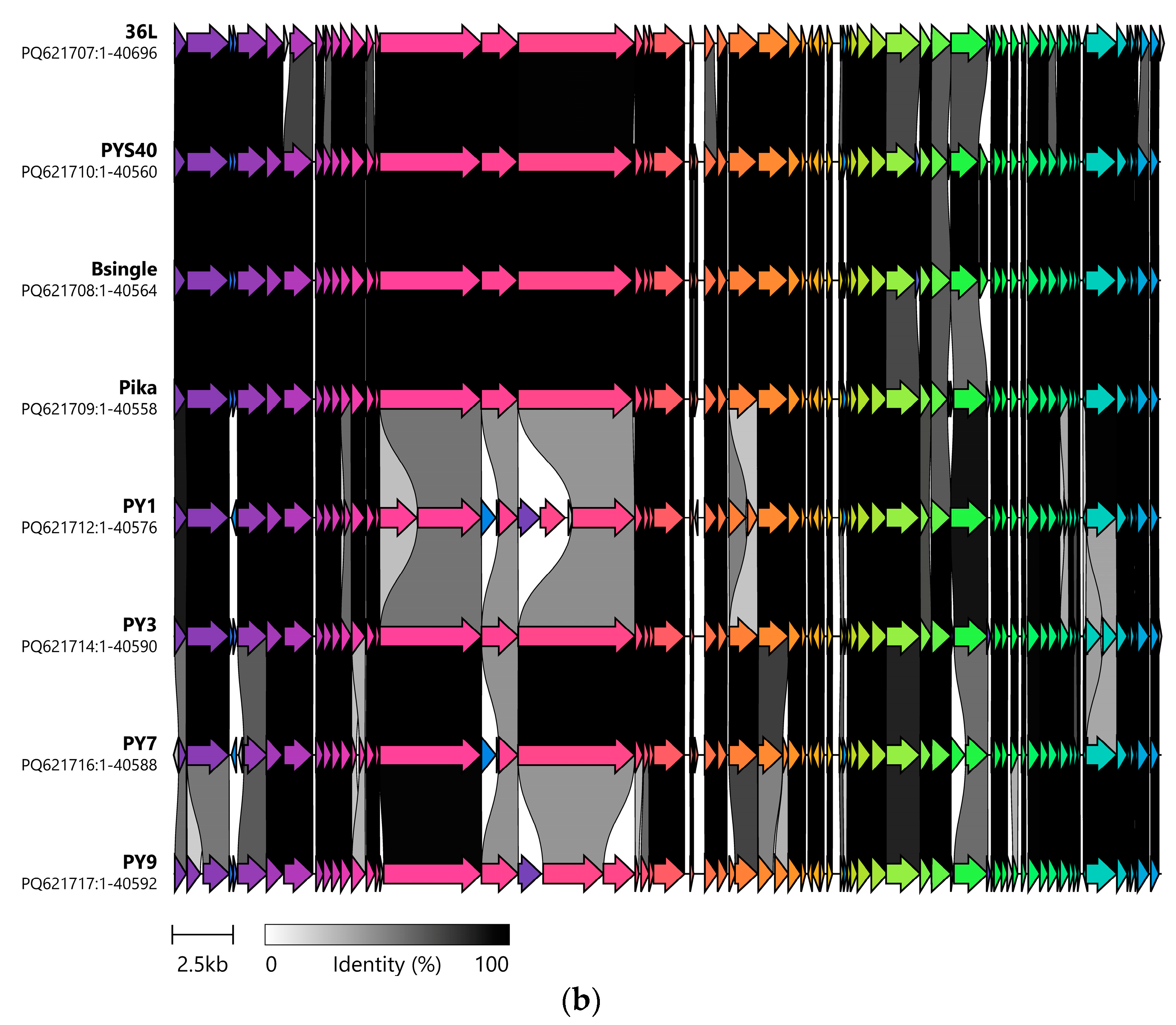
3.1. Genetic Analysis of Isolated Phages Reveals Two Clusters of Unique Phages That Have Strong Intercluster Homology

We isolated and sequenced 12 novel Streptococcus phages ranging between 33.8 and 40.5 Kbp in size, with GC content between 37% and 40% (Table 1). Annotation of the phage genomes showed clustering of genes by functional groups and all genes necessary for full phage functionality are present (Figure 1). Based on genomic similarity and phylogenetic relatedness, these phages segregated into two distinct clusters (Figure 2 and Figure 3).
Phylogenetic analysis, incorporating our 12 sequenced phages and previously sequenced Streptococcus phages (listed in Supplemental Material Table S2), grouped our phages in two different clusters and confirmed their novelty (Figure 2). All phages are predicted to belong to a common family along with other Streptococcus phages. Cluster 1, where Mushu, Taco and CSJC are, grouped them with high phylogenetic similarity with SB01 (S. equinus phage) and, to a lesser extent, the Streptococcus pasteurianus phages SG586P1 and SG586P3, all members of the SBSEC streptococci [94]. Notably, two Streptococcus thermophilus phages, D4446 and phage P738, also demonstrated close genetic relatedness to the Cluster 1 phages. Victor analysis assigned Cluster 1 phages into the same genus, though identifies them as different species. The next eight phages (as shown highlighted in Figure 2) sequenced all demonstrate high genetic similarity, leading us to consider them as a group, Cluster 2. All Cluster 2 phages share the same predicted family, genus, and species, as would be expected of highly genetically similar isolates. This group also shows some relatedness to the final phage, a singleton lysogenic phage Vroast which bears closest resemblance to the SBSEC prophage Javan 220 [78]. The SBSEC phages included in our analysis, vB_SbRt-pBovineB21, phage vB_SbRt-pBovineS21 and ImqsRe26_1, are closely related to phage C1 whose host belongs to the Lancefield group C of streptococci [75], a different clade than SBSEC. Unsurprisingly, other phages from different streptococcal species were found to have phylogenetic relatedness, such as phages ImqsRe26_1 (Streptococcus equi) and phage C1 (Group C streptococci). Altogether the species of the bacteriophage hosts did not strongly correlate with phylogenetic clustering.
Clinker analysis (Figure 3) aligning the annotations of the sequenced genomes showed that Cluster 1 phages have similar genomes and the arrangement of gene functional groups is maintained in orthologous blocks, though there were clear differences between the members of the group indicated by regions without homologues identified. Interestingly, this was not the case for Cluster 2 phages. The differences between the individual genomes spanned the entire length of the sequence and seemed to concentrate around functional genes, in particular, genes related to tail subunits and assembly.
Analysis using OrthoANI (Orthologous Average Nucleotide Identity) (Figure S3) matched the segregation of phage genomes in two clusters obtained from VICTOR phylogenomic analysis (Figure 2). OrthoANI measures the overall similarity between two genome sequences by fragmenting the genomes into 1020 bp-long pieces and calculating the average nucleotide identities in an orthologous manner and assigning a percentage similarity. According to this analysis, Mushu, CSJC and Taco are between 96.14 and 99.91% similar to each other, while cluster 2 phages are between 99.87 and 99.98% orthologically identical. Cluster 2 phages are about 86% OrthoANI similar to Javan 220, a temperate phage described elsewhere, which also showed high similarity to Vroast genome.
Imaging via TEM of a selection of the isolated phages revealed that they all present a siphovirus morphology (Figure 4), with capsids of icosahedral or almost round shape and long, thin, flexible tails. This was consistent with predictions provided by PhageTerm [82] as most siphoviruses are headful packaging phages. Analysis of phage genomes and raw sequencing reads using PhageTerm yielded predictions for packaging mechanism and classification (Supplemental Material Table S6). PhageTerm, predicted CSJC, Vroast and PY9 to be headful packaging phages. Interestingly, PY3 was classified as T5 type, Direct Terminal repeats (DTR) class whereas PY9 (which is 99.91% OrthoANI similar to PY3) was classified as P1 type and headful (PAC) class. All other phages were classified as new or unknown class and unknown type. PhageTerm determined all phage ends to be redundant and most to have permuted genomes, which is consistent with headful packaging phages.

3.2. The Isolated Streptococcus Bacteriophages Have a Limited Host Range but Are Generally Effective at Inhibiting Bacterial Growth In Vitro, and Retain Activity When Exposed to Ruminal Fluid In Vitro

The phages presented in this study demonstrated infectivity against various Streptococcus strains, including bacteria isolated from both related sources and from cattle separated by significant geographical and temporal distances. The host range profiles of all 12 bacteriophage isolates are presented in Figure 5.
We evaluated each bacteriophage’s ability to inhibit the growth of their isolation host by measuring the OD600 in liquid cultures at various MOIs (Figure 6, Supplemental Figure S1). All 12 fully sequenced bacteriophages demonstrated the ability to suppress the growth of their host to some extent, some for up to 20 h. Despite the genomic similarity found among phages, their ability to inhibit bacterial growth was observed to be very different. CSJC outperforms similar phages Mushu and Taco at inhibiting bacterial growth at all MOIs for 14 h and causing minimal growth for up to 20 h. Phage PY9 sustains better suppression of bacterial growth at all MOIs than genetically similar phages Pika, Bsingle, 36L, PY1, PY3 and PY7. All phages, except Vroast, completely inhibit growth at MOI = 1, MOI = 0.1 and sometimes even at MOI = 0.01, for at least 10 h while most perform poorly at MOI = 0.001. Excluding Taco, after 20 h the max OD600 or phage-treated cultures at MOI = 1 was only 32% of the control bacteria in the worst performing phage (PY3) and 2% in the best (CSJC) (Supplemental Material Table S4).
Stability of SBSEC phages in rumen fluid was evaluated in a subset of 8 of the 12 phages presented in this study. Phage suspensions in 5 mL sterile rumen fluid (1 × 10⁷ PFU/mL) were incubated at 37 °C under anaerobic atmosphere and the fraction of surviving phages was quantified. The titer of phages dropped 0 to 0.45 log after 2 h in rumen fluid and continued exposure for 4, 24 and 48 h further decreased phage viability but not by more than 1.36 log (Taco) and remained around less than 1 log drop for the other 7 phages tested (Figure S4). Despite this considerable loss in phage viability, >1 × 10⁶ PFU/mL remained after 48 h. In actual application of phages as a feed additive there would be additional forces at play, while some would be detrimental, phage infection of SBSEC hosts leading to the release of more phage particles is also anticipated. Animal studies would provide true insights into the viability of these phages to modulate SBSEC proliferation in the rumen. Although the shelf stability of the phages from this study was not evaluated, it was noted that titers of phage stocks stored for 4 months at 4 °C in SMB presented minimal titer loss, but longer storage had variable effects in phage stability sometimes dropping 3 log units in 10 months (Taco and PY7) and sometimes maintaining similar titers for up to 12 months (CSJC and 36L) (Supplemental Material Table S5).

3.3. All but One of the Sequenced Phages Are Lytic, but Further Analysis Is Required for Cluster 2 Phages

Analysis of phage genomes using PhaTYP and PhageAI yielded lifestyle predictions that varied among the different algorithms. With the exception of lysogenic phage Vroast, all phages were predicted to be virulent by at least one algorithm, with some receiving virulent predictions from both PhaTYP and PhageAI (Supplemental Material Table S7). Genomic analysis revealed no canonical lysogeny-associated genes (integrase, excisionase, repressor or Cro protein) in any of the 11 lytic phages. In contrast, Vroast carries an integrase, superinfection exclusion and transcriptional regulator genes encoded within the same region. In the 11 lytic phages, we found other genes that can potentially be associated to lysogeny however all of them are also found in strictly lytic phages. A comprehensive list of all the genes relevant for considerations of therapeutical use that were identified in the genomes of all the phages from this study is found in Supplemental Material Table S8. They include a transcriptional regulator upstream a HNH endonuclease in Cluster 2 phages, recombinase-like proteins in Cluster 1 and Cluster 2 phages, a single strand binding protein found in both Cluster 1 and Cluster 2 phages, and a holiday junction resolvase found in Cluster 2 phages. Interestingly, a ParB-like partition protein is present and highly conserved in the genomes of Cluster 2 phages. No antibiotic resistance genes or virulence genes were found in the genomes of the phages presented here. A sequence which is predicted to be a phosphoadenosine phosphosulfate (PAPS) reductase is also present in Cluster 2 phages.
A subset of 7 Streptococcus phages was used for the infection of host bacteria under conditions known to promote lysogeny [87,88]. The assessment included the temperate phage isolate Vroast as a positive control for lysogenization, CSJC and Mushu from Cluster 1 and PY1, PY7, 36L and Pika from Cluster 2. Five single colonies that survived infection were isolated per phage exposure. PCR screening using cluster-specific primers revealed no detectable phage DNA present in any infection survivors (Supplemental Material Table S6). Due to lack of sequencing information, no PCR-detection of phage Vroast could be done; however, mitomycin treatment to induce potential prophages resulted in 4 Vroast-exposed survivors producing infectious particles capable of lysing the naïve host. Phage sensitivity testing of surviving colonies provided additional insight into potential lysogenization. Of the 4 Vroast survivors that produced phages following mitomycin induction, 3 also showed phage immunity, confirming successful lysogenization. Some PY1 and Mushu survivors exhibited immunity to phage infection, despite negative PCR and mitomycin induction results, suggesting an alternative resistance mechanism present in 4 and 3 of the 5 survivor isolates, respectively.

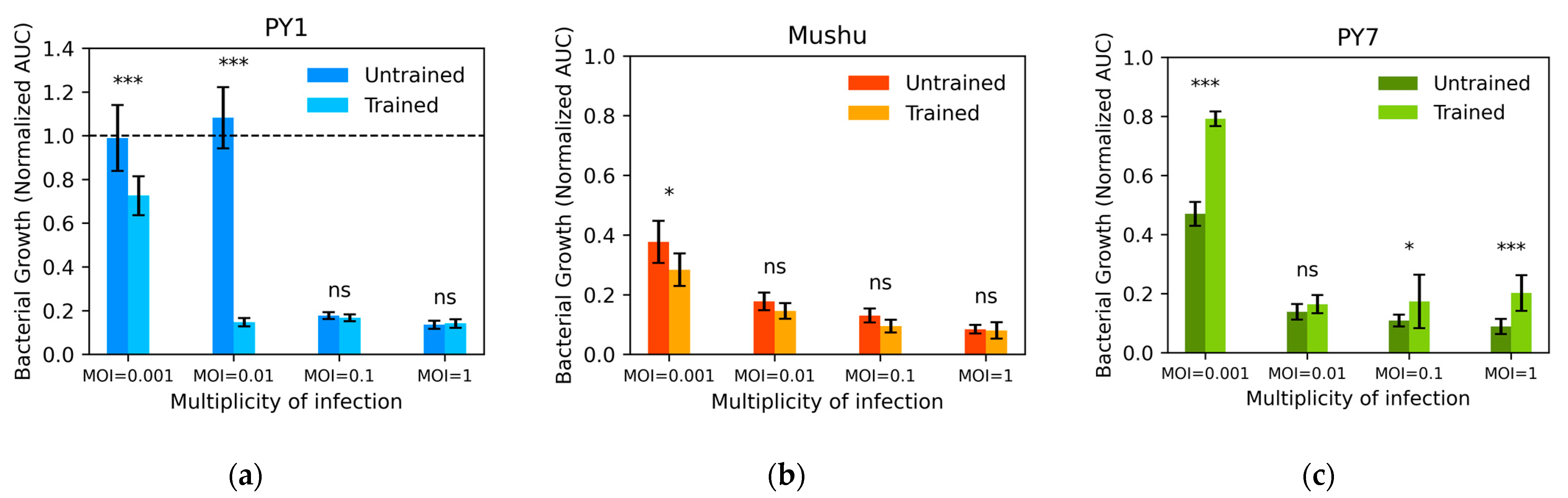
3.4. Phage Training by Serial Passaging Had Inconsistent Results

The phage training protocol of sequential co-culture by serial passaging that we applied to train three bacteriophages relies on the bacteria-bacteriophage arms-race dynamics that select for phages better at infecting bacteria, while at the same time selecting for bacteria that are better suited to survive phage infection. The ability of PY7 to inhibit bacteria growth after 16 rounds of training (after this PY7 lost its ability to plaque) was statistically worse than the original PY7 on naïve bacteria at MOIs of 0.001, 0.1 and 1 (p < 0.05). At the lowest MOI, a 32% reduction in bactericidal effect was observed. Mushu presented a 9% increase in killing efficiency at the lowest MOI, 0.001 (p < 0.05) and no difference at other MOIs. Growth kinetics of trained phage PY1 demonstrate a strong and significant improvement at the lowest MOIs of 0.001 and 0.01, improving performance as compared to untrained phage by 26% and 93%, respectively (p < 0.001) (Figure 7, Supplemental Material Figure S1).

4. Discussion

A total of 12 SBSEC bacteriophages were isolated from cattle feces or ruminal fluid and were comprehensively characterized. Despite great efforts to isolate lytic SBSEC phages, including processing of many ruminal fluid and fecal samples, only one lysogenic phage and 11 lytic phages belonging to two truly distinct phage types were isolated, congruent with findings by others and with the observation that despite the high numbers of phages in the rumen, most may exist in harmony with their hosts in a state of lysogeny or pseudolysogeny [95]. The host range of SBSEC phages presented here was limited, similar to what has been previously described by others for this bacterial species, however some phages lysed bacterial isolates from distinct geographic and temporal sources suggesting phage adaptation to common bacterial features of this species [44,46]. Most of the phages presented exerted long-term inhibition of bacterial growth in pure culture in some instances for up to 20 h, making them potential candidates as precise biocontrol agents. Although our investigation was limited to in vitro experiments, during actual applications, in vivo dynamics often amplify the effectiveness of phages due to limited resources, competitive exclusion, and environmental stresses [96,97]. While previous publications have reported the isolation and characterization of SBSEC phages, many of the early isolated SBSEC phages were lysogenic or lack full genomic sequence characterization [44,45,46,48,49,75,95,98,99]. However, these investigations laid a strong foundation in understanding the prevalence and role of bacteriophages in the ruminal ecosystem and how SBSEC phages could be applied to control bacterial proliferation in the rumen [42]. The phages isolated in this study contribute significant diversity to known Streptococcus phages and as such they may represent unique adaptations or functionality not previously described. Our results demonstrate that these phages are lytic and further investigation of their safety and performance in more complex systems or in vivo is warranted. Note, however, that phages exclusively infect bacteria and cannot infect animal or plant cells, making them inherently safe from that perspective.
Genomic sequencing along with phylogenetic and OrthoANI analysis showed that 11 of the 12 phages sequenced clustered in two different groups, here named Cluster 1 and Cluster 2, plus a singleton Vroast which were consistent with the observed differences in host range. Within each cluster there were high levels of genomic identity, particularly in Cluster 2, despite observed differences in the host range and the dissimilar ability of phages to control bacterial growth in vitro. Further resolution of single basepair differences between phage genomes within each cluster could be obtained by the use of hybrid assemblies, however assemblies generated using ONT data can be sufficient (achieving raw read accuracies of approximately 99%) especially when the goal is to generate de novo assemblies that allow identification of genes present or absent in a genome [62,100,101]. The observed phenotypic differences between highly similar phages are either related to the small genomic differences observed between genomes but could also be due to the differences in bacterial hosts used for amplification that could impart the phage lysate additional characteristics [102]. Other reports of SBSEC phages have also found high similarity between their isolated phages, perhaps indicating the prevalence of specific phage types within the ruminal ecosystem [48,49].
Similarities to other phages with lytic activity against other SBSEC bacteria were present, as expected, however phages with hosts not belonging to SBSEC were also clustered together. This similarity between Streptococcus species infecting phages has been observed previously and seems to indicate adaptation to features common to streptococci for phage attachment and to ensure protein function in closely related bacterial strains [47]. Similarities to other genus-specific phages is expected considering that phages show complex evolutionary relationships that do not follow hierarchical phylogeny due to prevalent mosaicism [103]. Phages CSJC, Mushu and Taco were grouped in Cluster 1 closely together with 3 Streptococcus phages, including S. equinus (SBSEC) phage Sb01 which is described to be lytic based on the production of clear plaques and clear bacterial cultures and due to the lack of integrase gene [47] and phages P738 and D4446 which are also deemed to be lytic [77]. The phages belonging to Cluster 2 are grouped separately from other Streptococcus phage sequences used in this analysis, but as expected from their genomic similarity, highly related to each other. Cluster 2 phages have phylogenetic relatedness and about 86% OrthoANI similarity to Streptococcus phage Javan 220. The full-length prophage Javan 220 was identified in the genome of S. equinus strain Sb02, but has not been isolated and propagated. In contrast to the 11 lytic phages from this study, Javan 220 carries an integrase and a repressor [78], as does Vroast, which are crucial functions for the temperate lifestyle of lysogenic phages.
None of the phage genomes presented in this study carry any antibiotic resistance genes or virulence genes that could render them unsuitable for any potential therapeutical applications. Cluster 1 phages carry a YopX-like protein. Although Yersinia outer proteins (Yops) are a well described group of virulence factors of plasmid origin, they are also found in the replication module of a variety of Streptococcus and Staphylococcus epidermidis bacteriophages and their function is considered to be unrelated to virulence [104,105]. Cluster 2 phages carry a potential moron (at term referring to an extra gene within a prophage genome which confers benefits the host [106]): a phosphoadenosine phosphosulfate (PAPs) reductase. Considering the importance of this protein to the metabolic function of bacteria it has been suggested that this gene could provide an advantage to its host by facilitating inorganic sulfate assimilation [107]. However, PAPs are widely distributed among phages and could also be used to recruit cysteine for phage assembly [108,109].
None of the typical hallmarks of lysogeny were identified in the genomes of the phages presented, specifically, no integrase, excisionase or repressor genes, with the exception of Vroast. However, the two tools used for lifestyle prediction, PhaTYP and PhageAI, showed conflicting results for most phages, possibly due to the identification of plausible bacterial genes. The various tools available are trained on different sets of data and as a result the outcomes can be dramatically different when analyzing the same type of data. It is important to note that all phages presented in this work were predicted to be virulent by at least one tool except for Vroast. Another consideration is the dearth of lifestyle classification of many available phage genome sequenced; therefore, the ambiguity of lifestyle classification tools may be due to flawed inputs yielding unreliable results. We looked for genes which could be indirectly related to maintaining a lysogenic lifestyle and found a limited number of CDS annotated as genes with functions that can be important for lysogeny, though they also may play a role in the lytic lifestyle of virulent phages (Supplemental Material Table S8).
Both Cluster 1 and Cluster 2 phages carry transcriptional regulators, which are most likely related to the temporal expression of phage genomes for effective replication assembly and lysis of the host, critical in both lytic and lysogenic phages [110]. In Cluster 2 phages there is a Holliday junction resolvase (HJRs), which is a key enzyme of DNA recombination rarely involved in the lysogenic lifestyle of bacteriophages [111]. In both Cluster 1 and 2 there are single-strand annealing proteins and DNA binding proteins that are frequently encoded in the genome of many bacteriophages playing a role in DNA repair and recombination, essential for phage replication and mosaicism [112]. Sak4 recombinases (found in Cluster 2 phages) are commonly found in temperate phages more than in lytic ones [113] but their recombinatorial activity is still unconfirmed [113,114]. Although Cluster 2 phages carry an HNH endonuclease, which can be associated to genome integration in lysogeny, they are also present in lytic phages and involved in DNA packaging and defense against co-infecting phages [115,116,117].
The ParB-like partition protein gene found in the genomes of Cluster 2 phages is usually an important gene for the replication and segregation of plasmid-like prophages. Although ParB-like genes can be found in strictly lytic phages this role is less frequently described [118,119]. In Cluster 2 phages, the ParB-like partition protein was highly conserved but no ParA (ATPase) could be found which is another essential gene for plasmid-like replication [120]. Likewise, no toxin/antitoxin system was evident in our phages which is often required for maintenance of plasmid phage genomes in the bacterial host [121,122]. Together with our inability to find lysogens of Cluster 1 or Cluster 2 phages in a subset of isolates it is likely that all 11 are lytic. However, the presence of ParB-like and Sak4 genes in Cluster 2 phages suggests further investigation into their lifestyle is warranted. Interestingly only survivors of PY1 and Mushu produced some bacterial clones resistant to subsequent infection, however their lack of detectable PY1 or Mushu via PCR and lack of mitomycin inducible prophages suggests alternative mechanisms for their resistance, which often involves mechanisms of reduced phage adsorption [90,123,124]. In general, the lack of phage resistance in concert with strong killing efficiencies in vitro is encouraging, and merits further investigations in more complex systems.
Evolutionary adaptation to train our bacteriophages for improved phenotypes yielded variable results. Ideally, phage training would improve bactericidal effect at the lowest MOI, allowing for the lowest dosage of phage to be applied while maintaining bacterial growth repression. Phage training by co-evolution with their host only markedly improved the bacteriolytic activity of phage PY1 at the two lowest MOIs (out of 3 phages trained) despite multiple reports of successful applications of similar protocols to generate phages with improved infectivity and biocontrol [125,126,127,128] and strong genetic similarities with phage PY7, which demonstrated a statistically significant reduction in host killing performance at the lowest MOI. However, publication biases towards positive results make it difficult to understand how often phage training results in deleterious adaptations. The antagonistic co-evolution accelerates molecular changes and generates genetic diversity intra and inter-population in which both the bacterium and the phage are allowed to evolve adaptations and counter-adaptations [90,123,129]. For this reason, it is not surprising that phages better adapted to prevailing in the co-culture environment but not better at suppressing bacterial growth in vitro can be isolated. Furthermore, there may be host dependent factors that reduce the effectiveness of these types of training strategies, which may be revealed with more comprehensive investigation in future.

5. Conclusions

This study characterizes 12 novel phages with significant bacteriolytic activity against SBSEC bacteria, demonstrating the potential of 11 lytic phages as precise and safe biocontrol tools to ameliorate ruminal acidosis and increase feed efficiency. Genomic analyses of the lytic phages confirmed their safety through the absence of virulence and antibiotic resistance genes, while the lack of canonical lysogeny-related genes and experimental evidence further supports their suitability for application. Despite the limited host ranges of individual lytic phage isolates and clear trends between the two clusters, some phages demonstrated cross-geographic efficacy, suggesting there is potential for broader application in cattle microbiome management. We also observed limited development of resistance during experiments.
Our findings contribute to the growing diversity and understanding of Streptococcus phages, enriching the genomic datasets that underpin phage ecology and evolution. These phages represent a significant step forward in advancing phage-based biocontrol strategies, particularly in mitigating challenges like ruminal acidosis and improving cattle feed efficiency in agricultural settings. Phage training via serial co-culturing yielded variable effects on bactericidal efficiency, which underscores the complexity of phage-host interactions and the necessity for further investigation into optimization strategies. Stability assessments in rumen fluid demonstrated a moderate decline in phage viability, with most isolates retaining high infectivity after prolonged exposure, an essential consideration for future in vivo applications. While shelf stability varied among isolates, select phages maintained high titers for up to a year under refrigeration, supporting their potential as viable feed additives, though investigation into stability under other methods of storage, e.g., lyophilization, remain to be pursued.
These results underscore the importance of continued research to assess efficacy, safety, and unintended consequences of phage application and manipulation, which may ultimately contribute to the development of tailored, sustainable biocontrol solutions. As the cattle industry seeks alternatives to traditional antibiotics for managing SBSEC and improving feed efficiency, further investigation into these and other reported phages offer promising alternatives for targeted microbiome modulation. Future research should focus on several key areas: evaluating phage stability and efficacy under various environmental conditions, investigating potential synergistic effects of phage combinations, and assessing the emergence of bacterial resistance in complex microbial communities. Understanding these aspects will be crucial for developing effective delivery systems and treatment protocols. This work expands the toolbox for addressing agricultural challenges and establishes a framework for future studies in microbiome engineering and pathogen control, contributing to the broader goal of sustainable livestock production.

Supplementary Materials

The following supporting information can be downloaded at: https://www.mdpi.com/article/10.3390/applmicrobiol5010028/s1, Table S1. List of bacterial strains used in this study; Table S2. Streptococcus phage genomes that were included in the phylogenetic analysis of the phage genomes generated in this study; Table S3. Phage-specific primers used in this study; Table S4. Percentage of the final OD600 of bacterial cultures exposed to phage for 20 h; Table S5. Shelf stability of phage stocks stored in SM buffer at 4 °C under aerobic atmosphere; Table S6. Characteristics of bacterial isolates that survived phage infection are indicative of lytic lifestyle for phages of Cluster 1 and Cluster 2; Table S7. Summary of the results of the analysis of the genomes of the phages presented in this study using PhaTYP, PhageAI and PhageTerm; Table S8. List of genes identified in the genomes of SBSEC phages characterized in this study with relevance for potential therapeutic use; Figure S1. Curves of bacterial growth (OD600) of SBSEC hosts exposed to different phages at various MOIs; Figure S2. Curves of bacterial growth (OD600) of SBSEC hosts exposed to different trained phages at various MOIs; Figure S3. Heatmap displaying the overall similarities including percentage values between newly isolated phages and a selection of reference genomes via OrthoANI (Orthologous Average Nucleotide Identity); Figure S4. Decrease in phage viability for 8 phages incubated in ruminal fluid at 37 °C under anaerobic conditions for 2, 4, 24, and 48 h.

Author Contributions

Conceptualization, J.M., C.S. and J.L.G.; methodology, J.L.G.; software, J.M., C.S. and J.L.G.; validation, J.M., C.S. and J.L.G.; formal analysis, J.M. and C.S.; investigation, M.T., C.H. and J.L.G.; resources, Y.J.T., J.M. and P.J.J.A.; data curation, J.M.; writing—original draft preparation, C.S. and J.L.G.; writing—review and editing, J.M., P.J.J.A. and Y.J.T.; visualization, C.S. and C.H.; supervision, J.M.; project administration, J.M.; funding acquisition, J.M. All authors have read and agreed to the published version of the manuscript.

Funding

This research was funded by USDA-NIFA award #2022-33610-37855.

Institutional Review Board Statement

Not applicable.

Informed Consent Statement

Not applicable.

Data Availability Statement

The genomes of the phages presented in this study are openly available in GenBank at the following accession numbers: PQ621712, PQ621714, PQ621716, PQ621717, PQ621707, PQ621708, PQ621709, PQ621710, PQ621704, PQ621705, PQ621706, PV146553.

Acknowledgments

The authors would like to express sincere gratitude to and acknowledge the contributions of Mitchell Clark, Megan M. Miller, and Luke Keenan, for their technical support and hard work over the course of this line of research.

Conflicts of Interest

The authors declare no conflicts of interest.

References

  1. Knight, R. USDA Economic Research Service. Cattle & Beef—Sector at a Glance. 2025. Available online: https://www.ers.usda.gov/topics/animal-products/cattle-beef/sector-at-a-glance (accessed on 30 December 2024).
  2. Ibidhi, R.; Ben Salem, H. Water footprint of livestock products and production systems: A review. Anim. Prod. Sci. 2020, 60, 1369–1380. [Google Scholar] [CrossRef]
  3. Godfray, H.C.J.; Aveyard, P.; Garnett, T.; Hall, J.W.; Key, T.J.; Lorimer, J.; Pierrehumbert, R.T.; Scarborough, P.; Springmann, M.; Jebb, S.A. Meat consumption, health, and the environment. Science 2018, 361, eaam5324. [Google Scholar] [CrossRef]
  4. Pelletier, N.; Tyedmers, P. Forecasting potential global environmental costs of livestock production 2000–2050. Proc. Natl. Acad. Sci. USA 2010, 107, 18371–18374. [Google Scholar] [CrossRef]
  5. Paz, H.A.; Hales, K.E.; Wells, J.E.; Kuehn, L.A.; Freetly, H.C.; Berry, E.D.; Flythe, M.D.; Spangler, M.L.; Fernando, S.C. Rumen bacterial community structure impacts feed efficiency in beef cattle. J. Anim. Sci. 2018, 96, 1045–1058. [Google Scholar] [CrossRef] [PubMed]
  6. Mottet, A.; de Haan, C.; Falcucci, A.; Tempio, G.; Opio, C.; Gerber, P. Livestock: On our plates or eating at our table? A new analysis of the feed/food debate. Glob. Food Secur. 2017, 14, 1–8. [Google Scholar] [CrossRef]
  7. de Vries, M.; de Boer, I.J. Comparing environmental impacts for livestock products: A review of life cycle assessments. Livest. Sci. 2010, 128, 1–11. [Google Scholar] [CrossRef]
  8. Rotz, C.A.; Asem-Hiablie, S.; Place, S.; Thoma, G. Environmental footprints of beef cattle production in the United States. Agric. Syst. 2019, 169, 1–13. [Google Scholar] [CrossRef]
  9. Wagner, J.J.; Archibeque, S.L.; Feuz, D.M. The modern feedlot for finishing cattle. Annu. Rev. Anim. Biosci. 2014, 2, 535–554. [Google Scholar] [CrossRef]
  10. Kahn, L.; Cottle, D. Beef Cattle Production and Trade; Csiro Publishing: Clayton, Australia, 2014. [Google Scholar]
  11. Coffey, D.; Dawson, K.; Ferket, P.; Connolly, A. Review of the feed industry from a historical perspective and implications for its future. J. Appl. Anim. Nutr. 2016, 4, e3. [Google Scholar] [CrossRef]
  12. Ojo, A.O.; Mulim, H.A.; Campos, G.S.; Junqueira, V.S.; Lemenager, R.P.; Schoonmaker, J.P.; Oliveira, H.R. Exploring Feed Efficiency in Beef Cattle: From Data Collection to Genetic and Nutritional Modeling. Animals 2024, 14, 3633. [Google Scholar] [CrossRef]
  13. Herd, R.; Oddy, V.; Richardson, E. Biological basis for variation in residual feed intake in beef cattle. 1. Review of potential mechanisms. Aust. J. Exp. Agric. 2004, 44, 423–430. [Google Scholar] [CrossRef]
  14. Fitzsimons, C.; McGee, M.; Keogh, K.; Waters, S.M.; Kenny, D.A. Molecular physiology of feed efficiency in beef cattle. In Biology of Domestic Animals; CRC Press: Boca Raton, FL, USA, 2017; pp. 122–165. [Google Scholar]
  15. Kenny, D.; Fitzsimons, C.; Waters, S.; McGee, M. Invited review: Improving feed efficiency of beef cattle–the current state of the art and future challenges. Animal 2018, 12, 1815–1826. [Google Scholar] [CrossRef]
  16. Nkrumah, J.; Okine, E.; Mathison, G.; Schmid, K.; Li, C.; Basarab, J.; Price, M.; Wang, Z.; Moore, S. Relationships of feedlot feed efficiency, performance, and feeding behavior with metabolic rate, methane production, and energy partitioning in beef cattle. J. Anim. Sci. 2006, 84, 145–153. [Google Scholar] [CrossRef]
  17. Herd, R.; Arthur, P. Physiological basis for residual feed intake. J. Anim. Sci. 2009, 87, E64–E71. [Google Scholar] [CrossRef] [PubMed]
  18. Moloney, A.P.; McGee, M. Factors influencing the growth of meat animals. In Lawrie’ s Meat Science; Elsevier: Amsterdam, The Netherlands, 2017; pp. 19–47. [Google Scholar]
  19. Zhou, M.; Hernandez-Sanabria, E. Characterization of variation in rumen methanogenic communities under different dietary and host feed efficiency conditions, as determined by PCR-denaturing gradient gel electrophoresis analysis. Appl. Environ. Microbiol. 2010, 76, 3776–3786. [Google Scholar] [CrossRef]
  20. Petri, R.; Schwaiger, T.; Penner, G.; Beauchemin, K.; Forster, R.; McKinnon, J.; McAllister, T. Changes in the rumen epimural bacterial diversity of beef cattle as affected by diet and induced ruminal acidosis. Appl. Environ. Microbiol. 2013, 79, 3744–3755. [Google Scholar] [CrossRef] [PubMed]
  21. Fernando, S.C.; Purvis, H.; Najar, F.; Sukharnikov, L.; Krehbiel, C.; Nagaraja, T.; Roe, B.; DeSilva, U. Rumen microbial population dynamics during adaptation to a high-grain diet. Appl. Environ. Microbiol. 2010, 76, 7482–7490. [Google Scholar] [CrossRef]
  22. Inda, M.E.; Broset, E.; Lu, T.K.; de la Fuente-Nunez, C. Emerging Frontiers in Microbiome Engineering. Trends Immunol. 2019, 40, 952–973. [Google Scholar] [CrossRef]
  23. Sheth, R.U.; Cabral, V.; Chen, S.P.; Wang, H.H. Manipulating bacterial communities by in situ microbiome engineering. Trends Genet. 2016, 32, 189–200. [Google Scholar] [CrossRef]
  24. Clemmons, B.A.; Voy, B.H.; Myer, P.R. Altering the gut microbiome of cattle: Considerations of host-microbiome interactions for persistent microbiome manipulation. Microb. Ecol. 2019, 77, 523–536. [Google Scholar] [CrossRef]
  25. DePeters, E.; George, L. Rumen transfaunation. Immunol. Lett. 2014, 162, 69–76. [Google Scholar] [CrossRef]
  26. McAllister, T.A.; Beauchemin, K.A.; Alazzeh, A.Y.; Baah, J.; Teather, R.M.; Stanford, K. Review: The use of direct fed microbials to mitigate pathogens and enhance production in cattle. Can. J. Anim. Sci. 2011, 91, 193–211. [Google Scholar] [CrossRef]
  27. Henderson, G.; Cox, F.; Ganesh, S.; Jonker, A.; Young, W.; Collaborators, G.R.C.; Abecia, L.; Angarita, E.; Aravena, P.; Arenas, G.N. Rumen microbial community composition varies with diet and host, but a core microbiome is found across a wide geographical range. Sci. Rep. 2015, 5, 14567. [Google Scholar] [CrossRef] [PubMed]
  28. Thomas, M.; Webb, M.; Ghimire, S.; Blair, A.; Olson, K.; Fenske, G.J.; Fonder, A.T.; Christopher-Hennings, J.; Brake, D.; Scaria, J. Metagenomic characterization of the effect of feed additives on the gut microbiome and antibiotic resistome of feedlot cattle. Sci. Rep. 2017, 7, 12257. [Google Scholar] [CrossRef]
  29. Callaway, T.; Edrington, T.; Anderson, R.; Harvey, R.; Genovese, K.; Kennedy, C.; Venn, D.; Nisbet, D. Probiotics, prebiotics and competitive exclusion for prophylaxis against bacterial disease. Anim. Health Res. Rev. 2008, 9, 217–225. [Google Scholar] [CrossRef] [PubMed]
  30. Gill, H.S.; Shu, Q.; Leng, R.A. Immunization with Streptococcus bovis protects against lactic acidosis in sheep. Vaccine 2000, 18, 2541–2548. [Google Scholar] [CrossRef] [PubMed]
  31. Wedlock, D.; Janssen, P.; Leahy, S.; Shu, D.; Buddle, B. Progress in the development of vaccines against rumen methanogens. Animal 2013, 7, 244–252. [Google Scholar] [CrossRef]
  32. Nagaraja, T.; Miller, G. Rumen Microbial Changes in Ionophore Antiobiotic-Treated Steers with Experimenally Induced Acidosis. Asian-Australas. J. Anim. Sci. 1989, 2, 465–468. [Google Scholar] [CrossRef]
  33. Jo, S.J.; Kwon, J.; Kim, S.G.; Lee, S.J. The Biotechnological Application of Bacteriophages: What to Do and Where to Go in the Middle of the Post-Antibiotic Era. Microorganisms 2023, 11, 2311. [Google Scholar] [CrossRef]
  34. D’Herelle, F. On an invisible microbe antagonistic toward dysenteric bacilli: Brief note by Mr. F. D’Herelle, presented by Mr. Roux. 1917. Res. Microbiol. 2007, 158, 553–554. [Google Scholar] [CrossRef]
  35. Schwarz, C.; Mathieu, J.; Laverde Gomez, J.A.; Yu, P.; Alvarez, P.J. Renaissance for Phage-Based Bacterial Control. Environ. Sci. Technol. 2021, 56, 4691–4701. [Google Scholar] [CrossRef]
  36. Schwarz, C.; Mathieu, J.; Laverde Gomez, J.; Miller, M.R.; Tikhonova, M.; Hamor, C.; Alvarez, P.J. Isolation and Characterization of Six Novel Fusobacterium necrophorum Phages. PHAGE 2024, 5, 63–75. [Google Scholar] [CrossRef] [PubMed]
  37. Schwarz, C. Ruminal Fusobacterium and Their Phages-Fundamentals and Applications; University of California: Berkeley, CA, USA, 2023. [Google Scholar]
  38. Russell, J.B.; Rychlik, J.L. Factors that alter rumen microbial ecology. Science 2001, 292, 1119–1122. [Google Scholar] [CrossRef] [PubMed]
  39. Cheng, K.-J.; McAllister, T.; Popp, J.; Hristov, A.; Mir, Z.; Shin, H. A review of bloat in feedlot cattle. J. Anim. Sci. 1998, 76, 299–308. [Google Scholar] [CrossRef]
  40. Goodrich, R.D.; Garrett, J.E.; Gast, D.R.; Kirick, M.A.; Larson, D.A.; Meiske, J.C. Influence of Monensin on the Performance of Cattle. J. Anim. Sci. 1984, 58, 1484–1498. [Google Scholar] [CrossRef]
  41. Marques, R.d.S.; Cooke, R.F. Effects of Ionophores on Ruminal Function of Beef Cattle. Animals 2021, 11, 2871. [Google Scholar] [CrossRef]
  42. Gilbert, R.A.; Klieve, A.V. Ruminal Viruses (Bacteriophages, Archaeaphages). In Rumen Microbiology: From Evolution to Revolution; Puniya, A.K., Singh, R., Kamra, D.N., Eds.; Springer: New Delhi, India, 2015; pp. 121–141. [Google Scholar]
  43. Tarakanov, B.V. Biology of lysogenic strains of Streptococcus bovis and virulent mutants of their temperate phages. Mikrobiologiia 1996, 65, 656–662. [Google Scholar] [PubMed]
  44. Klieve, A.V.; Heck, G.L.; Prance, M.A.; Shu, Q. Genetic homogeneity and phage susceptibility of ruminal strains of Streptococcus bovis isolated in Australia. Lett. Appl. Microbiol. 1999, 29, 108–112. [Google Scholar] [CrossRef]
  45. Iverson, W.G.; Millis, N.F. Characterization of Streptococcus bovis bacteriophages. Can. J. Microbiol. 1976, 22, 847–852. [Google Scholar] [CrossRef]
  46. Štyriak, I.; Španová, A.; Montagová, H.; Kmet, V. Isolation and characterization of a new ruminal bacteriophage lytic to Streptococcus bovis. Curr. Microbiol. 1994, 28, 355–358. [Google Scholar] [CrossRef]
  47. Gilbert, R.A.; Kelly, W.J.; Altermann, E.; Leahy, S.C.; Minchin, C.; Ouwerkerk, D.; Klieve, A.V. Toward Understanding Phage:Host Interactions in the Rumen; Complete Genome Sequences of Lytic Phages Infecting Rumen Bacteria. Front. Microbiol. 2017, 8, 2340. [Google Scholar] [CrossRef]
  48. Park, S.Y.; Kwon, H.; Kim, S.G.; Park, S.C.; Kim, J.H.; Seo, S. Characterization of two lytic bacteriophages, infecting Streptococcus bovis/equinus complex (SBSEC) from Korean ruminant. Sci. Rep. 2023, 13, 9110. [Google Scholar] [CrossRef]
  49. Köhne, M.; Hüsch, R.; Tönissen, A.; Schmidt, M.; Müsken, M.; Böttcher, D.; Hirnet, J.; Plötz, M.; Kittler, S.; Sieme, H. Isolation and characterization of bacteriophages specific to Streptococcus equi subspecies zooepidemicus and evaluation of efficacy ex vivo. Front. Microbiol. 2024, 15, 1448958. [Google Scholar] [CrossRef] [PubMed]
  50. Gilbert, R.A.; Townsend, E.M.; Crew, K.S.; Hitch, T.C.A.; Friedersdorff, J.C.A.; Creevey, C.J.; Pope, P.B.; Ouwerkerk, D.; Jameson, E. Rumen Virus Populations: Technological Advances Enhancing Current Understanding. Front. Microbiol. 2020, 11, 450. [Google Scholar] [CrossRef] [PubMed]
  51. McDaniel, Z.S.; Hales, K.E.; Nagaraja, T.G.; Lawrence, T.E.; Tennant, T.C.; Amachawadi, R.G.; Carroll, J.A.; Burdick Sanchez, N.C.; Galyean, M.L.; Davis, E.; et al. Validation of an experimental model to induce liver abscesses in Holstein steers using an acidotic diet challenge and intraruminal bacterial inoculation. Appl. Anim. Sci. 2024, 40, 398–413. [Google Scholar] [CrossRef]
  52. Schwarz, C.; Mathieu, J.; Gomez, J.L.; Miller, M.R.; Tikhonova, M.; Nagaraja, T.G.; Alvarez, P.J. Unexpected finding of Fusobacterium varium as the dominant Fusobacterium species in cattle rumen: Potential implications for liver abscess etiology and interventions. J. Anim. Sci. 2023, 101, skad130. [Google Scholar] [CrossRef]
  53. Russell, J.B.; Robinson, P.H. Compositions and characteristics of strains of Streptococcus bovis. J. Dairy Sci. 1984, 67, 1525–1531. [Google Scholar] [CrossRef]
  54. Martin, S.A.; Russell, J.B. Transport and phosphorylation of disaccharides by the ruminal bacterium Streptococcus bovis. Appl. Environ. Microbiol. 1987, 53, 2388–2393. [Google Scholar] [CrossRef]
  55. Heuer, H.; Krsek, M.; Baker, P.; Smalla, K.; Wellington, E.M. Analysis of actinomycete communities by specific amplification of genes encoding 16S rRNA and gel-electrophoretic separation in denaturing gradients. Appl. Environ. Microbiol. 1997, 63, 3233–3241. [Google Scholar] [CrossRef]
  56. Adams, M.H. Bacteriophages; Inter-Science Publishers: New York, NY, USA, 1959. [Google Scholar]
  57. Protocol: Phage DNA Extraction-Traditional; Center for Phage Technology, Texas A&M University: College Station, TX, USA, 2018.
  58. Shen, W.; Le, S.; Li, Y.; Hu, F. SeqKit: A cross-platform and ultrafast toolkit for FASTA/Q file manipulation. PLoS ONE 2016, 11, e0163962. [Google Scholar] [CrossRef]
  59. Shen, W.; Sipos, B.; Zhao, L. SeqKit2: A Swiss army knife for sequence and alignment processing. iMeta 2024, 3, e191. [Google Scholar] [CrossRef] [PubMed]
  60. Kolmogorov, M.; Yuan, J.; Lin, Y.; Pevzner, P.A. Assembly of long, error-prone reads using repeat graphs. Nat. Biotechnol. 2019, 37, 540–546. [Google Scholar] [CrossRef] [PubMed]
  61. Wick, R.R.; Judd, L.M.; Gorrie, C.L.; Holt, K.E. Unicycler: Resolving bacterial genome assemblies from short and long sequencing reads. PLoS Comput. Biol. 2017, 13, e1005595. [Google Scholar] [CrossRef]
  62. Wick, R.R.; Judd, L.M.; Gorrie, C.L.; Holt, K.E. Completing bacterial genome assemblies with multiplex MinION sequencing. Microb. Genom. 2017, 3, e000132. [Google Scholar] [CrossRef]
  63. Koren, S.; Walenz, B.P.; Berlin, K.; Miller, J.R.; Bergman, N.H.; Phillippy, A.M. Canu: Scalable and accurate long-read assembly via adaptive k-mer weighting and repeat separation. Genome Res. 2017, 27, 722–736. [Google Scholar] [CrossRef] [PubMed]
  64. Nurk, S.; Walenz, B.P.; Rhie, A.; Vollger, M.R.; Logsdon, G.A.; Grothe, R.; Miga, K.H.; Eichler, E.E.; Phillippy, A.M.; Koren, S. HiCanu: Accurate assembly of segmental duplications, satellites, and allelic variants from high-fidelity long reads. Genome Res. 2020, 30, 1291–1305. [Google Scholar] [CrossRef]
  65. Bouras, G.; Nepal, R.; Houtak, G.; Psaltis, A.J.; Wormald, P.-J.; Vreugde, S. Pharokka: A fast scalable bacteriophage annotation tool. Bioinformatics 2022, 39, btac776. [Google Scholar] [CrossRef] [PubMed]
  66. Camacho, C.; Coulouris, G.; Avagyan, V.; Ma, N.; Papadopoulos, J.; Bealer, K.; Madden, T.L. BLAST+: Architecture and applications. BMC Bioinform. 2009, 10, 421. [Google Scholar] [CrossRef]
  67. Grant, J.R.; Enns, E.; Marinier, E.; Mandal, A.; Herman, E.K.; Chen, C.-y.; Graham, M.; Van Domselaar, G.; Stothard, P. Proksee: In-depth characterization and visualization of bacterial genomes. Nucleic Acids Res. 2023, 51, W484–W492. [Google Scholar] [CrossRef]
  68. Gilchrist, C.L.M.; Chooi, Y.H. clinker & clustermap.js: Automatic generation of gene cluster comparison figures. Bioinformatics 2021, 37, 2473–2475. [Google Scholar] [CrossRef]
  69. Lee, I.; Ouk Kim, Y.; Park, S.C.; Chun, J. OrthoANI: An improved algorithm and software for calculating average nucleotide identity. Int. J. Syst. Evol. Microbiol. 2016, 66, 1100–1103. [Google Scholar] [CrossRef]
  70. Meier-Kolthoff, J.P.; Göker, M. VICTOR: Genome-based phylogeny and classification of prokaryotic viruses. Bioinformatics 2017, 33, 3396–3404. [Google Scholar] [CrossRef]
  71. Meier-Kolthoff, J.P.; Auch, A.F.; Klenk, H.-P.; Göker, M. Genome sequence-based species delimitation with confidence intervals and improved distance functions. BMC Bioinform. 2013, 14, 60. [Google Scholar] [CrossRef] [PubMed]
  72. Göker, M.; García-Blázquez, G.; Voglmayr, H.; Tellería, M.T.; Martín, M.P. Molecular taxonomy of phytopathogenic fungi: A case study in Peronospora. PLoS ONE 2009, 4, e6319. [Google Scholar] [CrossRef]
  73. Meier-Kolthoff, J.P.; Auch, A.F.; Klenk, H.P.; Göker, M. Highly parallelized inference of large genome-based phylogenies. Concurr. Comput. Pract. Exp. 2014, 26, 1715–1729. [Google Scholar] [CrossRef]
  74. Tiwari, R.; Artiushin, S.; Timoney, J.F. P9, a temperate bacteriophage of Streptococcus equi. Int. Congr. Ser. 2006, 1289, 165–168. [Google Scholar] [CrossRef]
  75. Nelson, D.; Schuch, R.; Zhu, S.; Tscherne, D.M.; Fischetti, V.A. Genomic Sequence of C1 the First Streptococcal Phage. J. Bacteriol. 2003, 185, 3325–3332. [Google Scholar] [CrossRef] [PubMed]
  76. Sabri, M.; Häuser, R.; Ouellette, M.; Liu, J.; Dehbi, M.; Moeck, G.; García, E.; Titz, B.; Uetz, P.; Moineau, S. Genome annotation and intraviral interactome for the Streptococcus pneumoniae virulent phage Dp-1. J. Bacteriol. 2011, 193, 551–562. [Google Scholar] [CrossRef]
  77. Philippe, C.; Levesque, S.; Dion, M.; Tremblay, D.; Horvath, P.; Lüth, N.; Cambillau, C.; Franz, C.; Neve, H.; Fremaux, C. Genomic and morphological characterization of a novel genus of phages infecting Streptococcus thermophilus. Appl. Environ. Microbiol. 2020, 86, e00227-20. [Google Scholar] [CrossRef]
  78. Rezaei Javan, R.; Ramos-Sevillano, E.; Akter, A.; Brown, J.; Brueggemann, A.B. Prophages and satellite prophages are widespread in Streptococcus and may play a role in pneumococcal pathogenesis. Nat. Commun. 2019, 10, 4852. [Google Scholar] [CrossRef]
  79. Blazanin, M.; Vasen, E.; Jolis, C.V.; An, W.; Turner, P.E. Theoretical validation of growth curves for quantifying phage-bacteria interactions. bioRxiv 2023. [Google Scholar] [CrossRef]
  80. Rajnovic, D.; Muñoz-Berbel, X.; Mas, J. Fast phage detection and quantification: An optical density-based approach. PLoS ONE 2019, 14, e0216292. [Google Scholar] [CrossRef]
  81. West, R.M. Best practice in statistics: Use the Welch t-test when testing the difference between two groups. Ann. Clin. Biochem. 2021, 58, 267–269. [Google Scholar] [CrossRef]
  82. Garneau, J.R.; Depardieu, F.; Fortier, L.-C.; Bikard, D.; Monot, M. PhageTerm: A tool for fast and accurate determination of phage termini and packaging mechanism using next-generation sequencing data. Sci. Rep. 2017, 7, 8292. [Google Scholar] [CrossRef]
  83. Shang, J.; Tang, X.; Sun, Y. PhaTYP: Predicting the lifestyle for bacteriophages using BERT. Brief. Bioinform. 2022, 24, bbac487. [Google Scholar] [CrossRef]
  84. Shang, J.; Peng, C.; Liao, H.; Tang, X.; Sun, Y. PhaBOX: A web server for identifying and characterizing phage contigs in metagenomic data. Bioinform. Adv. 2023, 3, vbad101. [Google Scholar] [CrossRef] [PubMed]
  85. Tynecki, P.; Guziński, A.; Kazimierczak, J.; Jadczuk, M.; Dastych, J.; Onisko, A. PhageAI—Bacteriophage Life Cycle Recognition with Machine Learning and Natural Language Processing. bioRxiv 2020. [Google Scholar] [CrossRef]
  86. Altamirano, F.L.G.; Barr, J.J. Screening for Lysogen Activity in Therapeutically Relevant Bacteriophages. Bio-Protocol 2021, 11, e3997. [Google Scholar] [CrossRef]
  87. Golding, I.; Coleman, S.; Nguyen, T.; Yao, T. Decision Making by Temperate Phages. Encycl. Virol. 2019, 1, 5. [Google Scholar]
  88. Luque, A.; Silveira, C.B. Quantification of Lysogeny Caused by Phage Coinfections in Microbial Communities from Biophysical Principles. mSystems 2020, 5, e00353-20. [Google Scholar] [CrossRef]
  89. Levine, M. Effect of mitomycin C on interactions between temperate phages and bacteria. Virology 1961, 13, 493–499. [Google Scholar] [CrossRef] [PubMed]
  90. Borin, J.M.; Avrani, S.; Barrick, J.E.; Petrie, K.L.; Meyer, J.R. Coevolutionary phage training leads to greater bacterial suppression and delays the evolution of phage resistance. Proc. Natl. Acad. Sci. USA 2021, 118, e2104592118. [Google Scholar] [CrossRef]
  91. Meyer, J.R.; Dobias, D.T.; Weitz, J.S.; Barrick, J.E.; Quick, R.T.; Lenski, R.E. Repeatability and contingency in the evolution of a key innovation in phage lambda. Science 2012, 335, 428–432. [Google Scholar] [CrossRef]
  92. Ngiam, L.; Weynberg, K.; Guo, J. Evolutionary and co-evolutionary phage training approaches enhance bacterial suppression and delay the emergence of phage resistance. ISME Commun. 2024, 4, ycae082. [Google Scholar] [CrossRef] [PubMed]
  93. Borges, A.L. How to train your bacteriophage. Proc. Natl. Acad. Sci. USA 2021, 118, e2109434118. [Google Scholar] [CrossRef]
  94. Gao, X.Y.; Zhi, X.Y.; Li, H.W.; Klenk, H.P.; Li, W.J. Comparative genomics of the bacterial genus Streptococcus illuminates evolutionary implications of species groups. PLoS ONE 2014, 9, e101229. [Google Scholar] [CrossRef] [PubMed]
  95. Klieve, A.V.; Swain, R.A.; Nolan, J. Bacteriophages in the rumen: Types present, population size and implications for the efficiency of feed utilisation. In Proceedings of the Australian Society of Animal Production, Brisbane, QLD, Australia, 8–12 July 1996; pp. 92–94. [Google Scholar]
  96. Sun, R.; Yu, P.; Zuo, P.; Villagrán, D.; Mathieu, J.; Alvarez, P.J.J. Biofilm Control in Flow-Through Systems Using Polyvalent Phages Delivered by Peptide-Modified M13 Coliphages with Enhanced Polysaccharide Affinity. Environ. Sci. Technol. 2022, 56, 17177–17187. [Google Scholar] [CrossRef]
  97. Yu, P.; Mathieu, J.; Yang, Y.; Alvarez, P.J.J. Suppression of Enteric Bacteria by Bacteriophages: Importance of Phage Polyvalence in the Presence of Soil Bacteria. Environ. Sci. Technol. 2017, 51, 5270–5278. [Google Scholar] [CrossRef]
  98. Klieve, A.V.; Bauchop, T. Phage resistance and altered growth habit in a strain of Streptococcus bovis. FEMS Microbiol. Lett. 1991, 80, 155–159. [Google Scholar] [CrossRef]
  99. Štyriak, I.; Španová, A.; Žitňan, R. Partial characterization of two ruminal bacteriophages with similar restriction patterns and different capsids morphology. Arch. Anim. Breed. 2005, 48, 572–579. [Google Scholar] [CrossRef]
  100. Wick, R.R.; Judd, L.M.; Holt, K.E. Assembling the perfect bacterial genome using Oxford Nanopore and Illumina sequencing. PLoS Comput. Biol. 2023, 19, e1010905. [Google Scholar] [CrossRef]
  101. Hernandez, S.I.; Berezin, C.-T.; Miller, K.M.; Peccoud, S.J.; Peccoud, J. Hybrid sequencing facilitates robust de novo plasmid assembly. bioRxiv 2024. [Google Scholar] [CrossRef]
  102. Peters, T.L.; Schow, J.; Spencer, E.; Leuven, J.T.V.; Wichman, H.; Miller, C. Directed evolution of bacteriophages: Thwarted by prolific prophage. Appl. Environ. Microbiol. 2024, 90, e00884-24. [Google Scholar] [CrossRef]
  103. Dion, M.B.; Oechslin, F.; Moineau, S. Phage diversity, genomics and phylogeny. Nat. Rev. Microbiol. 2020, 18, 125–138. [Google Scholar] [CrossRef] [PubMed]
  104. Philippe, C.; Chaïb, A.; Jaomanjaka, F.; Claisse, O.; Lucas, P.M.; Samot, J.; Cambillau, C.; Le Marrec, C. Characterization of the First Virulent Phage Infecting Oenococcus oeni, the Queen of the Cellars. Front. Microbiol. 2021, 11, 596541. [Google Scholar] [CrossRef] [PubMed]
  105. Gutiérrez, D.; Martínez, B.; Rodríguez, A.; García, P. Genomic characterization of two Staphylococcus epidermidis bacteriophages with anti-biofilm potential. BMC Genom. 2012, 13, 228. [Google Scholar] [CrossRef]
  106. Cumby, N.; Davidson, A.R.; Maxwell, K.L. The moron comes of age. Bacteriophage 2012, 2, 225–228. [Google Scholar] [CrossRef]
  107. Farlow, J.; Bolkvadze, D.; Leshkasheli, L.; Kusradze, I.; Kotorashvili, A.; Kotaria, N.; Balarjishvili, N.; Kvachadze, L.; Nikolich, M.; Kutateladze, M. Genomic characterization of three novel Basilisk-like phages infecting Bacillus anthracis. BMC Genom. 2018, 19, 685. [Google Scholar] [CrossRef]
  108. Wallace, B.A.; Varona, N.S.; Hesketh-Best, P.J.; Stiffler, A.K.; Silveira, C.B. Globally distributed bacteriophage genomes reveal mechanisms of tripartite phage–bacteria–coral interactions. ISME J. 2024, 18, wrae132. [Google Scholar] [CrossRef]
  109. Kieft, K.; Zhou, Z.; Anderson, R.E.; Buchan, A.; Campbell, B.J.; Hallam, S.J.; Hess, M.; Sullivan, M.B.; Walsh, D.A.; Roux, S.; et al. Ecology of inorganic sulfur auxiliary metabolism in widespread bacteriophages. Nat. Commun. 2021, 12, 3503. [Google Scholar] [CrossRef]
  110. Yang, H.; Ma, Y.; Wang, Y.; Yang, H.; Shen, W.; Chen, X. Transcription regulation mechanisms of bacteriophages: Recent advances and future prospects. Bioengineered 2014, 5, 300–304. [Google Scholar] [CrossRef]
  111. Aravind, L.; Makarova, K.S.; Koonin, E.V. SURVEY AND SUMMARY: Holliday junction resolvases and related nucleases: Identification of new families, phyletic distribution and evolutionary trajectories. Nucleic Acids Res. 2000, 28, 3417–3432. [Google Scholar] [CrossRef] [PubMed]
  112. Neamah, M.M.; Mir-Sanchis, I.; López-Sanz, M.; Acosta, S.; Baquedano, I.; Haag, A.F.; Marina, A.; Ayora, S.; Penadés, J.R. Sak and Sak4 recombinases are required for bacteriophage replication in Staphylococcus aureus. Nucleic Acids Res. 2017, 45, 6507–6519. [Google Scholar] [CrossRef] [PubMed]
  113. Lopes, A.; Amarir-Bouhram, J.; Faure, G.; Petit, M.-A.; Guerois, R. Detection of novel recombinases in bacteriophage genomes unveils Rad52, Rad51 and Gp2.5 remote homologs. Nucleic Acids Res. 2010, 38, 3952–3962. [Google Scholar] [CrossRef] [PubMed]
  114. Hutinet, G.; Besle, A.; Son, O.; McGovern, S.; Guerois, R.; Petit, M.-A.; Ochsenbein, F.; Lecointe, F. Sak4 of Phage HK620 Is a RecA Remote Homolog With Single-Strand Annealing Activity Stimulated by Its Cognate SSB Protein. Front. Microbiol. 2018, 9, 743. [Google Scholar] [CrossRef]
  115. Zhang, L.; Xu, D.; Huang, Y.; Zhu, X.; Rui, M.; Wan, T.; Zheng, X.; Shen, Y.; Chen, X.; Ma, K.; et al. Structural and functional characterization of deep-sea thermophilic bacteriophage GVE2 HNH endonuclease. Sci. Rep. 2017, 7, 42542. [Google Scholar] [CrossRef]
  116. Wilson, G.W.; Edgell, D.R. Phage T4 mobE promotes trans homing of the defunct homing endonuclease I-TevIII. Nucleic Acids Res. 2009, 37, 7110–7123. [Google Scholar] [CrossRef]
  117. Moodley, S.; Maxwell, K.L.; Kanelis, V. The protein gp74 from the bacteriophage HK97 functions as a HNH endonuclease. Protein Sci. 2012, 21, 809–818. [Google Scholar] [CrossRef]
  118. Bignell, C.; Thomas, C.M. The bacterial ParA-ParB partitioning proteins. J. Biotechnol. 2001, 91, 1–34. [Google Scholar] [CrossRef]
  119. Ludtke, D.N.; Eichorn, B.G.; Austin, S.J. Plasmid-partition functions of the P7 prophage. J. Mol. Biol. 1989, 209, 393–406. [Google Scholar] [CrossRef]
  120. Andrews, T.; Hoyer, J.S.; Ficken, K.; Fey, P.D.; Duffy, S.; Boyd, J.M. A Transducing Bacteriophage Infecting Staphylococcus epidermidis Contributes to the Expansion of a Novel Siphovirus Genus and Implies the Genus Is Inappropriate for Phage Therapy. Msphere 2023, 8, e00524-22. [Google Scholar] [CrossRef]
  121. Łobocka, M.B.; Rose, D.J.; Plunkett, G.; Rusin, M.; Samojedny, A.; Lehnherr, H.; Yarmolinsky, M.B.; Blattner, F.R. Genome of Bacteriophage P1. J. Bacteriol. 2004, 186, 7032–7068. [Google Scholar] [CrossRef] [PubMed]
  122. Lehnherr, H.; Maguin, E.; Jafri, S.; Yarmolinsky, M.B. Plasmid addiction genes of bacteriophage P1: Doc, which causes cell death on curing of prophage, and phd, which prevents host death when prophage is retained. J. Mol. Biol. 1993, 233, 414–428. [Google Scholar] [CrossRef] [PubMed]
  123. Bull, J.J.; Vegge, C.S.; Schmerer, M.; Chaudhry, W.N.; Levin, B.R. Phenotypic resistance and the dynamics of bacterial escape from phage control. PLoS ONE 2014, 9, e94690. [Google Scholar] [CrossRef] [PubMed]
  124. Egido, J.E.; Costa, A.R.; Aparicio-Maldonado, C.; Haas, P.-J.; Brouns, S.J.J. Mechanisms and clinical importance of bacteriophage resistance. FEMS Microbiol. Rev. 2021, 46, fuab048. [Google Scholar] [CrossRef]
  125. Habusha, M.; Tzipilevich, E.; Fiyaksel, O.; Ben-Yehuda, S. A mutant bacteriophage evolved to infect resistant bacteria gained a broader host range. Mol. Microbiol. 2019, 111, 1463–1475. [Google Scholar] [CrossRef]
  126. Favor, A.H.; Llanos, C.D.; Youngblut, M.D.; Bardales, J.A. Optimizing bacteriophage engineering through an accelerated evolution platform. Sci. Rep. 2020, 10, 13981. [Google Scholar] [CrossRef]
  127. Laanto, E.; Mäkelä, K.; Hoikkala, V.; Ravantti, J.J.; Sundberg, L.-R. Adapting a phage to combat phage resistance. Antibiotics 2020, 9, 291. [Google Scholar] [CrossRef]
  128. Peters, T.L.; Song, Y.; Bryan, D.W.; Hudson, L.K.; Denes, T.G. Mutant and recombinant phages selected from in vitro coevolution conditions overcome phage-resistant Listeria monocytogenes. Appl. Environ. Microbiol. 2020, 86, e02138-20. [Google Scholar] [CrossRef]
  129. Paterson, S.; Vogwill, T.; Buckling, A.; Benmayor, R.; Spiers, A.J.; Thomson, N.R.; Quail, M.; Smith, F.; Walker, D.; Libberton, B.; et al. Antagonistic coevolution accelerates molecular evolution. Nature 2010, 464, 275–278. [Google Scholar] [CrossRef]
Figure 1. Simplified gene annotations produced using Phrokka of one representative of each cluster of phages showing the canonical organization of genes in functional groups, (a) phage CSJC, of Cluster 1 (b) phage Pika of Cluster 2, and (c) Vroast, the single lysogenic phage presented in this cohort. Hypothetical genes identified using Phrokka (v. 1.2.0) of unknown function are displayed, but unlabeled. Gene maps were generated using Proksee (v1.0.0a6) [67].
Applmicrobiol 05 00028 g001aApplmicrobiol 05 00028 g001b
Figure 2. Nucleotide based phylogenomic analysis (Formula D0) using VICTOR shows that the phages from this study (highlighted in orange) cluster in two separate groups and are unique and novel. Phages of Cluster 1 (CSJC, Taco and Mushu) bear high proximity to three other Streptococcus phage sequences currently available in genomic databases. The other cluster (Cluster 2) encompasses the remainder of the phages reported in this study are grouped together, at high relative distance from deposited phages included in this analysis. The singleton lysogenic phage, Vroast, clustered more closely to the Cluster 2 group, though it was identified as a probable different species from this group, as well as from its closest BLASTn match in the Virus nt database (Javan 220). This phylogenetic analysis included characterized fully sequenced Streptococcus phage genomes listed in Supplemental Material Table S2.
Applmicrobiol 05 00028 g002
Figure 3. Alignments of genome annotations of phages generated with clinker in CAGECAT [68]. (a) Alignment of Cluster 1 phages shows that despite high orthologous similarity and differences were limited primarily to the coding sequences predicted to be the tail fiber and host specificity protein, indicated by the lower percent identities in and around this region (b) Alignment of Cluster 2 phages reveals a high degree of similarity along the entire genomes of isolates despite differences in their isolation source and host range, with small differences throughout.
Applmicrobiol 05 00028 g003aApplmicrobiol 05 00028 g003b
Figure 4. Transmission electron microscopy (TEM) of bacteriophages (a) Taco (b) Pika and (c) PY7. All phages exhibit characteristic siphovirus morphological features, including long, flexible, non-contractile tails (200, 300, and 300 nanometers in length, respectively) and isometric capsids. Uncropped copies of these images and additional images with measurements are available in the Supplementary Information.
Applmicrobiol 05 00028 g004
Figure 5. Host range of bacteriophages isolated in this study. The activity of phages on SBSEC isolates used for isolation as well as those obtained from unrelated geographically distant sources were tested. The SBSEC host used for isolation and amplification of each phage is in parenthesis next to the phage name. Numbers and intensity of blue shade within the matrix denotes level of sensitivity to phage infection observed by DLA spot test: 3, full lysis; 2, mild clearance; 1, slight or uncertain clearance; 0, insensitive (no evidence of plaquing or infection).
Applmicrobiol 05 00028 g005
Figure 6. All phages inhibited growth for a minimum of 10 h at least one MOI, except the single lysogenic phage, Vroast. Phages (a) CSJC, (b) Mushu belong to genomic Cluster 1, and show strong bactericidal effects at most MOIs, maintained for at minimum 14 h. Vroast, (c) demonstrated a statistically significant degree of growth repression in its host, but did not completely repress growth for any length of time. Phages (d) PY1 and (e) PY7 are part of Cluster 2, and also show robust growth inhibition, especially at higher MOIs. Additional growth kinetics for the remainder of characterized phages are found in Supplemental Material Figure S1.
Applmicrobiol 05 00028 g006
Figure 7. Phage training using co-evolutionary sequential passaging had variable effects on the ability of phages to control bacterial growth. Area Under the Curve (AUC) analysis, when normalized to untreated controls, can serve as a method of estimating the bacteriolytic ability at a phage over time, instead of at a single time point. A higher AUC value indicates less inhibition of bacterial growth, and thus poorer performance by the phage. (a) PY1 drastically improved its bacteriolytic activity at MOI 0.001 and 0.01, showing a 26 and 93% improvement in bacterial growth repression after training, respectively. (b) Phage Mushu showed improvement at MOI 0.001 and no change at other MOIs. (c) PY7 worsened its ability to control bacterial growth at MOIs 1, 0.1 and 0.001 to a statistically significant extent, as indicated by a higher AUC. Time course growth curves can be found in Supplemental Material Figure S1). Significance markers (***, *, ns) were added above bars based on the p-values (p < 0.001, p < 0.05, p ≥ 0.05).
Applmicrobiol 05 00028 g007
Table 1. A summary of genetic characteristics of novel Streptococcus bacteriophage isolates.
Isolation SourceClusterPhageProduction HostGenome (bp)GC Content (%)Predicted CDS CDS Hypothetical
Fecal1CSJCOC2C33,86837.165832
Fecal1MushuB+36,29337.16734
Fecal1TacoOC1D35,44537.166637
Rumen2PikaMEM3640,55839.56839
Rumen236LMEM3640,69639.416839
Rumen2B-singleC5D1040,56439.56738
Rumen2PYS40MEM3540,56039.496839
Rumen2PY1MEM740,57639.497241
Rumen2PY2MEM740,58939.497138
Rumen2PY3MEM740,59039.56738
Rumen2PY4MEM740,59539.57240
Rumen2PY7MEM3540,58839.57039
Rumen2PY9MEM740,59239.497237
Fecal-VroastMP2-732,96840.65831
Disclaimer/Publisher’s Note: The statements, opinions and data contained in all publications are solely those of the individual author(s) and contributor(s) and not of MDPI and/or the editor(s). MDPI and/or the editor(s) disclaim responsibility for any injury to people or property resulting from any ideas, methods, instructions or products referred to in the content.

