Article

Antibody Response against the SARS-CoV-2 Nucleocapsid Protein and Its Subdomains—Identification of Pre-Immunization Status by Human Coronaviruses with Multipanel Nucleocapsid Fragment Immunoblotting

1
Department of Medicine III, Division of Nephrology and Dialysis, Medical University of Vienna, A1090 Vienna, Austria
2
Unidad de Investigacion, Hospital Universitario de Canarias-FIISC, Ofra s/n, 38320 La Laguna, Spain
3
Instituto de Tecnologías Biomedicas, Universidad de La Laguna, 38200 La Laguna, Spain
4
Universidad Fernando Pessoa Canarias, 35450 Las Palmas de Gran Canaria, Spain
*
Authors to whom correspondence should be addressed.
COVID 2021, 1(1), 105-114; https://doi.org/10.3390/covid1010009
Submission received: 1 May 2021 / Revised: 19 June 2021 / Accepted: 5 July 2021 / Published: 7 July 2021

Abstract

:
A novel beta coronavirus that emerged in late December 2019 triggered a global pandemic. Diagnostic methods for rapid identification of infected individuals were established in new biotechnological approaches. Vaccine production and application to individuals and measurement of SARS-CoV-2 antibodies also began. Serum samples from 240 health care workers were collected at three-month intervals over nine months. Indirect SARS-CoV-2 nucleocapsid IgG ELISA tests were used to identify humoral immune responses. All seropositive individuals and those with borderline ELISA values were tested with a specifically generated multipanel nucleocapsid fragment immunoblot. Of the 240 individuals, 24 showed seroconversion in ELISA after experiencing COVID-19. All of them showed a positive reaction against the full-length nucleocapsid protein in the immunoblot. The highest reactivity was seen either against fragment N(100–300) or in a minority against the posterior part N(200–419). In general, the staining pattern of COVID-19 patients showed four phenotypes. In contrast, three individuals classified as borderline by ELISA reacted exclusively with fragments N(1–220) and N(100–300) containing the octamer amino acid sequence FYYLGTGP, which is identical in human coronaviruses sharing this sequence with SARS-CoV-2. These represent a unique and thus fifth phenotype. This work suggests the existence of distinct phenotypic patterns of IgG production towards N-protein subdomains.

1. Introduction

In early December 2019, an undefined novel coronavirus (CoV) caused a severe acute respiratory syndrome (SARS) outbreak in Wuhan province, China. This so-called SARS-CoV-2 has spread rapidly, causing a pandemic of enormous global impact and economic consequences. Recently, it has been shown that the nucleocapsid protein (N) is the dominant immunogen among spike (S) and membrane (M) protein of the virus proteome [1,2]. The N protein is immunogenic across all its subdomains including the RNA-binding domain, the linker region and the dimerization domain. Although the ability to neutralize SARS-CoV-2 antibodies has been attributed only to spike-specific antibodies, marked humoral immunity to the N protein exists in COVID-19 patients. [3]. Immunogenic peptides have been identified in the past by bioinformatics [4,5] as well as on peptide microarrays [6]. Despite this observation, all vaccines designed to this date use the spike protein as their main immunogenic target [7,8].
The biological function of the nucleocapsid protein has been studied [9] and has been shown to play an important role in suppressing the host cell’s self-defense and in viral RNA replication and packaging [10]. It has been proven essential to the viral life cycle and for the wrapping of the RNA that encodes the entire viral genome, most likely protecting it from RNA digesting enzymes. Its positively charged amino acids in the N-terminal and C-terminal domains are involved in nucleic acid binding [11] and packaging of the 30,000-base-long genome into virions [12]. The mechanism and biological function, such as disease prevention by antibody host response against the N protein, have so far only been studied in animal models using other coronaviruses. It has been shown that mice injected with a monoclonal antibody against the N protein survive a coronavirus infection [13]. These authors hypothesize that nucleocapsid peptides are presented at the cell surface of infected cells and immunocompetent killer cells succeed in eliminating the virus as well as infected cells. This among other observations has led to the assumption that the N protein should be included in vaccine design [14,15].
In this study, the humoral immune response against SARS-CoV-2 was investigated in health care workers at the Vienna General Hospital. An N protein serology was consecutively performed over a period of nine months. The focus of the study was the cross-reactivity of IgG antibodies with seasonal human coronaviruses (HCoV) and detection of silent SARS-CoV-2 immunization with absence of disease. As a screening method, anti N protein IgG testing by ELISA was performed in this cohort. On IgG positive donors for the N protein, multipanel immunoblotting against four variants (whole protein and three subdomains) of the nucleocapsid protein was employed using a specific manifold, multi-slot immunoblotting device. Together with ELISA testing, the IgG responses to N protein subdomains were evaluated by immunoblots for differentiating individual phenotypes and furthermore to distinguish between borderline reactions in ELISA due to cross-reactivity with HCoVs (HCoV-NL63, HCoV-229E, HCoV-OC43, HCoV-HKU1) and asymptomatic SARS-CoV-2 infection.

2. Materials and Methods

2.1. Blood Sampling

Eight milliliters of blood from 240 health care workers employed at the Department of Nephrology and Dialysis at the Medical University of Vienna were collected.
Following 1–2 h of coagulation, samples were centrifuged at 3000 rpm for 10 min. The resultant serum was aliquoted and stored frozen at −80 °C for further use or taken for immediate analysis. Blood collection was repeated for up to nine months in a 3-month interval. Serum samples were blinded to the analytic personnel by bar codes.
At each study visit, participants gave information on performed SARS-CoV-2 tests (PCR or antigen test) and course of any disease episode. Demographic data and inclusion criteria are described in earlier work [16].

2.2. Nucleocapsid ELISA Screening Test

A commercial ELISA test was purchased from ImmunoDiagnostics (SARS-CoV-2 IgG ELISA Kit, Cat No. 41A222R, Hong Kong, China), originally published earlier [3]. The test was performed as recommended in the test manual with the exception of using an automated ELISA wash station. In brief, the donor serum was thawed under air flow and diluted at 1/100 in assay buffer as provided by the test kit. The diluted serum was applied to the N protein pre-coated plate and incubated for 1 h at room temperature under constant shaking. Following the aspiration of the serum and four wash cycles using wash buffer as provided by the test kit, the detection antibody (diluted 1/100 in assay buffer) was applied to the plate and incubated for 1 h. Following reagent aspiration and 4 wash cycles, the chromogenic substrate (TMB) was applied and reacted for 15 min under light protection. The reaction was stopped after 15 min using the stop reagent provided and read at 450 nm using an ELISA plate reader (Synergy H1, BioTek Instruments, Inc; Winooski, VT, USA).
The mean level of duplicates was used for data analysis.

2.3. Molecular Cloning

The N protein gene sequence and its fragments coding for N(1–419), N(1–220), N(100–300) and N(200–419) were cloned into the pET30a (Novagen, Bloemfontein, South Africa) vector which was transformed into BL21 E.coli. Following induction with isopropyl-ß-D-thiogalactopyranosid (IPDG) the recombinant proteins were purified individually using nickel-affinity chromatography. This procedure was carried out under denaturing conditions using 7M urea at pH 7 buffers as described in detail in earlier work [1]. For obtaining equal protein concentrations for each fragment, recombinant protein solutions were concentrated in Centricon devices. N protein variants were quantified using the Pierce BCA Protein Assay Kit (Thermo Fisher Scientific).

2.4. Immunoblotting

Purified recombinant SARS-CoV-2 nucleocapsid protein and variants N(1–419), N(1–220), N(100–300), N(200–419) as described in the earlier work [1] were loaded onto a MINI-Protean TGX, 15-well comb Cat.#456–1045 (BioRad, Hercules, CA, USA) for individual fragment per lane immunoblotting. MINI-Protean TGX 12% IPG well comb, 7 cm strip Cat.#456–1041 (BioRad, Hercules, CA, USA) was used for individual donor per lane multipanel, multislot immunoblotting. The gels were run using tris-glycine-SDS buffer under reducing conditions. The gel was transferred by semidry blotting onto nitrocellulose and then placed into the MilliBlot-MP membrane processor (Merck KGaA, Darmstadt, Germany). Into each of the slots 200 µL of donor serum diluted 1/100 in assay buffer (purchased from ImmunoDiagnostics, Hongkong, China) were applied. Following incubation overnight at 4 °C under constant shaking the blot was transferred into an incubation tray after washing through the injection port. The blot was then further incubated with HRP-conjugated human IgG detection reagent diluted 1/2000 in assay buffer (1/7 diluted in PBS). For IgA and IgM detection, isotype Ab P0212 from DAKO (diluted 1/3000) was used. Following incubation for 60 min at room temperature under constant shaking and washing in TPBS for 10 min twice, the site of antibody binding was developed using chemiluminescence reagent (Merck KGaA, Darmstadt, Germany) and recorded by the lumi-imaging device Fusion FX Vilber Lourmat (Vilber, Eberhardzell, Germany). Recorded pictures were further processed with Adobe Photoshop 6.

2.5. Bioinformatics and In Silico Analysis

Multiple sequence alignment was performed with Clustal Omega. All sequences were retrieved from NCBI under the indicated reference numbers.

2.6. Ethics Approval

The study was approved by the Local Ethics Committee of the Medical University of Vienna under the number 1357/2020. Each participant provided oral and written informed consent.

3. Results

In this study, testing for antibodies against the nucleocapsid protein was chosen, as this protein had emerged as one of the dominant immunogens in recent literature. Of the 240 healthcare workers enrolled in March 2020, 24 developed high levels of N protein-specific IgG antibodies as measured by ELISA (>0.65 at OD = 450 nm). All of them experienced an episode of COVID-19, most of them during the massive outbreak of COVID-19 in autumn. In fact, there was already an increased rate of incidence in June and September.

3.1. Individual Fragment Per Lane Immunoblotting of Donor IgG Phenotype

A high anti-N IgG level was associated with a positive PCR result and clinical signs of COVID-19. To decipher the sub-regions of the N protein against specific IgG antibodies, immunoblotting was performed with the full-length SARS-CoV-2 N protein, N(1–419), and fragments containing subdomains, such as N(1–220), N(100–300) and N(200–419). As shown in Figure 1a–d, a divergent reaction pattern towards individual fragments was observed among different COVID-19 patients (A, B, C and D) shortly after recovery. The reaction pattern, as shown in the individual lane fragment blot of donor A, had a dominant reaction towards fragment N(100–300) (Figure 1a); in contrast donor B, showed dominant reaction towards N(200–419) (Figure 1b). Donor C showed a more dominant reaction towards N(1–220) with a low signal at N(200–419) (Figure 1c). Donor D had a dominant signal at N(200–419) with a much lower signal at N(1–220) (Figure 1d). All of the individuals who had undergone COVID-19 could be ascribed to one of these four phenotypes when testing shortly (2–3 weeks) after recovery from the disease.
Remarkable was the pattern of donor E, depicted in Figure 1e, who did not recognize the entire N(1–419) but stained N(1–220) and N(100–300) at low intensity. This donor was in the group of subjects with borderline ELISA test values who are described further down in the study. The blot had to be exposed ten times longer than for donors A, B, C and D. This can be seen from the high background intensity.

3.2. Individual Donor Per Lane Testing for the Immunodominant Region with a Multislot Immunoblotting Device

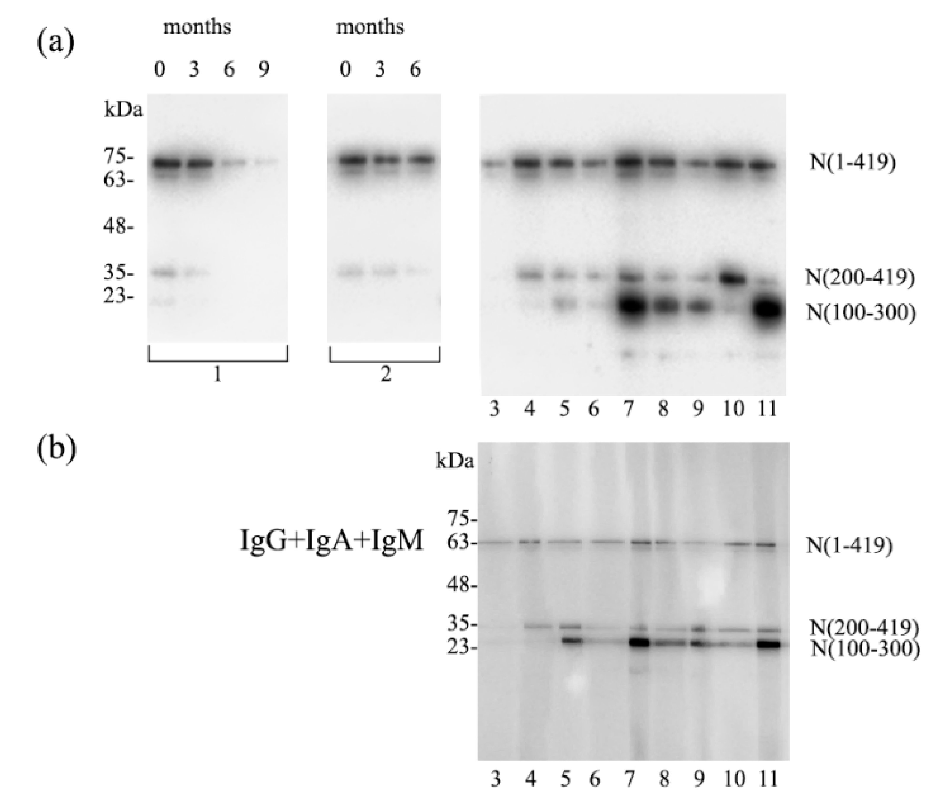
The variable reaction pattern (phenotypic pattern) regarding fragment N(200–419) versus N(100–300) was reproduced with a multislot blotting device using sera from donors with a recent history of COVID-19. As depicted in Figure 2a, donors 1, 2, 3, 4 and 10 resembled the phenotype pattern of donors B and D in Figure 1 with dominant N(200–419) fragment staining. In contrast, donors 5, 7, 8, 9 and 11 showed strong N(100–300) staining resembling the phenotype pattern of donor A in Figure 1. A remarkable reduction in IgG antibody staining was observed over the course of 9 months in donor 1 shown in Figure 2a (left). This dynamic was also present, albeit more slowly, in donor 2 (Figure 2a; middle). When the same donor sera (3–11) were developed for the three isotypes IgM, IgA and IgG, there was a clear immunodominance of N(100–300) in donors 5, 7, 8, 9 and 11 as depicted in Figure 2b.

3.3. Participants with Borderline IgG ELISA Results

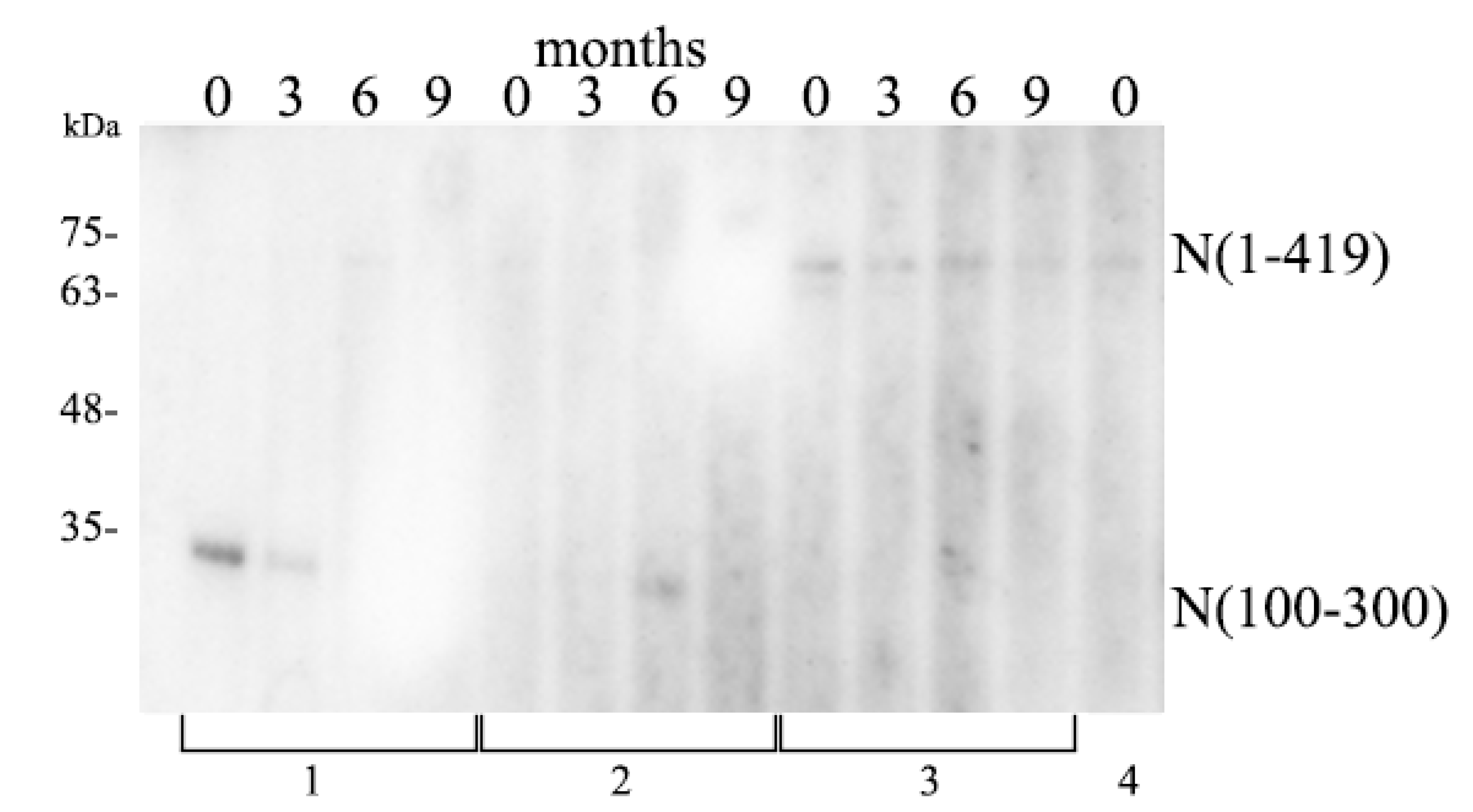
In contrast to the donors with a recent history of COVID-19, 15 individuals were classified as borderline positive in ELISA testing. These participants had no positive PCR tests, no SARS-CoV-2 related symptoms or history of COVID-19. In this subgroup IgG extinction levels reached from 0.2–0.45 (OD = 450 nm). We therefore extended work by screening borderline positive participants using the multislot and multipanel device and loaded the fragments N(100–300) together with the entire N protein N(1–419) onto a flat comb gel. As shown in Figure 3, two out of four individuals (representative out of 15) revealed reaction directed exclusively against the N(1–300). This fragment contains the octamer amino acid sequence FYYLGTGP, which SARS-CoV-2 shares with all HCoVs (Figure 4 and Figure 5). These patients were negative to the SARS-CoV-2 spike protein in immunoblots.
No other section of this length of shared amino acid sequences between HCoVs and SARS-CoV-2 was found in the entire N protein (Supplementary Figure S1). These immunoblot data have to be attributed to the fact that these two individuals (1 and 2) produced IgG against one or several of the human coronaviruses HCoV-NL63, HCoV-229E, HCoV-OC43 or HCoV-HKU1, but not against SARS-CoV-2. For this reason, further analysis of the IgG recognition pattern of donor 1 was performed. As depicted in Figure 1e the donor specific reaction was exclusively directed towards the fragments N(1–220) and N(100–300) both containing the octamer peptide FYYLGTGP as shown in Figure 4 and Figure 5. This donor 1 together with donor 2 in Figure 3 did not react with the entire protein N(1–419), due to its conformational structure, but only with fragments N(1–220) and N(100–300) in which the primary structure is unveiled and can be recognized by IgG antibodies. In contrast, donors 3 and 4 of the borderline positive participants showed a low but clearly discernable reaction against the entire N protein N(1–419) (Figure 3). These participants must have had exposition to low viral loads of SARS-CoV-2 that did not induce COVID-19. The overall N protein identity among HCoVs and SARS-CoV-2 is below 34% (Supplementary Table S1) and is thus not expected to induce IgG cross reactivity at this level of stringency immunoblotting when a recombinant SARS-CoV-2 N protein is used as target.

4. Discussion

In this observational study, 240 healthcare workers—doctors, nurses, auxiliary and administrative staff—were included and monitored for the humoral immune response against SARS-CoV-2 and occurrence of COVID-19. Accordingly, the anti-N protein IgG level was measured by ELISA as a screening method. Thereby an increase in N protein-specific IgG antibody prevalence from 0% up to 10% from study entry in March 2020 to follow-up at 9 months was detected. In-hospital containment measures seemed to prevent the spread of SARS-CoV-2 clusters within the personnel, however, the second outbreak in October caused a marked increase in community acquired COVID-19. The recorded antibody level increase reflected the disease incidence ascertained by positive PCR as described in the study participants’ medical histories. This observation is of importance as it confirms that the N protein represents a potent and consistent immunogen as reported in previous literature evaluating clinical data [1,17,18,19,20,21]. Here, it is shown that the immunogenic region of the N protein varies from individual to individual to some degree. We confirm previous findings [1] that the dominant immunogenic region is located within N(100–300). A minority of participants showed a phenotype with dominant reaction to the anterior (N1–220) and posterior part N(200–419).
The majority of participants with high N protein ELISA tests showed a patient-specific phenotypic staining pattern with dominant reaction against N(100–300) especially shortly after disease recovery. This suggests that only using specific parts of the N protein in ELISA methods would miss a certain percentage of positive individuals.
It seems meaningful to note that the individual participant-specific phenotypic response pattern was maintained throughout the 9-month observation period, albeit with decreasing intensity. The reaction pattern as shown in the individual lane fragment blot (Figure 1) was characterized by distinct staining composition of the fragments N(1–220), N(100–300) and N(200–419). Of note was the pattern of donor E depicted in Figure 1e who did not recognize the entire N(1–419) but stained positive for N(1–220) and N(100–300), which is representative of two further individuals with similar reaction patterns (one of them shown in Supplementary Figure S2). This demarcates the phenotype not related to SARS-CoV-2, as these two fragments contain the peptide sequence FYYLGTGP, which is identical in SARS-CoV-2 and seasonal HCoVs, and thus the responses of these donors can only be explained by a recent exposure to one of the HCoVs in the absence of a SARS-CoV-2 infection. Due to the highly positive charge in the anterior region of the N protein [22], the cross-reacting antibodies do not bind to the full-length protein.
Two hypotheses could explain the phenotypic reaction pattern of different individuals towards the nucleocapsid protein of SARS-CoV-2. First, the well-described observation that the octapeptide is identical in all HCoVs and SARS-CoV-2 supports the idea that there is a preexisting immunological memory in a high percentage of people derived from previous exposure to HCoVs. Infection with SARS-CoV-2 causes a strong flair up of antibody production, especially targeting this part of the N protein, to which individuals had been pre-immunized by HCoV exposition. There is de novo response to various antigenic regions, but not to the same extent as towards the pre-exposed fragment N(100–300), as it receives a booster. As a second hypothesis there might be an HLA dependent individual-specific N protein peptide antigen presentation and a resulting phenotypic immune response. In this line is the patient specific genetic background, which might influence the SARS-CoV-2 interaction with various host proteins such as accurately analyzed by earlier authors. [23]
The N protein is sensitive to peptide cleavage by several endopeptidases and is subject to proteasome cleavage. In the production of new monoclonal antibodies targeting the SARS-CoV-2 N protein, several degradation products of different molecular size have been detected by recent authors [24]. Some of these degradation products could be presented at the cell surface of infected cells and return to the blood stream [25]. Thus, they represent targets for killer cells, which are in the possession of an effective membrane damaging, pore-forming protein, through which they deliver signaling molecules switching on endoproteases and endogenous DNA cutting enzymes. This eventually leads to apoptosis [26], which causes the virus to disassemble. A similar assumption was suggested by earlier authors when they showed the life-saving protection of N protein-specific antibodies in mice [13]. Following this line of thought, it would be beneficial to include the N protein in a vaccine in order to harness the T-cell response to defend against infection with SARS-CoV-2.
We show different phenotypic patterns of IgG production against N protein subdomains, which could be attributed to individual genetic background resulting in divergent serologic responses. This finding is important, both for vaccine design and the interpretation of antibody ELISA data.

Supplementary Materials

The following are available online at https://www.mdpi.com/article/10.3390/covid1010009/s1, Table S1: N-protein percentage of identity (% of identity) among human corona viruses. Figure S1: Nucleocapsid homology among SARS-Cov-2 and HCoV-NL63, HCoV-229E, HCoV-OC43, HCoV-HKU1. Figure S2: Multiple slot immunoblotting against the N protein variants.

Author Contributions

Conceptualization, S.P., T.R., L.W., G.S.-P. and A.S.; Data curation, S.P. and S.K.; Investigation, S.P., S.K., T.R., V.A.J.S., L.W., D.G. and A.S.; Methodology, R.F., V.A.J.S. and L.W.; Resources, R.F., V.A.J.S. and L.W.; Supervision, A.S.; Writing—original draft, S.K. and L.W.; Writing—review and editing, R.F., W.W., D.G. and G.S.-P. All authors have read and agreed to the published version of the manuscript.

Funding

This research received no external funding.

Institutional Review Board Statement

The study was conducted according to the guidelines of the Declaration of Helsinki, and approved by the Ethics Committee of the Medical University of Vienna (1357/2020 and date of approval).

Informed Consent Statement

Informed consent was obtained from all subjects involved in the study.

Data Availability Statement

All data sets that have been generated within this study are provided in Figure 1, Figure 2 and Figure 3 and Supplementary Figure S2. Any further information will be available from the corresponding authors on request.

Conflicts of Interest

The authors declare no conflict of interest.

References

  1. Smits, V.A.J.; Hernández-Carralero, E.; Paz-Cabrera, M.C.; Cabrera, E.; Hernández-Reyes, Y.; Hernández-Fernaud, J.R.; Gillespie, D.A.; Salido, E.; Hernández-Porto, M.; Freire, R. The Nucleocapsid protein triggers the main humoral immune response in COVID-19 patients. Biochem. Biophys. Res. Commun. 2021, 543, 45–49. [Google Scholar] [CrossRef] [PubMed]
  2. Shrock, E.; Fujimura, E.; Kula, T.; Timms, R.T.; Lee, I.H.; Leng, Y.; Robinson, M.L.; Sie, B.M.; Li, M.Z.; Chen, Y.; et al. Viral epitope profiling of COVID-19 patients reveals cross-reactivity and correlates of severity. Science 2020, 370. [Google Scholar] [CrossRef] [PubMed]
  3. Guo, L.; Ren, L.; Yang, S.; Xiao, M.; Chang, D.; Yang, F.; Dela Cruz, C.S.; Wang, Y.; Wu, C.; Xiao, Y.; et al. Profiling Early Humoral Response to Diagnose Novel Coronavirus Disease (COVID-19). Clin. Infect. Dis. 2020, 71, 778–785. [Google Scholar] [CrossRef] [Green Version]
  4. Mukherjee, S.; Tworowski, D.; Detroja, R.; Mukherjee, S.B.; Frenkel-Morgenstern, M. Immunoinformatics and Structural Analysis for Identification of Immunodominant Epitopes in SARS-CoV-2 as Potential Vaccine Targets. Vaccines 2020, 8, 290. [Google Scholar] [CrossRef]
  5. Zamecnik, C.R.; Rajan, J.V.; Yamauchi, K.A.; Mann, S.A.; Loudermilk, R.P.; Sowa, G.M.; Zorn, K.C.; Alvarenga, B.D.; Gaebler, C.; Caskey, M.; et al. ReScan, a Multiplex Diagnostic Pipeline, Pans Human Sera for SARS-CoV-2 Antigens. Cell Rep. Med. 2020, 1, 100123. [Google Scholar] [CrossRef] [PubMed]
  6. Wang, H.; Hou, X.; Wu, X.; Liang, T.; Zhang, X.; Wang, D.; Teng, F.; Dai, J.; Duan, H.; Guo, S.; et al. SARS-CoV-2 proteome microarray for mapping COVID-19 antibody interactions at amino acid resolution. bioRxiv 2020, 6, 2238–2249. [Google Scholar] [CrossRef]
  7. Folegatti, P.M.; Ewer, K.J.; Aley, P.K.; Angus, B.; Becker, S.; Belij-Rammerstorfer, S.; Bellamy, D.; Bibi, S.; Bittaye, M.; Clutterbuck, E.A.; et al. Safety and immunogenicity of the ChAdOx1 nCoV-19 vaccine against SARS-CoV-2: A preliminary report of a phase 1/2, single-blind, randomised controlled trial. Lancet 2020, 396, 467–478. [Google Scholar] [CrossRef]
  8. Mulligan, M.J.; Lyke, K.E.; Kitchin, N.; Absalon, J.; Gurtman, A.; Lockhart, S.; Neuzil, K.; Raabe, V.; Bailey, R.; Swanson, K.A.; et al. Phase I/II study of COVID-19 RNA vaccine BNT162b1 in adults. Nature 2020, 586, 589–593. [Google Scholar] [CrossRef]
  9. McBride, R.; van Zyl, M.; Fielding, B.C. The coronavirus nucleocapsid is a multifunctional protein. Viruses 2014, 6, 2991–3018. [Google Scholar] [CrossRef] [PubMed] [Green Version]
  10. Cong, Y.; Ulasli, M.; Schepers, H.; Mauthe, M.; V’kovski, P.; Kriegenburg, F.; Thiel, V.; de Haan, C.A.M.; Reggiori, F. Nucleocapsid Protein Recruitment to Replication-Transcription Complexes Plays a Crucial Role in Coronaviral Life Cycle. J. Virol. 2020, 94, e01925-19. [Google Scholar] [CrossRef] [Green Version]
  11. Zeng, W.; Liu, G.; Ma, H.; Zhao, D.; Yang, Y.; Liu, M.; Mohammed, A.; Zhao, C.; Yang, Y.; Xie, J.; et al. Biochemical characterization of SARS-CoV-2 nucleocapsid protein. Biochem. Biophys. Res. Commun. 2020, 527, 618–623. [Google Scholar] [CrossRef]
  12. Yao, H.; Song, Y.; Chen, Y.; Wu, N.; Xu, J.; Sun, C.; Zhang, J.; Weng, T.; Zhang, Z.; Wu, Z.; et al. Molecular Architecture of the SARS-CoV-2 Virus. Cell 2020, 183, 730–738 e713. [Google Scholar] [CrossRef]
  13. Nakanaga, K.; Yamanouchi, K.; Fujiwara, K. Protective effect of monoclonal antibodies on lethal mouse hepatitis virus infection in mice. J. Virol. 1986, 59, 168–171. [Google Scholar] [CrossRef] [PubMed] [Green Version]
  14. Dutta, N.K.; Mazumdar, K.; Gordy, J.T. The Nucleocapsid Protein of SARS-CoV-2: A Target for Vaccine Development. J. Virol. 2020, 94, e00647-20. [Google Scholar] [CrossRef]
  15. Ahlen, G.; Frelin, L.; Nikouyan, N.; Weber, F.; Hoglund, U.; Larsson, O.; Westman, M.; Tuvesson, O.; Gidlund, E.K.; Cadossi, M.; et al. The SARS-CoV-2 N Protein Is a Good Component in a Vaccine. J. Virol. 2020, 94, e01279-20. [Google Scholar] [CrossRef] [PubMed]
  16. Reiter, T.; Pajenda, S.; Wagner, L.; Gaggl, M.; Atamaniuk, J.; Holzer, B.; Zimpernik, I.; Gerges, D.; Mayer, K.; Aigner, C.; et al. Covid-19 serology in nephrology health care workers. medRxiv 2020. [Google Scholar] [CrossRef]
  17. Sasisekharan, V.; Pentakota, N.; Jayaraman, A.; Tharakaraman, K.; Wogan, G.N.; Narayanasami, U. Orthogonal immunoassays for IgG antibodies to SARS-CoV-2 antigens reveal that immune response lasts beyond 4 mo post illness onset. Proc. Natl. Acad. Sci. USA 2021, 118. [Google Scholar] [CrossRef]
  18. Woo, P.C.; Lau, S.K.; Tsoi, H.W.; Chan, K.H.; Wong, B.H.; Che, X.Y.; Tam, V.K.; Tam, S.C.; Cheng, V.C.; Hung, I.F.; et al. Relative rates of non-pneumonic SARS coronavirus infection and SARS coronavirus pneumonia. Lancet 2004, 363, 841–845. [Google Scholar] [CrossRef] [Green Version]
  19. Yu, F.; Le, M.Q.; Inoue, S.; Thai, H.T.; Hasebe, F.; Del Carmen Parquet, M.; Morita, K. Evaluation of inapparent nosocomial severe acute respiratory syndrome coronavirus infection in Vietnam by use of highly specific recombinant truncated nucleocapsid protein-based enzyme-linked immunosorbent assay. Clin. Diagn. Lab. Immunol. 2005, 12, 848–854. [Google Scholar] [CrossRef] [Green Version]
  20. Yue, L.; Cao, H.; Xie, T.; Long, R.; Li, H.; Yang, T.; Yan, M.; Xie, Z. N-terminally truncated nucleocapsid protein of SARS-CoV-2 as a better serological marker than whole nucleocapsid protein in evaluating the immunogenicity of inactivated SARS-CoV-2. J. Med. Virol. 2021, 93, 1732–1738. [Google Scholar] [CrossRef]
  21. Burbelo, P.D.; Riedo, F.X.; Morishima, C.; Rawlings, S.; Smith, D.; Das, S.; Strich, J.R.; Chertow, D.S.; Davey, R.T., Jr.; Cohen, J.I. Detection of Nucleocapsid Antibody to SARS-CoV-2 is More Sensitive than Antibody to Spike Protein in COVID-19 Patients. medRxiv 2020. [Google Scholar] [CrossRef]
  22. Ye, Q.; West, A.M.V.; Silletti, S.; Corbett, K.D. Architecture and self-assembly of the SARS-CoV-2 nucleocapsid protein. Protein Sci. 2020, 29, 1890–1901. [Google Scholar] [CrossRef] [PubMed]
  23. Gordon, D.E.; Jang, G.M.; Bouhaddou, M.; Xu, J.; Obernier, K.; O’Meara, M.J.; Guo, J.Z.; Swaney, D.L.; Tummino, T.A.; Huttenhain, R.; et al. A SARS-CoV-2-Human Protein-Protein Interaction Map Reveals Drug Targets and Potential Drug-Repurposing. bioRxiv 2020. [Google Scholar] [CrossRef] [Green Version]
  24. Terry, J.S.; Anderson, L.B.R.; Scherman, M.S.; McAlister, C.E.; Perera, R.; Schountz, T.; Geiss, B.J. Development of a SARS-CoV-2 nucleocapsid specific monoclonal antibody. Virology 2021, 558, 28–37. [Google Scholar] [CrossRef] [PubMed]
  25. Diao, B.; Wen, K.; Chen, J.; Liu, Y.; Yuan, Z.; Han, C.; Chen, J.; Pan, Y.; Chen, L.; Dan, Y.; et al. Diagnosis of Acute Respiratory Syndrome Coronavirus 2 Infection by Detection of Nucleocapsid Protein. medRxiv 2020. [Google Scholar] [CrossRef]
  26. Lieberman, J.; Fan, Z. Nuclear war: The granzyme A-bomb. Curr. Opin. Immunol. 2003, 15, 553–559. [Google Scholar] [CrossRef]
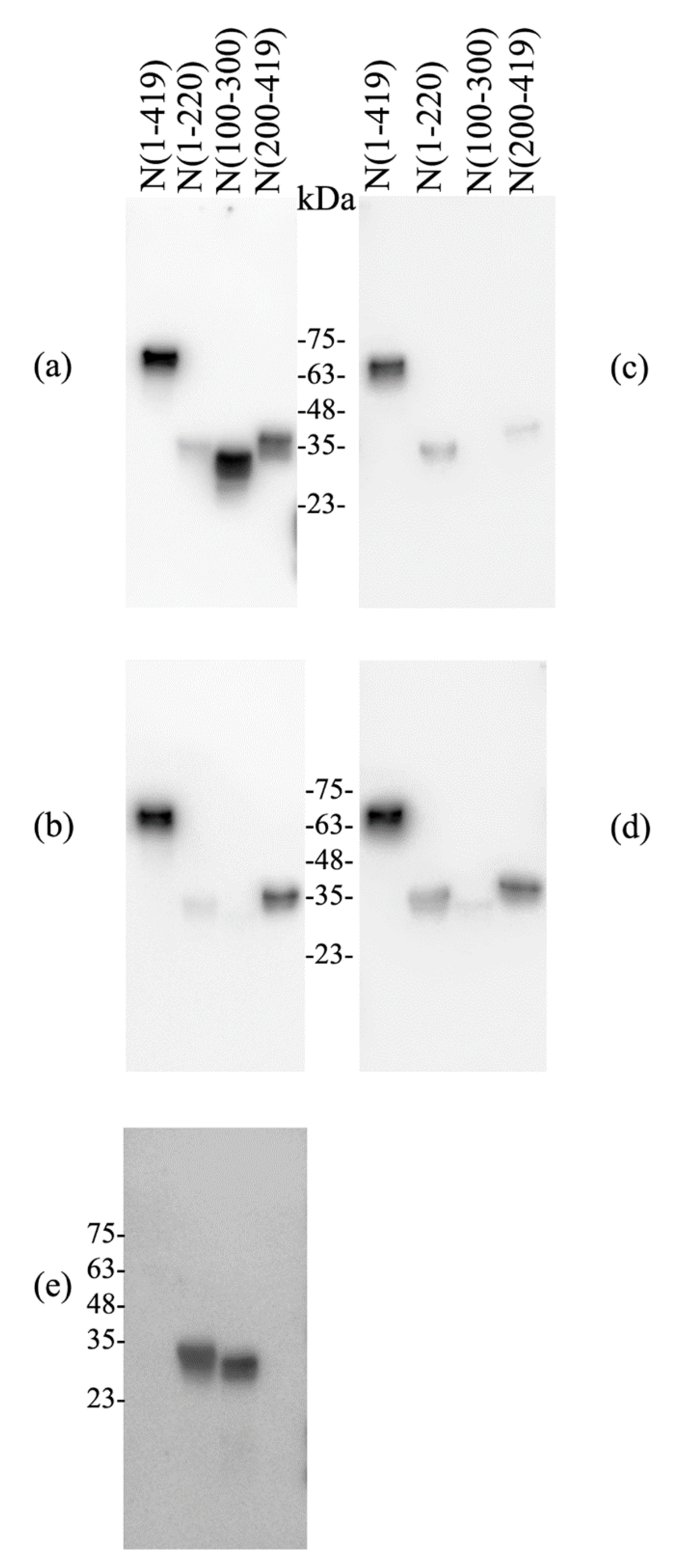
Figure 1. Participant-specific IgG reaction pattern against the entire N protein and its subdomains detected by immunoblotting. Each panel represents one individual donor; donor A (a) shows dominant IgG against N(100–300), donor B (b) shows dominant IgG against N(200–419), donor C (c) shows dominant IgG against N(1–220), donor D (d) shows a similar pattern as B but a more balanced reaction against N(1–220) and N(200–419) and absence of reaction against N(100–300). Donors A, B, C and D had experienced COVID-19 recently. Donor E (e) (with no evidence of disease and negative SARS-CoV-2-PCR testing) showed solely IgG against N(1–220) and N(100–300) with absence of reaction against the entire N(1–419). All sera were diluted 1/100.
Covid 01 00009 g001
Figure 2. Multislot, multipanel immunoblotting against the N protein variants N(1–419), N(100–300), N(200–419) and IgG level time course in two donors. (a) Each slot was loaded with different sera (diluted 1/100) from recovered COVID-19 patients, either from different time points (donor 1 and 2 shown on the left and middle panel) or from different donors taken shortly after disease recovery (donors 3–11 on the right panel). At the top of the blot is the time of blood collection in months. The numbers at the bottom indicate the individual donor. (b) The blot loaded with the same N protein fragments was developed for the same donors 3–11 as depicted above, visualizing isotypes of IgG, IgA and IgM conjointly.
Covid 01 00009 g002
Figure 3. Multiple slot immunoblotting against the N protein variants N(1–419) and N(100–300) in participants categorized as borderline positive by ELISA. Sera (diluted 1/100) from 4 donors at different time points (0, 3, 6 and 9 months) were loaded into each slot. At the top of the blot is the time point of sample collection. The N protein variant is shown on the right and the numbers at the bottom indicate the individual donors. Four representative donors out of 15 are depicted, further three patients are shown in Supplementary Figure S2.
Covid 01 00009 g003
Figure 4. N protein structure and its main subdomains. CTD, dimerization domain; NTD, nucleotide binding domain. N(1–419) represents the entire N protein according to NCBI Reference Sequence: YP_009724397.2. N(1–220) contains the NTD. N(100–300) contains part of the NTD and part of the CTD. N(200–419) represents the CTD.
Covid 01 00009 g004
Figure 5. Octamer peptide sequence identity among SARS-CoV-2, SARS-CoV and the four HCoVs. Amino acid numbering is indicated on the right and left side of the protein sequence.
Covid 01 00009 g005
Publisher’s Note: MDPI stays neutral with regard to jurisdictional claims in published maps and institutional affiliations.