Article

The Antiviral Activity of GcMAF in the Treatment of Experimental Animals Infected with SARS-CoV-2

by
Anastasia S. Proskurina
1,†,
Oleg S. Taranov
2,†,
Svetlana S. Kirikovich
1,
Svetlana V. Aidagulova
3,
Elena K. Ivleva
2,
Andrey V. Shipovalov
2,
Gleb A. Kudrov
2,
Sergei A. Bodnev
2,
Alena S. Ovchinnikova
2,
Anna V. Zaykovskaya
2,
Oleg V. Pyankov
2,
Evgeniy V. Levites
1,
Genrikh S. Ritter
1,
Vera S. Ruzanova
1,
Sofya G. Oshikhmina
1,
Evgeniya V. Dolgova
1,
Evgeniy L. Zavjalov
1,
Alexandr A. Ostanin
4,
Elena R. Chernykh
4,
Nikolay A. Kolchanov
1 and
Sergey S. Bogachev
1,*
add Show full author list remove Hide full author list
1
Institute of Cytology and Genetics of Siberian Branch of the Russian Academy of Sciences, Novosibirsk Region 630090, Russia
2
State Research Center of Virology and Biotechnology “Vector”, Koltsovo, Novosibirsk Region 630559, Russia
3
Department of Scientific Work, Novosibirsk State Medical University, Novosibirsk Region 630091, Russia
4
Research Institute of Fundamental and Clinical Immunology, Novosibirsk Region 630099, Russia
*
Author to whom correspondence should be addressed.
These authors contributed equally to this work.
COVID 2025, 5(3), 36; https://doi.org/10.3390/covid5030036
Submission received: 6 February 2025 / Revised: 5 March 2025 / Accepted: 7 March 2025 / Published: 8 March 2025
(This article belongs to the Special Issue New Antivirals against Coronaviruses)

Abstract

:
Despite the end of the COVID-19 pandemic, there still remain risks of new aggressive strains of coronavirus. As the human population increases progressively, it is mandatory to ensure both preventive measures and an immediate response to emerging infectious threats. Another essential component for rapidly restraining a new possible pandemic is the development of new anticoronaviral therapeutics. In the present study, the anticoronaviral capabilities of Gc protein-derived macrophage-activating factor (GcMAF) are characterized. It is demonstrated that the administration of GcMAF to Syrian hamsters infected with SARS-CoV-2 within the first phase of infection (six days postinfection) is accompanied by (i) a statistically significant reduction in the viral load of the lung tissue and (ii) the switching of the inflammatory status of the lung tissue to a neutral one in terms of mRNA expression levels of the groups of pro/anti-inflammatory cytokines and chemokines. The potential mechanism for this antiviral action and the containment of the inflammatory response by the drug associated with the engagement of terminal N-acetylgalactosamine GcMAF and C-type lectin domain containing 10A expressed at the surface of lung-infiltrating macrophages and pneumocytes, which simultaneously express angiotensin-converting enzyme 2, is discussed.

1. Introduction

The 2020–2023 coronavirus disease 2019 (COVID-19) pandemic caused by severe acute respiratory syndrome-related coronavirus 2 (SARS-CoV-2) led to death in many cases [1,2]. SARS-CoV-2 is a single-stranded RNA virus. The cellular targets of SARS-CoV-2 include the upper respiratory tract epithelium, alveolar epithelial type II cells, and epitheliocytes in the stomach and intestine. SARS-CoV-2 dissemination from the systemic blood flow may have an impact on the brain.
SARS-CoV-2 enters the cell after binding occurs between the receptor-binding domain of the spike protein, a trimeric glycoprotein forming the crown of the virus, and the specific cell surface receptor; angiotensin-converting enzyme 2 (ACE2) is one such receptor that has been well studied. ACE2 is present on many cell types in the body, including vascular endothelial cells and lung epitheliocytes [3,4]. After the initial binding to the ACE2 receptor, the spike protein of SARS-CoV-2 is proteolytically activated via enzymatic cleavage of the S1/S2 subunits. S1 is dissociated from S2, which further interacts with the host cell membrane and initiates the fusion of the viral envelope and the cell membrane [5].
The latest research on this topic has revealed that there are also other receptors that can ensure virus entry into the cell, which include neuropilin NPR-1 [6,7,8] and tyrosine kinase receptor AXL [9]. Other potential receptors and co-receptors facilitating coronavirus entry into the cell include integrins, chaperons, dipeptidyl peptidase 4, CD147, vimentin residing on the outer side of the plasma membrane, some TLRs, heparin sulfate, sialic acids, scavenger receptors, and high-density lipoprotein receptors, as well as the recently discovered Krm1 receptor, which is highly affine for SARS-CoV-2. It is believed that all the aforementioned factors can be involved in pathogen internalization, both independently and as co-receptors [4,10,11,12]. The spike protein forming the crown of the virus belongs to the glycoprotein family. Research on the pathogenesis of SARS-CoV-2 demonstrates that along with ACE2 and the factors listed above, the receptor-binding domain of the spike protein is highly affine for the carbohydrate recognition domain (CRD) of C-type lectin receptors, which include CD209 [13] and asialoglycoprotein receptor 1—ASGR1 (CLEC10A is another member of the C-type lectin receptor family functionally close to ASGR1) [10]. ASGR1 was found to bind to the receptor-binding domain with a KD of ~95 nM, which is substantially higher than that in the case of ACE2 or Krm1 [4].
Clinical observations have demonstrated that disease progression depends both on developing infection and on the mobilization capacity of the organism [14].
The key pathological manifestations of coronavirus infection are characterized by pulmonary changes presenting as ground-glass opacities on CT images. Furthermore, multiple hemorrhagic zones are formed in the lungs; their number directly correlates with a fatal outcome. The lungs are profoundly infiltrated by inflammatory cells, leukocytes, and alveolar macrophages. Immune inflammatory cells secrete an ultra-high amount of proinflammatory cytokines (mainly IL-6), resulting in COVID-19-associated hyperinflammatory syndrome or cytokine storm [15].
A new procedure for producing GcMAF has been developed, and the preparation has been characterized in the Laboratory of Induced Cell Processes of the Institute of Cytology and Genetics, Siberian Branch of the Russian Academy of Sciences [16,17,18,19,20]. A study focusing on the effect of GcMAF on murine peritoneal macrophages, macrophage culture, and human dendritic cell culture was conducted. The synergistic effect of GcMAF in combination with the Karanahan technology on tumor-associated stromal cells in a Lewis lung carcinoma model was characterized [21,22]. In combination with the Karanahan cancer treatment technology, the preparation “alleviates” the tumor-associated macrophages of the stroma, thus polarizing their phenotype toward M0 [21].
It has been found that the impact on macrophages is caused by the engagement of terminal N-acetylgalactosamine GcMAF and C-type lectin domain containing 10A (CLEC10A) [20,23]. The receptor is expressed on the surface of many cell types, including dendritic cells and macrophages, and regulates many immune responses depending on the microenvironment and ligand type [23]. This interaction is likely to be responsible for the numerous effects of macrophage-activating factor that have been reported in the research literature [18,24,25,26,27,28,29,30,31].
Its ability to interact with C-type lectin receptors, which are potential co-receptors for SARS-CoV-2, implies that GcMAF potentially blocks one of the possible pathways of SARS-CoV-2 entry into the cell.
The preparation has multidirectional effects on the pro- and anti-inflammatory responses of peritoneal macrophages and whole blood cells. It has been demonstrated that the direction of the inflammatory response of macrophages treated with GcMAF depends on degree and specificity of trisaccharide deglycosylation at position Tre 420 of the macrophage-activating factor [32]. The highly specific preparation to induce an anti-inflammatory macrophage response upon exposure to GcMAF can be obtained under selective deglycosylation conditions (data not published, in preparation). This fact implies that GcMAF can affect the proinflammatory status of alveolar macrophages and leukocytes infiltrating the lung parenchyma to arrest the cytokine storm syndrome as coronavirus infection develops.
Therefore, GcMAF can simultaneously perform two actions to block the pathological sequelae of virus entry into the cell: (1) impede pathogen entry into the cell and (2) induce anti-inflammatory responses in immune cells infiltrating the lungs, thus arresting the cytokine storm. Due to these properties, GcMAF can potentially be placed alongside the most effective anticoronaviral drugs.
This study focused on the antiviral activity of GcMAF. The findings demonstrate that GcMAF has a high therapeutic potential and can be further promoted for clinical practice.

2. Materials and Methods

2.1. Cell Cultures

Vero E6 cells were obtained from the Cell Culture Collection of the State Research Center of Virology and Biotechnology “Vector”, Federal Service for Surveillance on Consumer Rights Protection and Human Wellbeing, and grown in minimal essential medium (MEM) supplemented with 10% fetal bovine serum (Gibco, Baltimore, MD, USA), penicillin (100 IU/mL), and streptomycin (100 µg/mL; Gibco, USA) at 37 °C in an atmosphere of 5% CO2. The same medium supplemented with 2% fetal bovine serum was used after the cells had been infected.

2.2. Virus

The SARS-CoV-2 hCoV-19/Russia/Vologda-171613-1208/2020 strain from the National Collection of Microorganisms of the State Research Center of Virology and Biotechnology “Vector”, Federal Service for Surveillance on Consumer Rights Protection and Human Wellbeing, was used in this study. This strain belongs to the B1.1 lineage, is similar to the hCoV-19/Russia/OMS-121618-1707/2020 strain (GISAID EPI_ISL_6565010), and is highly homologous to the parental Wuhan strain (GISAID EPI_ISL_406844). The SARS-CoV-2 virus was isolated in the Vero E6 cell culture; aliquots from one virus stock were frozen and stored at −70 °C. The infectious titer of virus stocks was ≥106 TCID50/mL. A new aliquot from the same stock was used in each experiment. The titer of the virus suspension was quantified by the finite dilution method for Vero E6 cells using the Reed–Muench procedure [33].

2.3. GcMAF

GcMAF was produced using the original procedure employing affinity chromatography on an actin column, which is subject to industrial ownership of the LLC “ACTIVATOR MAF”. The analysis of macrophage activation assesses the phagocytic index of activated peritoneal macrophages compared to that induced by the standard macrophage-activating factor LPS and compared to the phagocytic activity of the macrophage-activating factor precursor DBP. The phagocytic activity index was 8.0 ± 0.8 for the GcMAF used in this study and 5.0 ± 0.7 for LPS. An GcMAF exhibiting anti-inflammatory properties, denoted as GcMAF LEV in ref. [20,34], was used in this study.

2.4. Animals

Male and female outbred Syrian hamsters (body weight, 80–100 g) were used in the experiment. The animals were procured from the Center for Collective Use “Genetic Resources Center of Laboratory Animals”, Institute of Cytology and Genetics, SB RAS (RFMEFI62119X0023). The hamsters were placed into individually ventilated cages (two animals per cage) with ad libitum access to food and water. The animals were acclimatized to the experimental conditions for seven days prior to infection. During the experiments, temperature in the cages was maintained at a level of 22–24 °C; relative humidity was 40–55%.
All the animal experiments were approved by the Bioethics Committee of the State Research Center of Virology and Biotechnology “Vector”, Federal Service for Surveillance on Consumer Rights Protection and Human Wellbeing (Protocol N 3 from 15 June 2021), and conducted in compliance with the national and international guidelines for the care and humane handling of laboratory animals.

2.5. Experimental Design

Anesthetized animals were infected with the virus 10 min after they had intramuscularly received Zoletil 100 (Virbac, France) at a dose of 1250 µg/100 g body weight. The animals were infected by intranasal inoculation of the virus using a pipette (volume, 50 µL; dose, 500 TCID50).
Three groups (five animals were group) were formed: the control group consisting of infected animals; the group of hamsters that received GcMAF 1 × dose; and the group of hamsters that received GcMAF 5 × dose. The study drug was injected alternately via the subcutaneous (200 µL once daily) and intranasal (100 µL twice daily) routes for 6 days (144 h). An intact control group (n = 3) consisting of non-infected animals was also used.
The analyzed GcMAF was used at two working doses (1× and 5×). The 1 × dose corresponded to 1.125 µg; the 5 × dose corresponded to 5.625 µg. The doses were selected based on the results of ex vivo experiments [20,34]. The preparation was injected alternately via the subcutaneous and intranasal routes; a single dose was given subcutaneously on day 1, two doses were given intranasally on day 2, and so on. The total dose during the entire treatment was 10.125 µg for the 1 × dose and 50.625 µg for the 5 × dose.
All the animals were euthanized by cervical dislocation 144 h postinfection. Dissection was subsequently performed, and tissues from the nasal passages and lungs were harvested. The 10% tissue homogenates obtained using a ball mill (Analytik Jena, Jena, Germany) were clarified by centrifugation at 10,000 rpm (SW28 rotor, Beckman Coulter, High Mycombe, UK). Aliquots of clarified samples were used to determine the viral RNA level in the samples by real-time RT-PCR and to quantify the concentration of infectious virus (in TCID50/mL) by titration in Vero E6 cell culture.

2.6. Histological Studies

The lungs were harvested from the infected animals 144 h postinfection. The samples were fixed in 10% buffered formalin for histological applications (BioVitrum, St. Petersburg, Russia) for 48 h. The material was treated using the conventional procedure in a Tissue Tek VIP 6 AI vacuum infiltration tissue processor (Sakura Finetek, Torrance, CA, USA), which involved sequential dehydration in alcohol solutions with increasing concentrations, impregnation in a xylene–paraffin mixture, and paraffin embedding. Paraffinized sections 4–5 µm thick were prepared on an HM-360 automatic rotary microtome (Microm International GmbH, Walldorf, Germany). The sections were stained with hematoxylin and eosin. Optical spectrometry and microimaging were performed on an AxioImager Z1 microscope (Zeiss, Oberkochen, Germany) using the AxioVision version 4.8.2 software. The number and intensity of pathological manifestations were recorded, and measurements were carried out by analyzing scans of serial sections recorded using an Olympus SlideView VS200 digital slide scanner (Olympus, Hamburg, Germany; VS200ASW 3.2 software package). A PlanXApo 20×/0.80 lens was used to obtain the scanned images of microslides.
To obtain representative data, the lungs were dissected into five parts (two parts for the left lung and three parts for the right lung). Therefore, five histopathology specimens representing all the lung portions were obtained from each animal. Assessment was performed using a three-point scale, where 0 corresponded to no manifestations, 1 to a mild manifestation, 2 to a moderate manifestation, and 3 to a severe manifestation. The inflammatory cell infiltration intensity and manifestations of the hemorrhagic syndrome were quantified using the following formula:
[sign intensity according to the three-point scale] × [area of the lesion]/[area of the cross-section]

2.7. RT-PCR Quantification of SARS-CoV-2 Viral RNA in Bodily Fluids

RNA was isolated using a RIBO-prep kit (AmpliSens, Moskow, Russia). cDNA was synthesized from the isolated RNA using a Reverta-L reverse transcription reagent kit (Central Research Institute for Epidemiology, Moskow, Russia). Fragments of SARS-CoV-2 cDNA pre-synthesized on the SARS-CoV-2 RNA template by RT-PCR were amplified using the Vector-PCRrv-COVID19-RG test kit (State Research Center of Virology and Biotechnology “Vector”, Koltsovo, Novosibirsk region, Russia). The level of SARS-CoV-2 RNA in the samples was determined. The detection limit of this test system is considered a CT value of 36, with the amount of RNA being 1955 copies.

2.8. Virus Titration

The infectious activity of the virus in stocks, nasal turbinate tissue, and the lungs of infected animals was determined by analyzing the 50% tissue culture infectious dose. A modified method previously used to obtain viral material from the nasal turbinate tissue of laboratory ferrets was used to collect nasal swabs [35]. Vero E6 cells were seeded into 96-well plates 24 h prior to infection at a density of 1.5 × 10⁴ cell/well. Tenfold serial dilutions of the virus were prepared the same day the experiment was conducted. Then, 6 wells of the 96-well plate were infected with each virus dilution. After 72 hr incubation in an atmosphere of 5% CO2 at 37 °C, the cells were fixed in 4% paraformaldehyde solution, followed by staining with 0.1% crystal violet dye. Specific damage to the cell culture monolayer in the well was measured and expressed using the parameter of TCID50/mL. The infectious dose (TCID50) for intranasal infection was calculated using the Reed–Muench method [33].

2.9. Obtaining cDNA from Lung Tissue to Analyze the Synthesis of Pro- and Anti-Inflammatory Cytokine mRNA

When collecting lungs for histological studies, lung portions were simultaneously sampled to analyze the synthesis of pro- and anti-inflammatory cytokine mRNA. Lung samples were lysed in TRIzol Reagent (Thermo Fisher Scientific, Waltham, MA, USA) following the manufacturer’s instructions to obtain total RNA. The amount of RNA was measured on a Qubit 4 fluorometer (Thermo Fisher Scientific, Waltham, MA, USA). Reverse transcription PCR was carried out on a poly-A mRNA template using a T100 Thermal Cycler amplifier (Bio-Rad Laboratories, Inc., Hercules, CA, USA) and an MMLV RT kit (Evrogen, Moscow, Russia) according to the manufacturer’s protocol.

2.10. Cytokine Real-Time PCR

Real-time PCR was carried out in 96-well plates using BioMaster HS-qPCR SYBR (2×) (BIOLABMIX LLC, Novosibirsk, Russia) on a QuantStudio5 PCR system (Thermo Fisher Scientific, Waltham, USA) according to the manufacturer’s protocol. The cycling parameters were as follows: 95 °C for 10 min; 40 cycles of 95 °C for 30 s, 50 °C (CXCL10, GAPDH)/53 °C (IFN-γ, IL-1β, TNF-α, IL-6, TGF-β, GAPDH)/56 °C (CCL3, GAPDH)/58 °C (ARG, iNOS, GAPDH) for 30 s, and 72 °C for 30 s; and a final melting step involving slow heating from 6 to 95 °C.
Real-time qPCR analysis of each sample was performed in three replicates. The relative expression level was determined using the 2−ΔΔCt method [36]. The lungs of infected untreated hamsters were used as the control group; the expression level of the target gene in them was assumed to be equal to 1. The GAPDH gene was used as a reference.
PCR primers for the coding regions of each proinflammatory and anti-inflammatory cytokine gene were taken from the literature—IFN-γ, TNF-α, IL-6, TGF-β [37], arginase (ARG), inducible nitric acid synthase (iNOS) [38], IL-1β, CCL3, and CXCL10 [39]—and synthesized by BIOSSET Ltd. (BIOSSET, Novosibirsk, Russia). The sequences of primers used in this study are listed in Table 1.

2.11. Statistical Analysis

Statistical analysis was performed using Statistica 10 software (StatSoft, Tulsa, OK, USA). The graphs were constructed using GraphPad Prism 9.3.1 software (GraphPad Software, San Diego, CA, USA). The graphs show the median values, the interquartile range, and the minimum and maximum values. The validity of differences was evaluated using the Mann–Whitney U test. The revealed differences were considered statistically significant at p < 0.05 or p < 0.005 (indicated in figure legends).

3. Results

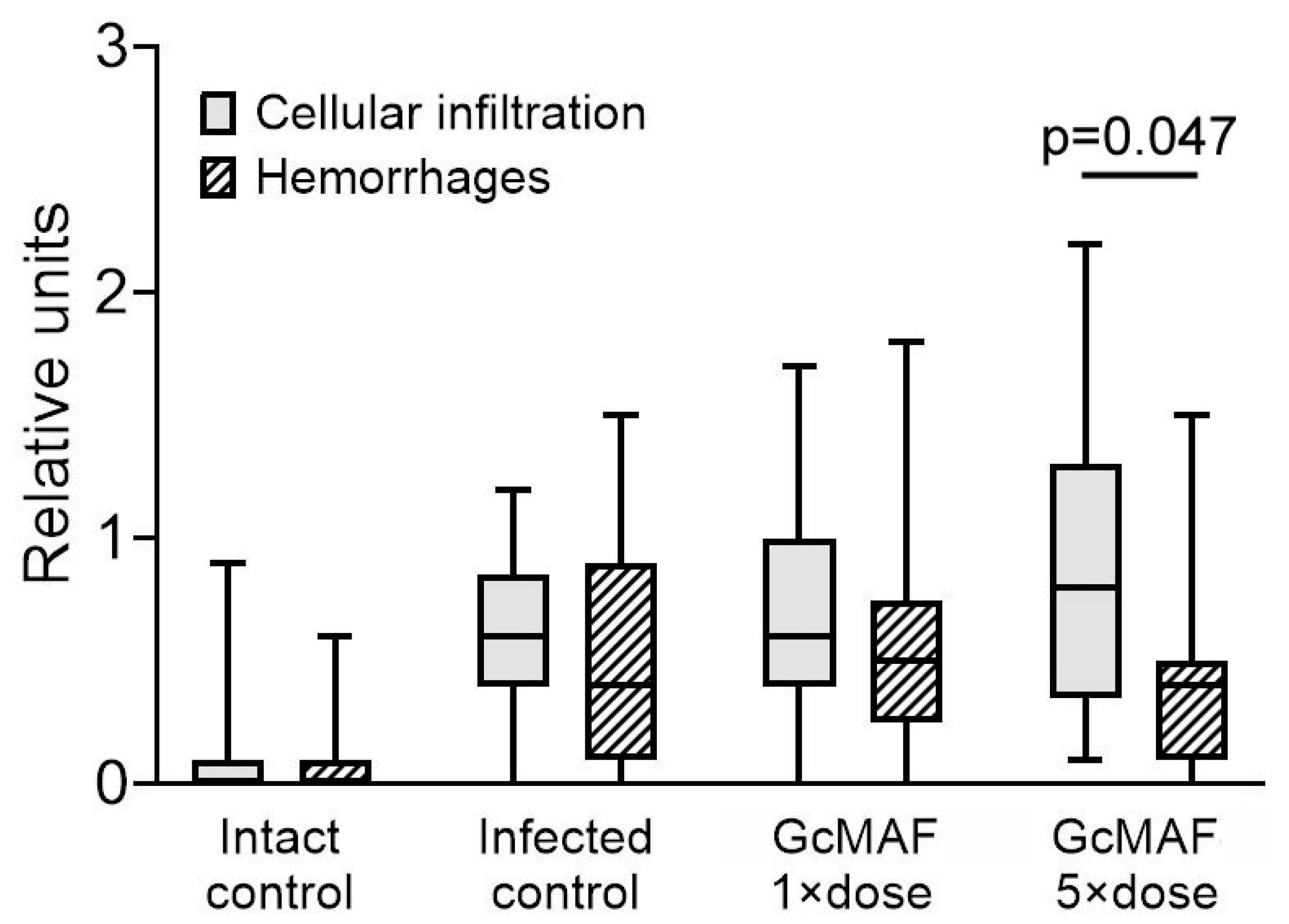
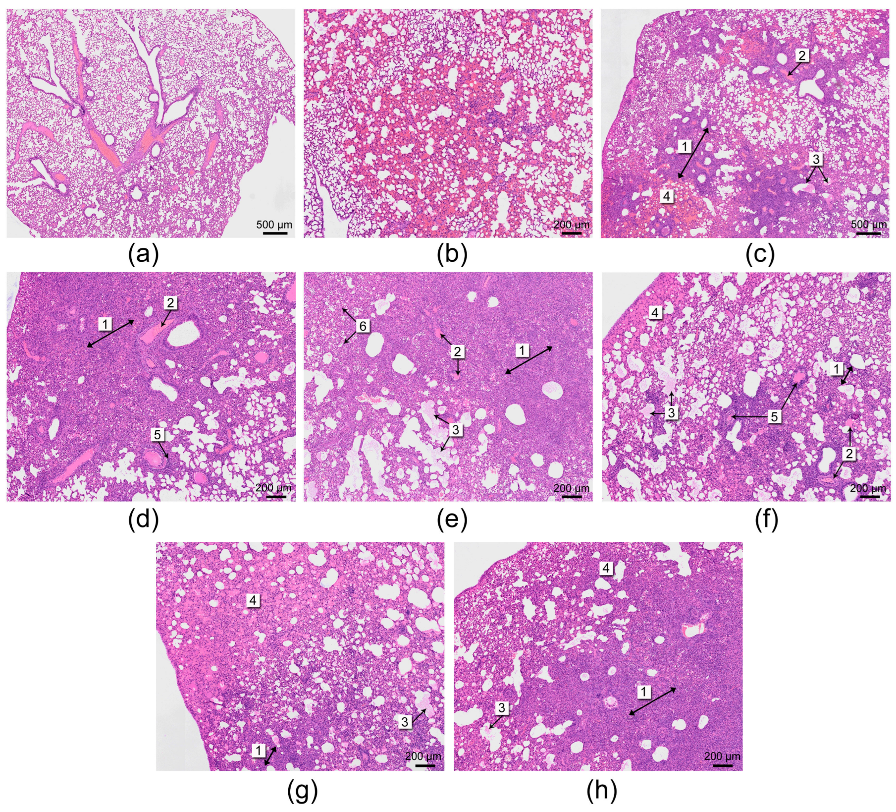
3.1. Histological Analysis

The anatomopathological changes in the lungs of hamsters in the control groups and in hamsters receiving treatment against COVID-19 with the GcMAF 1× or 5× dose for six days were characterized by decreased airiness of the lung parenchyma (dystelectasis and atelectasis); plasmorrhage and hemorrhage (as a result of increased vascular permeability of different severity); and diffuse inflammatory cell infiltration (often perivascular and peribronchial infiltrates) that mainly consisted of lymphoid cells and a small amount of neutrophilic granulocytes.
A comparative analysis of the severity of cellular infiltration and hemorrhagic manifestations, assessed on a three-point scale based on pathological analysis data, was carried out. In the intact control group, only few lung lesions were detected and had an artificial nature. No statistically significant differences between the study groups treated with GcMAF and the infected control samples were revealed, and the data showed only trends. Maximum inflammatory infiltration intensity was revealed in the infected control group. Peribronchial infiltration was observed in sporadic cases. Perivascular infiltration intensity was much lower in the group treated with GcMAF 5 × dose. Hemorrhage intensity was higher in the infected control group. Intrabronchial hemorrhage was observed in sporadic cases. Red blood cell (RBC) aggregation (blood sludge) and perivascular edema were least intense in the group treated with the GcMAF 5 × dose. Desquamated epithelial cells and leukocytes in the alveolar lumen were detected in all the groups except for the intact control. A single observation (fine neutrophilic infiltrate) was made in the infected control group.
The inflammatory cell infiltration intensity and hemorrhagic syndrome manifestations with allowance for the lesion area with respect to the cross-section area were also compared (Figure 1). The group treated with the GcMAF 5 × dose was characterized by the highest intensity and area of inflammatory cellular infiltration; however, the hemorrhage intensity was the lowest in this group.
Figure 2 illustrates the main phenomena observed during histological analysis of the lungs of control and experimental animals.

3.2. Quantification of the Viral Load (Infectious Titer) in the Nasal Cavity and Lungs of Experimental Animals

Viral load was quantified in the lung homogenates and nasal cavity of the infected animals 144 h post infection by real-time RT-PCR (Figure 3a) and by infectious virus titration in Vero E6 cell culture according to TCID50 (Figure 3b). Both methods revealed a statistically significant reduction in viral load in the groups treated with GcMAF at a 1 × dose and 5 × dose compared to the control group (p < 0.05, Mann–Whitney U test).

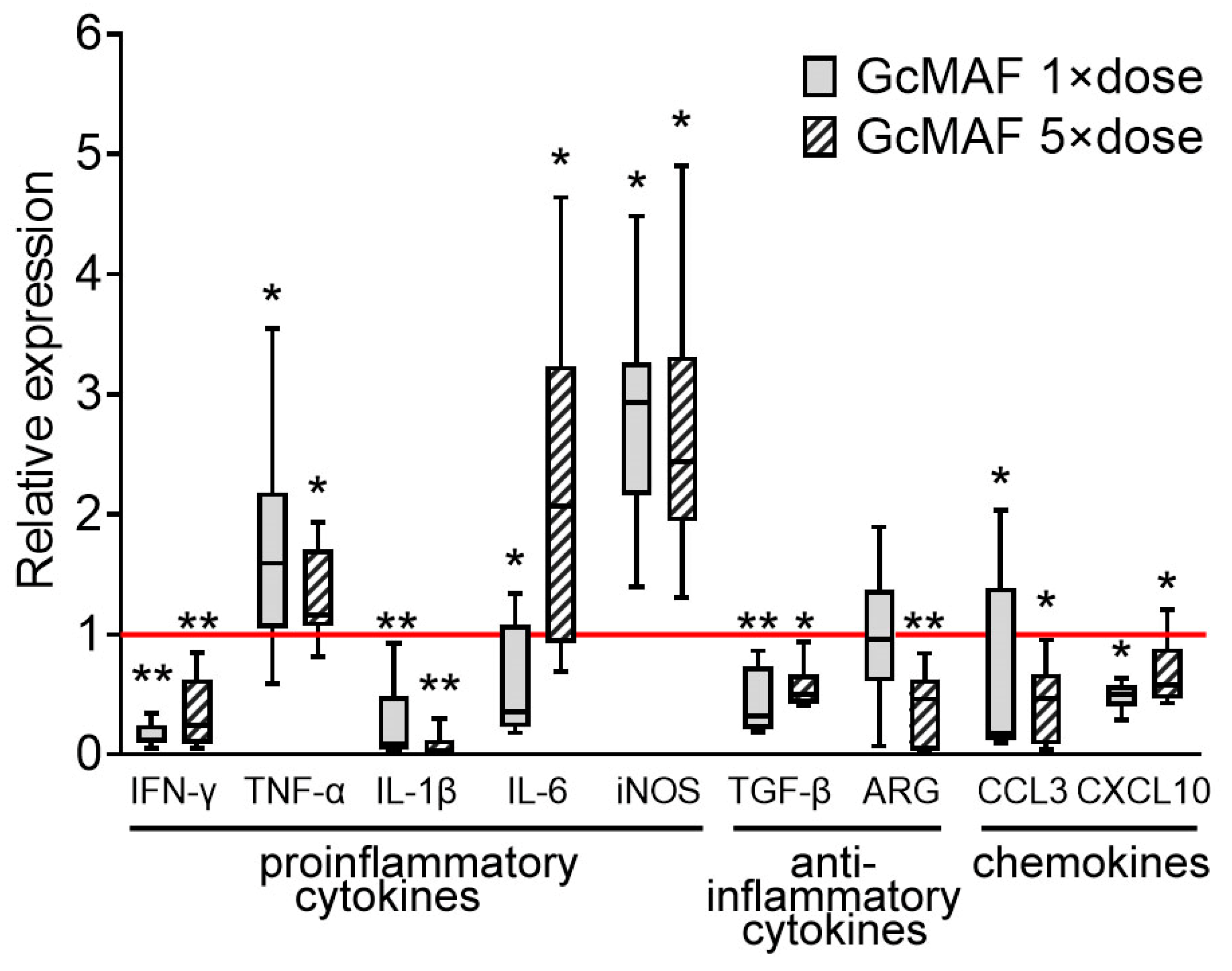
3.3. Analysis of the Synthesis of mRNA of Certain Pro- and Anti-Inflammatory Cytokines

The development of the inflammatory response can be assessed in various ways, for example, by assessing changes in the amount of cytokines in the peripheral blood or the synthesis of cytokines related to various inflammatory vectors in the tissue that is the source of inflammation, in this case, lung tissue. For lung tissue, as the most blood-supplied organ affected by SARS-CoV-2, such an assessment will generally reflect the general inflammation in the body associated with infection by the virus. This is the approach used in this study.
mRNA expression of the major pro- and anti-inflammatory cytokines was analyzed to quantify inflammation intensity in the lungs of experimental animals (Figure 4). Lung samples were lysed in TRIzol reagent on the final day of the experiment. Lung tissue samples of non-treated infected animals were used as control.
According to the findings, the synthesis of the major proinflammatory cytokines IFN-γ and IL-1β was suppressed in the lung tissue of treated animals for both doses. The synthesis of mRNA of the proinflammatory cytokine IL-6 was modulated depending on dose: the 1 × dose statistically significantly reduced the synthesis of IL-6 mRNA, while the 5 × dose statistically significantly increased it. Both doses statistically significantly increased the synthesis of mRNA of TNF-α and iNOS. The expression of anti-inflammatory TGF-β was reliably suppressed. The synthesis of ARG mRNA was statistically significantly inhibited by using the 5 × dose, while the 1 × dose had no effect on synthesis of mRNA of this factor. The synthesis of mRNA of the chemokines responsible for peripheral immune cell recruitment to the analyzed tissue was also suppressed. The overall picture of mRNA expression of some major pro- and anti-inflammatory cytokines and chemokines suggests that both the pro- and anti-inflammatory processes were simultaneously suppressed in the lung tissue of treated animals. In other words, the tissue acquired a neutral phenotype with respect to inflammation.

4. Discussion

There are two equally important trends in the approach to COVID-19 treatment. First, the viral load needs to be reduced using all appropriate measures; second, a set of actions aiming at preventing or eliminating the sequelae of the pathological effect of the virus at the level of the body’s functional systems needs to be taken. Various agents inhibiting a virus’s activity at different stages of its interaction with the body and the cell have been developed and are successfully used to solve the first part of the aforementioned problem [4]. The second part of the problem can be solved by taking measures aiming to reduce the pathophysiological sequelae of the viral action and, primarily, measures suppressing lung tissue destruction [4]. Along with the destruction of pneumocytes via direct cytolysis by the virus replicated at high copy numbers, the lethal pathological impact of the pathogen is related to the induction of an uncontrolled immune response by hyperactivated alveolar macrophages, which is known as the cytokine storm. The key feature of the cytokine storm involves the release of an enormous number of proinflammatory factors, activation of the cytolytic mechanisms and neutrophil NETosis, induction of microvascular thrombosis, and development of systemic inflammatory response syndrome causing multiple-organ failure [40].
Findings demonstrating that therapy with the macrophage-activating factor GcMAF simultaneously reduces viral load and shuts down proinflammatory response in lung tissue were obtained in this study. Therefore, we propose a mechanistic event map resulting from the molecular features of the structure of GcMAF and the known receptors with high affinity for the spike protein, ACE2 and ASGR1 (CLEC10A), which are responsible for the interaction between the SARS-CoV-2 virus and the cell.

4.1. The Structure and Functions of the Macrophage-Activating Factor GcMAF

Vitamin D-binding protein (DBP) is a multifunctional glycoprotein belonging to the family of blood proteins (group-specific component, Gc proteins sized 51–58 kDa). DBP is synthesized by hepatocytes and enters the bloodstream as a mature monomer carrying three functional domains. The DBP domain, the actin-binding domain, and the site of binding to the neutrophil cell membrane reside at the N- and C-termini of a glycoprotein molecule [41,42,43]. The key function of an macrophage-activating factor is its ability to activate macrophages. The DBP precursor acquires this ability due to site-specific selective deglycosylation and is converted to the specific macrophage-activating factor GcMAF. Glycosylated DBP carries one trisaccharide that is covalently bound to Thr420 and consists of GalNAs with two branched galactose and sialic acid residues. DBP is converted to GcMAF under the action of β-galactosidase and sialidase enzymes located on the cell membranes of activated B and T cells, respectively. Active GcMAF protein contains the residual saccharide N-acetylgalactosamine, either as a terminal saccharide residue or as part of the complex with galactose or sialic acid residue. It is the structure of the glycosylation site of the macrophage-activating factor after enzymatic treatment that is responsible for the inflammatory polarization of macrophages affected by it [20,44,45]. This selective deglycosylation of DBP takes place naturally during the development of the inflammatory response and is responsible for the inflammatory polarization of macrophages (proinflammatory vs. anti-inflammatory) affected by it.

4.2. Factors Ensuring Cell Infection by the Virus

4.2.1. ACE2

According to modern views, ACE2 is the major receptor responsible for SARS-CoV-2 internalization. ACE2 is abundantly expressed on alveolar epithelial and endothelial cells, alveolar macrophages, dendritic cells, neutrophils, and lymphocytes of the lung tissue [9,46,47,48]. Therefore, the lung tissue is most vulnerable to viral attack. Upon interaction with the SARS-CoV-2 virus, the receptor content on epitheliocytes drops while generally increasing in the lung tissue [49]. This fact may indicate that it is additionally expressed on activated proinflammatory macrophages that appear in the lung tissue being destroyed by the virus [48]. Such an increase in the content of the major virus-binding receptor on antigen-presenting cells sets the stage for the enhancement of viral load and cytokine storm induction. Lung tissue destruction triggers inflammation and the recruitment of numerous immune cells, thus intensifying the pathological process [50,51].

4.2.2. C-Type Lectin Receptors: CLEC10A

The data on the involvement of ASGR1 in interactions with SARS-CoV-2 as a co-receptor (a prototype of the members of the large family of C-type lectin (CLEC) receptors) are rather interesting for interpreting the findings obtained in this study. All the CLEC receptor family members carry the CRD, which binds the ligand in association with three Ca2+ molecules. Various C-type lectin receptors bind different carbohydrates. Two members of the large CLEC receptor family have a CRD (QPD) structure recognizing and binding to the free terminal GalNAc: ASGR1 (CLEC4H1) and CLEC10A (MGL or CD301). The resulting findings can be attributed to this very property of CLEC receptors.
In the absence of pathological manifestations, CLEC10A is expressed on tolerogenic dendritic cells, dermal and lung macrophages, and peritoneal macrophages. After various inducing events, the expression of C-lectin receptor increases significantly, and tolerogenic antigen-presenting cells induce either the development of Tregs or the anergy of immune cells (and T cells in particular) via an MGL (CLEC10A)-dependent mechanism upon ligand engagement [52,53].

4.3. The Putative Conceptual Events Occurring When Hamsters Are Infected with the SARS-CoV-2 Virus and Simultaneously Treated with GcMAF

The results of our study can be summarized as follows:
(1) GcMAF treatment statistically significantly (p < 0.05, Mann–Whitney U test) reduced the viral load in the lung tissue, which was demonstrated using two independent approaches (Figure 3);
(2) The lung tissue was massively infiltrated with leukocytes while being characterized by a non-proinflammatory response at the level of cytokine mRNA synthesis, indicating that the proinflammatory responses of leukocytes had been lost;
(3) Pulmonary hemorrhagic manifestations decreased.
We propose the following mechanistic explanation for the obtained results in light of the above-described properties of the main participants in the infectious process, which should be considered a hypothesis.

4.3.1. Reduction in Viral Load in the Lungs of Infected Hamsters Treated with GcMAF

The destruction of epithelial cells through the ACE2/SARS-CoV-2 mechanism leads to numerous necrotic lesions and induces the infectious process. Some alveolar macrophages acquire the M1 proinflammatory phenotype, and ACE2 (and probably CLEC10A) becomes exposed on the plasma membrane [48,49,52,53]. Such a rise in the number of potential coronavirus acceptors increases the number of targets used by the virus upon internalization, thus leading to the large-scale destruction of alveolar antigen-presenting cells and aggravating inflammation. A proinflammatory cell-mediated immune response is elicited in some antigen-presenting immune cells not affected by the virus; one of the characteristics of this response is chemokine secretion accompanied by the recruitment of peripheral immune cells to the inflammation site (i.e., to the lungs). A cytokine storm occurs.
As it follows from the GcMAF structure, the polypeptide is a specific ligand for CLEC10A. We hypothesize that CLEC10A is an ACE2 co-receptor and that both factors are needed for virus internalization; the blocking of CLEC10A by its specific ligand GcMAF will reduce the probability of virus entry into the cell, as well as the number of potential viral infection targets. This hypothesis can be used to explain the statistically significant reduction in viral load in the lung tissue for both GcMAF doses (Figure 3).

4.3.2. Hypothesized Mechanism of Massive Infiltration of the Lungs by Lymphocytes and the Acquisition of Non-Inflammatory Reactions by Lung Tissue at the Level of Cytokine mRNA Synthesis

Simultaneously with blocking the pathway for virus entry into the cell, GcMAF and CLEC10A engagement on alveolar macrophages and dendritic cells, as suggested in the analyzed literature [23,54], elicits an anti-inflammatory cell-mediated immune response in these cells, which is characterized by the synthesis of proinflammatory factors and IL-10 in particular. The secreted cytokines affect the numerous immune cells recruited to the inflammation site by inducing immune tolerance or complete anergy in them [54].
Previous results demonstrate that the original GcMAF can induce the polarization of macrophages toward the immune-tolerant M0 phenotype in uninfected animals (mice), thus arresting the synthesis of both pro- and anti-inflammatory cytokines [20,21].
The resulting findings indicate that the lung tissue of treated animals is infiltrated by numerous immune cells and is characterized by significant inhibition of synthesis of the major pro- and anti-inflammatory cytokines/factors, as well as the major chemoattractants IFN-γ, IL-1β, IL-6 (1 × dose), TGF-β, ARG (5 × dose), and chemokines. The synthesis of mRNA of two factors was increased statistically significantly for TNF-α, IL-6 (5 × dose), and iNOS. iNOS can participate in both directions of the inflammatory response depending on cofactors; therefore, its expression should be viewed in the context of the overall inflammatory response.
It is known that at high doses of the ligand (GcMAF), the aggregation of receptors (CLEC10A) occurs, and the synthesis of any cytokines stops [20,34]. In the experiments in the present study, doses of the preparation (1 × dose − 1.125 μg, 5 × dose − 5.625 μg) were used that were on the border of equimolar/super-excessive in relation to the CLEC10A receptor. We believe that this circumstance is the reason for the inhibition of the synthesis of mRNA of the analyzed cytokines.
Thus, alveolar tissue acquires the neutral phenotype with respect to inflammation. This means that all the recruited immune cells constituting the major portion of the infected lung exist in the non-inflammation state. This result is important evidence for the fact that GcMAF can potentially prevent the formation of conditions for cytokine storm development.
Hence, it is fair to believe that treatment is accompanied by two processes simultaneously occurring in the lungs: (1) the primary massive proinflammatory process caused by the lysis of infected alveolar cells and the accumulation of a large number of immune cells in the affected area and (2) a secondary massive “inflammation-alleviating response” caused by the impact of GcMAF on antigen-presenting alveolar cells, where the development of tolerance of leukocytes infiltrating the lungs becomes predominant.

4.3.3. A Mechanistic, Putative Mechanism for Relieving Hemorrhagic Manifestations in the Lungs of Hamsters Infected with SARS-CoV-2

Hemorrhagic syndrome is an appreciably rare phenomenon accompanying COVID-19, but it is characterized by high mortality [55,56,57]. Hemorrhage in COVID-19 patients is believed to be related to a disturbance in the coagulation system and excessive clotting in the pulmonary or cerebral vessels, as well as vessels in any other location. Decreased fibrinolysis, vascular endothelial dysfunction, and triggering of the procoagulant pathway because of the virus-induced inflammatory immune response are among the reasons for hypercoagulation in COVID-19 patients [58].
Our study revealed that the number of hemorrhagic manifestations was inversely proportional to the count of leukocytes infiltrating the lung tissue in the group treated with GcMAF 5 × dose. The number of hemorrhagic manifestations decreased as the count of cells infiltrating the lungs increased (Figure 1).
It was demonstrated in this study that adding GcMAF to the treatment regimen increases the number of immune cells recruited to the inflammation site in a dose-dependent manner; no difference from the infected control was observed for chemokine mRNA synthesis at the endpoint (day 6 after the experiment initiation). This can be attributed to the time delay between chemokine synthesis during the beginning of the inflammatory phase, as well as to immune cell migration to the lungs and later repolarization of the lung tissue and chemokine synthesis suppression after GcMAF treatment. In this case, GcMAF and CLEC10A engagement on alveolar immune cells within the first days of treatment additionally stimulates antigen-presenting cells to secrete attractant chemokines. This response enhances the migration of immune cells into the lung tissue, where, under the already developed conditions of proinflammatory response inhibition, they acquire the immune-tolerant phenotype or become anergic. The recruited and inflamed cells stop massively secreting inflammatory factors. In other words, all the recruited and inflamed cells become a neutral cell mass.
Pulmonary hemorrhage is believed to be primarily related to coagulation disturbance and excessive clotting both in capillaries and large vessels. One of the potential reasons for small vessel thrombosis is neutrophil activity (above all, the formation of neutrophil extracellular traps). During a developing inflammatory process, the numerous neutrophils recruited to the lung tissue induce extensive small vessel thrombosis, thus causing damage to capillaries and larger vessels, which is accompanied by hemorrhages [40].
According to our findings, when GcMAF is added to therapy, the proinflammatory status of the lung tissue is polarized toward a neutral one without the explicit anti-inflammatory mode. We suggest that this very fact causes a reduction in neutrophil activity and clotting and decreases the risk of local hemorrhage. In other words, the thrombogenic potential of the cell systems infiltrating the lung tissue decreases, which leads to a decrease in the number of hemorrhagic manifestations (as a tendency) observed in the lungs of experimental animals.
We believe that the “neutral” cell mass of recruited immune cells is an additional factor for the reduction in the number of hemorrhagic manifestations in the group treated with the GcMAF 5 × dose, as follows from the results shown in Figure 1. The external pressure exerted on the walls of small alveolar vessels by the enormous number of recruited leukocytes compensates for the intravascular blood pressure that is increased because of clotting and prevents blood vessel rupture. In other words, a natural external cellular framework maintaining the integrity of alveolar blood vessels is formed.

5. Conclusions

Our findings demonstrate that treatment of SARS-CoV-2-infected hamsters with GcMAF statistically significantly reduces the viral load in the lung tissue. The lung tissue of the animals receiving GcMAF therapy is massively infiltrated with leukocytes while having a neutral inflammatory phenotype at the level of cytokine mRNA synthesis. Treatment with GcMAF creates conditions for reduced pulmonary hemorrhagic events. Overall, it follows that GcMAF slows down coronavirus replication in the lung tissue and simultaneously mitigates the inflammation induced by coronavirus infection in the lungs.
Our study is the first attempt to discover an approach allowing for such an impact on SARS-CoV-2 that would simultaneously prevent the infection of alveolar cells by the virus and inhibit the proinflammatory reactivity of immune cells infiltrating the lungs. This approach can be regarded as a universal modality for treating infectious lung diseases that combines such components as blocking the infection invasion and regulating the pro- and anti-inflammatory status of alveolar macrophages.

Author Contributions

Conceptualization, S.S.B.; methodology, S.S.K., O.V.P., E.V.L. and E.L.Z.; validation, S.S.K., A.V.S., G.A.K., S.A.B., A.S.O., A.V.Z., O.V.P., G.S.R., V.S.R., S.G.O. and E.V.D.; formal analysis, A.S.P., G.S.R., V.S.R., S.G.O. and E.V.D.; investigation, O.S.T., S.S.K., S.V.A., E.K.I., A.V.S., G.A.K., S.A.B., A.S.O., A.V.Z., E.V.L., A.A.O. and E.R.C.; resources, E.L.Z.; writing—original draft preparation, O.S.T. and S.S.B.; writing—review and editing, A.S.P.; visualization, A.S.P., O.S.T. and E.K.I.; supervision, A.A.O., E.R.C. and N.A.K.; project administration, S.V.A., N.A.K. and S.S.B.; funding acquisition, S.S.B. All authors have read and agreed to the published version of the manuscript.

Funding

This research was funded by the Russian Ministry of Science and High Education via the Institute of Cytology and Genetics [State Budget Project No. FWNR-2022-0016], as well as Inga N. Zaitseva.

Institutional Review Board Statement

All the animal experiments were approved by the Bioethics Committee of the State Research Center of Virology and Biotechnology “Vector”, Federal Service for Surveillance on Consumer Rights Protection and Human Wellbeing (Protocol N 3 from 15 June 2021), and conducted in compliance with the national and international guidelines for the care and humane handling of laboratory animals.

Informed Consent Statement

Not applicable.

Data Availability Statement

The original contributions presented in this study are included in the article. Further inquiries can be directed to the corresponding author.

Acknowledgments

The authors express their gratitude to the Common Use Center Vivarium for Conventional Animals of the Institute of Cytology and Genetics, SB RAS, for providing mice.

Conflicts of Interest

The authors declare no conflicts of interest.

Abbreviations

The following abbreviations are used in this manuscript:
ACE2Angiotensin-converting enzyme 2
ARGArginase
CLEC10AC-Type lectin domain containing 10A
COVID-19Coronavirus disease 2019
CRDCarbohydrate recognition domain
DBPVitamin D-binding protein
GcMAFGc protein-derived macrophage-activating factor
iNOSInducible nitric acid synthase
SARS-CoV-2Severe acute respiratory syndrome-related coronavirus 2

References

  1. Acuti Martellucci, C.; Flacco, M.E.; Cappadona, R.; Bravi, F.; Mantovani, L.; Manzoli, L. SARS-CoV-2 Pandemic: An Overview. Adv. Biol. Regul. 2020, 77, 100736. [Google Scholar] [CrossRef]
  2. Adil, M.T.; Rahman, R.; Whitelaw, D.; Jain, V.; Al-Taan, O.; Rashid, F.; Munasinghe, A.; Jambulingam, P. SARS-CoV-2 and the Pandemic of COVID-19. Postgrad. Med. J. 2021, 97, 110–116. [Google Scholar] [CrossRef] [PubMed]
  3. Hamming, I.; Timens, W.; Bulthuis, M.L.C.; Lely, A.T.; Navis, G.J.; van Goor, H. Tissue Distribution of ACE2 Protein, the Functional Receptor for SARS Coronavirus. A First Step in Understanding SARS Pathogenesis. J. Pathol. 2004, 203, 631–637. [Google Scholar] [CrossRef] [PubMed]
  4. Matusewicz, L.; Golec, M.; Czogalla, A.; Kuliczkowski, K.; Konka, A.; Zembala-John, J.; Sikorski, A.F. COVID-19 Therapies: Do We See Substantial Progress? Cell. Mol. Biol. Lett. 2022, 27, 42. [Google Scholar] [CrossRef]
  5. Jackson, C.B.; Farzan, M.; Chen, B.; Choe, H. Mechanisms of SARS-CoV-2 Entry into Cells. Nat. Rev. Mol. Cell. Biol. 2022, 23, 3–20. [Google Scholar] [CrossRef]
  6. Cantuti-Castelvetri, L.; Ojha, R.; Pedro, L.D.; Djannatian, M.; Franz, J.; Kuivanen, S.; van der Meer, F.; Kallio, K.; Kaya, T.; Anastasina, M.; et al. Neuropilin-1 Facilitates SARS-CoV-2 Cell Entry and Infectivity. Science 2020, 370, 856–860. [Google Scholar] [CrossRef]
  7. Zalpoor, H.; Akbari, A.; Samei, A.; Forghaniesfidvajani, R.; Kamali, M.; Afzalnia, A.; Manshouri, S.; Heidari, F.; Pornour, M.; Khoshmirsafa, M.; et al. The Roles of Eph Receptors, Neuropilin-1, P2X7, and CD147 in COVID-19-Associated Neurodegenerative Diseases: Inflammasome and JaK Inhibitors as Potential Promising Therapies. Cell Mol. Biol. Lett. 2022, 27, 10. [Google Scholar] [CrossRef] [PubMed]
  8. Daly, J.L.; Simonetti, B.; Klein, K.; Chen, K.E.; Williamson, M.K.; Antón-Plágaro, C.; Shoemark, D.K.; Simón-Gracia, L.; Bauer, M.; Hollandi, R.; et al. Neuropilin-1 Is a Host Factor for SARS-CoV-2 Infection. Science 2020, 370, 861–865. [Google Scholar] [CrossRef]
  9. Wang, S.; Qiu, Z.; Hou, Y.; Deng, X.; Xu, W.; Zheng, T.; Wu, P.; Xie, S.; Bian, W.; Zhang, C.; et al. AXL Is a Candidate Receptor for SARS-CoV-2 That Promotes Infection of Pulmonary and Bronchial Epithelial Cells. Cell Res. 2021, 31, 126–140. [Google Scholar] [CrossRef]
  10. Gu, Y.; Cao, J.; Zhang, X.; Gao, H.; Wang, Y.; Wang, J.; He, J.; Jiang, X.; Zhang, J.; Shen, G.; et al. Receptome Profiling Identifies KREMEN1 and ASGR1 as Alternative Functional Receptors of SARS-CoV-2. Cell Res. 2022, 32, 24–37. [Google Scholar] [CrossRef]
  11. Eslami, N.; Aghbash, P.S.; Shamekh, A.; Entezari-Maleki, T.; Nahand, J.S.; Sales, A.J.; Baghi, H.B. SARS-CoV-2: Receptor and Co-Receptor Tropism Probability. Curr. Microbiol. 2022, 79, 133. [Google Scholar] [CrossRef] [PubMed]
  12. Avdonin, P.P.; Rybakova, E.Y.; Trufanov, S.K.; Avdonin, P.V. SARS-CoV-2 Receptors and Their Involvement in Cell Infection. Biochem. (Mosc) Suppl. Ser. A Membr. Cell. Biol. 2023, 17, 1–11. [Google Scholar] [CrossRef]
  13. Rahimi, N. C-Type Lectin CD209L/L-SIGN and CD209/DC-SIGN: Cell Adhesion Molecules Turned to Pathogen Recognition Receptors. Biology 2020, 10, 1. [Google Scholar] [CrossRef]
  14. Tay, M.Z.; Poh, C.M.; Rénia, L.; MacAry, P.A.; Ng, L.F.P. The Trinity of COVID-19: Immunity, Inflammation and Intervention. Nat. Rev. Immunol. 2020, 20, 363–374. [Google Scholar] [CrossRef]
  15. Cron, R.Q.; Schulert, G.S.; Tattersall, R.S. Defining the Scourge of COVID-19 Hyperinflammatory Syndrome. Lancet Rheumatol. 2020, 2, e727–e729. [Google Scholar] [CrossRef] [PubMed]
  16. Mohamad, S.B.; Nagasawa, H.; Uto, Y.; Hori, H. Preparation of Gc Protein-Derived Macrophage Activating Factor (GcMAF) and Its Structural Characterization and Biological Activities. Anticancer Res. 2002, 22, 4297–4300. [Google Scholar] [PubMed]
  17. Naraparaju, V.R.; Yamamoto, N. Roles of β-Galactosidase of B Lymphocytes and Sialidase of T Lymphocytes in Inflammation-Primed Activation of Macrophages. Immunol. Lett. 1994, 43, 143–148. [Google Scholar] [CrossRef]
  18. Saburi, E.; Saburi, A.; Ghanei, M. Promising Role for Gc-MAF in Cancer Immunotherapy: From Bench to Bedside. Casp. J. Intern. Med. 2017, 8, 228–238. [Google Scholar]
  19. Ostanin, A.A.; Kirikovich, S.S.; Dolgova, E.V.; Proskurina, A.S.; Chernykh, E.R.; Bogachev, S.S. A Thorny Pathway of Macrophage Activating Factor (GcMAF): From Bench to Bedside. Vavilov J. Genet. Breed. 2019, 23, 624–631. [Google Scholar] [CrossRef]
  20. Kirikovich, S.S.; Levites, E.V.; Proskurina, A.S.; Ritter, G.S.; Peltek, S.E.; Vasilieva, A.R.; Ruzanova, V.S.; Dolgova, E.V.; Oshihmina, S.G.; Sysoev, A.V.; et al. The Molecular Aspects of Functional Activity of Macrophage-Activating Factor GcMAF. Int. J. Mol. Sci. 2023, 24, 17396. [Google Scholar] [CrossRef]
  21. Dolgova, E.V.; Kirikovich, S.S.; Levites, E.V.; Ruzanova, V.S.; Proskurina, A.S.; Ritter, G.S.; Taranov, O.S.; Varaksin, N.A.; Ryabicheva, T.G.; Leplina, O.Y.; et al. Analysis of the Biological Properties of Blood Plasma Protein with GcMAF Functional Activity. Int. J. Mol. Sci. 2022, 23, 8075. [Google Scholar] [CrossRef] [PubMed]
  22. Ruzanova, V.S.; Kirikovich, S.S.; Levites, E.V.; Proskurina, A.S.; Dolgova, E.V.; Ritter, G.S.; Efremov, Y.R.; Dubatolova, T.D.; Sysoev, A.V.; Koleno, D.I.; et al. The Macrophage Activator GcMAF-RF Enhances the Antitumor Effect of Karanahan Technology Through Induction of M2–M1 Macrophage Reprogramming. J. Immunol. Res. 2024, 2024, 7484490. [Google Scholar] [CrossRef] [PubMed]
  23. Kenneth Hoober, J. ASGR1 and Its Enigmatic Relative, CLEC10A. Int. J. Mol. Sci. 2020, 21, 4818. [Google Scholar] [CrossRef]
  24. Inui, T.; Kuchiike, D.; Kubo, K.; Mette, M.; Uto, Y.; Hori, H.; Sakamoto, N. Clinical Experience of Integrative Cancer Immunotherapy with GcMAF. Anticancer Res. 2013, 33, 2917–2920. [Google Scholar] [PubMed]
  25. Kisker, O.; Onizuka, S.; Becker, C.M.; Fannon, M.; Flynn, E.; D’Amato, R.; Zetter, B.; Folkman, J.; Ray, R.; Swamy, N.; et al. Vitamin D Binding Protein-Macrophage Activating Factor (DBP-Maf) Inhibits Angiogenesis and Tumor Growth in Mice. Neoplasia 2003, 5, 32–40. [Google Scholar] [CrossRef]
  26. Korbelik, M.; Naraparaju, V.R.; Yamamoto, N. Macrophage-Directed Immunotherapy as Adjuvant to Photodynamic Therapy of Cancer. Br. J. Cancer 1997, 75, 202–207. [Google Scholar] [CrossRef]
  27. Kuchiike, D.; Uto, Y.; Mukai, H.; Ishiyama, N.; Abe, C.; Tanaka, D.; Kawai, T.; Kubo, K.; Mette, M.; Inui, T.; et al. Degalactosylated/Desialylated Human Serum Containing GcMAF Induces Macrophage Phagocytic Activity and in Vivo Antitumor Activity. Anticancer Res. 2013, 33, 2881–2885. [Google Scholar]
  28. Pacini, S.; Morucci, G.; Punzi, T.; Gulisano, M.; Ruggiero, M.; Amato, M.; Aterini, S. Effect of Paricalcitol and GcMAF on Angiogenesis and Human Peripheral Blood Mononuclear Cell Proliferation and Signaling. J. Nephrol. 2012, 25, 577–581. [Google Scholar] [CrossRef]
  29. Thyer, L.; Ward, E.; Smith, R.; Branca, J.J.V.; Morucci, G.; Gulisano, M.; Noakes, D.; Eslinger, R.; Pacini, S. GC Protein-Derived Macrophage-Activating Factor Decreases α-N-Acetylgalactosaminidase Levels in Advanced Cancer Patients. Oncoimmunology 2013, 2, e25769. [Google Scholar] [CrossRef]
  30. Thyer, L.; Ward, E.; Smith, R.; Fiore, M.; Magherini, S.; Branca, J.; Morucci, G.; Gulisano, M.; Ruggiero, M.; Pacini, S. A Novel Role for a Major Component of the Vitamin D Axis: Vitamin D Binding Protein-Derived Macrophage Activating Factor Induces Human Breast Cancer Cell Apoptosis through Stimulation of Macrophages. Nutrients 2013, 5, 2577–2589. [Google Scholar] [CrossRef]
  31. Toyohara, Y.; Hashitani, S.; Kishimoto, H.; Noguchi, K.; Yamamoto, N.; Urade, M. Inhibitory Effect of Vitamin D-Binding Protein-Derived Macrophage Activating Factor on DMBA-Induced Hamster Cheek Pouch Carcinogenesis and Its Derived Carcinoma Cell Line. Oncol. Lett. 2011, 2, 685–691. [Google Scholar] [CrossRef] [PubMed]
  32. Rehder, D.S.; Nelson, R.W.; Borges, C.R. Glycosylation Status of Vitamin D Binding Protein in Cancer Patients. Protein Sci. 2009, 18, 2036–2042. [Google Scholar] [CrossRef]
  33. Reed, L.J.; Muench, H. A Simple Method of Estimating Fifty Percent Endpoints. Am. J. Hyg. 1938, 27, 493–497. [Google Scholar] [CrossRef]
  34. Kirikovich, S.S.; Levites, E.V.; Proskurina, A.S.; Ritter, G.S.; Dolgova, E.V.; Ruzanova, V.S.; Oshihmina, S.G.; Snegireva, J.S.; Gamaley, S.G.; Sysoeva, G.M.; et al. Production of GcMAF with anti-inflammatory properties and its effect on models of induced arthritis in mice and cystitis in rats. Curr. Issues Mol. Biol. 2024, 46, 10934–10959. [Google Scholar] [CrossRef] [PubMed]
  35. Matchett, C.A.; Marr, R.; Berard, F.M.; Cawthon, A.G.; Swing, S.P. The Laboratory Ferret; CRC Press: Boca Raton, FL, USA, 2012; 123p. [Google Scholar]
  36. Li, X.; Wang, Y.; Li, J.; Mei, X.; Liu, Y.; Huang, H. qPCRtools: An R package for qPCR data processing and visualization. Front. Genet. 2022, 13, 1002704. [Google Scholar] [CrossRef]
  37. Vernel-Pauillac, F.R.; Goarant, C. Differential Cytokine Gene Expression According to Outcome in a Hamster Model of Leptospirosis. PLoS Negl. Trop. Dis. 2010, 4, e582. [Google Scholar] [CrossRef]
  38. Osorio, Y.; Melby, P.C.; Pirmez, C.; Chandrasekar, B.; Guarín, N.; Travi, B.L. The Site of Cutaneous Infection Influences the Immunological Response and Clinical Outcome of Hamsters Infected with Leishmania Panamensis. Parasite Immunol. 2003, 25, 139–148. [Google Scholar] [CrossRef]
  39. Matsui, M.; Rouleau, V.; Bruyère-Ostells, L.; Goarant, C. Gene Expression Profiles of Immune Mediators and Histopathological Findings in Animal Models of Leptospirosis: Comparison between Susceptible Hamsters and Resistant Mice. Infect. Immun. 2011, 79, 4480–4492. [Google Scholar] [CrossRef]
  40. Fajgenbaum, D.C.; June, C.H. Cytokine Storm. N. Engl. J. Med. 2020, 383, 2255–2273. [Google Scholar] [CrossRef]
  41. Malik, S.; Fu, L.; Juras, D.J.; Karmali, M.; Wong, B.Y.L.; Gozdzik, A.; Cole, D.E.C. Common Variants of the Vitamin D Binding Protein Gene and Adverse Health Outcomes. Crit. Rev. Clin. Lab. Sci. 2013, 50, 1–22. [Google Scholar] [CrossRef]
  42. Otterbein, L.R.; Cosio, C.; Graceffa, P.; Dominguez, R. Crystal Structures of the Vitamin D-Binding Protein and Its Complex with Actin: Structural Basis of the Actin-Scavenger System. Proc. Natl. Acad. Sci. USA 2002, 99, 8003–8008. [Google Scholar] [CrossRef] [PubMed]
  43. Verboven, C.; Rabijns, A.; De Maeyer, M.; Van Baelen, H.; Bouillon, R.; De Ranter, C. A Structural Basis for the Unique Binding Features of the Human Vitamin D-Binding Protein. Nat. Struct. Biol. 2002, 9, 131–136. [Google Scholar] [CrossRef]
  44. Yamamoto, N.; Suyama, H.; Yamamoto, N. Immunotherapy for Prostate Cancer with Gc Protein-Derived Macrophage-Activating Factor, GcMAF. Transl. Oncol. 2008, 1, 65–72. [Google Scholar] [CrossRef]
  45. Yamamoto, N.; Kumashiro, R. Conversion of Vitamin D3 Binding Protein (Group-Specific Component) to a Macrophage Activating Factor by the Stepwise Action of Beta-Galactosidase of B Cells and Sialidase of T Cells. J. Immunol. 1993, 151, 2794–2802. [Google Scholar] [CrossRef]
  46. McCracken, I.R.; Saginc, G.; He, L.; Huseynov, A.; Daniels, A.; Fletcher, S.; Peghaire, C.; Kalna, V.; Andaloussi-Mäe, M.; Muhl, L.; et al. Lack of Evidence of Angiotensin-Converting Enzyme 2 Expression and Replicative Infection by SARS-CoV-2 in Human Endothelial Cells. Circulation 2021, 143, 865–868. [Google Scholar] [CrossRef]
  47. Muus, C.; Luecken, M.D.; Eraslan, G.; Sikkema, L.; Waghray, A.; Heimberg, G.; Kobayashi, Y.; Vaishnav, E.D.; Subramanian, A.; Smillie, C.; et al. Single-Cell Meta-Analysis of SARS-CoV-2 Entry Genes across Tissues and Demographics. Nat. Med. 2021, 27, 546–559. [Google Scholar] [CrossRef] [PubMed]
  48. Song, X.; Hu, W.; Yu, H.; Zhao, L.; Zhao, Y.; Zhao, X.; Xue, H.H.; Zhao, Y. Little to No Expression of Angiotensin-Converting Enzyme-2 on Most Human Peripheral Blood Immune Cells but Highly Expressed on Tissue Macrophages. Cytometry A 2020, 103, 136–145. [Google Scholar] [CrossRef] [PubMed]
  49. Gerard, L.; Lecocq, M.; Bouzin, C.; Hoton, D.; Schmit, G.; Pereira, J.P.; Montiel, V.; Plante-Bordeneuve, T.; Laterre, P.F.; Pilette, C. Increased Angiotensin-Converting Enzyme 2 and Loss of Alveolar Type II Cells in COVID-19-Related Acute Respiratory Distress Syndrome. Am. J. Respir. Crit. Care Med. 2021, 204, 1024–1034. [Google Scholar] [CrossRef]
  50. Sharif-Askari, F.S.; Sharif-Askari, N.S.; Goel, S.; Mahboub, B.; Ansari, A.W.; Temsah, M.H.; Zakri, A.M.; Ratemi, E.; Hamoudi, R.; Hamid, Q.; et al. Upregulation of Interleukin-19 in Severe Asthma: A Potential Saliva Biomarker for Asthma Severity. ERJ Open Res. 2021, 7, 00984–2020. [Google Scholar] [CrossRef]
  51. Qu, L.; Chen, C.; Yin, T.; Fang, Q.; Hong, Z.; Zhou, R.; Tang, H.; Dong, H. ACE2 and Innate Immunity in the Regulation of SARS-CoV-2-Induced Acute Lung Injury: A Review. Int. J. Mol. Sci. 2021, 22, 11483. [Google Scholar] [CrossRef]
  52. Raes, G.; Brys, L.; Dahal, B.K.; Brandt, J.; Grooten, J.; Brombacher, F.; Vanham, G.; Noël, W.; Bogaert, P.; Boonefaes, T.; et al. Macrophage Galactose-Type C-Type Lectins as Novel Markers for Alternatively Activated Macrophages Elicited by Parasitic Infections and Allergic Airway Inflammation. J. Leukoc. Biol. 2005, 77, 321–327. [Google Scholar] [CrossRef] [PubMed]
  53. van Vliet, S.J.; Saeland, E.; van Kooyk, Y. Sweet Preferences of MGL: Carbohydrate Specificity and Function. Trends Immunol. 2008, 29, 83–90. [Google Scholar] [CrossRef]
  54. van Kooyk, Y.; Ilarregui, J.M.; van Vliet, S.J. Novel Insights into the Immunomodulatory Role of the Dendritic Cell and Macrophage-Expressed C-Type Lectin MGL. Immunobiology 2015, 220, 185–192. [Google Scholar] [CrossRef] [PubMed]
  55. Santana, M.F.; Frank, C.H.M.; Almeida, T.V.R.; Jeronimo, C.M.P.; de Araújo Pinto, R.A.; Martins, Y.F.; de Farias, M.E.L.; Dutra, B.G.; Brito-Sousa, J.D.; Baía-Da-Silva, D.C.; et al. Hemorrhagic and Thrombotic Manifestations in the Central Nervous System in COVID-19: A Large Observational Study in the Brazilian Amazon with a Complete Autopsy Series. PLoS ONE 2021, 16, e0255950. [Google Scholar] [CrossRef] [PubMed]
  56. Lodigiani, C.; Iapichino, G.; Carenzo, L.; Cecconi, M.; Ferrazzi, P.; Sebastian, T.; Kucher, N.; Studt, J.D.; Sacco, C.; Alexia, B.; et al. Venous and Arterial Thromboembolic Complications in COVID-19 Patients Admitted to an Academic Hospital in Milan, Italy. Thromb. Res. 2020, 191, 9–14. [Google Scholar] [CrossRef]
  57. Tan, B.K.; Mainbourg, S.; Friggeri, A.; Bertoletti, L.; Douplat, M.; Dargaud, Y.; Grange, C.; Lobbes, H.; Provencher, S.; Lega, J.C. Arterial and Venous Thromboembolism in COVID-19: A Study-Level Meta-Analysis. Thorax 2021, 76, 970–979. [Google Scholar] [CrossRef]
  58. Sreedharan, R.; Factora, F.; Trombetta, C.; Khanna, S. Hypercoagulability Resulting in Adrenal Hemorrhage in COVID-19. Colomb. J. Anesthesiol. 2022, 50, e992. [Google Scholar] [CrossRef]
Figure 1. A comparative analysis of inflammatory cellular infiltration intensity and manifestations of hemorrhagic syndrome with allowance for the lesion area with respect to the cross-sectional area according to the histopathological analysis of hamster lungs in control groups and groups receiving COVID-19 therapy with GcMAF at a 1× or 5 × dose for six days. The median values, the interquartile range, and the minimum and maximum values are provided. n = 25 in each group, except for the intact control, where n = 15. After therapy with the GcMAF 5 × dose, a significant difference was observed between the groups (p = 0.047, Mann–Whitney U test).
Covid 05 00036 g001
Figure 2. Histological sections of the lungs of Syrian hamsters stained with hematoxylin and eosin. (a) Intact control. Normal lung structure. (bd) Infected control. (b) Alveolar hemorrhage syndrome, with hemorrhage zone. (e,f) GcMAF 1 × dose. (g,h) GcMAF 5 × dose. The following denotations are used: 1 (double-headed arrows)—Lung parenchymal consolidation, thickening of the interalveolar septa caused by edema and inflammatory cell infiltration. 2—The blood congestion phenomenon: hyperemia in blood vessels, including capillaries; the vascular lumina contain plasma. 3—Detritus, signs of transudate in the alveolar lumen. 4—Plasmorrhage phenomena. 5—Perivascular inflammatory cell infiltration. 6—Desquamated alveolar epithelium and leukocytes.
Covid 05 00036 g002
Figure 3. Quantification of viral load in the lung homogenates and nasal cavity in the infected control animals and animals administered COVID-19 therapy with GcMAF at a 1 × dose or 5 × dose using two different methods. (a) Real-time RT-PCR. The copy number of SARS-CoV-2 RNA relative to the control infected group (taken as “1”, red line) is provided. (b) Infectious virus titration in Vero E6 cell culture. The values of TCID50 relative to the control infected group (taken as “1”, red line) are presented. The median values, the interquartile range, and the minimum and maximum values are provided. n = 5 in each group. The statistically significant differences compared to the control infected group are denoted by *—p < 0.05, Mann–Whitney U test.
Covid 05 00036 g003
Figure 4. Quantification of cytokines/chemokines in the lung homogenates of SARS-CoV-2-infected hamsters treated with GcMAF at a 1 × dose and 5 × dose. The diagram shows the mRNA expression of the cytokine/chemokine genes in lung homogenates of hamsters with respect to the infected control, the expression level of which was taken as “1” (red line). The median values, the interquartile range, and the minimum and maximum values are provided. n = 15 in each group. The statistically significant differences compared to the control infected group are denoted by *—p < 0.05; **—p < 0.005, Mann–Whitney U test.
Covid 05 00036 g004
Table 1. The sequences of primers used in this study (for—forward primer; rev—reverse primer).
PrimerOligonucleotide Sequence
IFN-γ for5′-GACAACCAGGCCATCC-3′
IFN-γ rev5′-CAAAACAGCACCGACT-3′
TNF-α for5′-AACGGCATGTCTCTCAA-3′
TNF-α rev5′-AGTCGGTCACCTTTCT-3′
IL-1β for5′-ATCTTCTGTGACTCCTGG-3′
IL-1β rev5′-GGTTTATGTTCTGTCCGT-3′
IL-6 for5′-AGACAAAGCCAGAGTCATT-3′
IL-6 rev5′-TCGGTATGCTAAGGCACAG-3′
iNOS for5′-TGAGCCACTGAGTTCTCCTAAGG-3′
iNOS rev5′-TCCTATTTCAACTCCAAGATGTTCTG-3′
TGF-β for5′-ACGGAGAAGAACTGCT-3′
TGF-β rev5′-ACGTAGTACACGATGGG-3′
ARG for5′-ACCTATGTGTCATTTGGGTGGA-3′
ARG rev5′-GCAGATATGCAGGGAGTCACC-3′
CCL3 for5′-CTCCTGCTGCTTCTTCTA-3′
CCL3 rev5′-TGGGTTCCTCACTGACTC-3′
CXCL10 for5′-CTCTACTAAGAGCTGGTCC-3′
CXCL10 rev5′-CTAACACACTTTAAGGTGGG-3′
GAPDH for5′-GCAGTTCAAAGGCACAGTCA-3′
GAPDH rev5′-TGGTGGTGAAGATGCCAGTA-3′
Disclaimer/Publisher’s Note: The statements, opinions and data contained in all publications are solely those of the individual author(s) and contributor(s) and not of MDPI and/or the editor(s). MDPI and/or the editor(s) disclaim responsibility for any injury to people or property resulting from any ideas, methods, instructions or products referred to in the content.

