
Solid Lipid Nanoparticles


College of Pharmacy, Gachon University, 191 Hambakmoe-ro, Yeonsu-gu, Incheon 21936, Korea
*
Author to whom correspondence should be addressed.
Encyclopedia 2022, 2(2), 952-973; https://doi.org/10.3390/encyclopedia2020063
Submission received: 21 April 2022
/
Revised: 11 May 2022
/
Accepted: 16 May 2022
/
Published: 18 May 2022
(This article belongs to the Section Medicine & Pharmacology)
Definition
:Solid lipid nanoparticles (SLNs) are produced from physiologically biocompatible lipids. They have been proven to improve solubility, cellular uptake, and stability, reduce enzyme degradation, and prolong the circulation time of various drugs. SLNs have been applied in the oral, parenteral, transdermal, intranasal, ocular, and pulmonary drug delivery of different drugs, with enhanced safety, bioavailability, and overall therapeutic effects. In this entry, the authors summarize the primary features of SLNs, methods to prepare SLNs, and recent applications of SLNs in drug delivery. Owing to their advantages, SLNs are potential drug delivery systems to improve the management of various diseases and will, soon, be available for clinical use.
1. Introduction
Nanotechnology-based drug delivery systems have increasingly been developed in the last decades and have significantly impacted the pharmaceutical sciences [1,2,3]. They play a critical role in delivering hydrophobic drugs, which comprise more than 40% of approved drugs [4]. The poor water solubility of these hydrophobic drugs is one of the major limiting steps that considerably affect drug release and bioavailability, which can be resolved using nanotechnology-based drug delivery systems. In addition, the incorporation of drugs in nanoparticles (NPs) increases drug stability, reduces enzyme degradation, prolongs circulation time, and improves the uptake of target cells, which, thereby, enhances the overall effectiveness and safety [5,6,7].
According to their chemical compositions and structure, nanotechnology-based drug delivery systems can be classified as inorganic NPs, polymeric NPs, and lipid-based NPs. Inorganic NPs are made up of inorganic materials, such as gold, silver, iron, and silica. Owing to some of the unique properties of the materials, these inorganic NPs may have distinct electrical, physical, optical, or magnetic features, such as the photothermal effects of gold NPs [8] or superparamagnetic properties of iron oxide NPs [9]. Inorganic NPs have good stability and are potential drug delivery systems in photothermal therapies, imaging, and diagnostics. However, as a result of their low water solubility and toxicity issues, they are not widely used in clinical applications [10,11]. Polymeric NPs are produced from a wide variety of natural or synthetic polymers. They include dendrimers, polymeric micelles, nanospheres, and polymersomes. Polymeric NPs can incorporate hydrophobic and hydrophilic drugs with different molecular weights, including small molecules, biological macromolecules, and proteins [12]. The advantages of polymeric NPs include biodegradability, biocompatibility, and co-delivery of different drugs. In addition, they can deliver drugs to targeted tissues with suitable surface modifications [13,14,15]. However, polymeric NPs have the disadvantages of toxicity and particle aggregation [5]. Lipid-based NPs have been widely investigated in the last decades. They have various advantages, such as biocompatibility, high bioavailability, high drug payloads, formulation simplicity, and self-assembly [5]. Lipid-based NPs include emulsions, liposomes, lipid NPs, and solid lipid nanoparticles (SLNs). They can be fabricated from biodegradable and non-toxic materials, making them ideal systems for clinical applications [16,17]. The second generation of SLNs is sometimes termed nanostructured lipid carriers (NLCs). However, SLNs can be used to mention both SLNs and NLCs.
SLNs were developed in the mid-1990s [18] and have been considered alternative systems to liposomes, emulsions, micelles, and polymeric NPs, due to various advantages. They are produced from physiologically biocompatible and biodegradable lipids as well as other materials that are generally recognized as safe (GRAS), and, therefore, they are safe nanotechnology-based drug delivery systems [19,20]. The solid matrices can protect the drugs incorporated in SLNs, and, thereby, the drug stability is efficiently improved [21]. Both hydrophilic and hydrophobic drugs can be encapsulated in SLNs with higher entrapment efficiencies than liposomes [22]. The drug release from SLNs can be controlled by altering the lipid components [23,24]. The surface of SLNs can be modified to target specific tissues and enhance stability [25]. SLNs can be produced using non-solvent techniques, such as high-pressure homogenization and high-speed stirring [26]. These advantages enable the wide applications of SLNs in oral, parenteral, transdermal, intranasal, ocular, and pulmonary drug delivery [27,28].
Considering the increasing interest in SLNs, in this entry, we provide an overview of SLNs. This entry is structured as follows: the primary features of SLNs, methods to prepare SLNs, recent applications of SLNs in drug delivery, and conclusions.
2. Features of SLNs
2.1. Structural Features and General Components of SLNs
SLNs are spherical particles with a solid lipid matrix containing drug molecules and a surfactant layer to stabilize the SLNs in an aqueous phase (Figure 1). SLNs and NLCs are different regarding their lipid components. SLNs are produced from solid lipids, while the lipid component of NLCs is a mixture of solid and liquid lipids [29,30]. Matrices of SLNs are in α and β’ forms (high energy modifications) after preparation. These matrices consist of similar lipid molecules, which tend to rearrange to β form, a more perfect modification. Therefore, during storage, drugs incorporated in SLNs may be expelled. The matrices of similar lipid molecules, also, have limited space for drug accommodation [31,32]. In contrast, the matrices of NLCs consist of different lipid molecules, which are imperfect or amorphous structures [33,34]. Among these lipid molecules, more space is available to accommodate drugs [35]. Therefore, compared with SLNs, NLCs may increase drug loading and reduce the drug expelled during storage.
General ingredients to produce SLNs include solid lipids, liquid lipids (oils), and emulsifiers. Typical ingredients are listed in Table 1. The solid and liquid lipids commonly used in the preparation of SLNs are, usually, approved by American and European regulatory authorities for clinical applications [18]. Solubility of drugs in different lipids is sometimes determined, by which the lipids that can solubilize more drugs are selected for the preparation of SLNs [36]. Emulsifiers are crucial to stabilize the NPs. In SLNs, most of the emulsifiers are hydrophilic, such as polysorbate 80 (Tween 80), lecithin, poloxamer 407 (Pluronic F127), poloxamer 188 (Pluronic F68), phosphatidylcholine, PEG-40 castor oil (Cremophor®® RH40), sodium deoxycholate, and sodium dodecyl sulfate [18]. Emulsifiers can be screened to determine the optimum concentration. Typically, they are used in a range of 0.1–5% (w/v).

Table 1.
Ingredients used for SLN preparation.
| Ingredient | Examples | References |
|---|---|---|
| Solid lipid | Glyceryl palmitostearate (Precirol®® ATO 5) | [37,38,39] |
| Glyceryl behenate (Compritol®® 888 ATO) | [37,40] | |
| Stearic acid | [41] | |
| Palmitic acid | [42] | |
| Tristearin | [39,43,44] | |
| Tripalmitin (Dynasan®® 116) | [45] | |
| Trimyristin (Dynasan®® 114) | [20] | |
| Cetyl palmitate | [46] | |
| Cholesterol | [47] | |
| Triolein | [44] | |
| Tricaprylin | [44] | |
| Liquid lipid | MCT (Miglyol®® 812) | [37,48] |
| Propylene glycol dicaprylocaprate (Labrafac®®) | [40] | |
| Caprylocaproyl Polyoxyl-8 glycerides (Labrasol®®) | [38] | |
| Propylene glycol monocaprylate (Capryol™ 90) | [39] | |
| Isopropyl myristate | [39] | |
| Oleic acid | [42,43,44] | |
| Squalene | [49,50] | |
| α-tocopherol | [51] | |
| Emulsifier | Poloxamer 188 | [37,40,47,50] |
| Poloxamer 407 | [39] | |
| Soybean lecithin, phosphatidylcholine | [20,43] | |
| Polysorbate 80 | [20,42,43] | |
| Polysorbate 60 | [44] | |
| PEG-40 castor oil (Cremophor®® RH40) | [38] | |
| Sodium deoxycholate | [51] | |
| Sodium dodecyl sulfate | [41] |
2.2. Physicochemical Characterization
2.2.1. Particle Size and Polydispersity Index
The particle size is an essential parameter in the process control and quality assurance of the production of SLNs. The particle size affects the total surface area of the nanodispersion system and its physical stability. Particle size can be determined by dynamic light scattering (DLS) (also known as photon correlation spectroscopy) and laser diffraction [52]. DLS measures the particle size, based on the fluctuation of the scattered light intensity caused by the particles’ movement. It is relatively sensitive and accurate, and its size ranges from a few nanometers to 3 µm [53]. Laser diffraction is based on the diffraction angle that relates to the particle radius in the nanodispersion. It can measure larger particle sizes than DLS [53,54]. Therefore, DLS is usually used to determine the particle size of SLNs. Light-weight microscopy is an alternative choice for samples that contain many size ranges [55].
The polydispersity index (PI) reflects the size distribution of a nanodispersion. A lower PI value indicates a more monodispersed nanodispersion [52]. Usually, a nanodispersion with a PI value < 0.5 is monodispersed and homogenous. A PI value > 0.5 indicates the non-homogeneity and polydispersity of a nanodispersion [55,56]. However, most studies suggest a PI value < 0.3 as an indication of a good size distribution [56,57]. PI can be measured using DLS [58].
2.2.2. Zeta Potential
Zeta potential is the electric potential of NPs that comes from the adsorption of ions or the ionization of surface groups of the NPs. It depends on the surface chemistry of the NPs and the media surrounding them. The zeta potential of a nanodispersion is, usually, determined using DLS [58,59]. Zeta potential is used to predict the physical stability of a nanodispersion. A high zeta potential indicates a strong repulsion between the NPs, which can prevent particle aggregation. On the other hand, in a nanodispersion with a low zeta potential, the repulsion between the particles is exceeded by the attraction, resulting in the flocculation or coagulation of the nanodispersion [52]. Generally, an absolute zeta potential value higher than 30 mV is considered appropriate for the stabilization of a nanodispersion.
2.2.3. Entrapment Efficiency
Entrapment efficiency (EE) is the ratio of the drug entrapped into NPs and the total amount of drug used for production [60,61].
During the preparation of SLNs, different parameters of the process and formulation are usually varied to increase EE as high as possible [62]. The amount of the entrapped drug can be determined after separating the unentrapped drug from the SLN dispersions. Several methods are currently used, such as gel filtration chromatography [63], dialysis [64], ultracentrifugation [65,66], and filter membrane (MWCO 10–20 kDa) [67]. Entrapped-drug SLNs can be extracted and quantified. The unentrapped drug can, also, be quantified, and the amount of the entrapped drug is, then, indirectly determined [68].
2.2.4. Differential Scanning Calorimetry (DSC) and X-ray Diffraction (XRD)
DSC and XRD are widely used to investigate the crystallinity and polymorphic behavior of the components of SLNs [58]. It is essential, since the lipid matrix and the entrapped drug may undergo a polymorphic transition, which can result in an undesirable drug expulsion during storage [51]. Following the order super-cooled melt < α-modification < β’-modification < β-modification, the lipid-packing density and the thermodynamic stability increase, while the drug-incorporation rates decrease [23]. DSC is a technique that measures the heat loss or gain of a sample, as a function of the temperature. It reflects the physical and chemical changes of the samples, due to temperature changes. DSC is used to determine the crystallinity degree of an SLN sample, by comparing the melting enthalpy/g of the samples with that of the bulk material [52]. For example, the DSC thermograms exhibited a slight shift to a lower temperature of the Compritol 888 ATO endothermic peak in SLNs, due to the decreased crystalline structure. In addition, a reduction in the enthalpy of SLNs was evidence of the formation of a less-ordered state in the SLNs [35].
XRD can identify specific crystalline compounds, based on their crystal structure. It is based on the diffracted angles of the X-ray beam, which depends on the distance between the crystals’ planes and the arrangement of the atoms [52]. Each type of crystalline material has a unique XRD pattern, including the position and the intensity of the diffractions. XRD can be used to predict the phase behavior and the arrangement manner of lipid molecules as well as characterize the structure of lipid and drug molecules [69]. Generally, DSC and XRD analyses are simultaneously performed, to evaluate the crystallinity and polymorphic behavior of SLNs.
2.2.5. Scanning Electron Microscopy (SEM), Transmission Electron Microscopy (TEM), and Atomic Force Microscopy (AFM)
SEM, TEM, and AFM are, generally, used to investigate the shape and morphology of SLNs, as well as determine particle size and size distribution [55]. SEM and TEM provide two-dimensional images of samples, whereas AFM enables the visualization of the three-dimensional surface. For SEM, an aqueous SLN dispersion is spread on a thin carbon film (sample holder). The sample is, then, placed inside of the vacuum column of the electron microscope. An electron beam at high energy hits the sample surface, and the secondary electrons are emitted. A detector counts the secondary electrons and transforms them into signals [52]. In the case of TEM, the electron beam is transmitted through the samples. SEM is not sensitive to the nano-size range; however, the use of field-emission SEM (FESEM) can solve this problem. Cryogenic FESEM may be useful to take the micrographs in the frozen condition, to prevent changes in the particle shape and morphology during the sample preparation process [55]. In AFM, the forces between a probing tip and a sample are measured and used to form a three-dimensional shape of the sample surface. It shows mechanical, functional, structural, and topographical information about surfaces, at a resolution from nanometer to angstrom [70,71]. Compared with SEM and TEM, AFM requires a simple sample preparation without the need of a vacuum.
Table 2 summarizes the physicochemical characterization of SLNs.
2.3. Drug Incorporation Models and Drug Release
There are three different models of drug incorporation in the lipid matrix of SLNs, including a homogenous matrix, drug-enriched shell, and drug-enriched core [24,52]. In the homogenous matrix model, the drug and lipid are homogeneously dispersed. This type of drug incorporation can be obtained for SLNs of highly lipophilic drugs, prepared by the hot homogenization method. In addition, the cold homogenization method can also lead to the homogenous matrix model. In this model, the release of the drug is due to its diffusion from the solid lipid matrix and the degradation of the lipid. This model was assumed valid for the lipid NPs loaded with prednisolone [72].
The drug-enriched shell model can be obtained, if the lipids precipitate faster than the drug during the cooling process. In this case, a lipid core is formed first, followed by the precipitation of a lipid–drug mixture, which forms the outer shell of the particles. Thus, most drug molecules are localized in the particle shell, whereas the lipid core of SLNs contains less or, even, none of a drug. SLNs with the drug-enriched shell usually show a burst release of the drug, as observed in the case of tetracaine [72].
In contrast, the drug-enriched core model is due to the faster precipitation of the drug compared with the lipids. This occurs when the drug concentration in the lipid is beyond its saturation solubility at production temperature. As a result, the lipid core contains more drug than the outer shell. The drug in the lipid core is, usually, associated with a prolonged release phase of the entrapped drug. It was reported that a low concentration of oxybenzone resulted in drug-enriched shell SLNs, with a fast release of the drug from the SLNs. When the concentration of oxybenzone was increased, drug-enriched core SLNs were formed, and a sustained drug-release profile was observed [73].
During the preparation SLNs, several alternative drug incorporation types may exist, such as liposome, micelle, mixed micelle, and drug nanosuspension, due to liquid lipids [67,74]. These incorporation types are usually neglected. Besides the three major drug incorporation models discussed above, some mixed types can be formed. Thus, the structure of SLNs depends on the chemical nature of the drugs and lipids, their interactions, and the production conditions [68].
In vitro drug-release studies of SLNs can be carried out, using the dialysis bag and Franz diffusion cell method. The drug release profile is often biphasic, with an initial burst release phase, followed by a sustained release phase. The burst release phase is due to the release of drug molecules located on the surface or in the outer shell of the NPs. The sustained release phase results from the gradual release of the drug molecules entrapped in the lipid core [75]. Many factors may affect the drug release from SLNs, including the production temperature, lipid matrix, drug properties, surfactant, and drug concentration in the lipid matrix [62]. For example, a high temperature in the hot homogenization method and a high surfactant concentration induced burst release [76].
2.4. Stability and Safety
The lipids in SLNs are, generally, present in a mixture of α and βʹ polymorphs. By kinetic energy (e.g., light and temperature), they can be transformed to β polymorph accompanied by gel formation. The physical stability of SLN dispersions can be evaluated by changes in particle size, PI, and zeta potential. Increases in particle size and PI as well as a decrease in zeta potential are indicators of instability. Light and temperature can induce particle growth and reduce zeta potential. The physical stability of SLN dispersions is considerably improved by storing them at a low temperature and protecting them from light [77]. For example, three-month storage under light resulted in the gelation of an SLN dispersion and a decrease in zeta potential (from −24.7 to −18 mV). At elevated temperatures, the particle size of the SLNs increased, and zeta potential decreased to −15 mV. However, in cold conditions, the particle size of the SLNs was unchanged after three months [77]. Several studies indicated that SLN dispersions were physically stable for more than one year [78,79].
The transformation of SLN aqueous dispersions into dried states can prevent hydrolytic reactions and the Oswald ripening, by which the stability of SLN dispersions is improved. Lyophilization and spray-drying are promising methods to improve the chemical and physical stability of SLNs over an extended period. Lyophilization (also known as freeze-drying) is a low-temperature dehydration process, in which samples are frozen before the sublimation of ice under low pressure [80]. The low temperature allows the dried SLNs to maintain their original shape. Some cryoprotectant agents, such as glucose, sucrose, maltose, mannose, trehalose, and sorbitol, are used to prevent particle aggregation during lyophilization and improve the properties of SLNs after reconstitution [52]. Spray-drying is an alternative method to lyophilization to preserve SLNs [81]. In spray-drying, a feeding fluid is sprayed into a hot gas, by which the sprayed droplets are quickly dried [82,83]. However, the spray-drying of SLNs is not widely used, since the drying temperature and shear force might, potentially, cause lipid melting and particle aggregation. Therefore, the drying temperature should be lower than the melting point of the solid lipid used in SLNs. The ethanol–water mixtures can be used to disperse SLNs before spray-drying, to minimize lipid melting [18,24].
SLNs are usually produced from GRAS excipients. Therefore, they are well tolerated by animals and humans. These excipients should be used in acceptable concentrations. If a higher concentration is used, a toxicity study should be carried out to prove the safety of the excipients [52]. The oral, transdermal, intramuscular, and subcutaneous administration of SLNs is generally safe. The particle size is not a critical issue for these administration routes; a small proportion of microparticles might reduce the performance of the SLNs, but will not result in toxic events [18]. However, the size distribution is crucial in intravenous injection, since the large particles may cause capillary blockage. The fine capillaries in humans have a diameter of about 9 µm; therefore, the particle size of SLNs should be in the submicron range [18]. Following intravenous injection, SLNs can bind to erythrocytes, affect blood clotting [24], and interact with albumin, to form a capping layer of albumin onto the surface of the NPs [84]. Many studies have reported that intravenous injection of SLNs was nontoxic in animals [85,86] and less toxic than polymeric NPs [87].
3. Methods for SLNs Preparation
SLNs have been produced using numerous methods. They can be classified into solvent-based, non-solvent, and other methods (Figure 2).
3.1. Solvent-Based Methods
3.1.1. Solvent Emulsification-Evaporation Method
The solvent emulsification-evaporation method includes two steps: (i) preparation of oil/water nanoemulsions and (ii) solvent evaporation. In this method, lipids and a drug are dissolved in a solvent or a solvent mixture to form the oil phase, which is, subsequently, emulsified in an aqueous phase. The solvents are water-immiscible organic solvents, such as dichloromethane, chloroform, cyclohexane, and toluene [88,89]. After oil-in-water nanoemulsions are formed, the organic solvent is evaporated. The solvent evaporation is, usually, carried out using a rotary evaporator or mechanical stirring. During solvent evaporation, the concentration of lipids in droplets increases gradually, resulting in lipid precipitation and the formation of SLNs [90].
This method is involved in the use of toxic organic solvents; therefore, it requires additional steps to remove the solvents and evaluate the toxicity of the formulations in vitro and in vivo. The resulting SLNs are, usually, in a high amount of water and should be concentrated by ultrafiltration or evaporation. On the other hand, the solvent emulsification-evaporation method is suitable for highly thermo-labile drugs, since it does not require high temperatures and physical stress, such as high-pressure homogenization and high-speed stirring. The prepared SLNs, generally, exhibit a narrow size distribution, with a mean particle size of approximately 100 nm [3]. This method has been used to prepare SLNs loaded with some drugs, such as lupinifolin [91], cinnarizine [92], naloxone [93], progesterone [94], eplerenone [95], and perphenazine [96].
3.1.2. Solvent Emulsification-Diffusion Method
The solvent emulsification-diffusion method includes four steps: (i) mutual saturation of water and an organic solvent with each other, (ii) preparation of oil/water nanoemulsions, (iii) dilution with water, and (iv) solvent elimination. The first step produces water-saturated solvent and solvent-saturated water, to obtain an initial thermodynamic equilibrium of the water and organic phases. Therefore, in this method, organic solvents, such as benzyl alcohol, butyl lactate, methyl acetate, ethyl acetate, and isopropyl acetate are used, due to their partial miscibility with water. In the second step, drugs and lipids are dissolved in the solvent, followed by the emulsification of both phases to form an oil/water emulsion. In the third step, the emulsion is diluted with water by 5–10-fold. Upon dilution with water, the solvent diffuses into water, leading to the precipitation of lipids. As a result, SLNs are formed. The fourth step is to eliminate the solvent, usually by vacuum drying or lyophilization [97,98].
An obvious disadvantage of this method is the use of organic solvents, which results in a purification step to remove them [99]. The resulting dispersion contains a high amount of water and a low concentration of SLNs, due to the substantial dilution with water in the third step [52,97]. On the other hand, the solvent emulsification-diffusion method is feasible to scale up [100]. Similar to the solvent emulsification-evaporation method, this method can prevent drug exposure to high temperatures and physical stress. It enables the encapsulation of both hydrophilic and hydrophobic drugs, such as sesamol [99], lorazepam [101], tenofovir [102], sumatriptan [103], and uspirone [104].
3.1.3. Solvent Injection Method
The solvent injection method was first used to prepare SLNs in 2003 [105]. It includes three steps: (i) preparation of the water phase and oil phase, (ii) solvent injection, and (iii) solvent removal. In this method, the oil phase is prepared by dissolving lipids and drugs in a water-miscible solvent, such as ethanol, methanol, isopropanol, acetone, or a water-miscible solvent mixture. The aqueous phase is a water solution of an emulsifier or an emulsifier mixture. In the second step, the organic phase is loaded into a syringe with a needle and quickly injected into the aqueous phase, under continuous mechanical stirring. Oil droplets are, immediately, formed at the injection site. Under solvent diffusion, lipid concentration within these droplets increases, leading to the formation of SLNs stabilized by the emulsifier [67,105]. The third step is the removal of the solvent. Some modifications of the solvent injection method include a micro-channel with a cross-shaped junction [106] and a co-flowing micro-channel system [107]. Some drugs have been loaded into SLNs using the solvent injection method, such as pueraria flavones [108], ondansetron [67], nalbuphine [109], and resveratrol [110].
3.2. Non-Solvent Methods
3.2.1. High-Pressure Homogenization Method
The high-pressure homogenization (HPH) method is used to reduce the size of droplets and solid particles under extreme pressure conditions [111,112,113,114]. The HPH method has the advantages of organic solvent-free operation, short production time, and scale-up feasibility [18,115,116]. This method can be classified into hot and cold homogenization. The hot HPH method includes three steps: (i) preparation of a coarse emulsion, (ii) homogenization under high pressure, and (iii) cooling. In the first step, drugs and lipids are melted at a temperature typically 5–10 °C higher than the melting point of solid lipids. An aqueous phase containing emulsifiers is preheated to the same temperature as the lipid melt [117]. Two phases are mixed to produce a hot coarse emulsion. In the second step, the emulsion is homogenized at the same temperature, using a homogenizer at 500–1500 bars for 3–5 cycles [118]. Under high pressure, the liquid mixtures are pushed through a narrow gap (few microns) of the homogenizer, at a high velocity (~1000 km/h), which results in high shear stresses and cavitational forces, to reduce the size of the droplets [119]. The third step is to cool down the nanoemulsions to form SLNs. This method has been used to prepare SLNs loaded with various drugs, such as celecoxib [120], atorvastatin [121], ketoconazole [122], fluoxetine [123], and ropinirole-dextran sulphate [124]. The hot HPH method is unsuitable for preparing SLNs loaded with heat-sensitive or hydrophilic drugs [52].
The cold HPH includes two steps: (i) preparation of lipid microparticles and (ii) homogenization under high pressure. In the first step, drugs and lipids are mixed at a high temperature, to prepare a homogeneous dispersion of drugs in lipid matrices [18]. The mixture is rapidly cooled down by dry ice or liquid nitrogen and, then, pulverized by a ball mill or a mortar to produce lipid microparticles (with size of ~50–100 µm). In the second step, the lipid microparticles are suspended in a cold aqueous solution containing surfactants. This suspension is, then, homogenized at a cold condition (0–4 °C) over 5–10 cycles at 500 bars [125]. This method is suitable for water-soluble drugs, to prevent drug loss during homogenization [18]. Some methods have been used to minimize drug loss, such as adjustment of the pH of the aqueous phase, in the cases of drugs with pH-dependent solubility [126,127] or preparation of dug-lipid conjugates [128]. The disadvantages of the cold HPH method include large particles and laborious processes [18]. This method has been used successfully to prepare SLNs loaded with toad venom extract [129], calf-thymus DNA and TRPsiRNA [130], and ondansetron [126].
3.2.2. High-Speed Stirring and Ultra-Sonication Methods
High-speed stirring (high-shear homogenization) is a straightforward and cost-effective method to prepare SLNs [52]. This method includes three steps: (i) preparation of water and lipid phases, (ii) homogenization, and (iii) cooling. In the first step, lipids and a drug are dispersed homogeneously at a high temperature (5–10 °C higher than the melting point of solid lipids), whereas an aqueous phase containing surfactants is prepared at the same temperature. In the second step, the two phases are mixed and homogeneously dispersed by a high-shear mixer, to form a hot oil/water emulsion. The third step is to cool down this emulsion to form SLNs [131,132,133]. This method is, usually, combined with ultra-sonication at the end of the second step, to reduce the size of the emulsion [134,135].
The high-speed stirring and ultra-sonication methods have the advantages of organic solvent-free operation and ease of implementation [136]. However, these methods are involved in high surfactant amounts [24], the exposal of drugs to high temperatures [137], and the contamination of metals originating from sonicator probes [18]. These methods have been successfully used to incorporate various drugs into SLNs, such as linagliptin [138], quercetin and resveratrol [139], amphotericin B [140], buspirone [141], clozapine [142], piribedil [143], primaquine [35], and astaxanthin [144].
3.2.3. Microemulsion Method
The microemulsion method includes two steps: (i) preparation of a microemulsion and (ii) dilution. In the first step, drug and lipids are mixed at a temperature above the lipids’ melting point. An aqueous phase containing surfactant is preheated to the same temperature and, then, added to the lipid phase under mild stirring, to form a microemulsion [145,146]. In the second step, the microemulsion is poured into a cold aqueous solution, under mechanical stirring. This process results in the formation of SLNs, due to lipid precipitation [147]. This method is straightforward, reproducible, solvent-free, and feasible to scale up. However, it uses a large amount of surfactant and water, and, thus, a water-removal step is required [148]. The microemulsion method has been used to prepare SLNs loaded with curcumin [147], isotretinoin and α-tocopherol acetate [149], carbamazepine [150], artemether and lumefantrine [151], and cannabidiol [152].
3.2.4. Phase Inversion Temperature (PIT) Method
The PIT method is based on the use of non-ionic polyoxyethylated surfactants that have temperature-dependent properties. The ethoxy groups are highly hydrated at low temperatures, and, thus, the surfactants have a high hydrophilic-lipophilic-balance (HLB) value. At high temperatures, the ethoxy groups are dehydrated, which decreases the HLB value of the surfactants and increases their lipophilicity [153]. PIT is the temperature at which the surfactants have an equal affinity for aqueous and lipid phases [154,155]. The PIT method includes three steps: (i) heating, (ii) cooling, and (iii) the precipitation of lipids. In the first step, drugs, lipids, water, and surfactant are heated to a temperature > PIT, to form a water/oil emulsion. In the second step, the water/oil emulsion is rapidly cooled, to induce the formation of an oil/water nanoemulsion. The heating and cooling process can be carried out for several cycles (e.g., three cycles between 60 and 90 °C) [156,157]. The third step is to cool the oil/water nanoemulsion, to precipitate lipids and form SLNs. The PIT method is solvent-free and requires little energy input. However, the nanoemulsion has low stability. This method was used to prepare SLNs loaded with idebenone [158], metronidazole [159], and loratadine [160].
3.2.5. Membrane Contactor Method
The membrane contactor method requires the use of a specific membrane contactor. This method includes two steps: (i) preparation of a hot nanoemulsion by a membrane contactor and (ii) cooling. In the first step, drugs and lipids are mixed at a temperature above the solid lipids’ melting point. The lipid phase is, then, pressed through the pores of a membrane under the same temperature, to produce small lipid droplets. On the other side of the membrane, an aqueous phase containing surfactants flows tangentially to the membrane surface and sweeps the lipid droplets away, to form a hot nanoemulsion. The second step is to cool down the nanoemulsion, to form SLNs [161]. The membrane contactor method has the advantages of scale-up feasibility and particle-size controllability [162,163]. However, its disadvantages include the requirement of a sophisticated system and the high clogging risk of the membrane. This method has been used to prepare SLNs loaded with vitamin E [164].
3.2.6. Coacervation Method
The coacervation method includes three steps: (i) preparation of a micellar solution, (ii) addition of a coacervating solution, and (iii) cooling. This method uses alkaline salts of fatty acids (e.g., sodium behenate and sodium stearate) as lipids. In the first step, lipids and drugs are dispersed in an aqueous solution of a polymeric stabilizer and heated, to form a clear micellar solution of the lipid alkaline salts [165,166]. In the second step, a coacervating solution is added dropwise, to precipitate the lipids. The third step is to cool down the suspension, for complete lipid precipitation [167]. The coacervation method is straightforward and solvent-free. However, it is not suitable for pH-sensitive drugs and only applicable to alkaline-salt lipids [167]. Some drugs have been loaded into SLNs using this method, such as temozolomide [168] and insulin [169].
3.3. Other Methods
3.3.1. Double Emulsion Method
The double emulsion method can be carried out with or without organic solvents. This method includes three steps: (i) preparation of a water/oil emulsion, (ii) preparation of a water/oi/water double emulsion, and (iii) precipitation of lipids. In the first step, an aqueous solution containing drugs and stabilizers is emulsified in a water-immiscible organic phase containing lipids [170,171] or in solvent-free molten lipids [172,173], to form a water/oil emulsion. In the second step, this emulsion is dispersed in an aqueous phase, to form a water/oil/water double emulsion. In the third step, the double emulsion is cooled down, to form SLNs [173]. If an organic solvent is used, it is evaporated to produce SLNs [174]. The double emulsion can be used to incorporate hydrophilic drugs and biomolecules into SLNs [175,176]. However, it has the disadvantages of high drug loss and large particle size [52,137]. This method has been used to prepare SLNs loaded with some drugs, such as insulin [177], sulforhodamine 101 [173], raloxifene [178], and diethyldithiocarbamate [172].
3.3.2. Supercritical-Fluid-Based Methods
Supercritical fluid (e.g., supercritical CO2) can be used to aid the preparation of SLNs. In the supercritical-assisted-injection method, supercritical CO2 is added in an organic phase, before solvent injection into an aqueous phase. Upon injection, lipids rapidly precipitate to form SLNs [179]. Instead of solvent injection, the organic phase–supercritical CO2 mixture can be expanded through a nozzle, to form SLNs [180]. In the supercritical fluid extraction of emulsion method, an oil/water emulsion and supercritical CO2 are added to an extraction column, in a counter-current manner from the top and bottom, respectively. The supercritical CO2, quickly and entirely, extracts solvents in the oil phase of the emulsion, to form SLNs [181,182]. These supercritical-fluid-based methods can provide uniform-particle-size distributions and high solvent-extraction efficiencies [181,183]. However, these methods require organic solvents and expensive supercritical fluids [182]. Some drugs have been loaded into SLNs by supercritical-fluid-based methods, such as camptothecin [180] and praziquantel [183].
Table 3 summarizes the primary features of each preparation method.
4. Recent Applications of SLNs in Drug Delivery
4.1. Oral Delivery
Following oral administration, drug-loaded SLNs can enter the bloodstream by various mechanisms, including lymphatic absorption [184,185], M cell uptake [140], paracellular transport [186], and receptor-mediated endocytosis and transcytosis [187,188]. Some lipid and surfactant components of SLNs may act as P-glycoprotein inhibitors, to increase drug permeation [189]. SLNs can be coated by some polymers (e.g., by chitosan), to increase mucoadhesion and, thereby, enhance drug permeation [140]. Various studies have demonstrated that SLNs effectively increase drug solubility, absorption, and oral bioavailability. Musika et al. showed that the encapsulation of lupinifolin into SLNs increased its absorption significantly [91]. Primaquine-loaded SLNs showed a sustained release of drug in vitro and decreased erythrocyte hemolysis ~four–five-fold, compared with the free drug solution [35]. Eplerenone-loaded SLNs increased drug permeation through the rabbit intestine two-fold, compared with the aqueous drug suspension [95]. However, in vivo pharmacokinetics have not been reported in these studies.
Various studies demonstrated that SLNs improved the bioavailability of the drug loaded in animals. In a previous study, non-coated and chitosan-coated amphotericin B-loaded SLNs were developed, which exhibited a 1.92- and 2.36-fold increase in oral bioavailability of amphotericin B, respectively, compared to the marketed Amphotret® in rats. The positive-charge chitosan-coating layer increased penetration of SLNs into the negatively charged mucosal layer, to improve drug absorption [140]. Linagliptin-loaded SLNs increased drug permeability and oral bioavailability by ~three-fold, compared with linagliptin solution in rats [138]. Glargine insulin-loaded SLNs enhanced drug uptake ex vivo and, effectively, reduced blood glucose levels in healthy rats [169]. Pentazocine-loaded SLNs showed higher cellular permeation and a 1.8-fold increase in rat oral bioavailability, compared with a marketed tablet. The SLNs, also, reduced oxidative stress in rats with carrageenan-induced inflammatory pain [170]. Perphenazine-loaded SLNs exhibited 2- and 16-fold higher bioavailability in plasma and the brain, respectively, than the pure drug suspension. This formulation can be an effective system for brain-targeted delivery of perphenazine, to treat schizophrenia [96].
4.2. Parenteral Delivery
Following injection, SLNs may prolong the plasma circulation of drugs and thereby increase drug bioavailability. For example, SLNs loaded with 5-fluorouracil increased bioavailability and half-life ~3.6- and three-fold, respectively, compared with free 5-fluorouracil upon intraperitoneal injection [190]. The SLNs showed better tumor-growth inhibition in a subcutaneous xenograft than free 5-fluorouracil. SLNs loaded with ondansetron exhibited the sustained-release characteristic in rats, following subcutaneous administration [67,126]. The mean residence time and systemic exposure of the SLNs were higher than those of a drug solution. In another study, resveratrol-loaded SLNs increased the cellular uptake of resveratrol, induced mitochondrial dysfunction and apoptosis, and, thereby, showed better efficacy in treating breast cancer in xenograft tumor models than the free resveratrol [110].
4.3. Transdermal Delivery
SLNs are effective in transdermal delivery of both hydrophilic and hydrophobic drugs. SLNs are physiologically safe and able to hydrate the skin [191]. Some studies reported in vitro, ex vivo, or in vivo evaluation of SLNs or SLN gels. For example, tacrolimus-loaded SLNs showed 25–40% skin permeation, while SLN gel reduced skin permeation. However, SLN gels improved skin drug retention, which was suitable for the treatment of atopic dermatitis [192]. In a previous study, quercetin and resveratrol were co-loaded into SLN gel, to improve the drug disposition in the epidermal layers [139]. The permeation of these drugs from the SLN gel demonstrated was higher than that from the conventional gel, with an enhancer index of 1.95 and 1.50, respectively. The SLN gel, also, showed higher maximal drug concentration in the skin and area under the curve (AUC), for both quercetin and resveratrol, in the epidermal and dermal layers, suggesting the potential of SLN gel in treating skin cancer. In another study, SLNs loaded with isotretinoin and α-tocopherol acetate showed sustained drug release for 24 h and potent anti-acne action, without skin irritation [149]. In another study, tacrolimus-loaded thermosensitive SLN gel showed the penetration of SLNs into a deeper layer of skin, compared with a reference product in both ex vivo and in vivo studies [193].
4.4. Intranasal Delivery
SLNs are able to directly deliver drugs to the brain, to enhance the brain bioavailability of drugs [194]. It is attributed to the improvement in drug solubility, drug permeation, stability, and drug retention [143,195]. Levofloxacin and doxycycline co-loaded SLN gel showed effective brain targeting, compared to the drug solution, as indicated by the drug targeting efficiency (DTE%) > 100% and drug transport percentage (DTP%) > 0 [133]. In another study, piribedil-loaded SLN gel and piribedil-loaded SLN suspension exhibited higher brain AUC (four- and 3.1-fold, respectively) than a piribedil suspension [143]. SLNs loaded with sesamol showed DTE% of 764 and DTP% of 86.1, indicating effective brain targeting [99]. Ziprasidone-loaded SLNs were prepared to treat schizophrenia via nose-to-brain delivery [196]. The brain–blood concentration ratios for SLNs were higher than those for ziprasidone solution, at all time points. The SLNs, also, exhibited faster onset and increased brain targeting (DTE% = 476.8).
4.5. Ocular Delivery
SLNs are potential drug delivery systems to treat various ocular diseases. SLNs are compatible with biological membranes and have high mucoadhesiveness. They can improve the drug retention and uptake, reduce clearance by the eye’s protective mechanisms, and, thereby, increase drug efficacy [197]. Previously, myriocin-loaded SLNs exhibited effective myriocin levels, in the back of the eye, in rabbits and mice [198]. The SLN ophthalmic formulation demonstrated potential for the treatment of retinitis pigmentosa. In another study, atorvastatin-loaded SLNs showed higher bioavailability in aqueous and vitreous humor than free atorvastatin [121]. The SLNs could be a potential self-administrable eye drop formulation, to treat age-related macular degeneration. In a previous study, epalrestat-loaded-SLNs-laden contact lenses were developed, which showed a high epalrestat accumulation in different ocular tissues, including the retina, in rabbits. Thus, the SLNs-laden contact lenses demonstrated the potential to deliver epalrestat to the posterior side of the eye for the treatment of some diabetic eye diseases, such as cataracts or diabetic retinopathy [199]. Recently, Nair et al. developed clarithromycin-loaded SLNs to improve the treatment of endophthalmitis [200]. The SLNs showed higher ex vivo permeation (~3-fold) and AUC (2.8-fold) in rabbits, compared with a drug solution.
4.6. Pulmonary Delivery
SLNs are used to deliver some drugs by inhalation, which can be employed to treat some lung diseases. For example, in a previous study, a prodrug of isoniazid (isonicotinic acid octylidene-hydrazide) was loaded into SLNs to treat Mycobacterium tuberculosis by inhalation [201]. The SLNs showed increases in intracellular antibiotic efficacy and macrophage uptake. The SLNs, also, exhibited better in vivo antibiotic effects in the lungs of rats. In another study, rapamycin-loaded SLNs were developed to treat extrapulmonary lymphangioleiomyomatosis [202]. The inhaled negatively charged rapamycin-loaded SLNs could, quickly, enter the lymphatic endothelium and, effectively, inhibit lymphangiogenesis. Inhalable SLNs can be employed for systemic or local delivery, to treat non-lung diseases. For example, dimethyl fumarate-loaded SLNs were, recently, prepared for pulmonary delivery, to improve the treatment of multiple sclerosis [203]. Mice with experimental autoimmune encephalomyelitis were treated with the SLNs by inhalation, which showed decreased brain and spinal cord injury as well as inflammation after 21 days of treatment.
4.7. Clinical Application State
Some lipid-based NP formulations have been approved by the United States Food and Drug Administration, including Doxil, DaunoXome, AmBisome, Visudyne, Marqibo, Onivyde, Vyxeos, and Onpattro [5]. They are liposomes and lipid NPs. Besides, lipid-based NPs have been successfully used to deliver mRNA. Two effective mRNA-based vaccines (BioNTech/Pfizer’s COMIRNATY (New York, NY, USA) and Moderna’s Spikevax (Cambridge, MA, USA)) to fight against the coronavirus disease of 2019 (COVID-19) are based on lipid NPs [204,205]. Despite the rapid development, they are safe and highly effective. This success demonstrates the potential of lipid-based NPs in the delivery of drugs and biomolecules. To the best of our knowledge, only one SLN formulation (oxiconazole nitrate SLN-loaded gel) entered clinical trials (NCT03823040). Thus, more effort is required to make SLN formulations available for clinical use.
5. Conclusions and Prospects
SLNs have emerged as potential drug delivery systems to enhance the safety, bioavailability, and therapeutic effects of various drugs, during the last two decades. They have been widely investigated for oral, parenteral, transdermal, intranasal, ocular, and pulmonary drug delivery. SLNs will soon be available for clinical use, to improve the management of various diseases with different administration routes.
Author Contributions
Conceptualization, T.-T.-L.N. and V.-A.D.; writing–original draft preparation, T.-T.-L.N.; writing–review and editing, V.-A.D.; visualization, T.-T.-L.N. and V.-A.D. All authors have read and agreed to the published version of the manuscript.
Funding
This research received no external funding.
Data Availability Statement
Not applicable.
Conflicts of Interest
The authors declare no conflict of interest.
References
- Lin, C.; Gao, H.; Ouyang, L. Advance cardiac nanomedicine by targeting the pathophysiological characteristics of heart failure. J. Control. Release 2021, 337, 494–504. [Google Scholar] [CrossRef] [PubMed]
- Rastegari, E.; Hsiao, Y.-J.; Lai, W.-Y.; Lai, Y.-H.; Yang, T.-C.; Chen, S.-J.; Huang, P.-I.; Chiou, S.-H.; Mou, C.-Y.; Chien, Y. An update on mesoporous silica nanoparticle applications in nanomedicine. Pharmaceutics 2021, 13, 1067. [Google Scholar] [CrossRef] [PubMed]
- van Alem, C.M.A.; Metselaar, J.M.; van Kooten, C.; Rotmans, J.I. Recent advances in liposomal-based anti-inflammatory therapy. Pharmaceutics 2021, 13, 1004. [Google Scholar] [CrossRef] [PubMed]
- Yang, G.; Liu, Y.; Wang, H.; Wilson, R.; Hui, Y.; Yu, L.; Wibowo, D.; Zhang, C.; Whittaker, A.K.; Middelberg AP, J.; et al. Bioinspired core–shell nanoparticles for hydrophobic drug delivery. Angew. Chem. Int. Ed. 2019, 58, 14357–14364. [Google Scholar] [CrossRef]
- Mitchell, M.J.; Billingsley, M.M.; Haley, R.M.; Wechsler, M.E.; Peppas, N.A.; Langer, R. Engineering precision nanoparticles for drug delivery. Nat. Rev. Drug Discov. 2021, 20, 101–124. [Google Scholar] [CrossRef]
- Patra, J.K.; Das, G.; Fraceto, L.F.; Campos, E.V.R.; del Pilar Rodriguez-Torres, M.; Acosta-Torres, L.S.; Diaz-Torres, L.A.; Grillo, R.; Swamy, M.K.; Sharma, S.; et al. Nano based drug delivery systems: Recent developments and future prospects. J. Nanobiotechnol. 2018, 16, 71. [Google Scholar] [CrossRef]
- Le, Q.-V.; Choi, J.; Oh, Y.-K. Nano delivery systems and cancer immunotherapy. J. Pharm. Investig. 2018, 48, 527–539. [Google Scholar] [CrossRef]
- Hu, X.; Zhang, Y.; Ding, T.; Liu, J.; Zhao, H. Multifunctional gold nanoparticles: A novel nanomaterial for various medical applications and biological activities. Front. Bioeng. Biotechnol. 2020, 8, 990. [Google Scholar] [CrossRef]
- Xu, W.; Yang, T.; Liu, S.; Du, L.; Chen, Q.; Li, X.; Dong, J.; Zhang, Z.; Lu, S.; Gong, Y.; et al. Insights into the Synthesis, types and application of iron Nanoparticles: The overlooked significance of environmental effects. Environ. Int. 2022, 158, 106980. [Google Scholar] [CrossRef]
- Arias, L.S.; Pessan, J.P.; Vieira, A.P.M.; Lima, T.M.T.D.; Monteiro, D.R. Iron oxide nanoparticles for biomedical applications: A perspective on synthesis, drugs, antimicrobial activity, and toxicity. Antibiotics 2018, 7, 46. [Google Scholar] [CrossRef]
- Ghosn, Y.; Kamareddine, M.H.; Tawk, A.; Elia, C.; El Mahmoud, A.; Terro, K.; El Harake, N.; El-Baba, B.; Makdessi, J.; Farhat, S. Inorganic nanoparticles as drug delivery systems and their potential role in the treatment of chronic myelogenous leukaemia. Technol. Cancer Res. Treat. 2019, 18, 1533033819853241. [Google Scholar] [CrossRef] [PubMed]
- Caldorera-Moore, M.; Vela Ramirez, J.E.; Peppas, N.A. Transport and delivery of interferon-α through epithelial tight junctions via pH-responsive poly(methacrylic acid-grafted-ethylene glycol) nanoparticles. J. Drug Target. 2019, 27, 582–589. [Google Scholar] [CrossRef] [PubMed]
- Valcourt, D.M.; Dang, M.N.; Scully, M.A.; Day, E.S. Nanoparticle-mediated co-delivery of Notch-1 antibodies and ABT-737 as a potent treatment strategy for triple-negative breast cancer. ACS Nano 2020, 14, 3378–3388. [Google Scholar] [CrossRef] [PubMed]
- Le, Q.-V.; Suh, J.; Choi, J.J.; Park, G.T.; Lee, J.W.; Shim, G.; Oh, Y.-K. In situ nanoadjuvant-assembled tumor vaccine for preventing long-term recurrence. ACS Nano 2019, 13, 7442–7462. [Google Scholar] [CrossRef] [PubMed]
- Nguyen, H.V.; Campbell, K.; Painter, G.F.; Young, S.L.; Walker, G.F. Nanoparticle system based on amino-dextran as a drug delivery vehicle: Immune-stimulatory cpg-oligonucleotide loading and delivery. Pharmaceutics 2020, 12, 1150. [Google Scholar] [CrossRef]
- Lorente, C.; Cabeza, L.; Clares, B.; Ortiz, R.; Halbaut, L.; Delgado, Á.V.; Perazzoli, G.; Prados, J.; Arias, J.L.; Melguizo, C. Formulation and in vitro evaluation of magnetoliposomes as a potential nanotool in colorectal cancer therapy. Colloids Surf. B Biointerfaces 2018, 171, 553–565. [Google Scholar] [CrossRef]
- El-Hammadi, M.M.; Delgado, Á.V.; Melguizo, C.; Prados, J.C.; Arias, J.L. Folic acid-decorated and PEGylated PLGA nanoparticles for improving the antitumour activity of 5-fluorouracil. Int. J. Pharm. 2017, 516, 61–70. [Google Scholar] [CrossRef]
- Mehnert, W.; Mäder, K. Solid lipid nanoparticles: Production, characterization and applications. Adv. Drug Deliv. 2001, 47, 165–196. [Google Scholar] [CrossRef]
- Pardeike, J.; Hommoss, A.; Müller, R.H. Lipid nanoparticles (SLN, NLC) in cosmetic and pharmaceutical dermal products. Int. J. Pharm. 2009, 366, 170–184. [Google Scholar] [CrossRef]
- Bhaskar, K.; Anbu, J.; Ravichandiran, V.; Venkateswarlu, V.; Rao, Y.M. Lipid nanoparticles for transdermal delivery of flurbiprofen: Formulation, in vitro, ex vivo and in vivo studies. Lipids Health Dis. 2009, 8, 6. [Google Scholar] [CrossRef]
- Qushawy, M.; Prabahar, K.; Abd-Alhaseeb, M.; Swidan, S.; Nasr, A. Preparation and evaluation of carbamazepine solid lipid nanoparticle for alleviating seizure activity in pentylenetetrazole-kindled mice. Molecules 2019, 24, 3971. [Google Scholar] [CrossRef] [PubMed]
- Joshi, M.D.; Müller, R.H. Lipid nanoparticles for parenteral delivery of actives. Eur. J. Pharm. Biopharm. 2009, 71, 161–172. [Google Scholar] [CrossRef] [PubMed]
- Müller, R.H.; Mäder, K.; Gohla, S. Solid lipid nanoparticles (SLN) for controlled drug delivery—A review of the state of the art. Eur. J. Pharm. Biopharm. 2000, 50, 161–177. [Google Scholar] [CrossRef]
- Wissing, S.A.; Kayser, O.; Müller, R.H. Solid lipid nanoparticles for parenteral drug delivery. Adv. Drug Deliv. Rev. 2004, 56, 1257–1272. [Google Scholar] [CrossRef]
- Baek, J.-S.; Cho, C.-W. Surface modification of solid lipid nanoparticles for oral delivery of curcumin: Improvement of bioavailability through enhanced cellular uptake, and lymphatic uptake. Eur. J. Pharm. Biopharm. 2017, 117, 132–140. [Google Scholar] [CrossRef]
- Khatri, H.; Chokshi, N.; Rawal, S.; Patel, B.M.; Badanthadka, M.; Patel, M.M. Fabrication and in vivo evaluation of ligand appended paclitaxel and artemether loaded lipid nanoparticulate systems for the treatment of NSCLC: A nanoparticle assisted combination oncotherapy. Int. J. Pharm. 2020, 583, 119386. [Google Scholar] [CrossRef]
- Dhiman, N.; Awasthi, R.; Sharma, B.; Kharkwal, H.; Kulkarni, G.T. Lipid nanoparticles as carriers for bioactive delivery. Front. Chem. 2021, 9, 580118. [Google Scholar] [CrossRef]
- Scioli Montoto, S.; Muraca, G.; Ruiz, M.E. Solid lipid nanoparticles for drug delivery: Pharmacological and biopharmaceutical aspects. Front. Mol. Biosci. 2020, 7, 587997. [Google Scholar] [CrossRef]
- Taratula, O.; Kuzmov, A.; Shah, M.; Garbuzenko, O.B.; Minko, T. Nanostructured lipid carriers as multifunctional nanomedicine platform for pulmonary co-delivery of anticancer drugs and siRNA. J. Control. Release 2013, 171, 349–357. [Google Scholar] [CrossRef]
- Fathi, H.A.; Allam, A.; Elsabahy, M.; Fetih, G.; El-Badry, M. Nanostructured lipid carriers for improved oral delivery and prolonged antihyperlipidemic effect of simvastatin. Colloids Surf. B Biointerfaces 2018, 162, 236–245. [Google Scholar] [CrossRef]
- Müller, R.H.; Radtke, M.; Wissing, S.A. Nanostructured lipid matrices for improved microencapsulation of drugs. Int. J. Pharm. 2002, 242, 121–128. [Google Scholar] [CrossRef]
- Westesen, K.; Bunjes, H.; Koch, M.H.J. Physicochemical characterization of lipid nanoparticles and evaluation of their drug loading capacity and sustained release potential. J. Control. Release 1997, 48, 223–236. [Google Scholar] [CrossRef]
- Luan, J.; Zheng, F.; Yang, X.; Yu, A.; Zhai, G. Nanostructured lipid carriers for oral delivery of baicalin: In vitro and in vivo evaluation. Colloids Surf. A Physicochem. Eng. Asp. 2015, 466, 154–159. [Google Scholar] [CrossRef]
- Araújo, J.; Nikolic, S.; Egea, M.A.; Souto, E.B.; Garcia, M.L. Nanostructured lipid carriers for triamcinolone acetonide delivery to the posterior segment of the eye. Colloids Surf. B Biointerfaces 2011, 88, 150–157. [Google Scholar] [CrossRef]
- Wu, K.-W.; Sweeney, C.; Dudhipala, N.; Lakhani, P.; Chaurasiya, N.D.; Tekwani, B.L.; Majumdar, S. Primaquine loaded solid lipid nanoparticles (SLN), nanostructured lipid carriers (NLC), and nanoemulsion (NE): Effect of lipid matrix and surfactant on drug entrapment, in vitro release, and ex vivo hemolysis. AAPS PharmSciTech 2021, 22, 240. [Google Scholar] [CrossRef]
- Baek, J.-S.; Pham, C.V.; Myung, C.-S.; Cho, C.-W. Tadalafil-loaded nanostructured lipid carriers using permeation enhancers. Int. J. Pharm. 2015, 495, 701–709. [Google Scholar] [CrossRef]
- Khalil, R.M.; Abd-Elbary, A.; Kassem, M.A.; Ghorab, M.M.; Basha, M. Nanostructured lipid carriers (NLCs) versus solid lipid nanoparticles (SLNs) for topical delivery of meloxicam. Pharm. Dev. Technol. 2014, 19, 304–314. [Google Scholar] [CrossRef]
- Guo, T.; Zhang, Y.; Zhao, J.; Zhu, C.; Feng, N. Nanostructured lipid carriers for percutaneous administration of alkaloids isolated from Aconitum sinomontanum. J. Nanobiotechnol. 2015, 13, 47. [Google Scholar] [CrossRef]
- Abdel-Salam, F.S.; Mahmoud, A.A.; Ammar, H.O.; Elkheshen, S.A. Nanostructured lipid carriers as semisolid topical delivery formulations for diflucortolone valerate. J. Liposome Res. 2017, 27, 41–55. [Google Scholar] [CrossRef]
- Cirri, M.; Bragagni, M.; Mennini, N.; Mura, P. Development of a new delivery system consisting in “drug—In cyclodextrin—In nanostructured lipid carriers” for ketoprofen topical delivery. Eur. J. Pharm. Biopharm. 2012, 80, 46–53. [Google Scholar] [CrossRef]
- Hu, F.-Q.; Jiang, S.-P.; Du, Y.-Z.; Yuan, H.; Ye, Y.-Q.; Zeng, S. Preparation and characterization of stearic acid nanostructured lipid carriers by solvent diffusion method in an aqueous system. Colloids Surf. B Biointerfaces 2005, 45, 167–173. [Google Scholar] [CrossRef] [PubMed]
- Tetyczka, C.; Griesbacher, M.; Absenger-Novak, M.; Fröhlich, E.; Roblegg, E. Development of nanostructured lipid carriers for intraoral delivery of Domperidone. Int. J. Pharm. 2017, 526, 188–198. [Google Scholar] [CrossRef] [PubMed]
- Uprit, S.; Kumar Sahu, R.; Roy, A.; Pare, A. Preparation and characterization of minoxidil loaded nanostructured lipid carrier gel for effective treatment of alopecia. Saudi Pharm. J. 2013, 21, 379–385. [Google Scholar] [CrossRef] [PubMed]
- Yang, Y.; Corona, A.; Schubert, B.; Reeder, R.; Henson, M.A. The effect of oil type on the aggregation stability of nanostructured lipid carriers. J. Colloid Interface Sci. 2014, 418, 261–272. [Google Scholar] [CrossRef]
- Souto, E.B.; Wissing, S.A.; Barbosa, C.M.; Müller, R.H. Evaluation of the physical stability of SLN and NLC before and after incorporation into hydrogel formulations. Eur. J. Pharm. Biopharm. 2004, 58, 83–90. [Google Scholar] [CrossRef]
- Teeranachaideekul, V.; Souto, E.B.; Junyaprasert, V.B.; Müller, R.H. Cetyl palmitate-based NLC for topical delivery of Coenzyme Q10—Development, physicochemical characterization and in vitro release studies. Eur. J. Pharm. Biopharm. 2007, 67, 141–148. [Google Scholar] [CrossRef]
- Emami, J.; Rezazadeh, M.; Varshosaz, J.; Tabbakhian, M.; Aslani, A. formulation of ldl targeted nanostructured lipid carriers loaded with paclitaxel: A detailed study of preparation, freeze drying condition, and in vitro cytotoxicity. J. Nanomater. 2012, 2012, 10. [Google Scholar] [CrossRef]
- Štecová, J.; Mehnert, W.; Blaschke, T.; Kleuser, B.; Sivaramakrishnan, R.; Zouboulis, C.C.; Seltmann, H.; Korting, H.C.; Kramer, K.D.; Schäfer-Korting, M. Cyproterone acetate loading to lipid nanoparticles for topical acne treatment: Particle characterisation and skin uptake. Pharm. Res. 2007, 24, 991–1000. [Google Scholar] [CrossRef]
- Fang, J.-Y.; Fang, C.-L.; Liu, C.-H.; Su, Y.-H. Lipid nanoparticles as vehicles for topical psoralen delivery: Solid lipid nanoparticles (SLN) versus nanostructured lipid carriers (NLC). Eur. J. Pharm. Biopharm. 2008, 70, 633–640. [Google Scholar] [CrossRef]
- Chen, C.-C.; Tsai, T.-H.; Huang, Z.-R.; Fang, J.-Y. Effects of lipophilic emulsifiers on the oral administration of lovastatin from nanostructured lipid carriers: Physicochemical characterization and pharmacokinetics. Eur. J. Pharm. Biopharm. 2010, 74, 474–482. [Google Scholar] [CrossRef]
- Souto, E.B.; Mehnert, W.; Müller, R.H. Polymorphic behaviour of Compritol®888 ATO as bulk lipid and as SLN and NLC. J. Microencapsul. 2006, 23, 417–433. [Google Scholar] [CrossRef] [PubMed]
- Das, S.; Chaudhury, A. Recent advances in lipid nanoparticle formulations with solid matrix for oral drug delivery. AAPS PharmSciTech 2011, 12, 62–76. [Google Scholar] [CrossRef]
- Müller, R.H.; Runge, S.; Ravelli, V.; Mehnert, W.; Thünemann, A.F.; Souto, E.B. Oral bioavailability of cyclosporine: Solid lipid nanoparticles (SLN®) versus drug nanocrystals. Int. J. Pharm. 2006, 317, 82–89. [Google Scholar] [CrossRef] [PubMed]
- Müller, R.H.; Runge, S.A.; Ravelli, V.; Thünemann, A.F.; Mehnert, W.; Souto, E.B. Cyclosporine-loaded solid lipid nanoparticles (SLN®): Drug–lipid physicochemical interactions and characterization of drug incorporation. Eur. J. Pharm. Biopharm. 2008, 68, 535–544. [Google Scholar] [CrossRef] [PubMed]
- Iqbal, M.A.; Md, S.; Sahni, J.K.; Baboota, S.; Dang, S.; Ali, J. Nanostructured lipid carriers system: Recent advances in drug delivery. J. Drug Target. 2012, 20, 813–830. [Google Scholar] [CrossRef] [PubMed]
- Anton, N.; Benoit, J.-P.; Saulnier, P. Design and production of nanoparticles formulated from nano-emulsion templates—A review. J. Control. Release 2008, 128, 185–199. [Google Scholar] [CrossRef]
- Zhang, J.; Fan, Y.; Smith, E. Experimental design for the optimization of lipid nanoparticles. J. Pharm. Sci. 2009, 98, 1813–1819. [Google Scholar] [CrossRef]
- Sanjula, B.; Shah, F.M.; Javed, A.; Alka, A. Effect of poloxamer 188 on lymphatic uptake of carvedilol-loaded solid lipid nanoparticles for bioavailability enhancement. J. Drug Target. 2009, 17, 249–256. [Google Scholar] [CrossRef]
- Paliwal, R.; Rai, S.; Vaidya, B.; Khatri, K.; Goyal, A.K.; Mishra, N.; Mehta, A.; Vyas, S.P. Effect of lipid core material on characteristics of solid lipid nanoparticles designed for oral lymphatic delivery. Nanomed. Nanotechnol. Biol. Med. 2009, 5, 184–191. [Google Scholar] [CrossRef]
- Liu, D.; Jiang, S.; Shen, H.; Qin, S.; Liu, J.; Zhang, Q.; Li, R.; Xu, Q. Diclofenac sodium-loaded solid lipid nanoparticles prepared by emulsion/solvent evaporation method. J. Nanoparticle Res. 2011, 13, 2375–2386. [Google Scholar] [CrossRef]
- Wang, T.; Wang, N.; Zhang, Y.; Shen, W.; Gao, X.; Li, T. Solvent injection-lyophilization of tert-butyl alcohol/water cosolvent systems for the preparation of drug-loaded solid lipid nanoparticles. Colloids Surf. B Biointerfaces 2010, 79, 254–261. [Google Scholar] [CrossRef] [PubMed]
- Duong, V.-A.; Nguyen, T.-T.-L.; Maeng, H.-J. Preparation of solid lipid nanoparticles and nanostructured lipid carriers for drug delivery and the effects of preparation parameters of solvent injection method. Molecules 2020, 25, 4781. [Google Scholar] [CrossRef]
- Lingling, G.; Yuan, Z.; Weigen, L. Preparation, optimization, characterization and in vivo pharmacokinetic study of asiatic acid tromethamine salt-loaded solid lipid nanoparticles. Drug Dev. Ind. Pharm. 2016, 42, 1325–1333. [Google Scholar] [CrossRef] [PubMed]
- Kanwar, R.; Gradzielski, M.; Mehta, S.K. Biomimetic Solid lipid nanoparticles of sophorolipids designed for antileprosy drugs. J. Phys. Chem. B 2018, 122, 6837–6845. [Google Scholar] [CrossRef] [PubMed]
- Hansraj, G.P.; Singh, S.K.; Kumar, P. Sumatriptan succinate loaded chitosan solid lipid nanoparticles for enhanced anti-migraine potential. Int. J. Biol. Macromol. 2015, 81, 467–476. [Google Scholar] [CrossRef]
- Shafique, H.; Ahad, A.; Khan, W.; Want, M.Y.; Bhatt, P.C.; Ahmad, S.; Panda, B.P.; Mujeeb, M. Ganoderic acid -loaded solid lipid nanoparticles ameliorate d-galactosamine induced hepatotoxicity in Wistar rats. J. Drug Deliv. Sci. Technol. 2019, 50, 48–56. [Google Scholar] [CrossRef]
- Duong, V.-A.; Nguyen, T.-T.-L.; Maeng, H.-J.; Chi, S.-C. Preparation of ondansetron hydrochloride-loaded nanostructured lipid carriers using solvent injection method for enhancement of pharmacokinetic properties. Pharm. Res. 2019, 36, 138. [Google Scholar] [CrossRef]
- Singh, A.; Ahmad, I.; Akhter, S.; Jain, G.K.; Iqbal, Z.; Talegaonkar, S.; Ahmad, F.J. Nanocarrier based formulation of Thymoquinone improves oral delivery: Stability assessment, in vitro and in vivo studies. Colloids Surf. B Biointerfaces 2013, 102, 822–832. [Google Scholar] [CrossRef]
- Estella-Hermoso de Mendoza, A.; Rayo, M.; Mollinedo, F.; Blanco-Prieto, M.J. Lipid nanoparticles for alkyl lysophospholipid edelfosine encapsulation: Development and in vitro characterization. Eur. J. Pharm. Biopharm. 2008, 68, 207–213. [Google Scholar] [CrossRef]
- Mühlen A z Mühlen E z Niehus, H.; Mehnert, W. Atomic force microscopy studies of solid lipid nanoparticles. Pharm. Res. 1996, 13, 1411–1416. [Google Scholar] [CrossRef]
- Dubes, A.; Parrot-Lopez, H.; Abdelwahed, W.; Degobert, G.; Fessi, H.; Shahgaldian, P.; Coleman, A.W. Scanning electron microscopy and atomic force microscopy imaging of solid lipid nanoparticles derived from amphiphilic cyclodextrins. Eur. J. Pharm. Biopharm. 2003, 55, 279–282. [Google Scholar] [CrossRef]
- zur Mühlen, A.; Schwarz, C.; Mehnert, W. Solid lipid nanoparticles (SLN) for controlled drug delivery—Drug release and release mechanism. Eur. J. Pharm. Biopharm. 1998, 45, 149–155. [Google Scholar] [CrossRef]
- Wissing, S.A.; Müller, R.H. Solid lipid nanoparticles as carrier for sunscreens: In vitro release and in vivo skin penetration. J. Control. Release 2002, 81, 225–233. [Google Scholar] [CrossRef]
- Jain, S.; Jain, S.; Khare, P.; Gulbake, A.; Bansal, D.; Jain, S.K. Design and development of solid lipid nanoparticles for topical delivery of an anti-fungal agent. Drug Deliv. 2010, 17, 443–451. [Google Scholar] [CrossRef] [PubMed]
- Castelli, F.; Puglia, C.; Sarpietro, M.G.; Rizza, L.; Bonina, F. Characterization of indomethacin-loaded lipid nanoparticles by differential scanning calorimetry. Int. J. Pharm. 2005, 304, 231–238. [Google Scholar] [CrossRef]
- Müller, R.H.; Radtke, M.; Wissing, S.A. Solid lipid nanoparticles (SLN) and nanostructured lipid carriers (NLC) in cosmetic and dermatological preparations. Adv. Drug Deliv. Rev. 2002, 54, S131–S155. [Google Scholar] [CrossRef]
- Freitas, C.; Müller, R.H. Effect of light and temperature on zeta potential and physical stability in solid lipid nanoparticle (SLN™) dispersions. Int. J. Pharm. 1998, 168, 221–229. [Google Scholar] [CrossRef]
- Kakkar, V.; Singh, S.; Singla, D.; Kaur, I.P. Exploring solid lipid nanoparticles to enhance the oral bioavailability of curcumin. Mol. Nutr. Food Res. 2011, 55, 495–503. [Google Scholar] [CrossRef]
- Shahgaldian, P.; Da Silva, E.; Coleman, A.W.; Rather, B.; Zaworotko, M.J. Para-acyl-calix-arene based solid lipid nanoparticles (SLNs): A detailed study of preparation and stability parameters. Int. J. Pharm. 2003, 253, 23–38. [Google Scholar] [CrossRef]
- Degobert, G.; Aydin, D. Lyophilization of nanocapsules: Instability sources, formulation and process parameters. Pharmaceutics 2021, 13, 1112. [Google Scholar] [CrossRef]
- Satari, N.; Taymouri, S.; Varshosaz, J.; Rostami, M.; Mirian, M. Preparation and evaluation of inhalable dry powder containing glucosamine-conjugated gefitinib SLNs for lung cancer therapy. Drug Dev. Ind. Pharm. 2020, 46, 1265–1277. [Google Scholar] [CrossRef] [PubMed]
- Nguyen, T.-T.-L.; Duong, V.-A.; Maeng, H.-J.; Chi, S.-C. Development of an oil suspension containing granisetron hydrochloride as a sustained-release parenteral formulation for enhancement of pharmacokinetic properties. J. Drug Deliv. Sci. Technol. 2019, 51, 643–650. [Google Scholar] [CrossRef]
- Nguyen, T.-T.-L.; Duong, V.-A.; Maeng, H.-J.; Chi, S.-C. Preparation of an oil suspension containing ondansetron hydrochloride as a sustained release parenteral formulation. Drug Deliv. Transl. Res. 2019, 10, 282–295. [Google Scholar] [CrossRef] [PubMed]
- Gualbert, J.; Shahgaldian, P.; Coleman, A.W. Interactions of amphiphilic calix[4]arene-based solid lipid nanoparticles with bovine serum albumin. Int. J. Pharm. 2003, 257, 69–73. [Google Scholar] [CrossRef]
- Shen, H.; Shi, S.; Zhang, Z.; Gong, T.; Sun, X. Coating solid lipid nanoparticles with hyaluronic acid enhances antitumor activity against melanoma stem-like cells. Theranostics 2015, 5, 755–771. [Google Scholar] [CrossRef]
- Tran, T.H.; Choi, J.Y.; Ramasamy, T.; Truong, D.H.; Nguyen, C.N.; Choi, H.-G.; Yong, C.S.; Kim, J.O. Hyaluronic acid-coated solid lipid nanoparticles for targeted delivery of vorinostat to CD44 overexpressing cancer cells. Carbohydr. Polym. 2014, 114, 407–415. [Google Scholar] [CrossRef]
- Müller, R.H.; Maaβen, S.; Weyhers, H.; Specht, F.; Lucks, J.S. Cytotoxicity of magnetite-loaded polylactide, polylactide/glycolide particles and solid lipid nanoparticles. Int. J. Pharm. 1996, 138, 85–94. [Google Scholar] [CrossRef]
- Pooja, D.; Tunki, L.; Kulhari, H.; Reddy, B.B.; Sistla, R. Characterization, biorecognitive activity and stability of WGA grafted lipid nanostructures for the controlled delivery of Rifampicin. Chem. Phys. Lipids 2015, 193, 11–17. [Google Scholar] [CrossRef]
- De, K. Decapeptide modified doxorubicin loaded solid lipid nanoparticles as targeted drug delivery system against prostate cancer. Langmuir 2021, 37, 13194–13207. [Google Scholar] [CrossRef]
- Farsani, P.A.; Mahjub, R.; Mohammadi, M.; Oliaei, S.S.; Mahboobian, M.M. Development of perphenazine-loaded solid lipid nanoparticles: Statistical optimization and cytotoxicity studies. BioMed Res. Int. 2021, 2021, 6619195. [Google Scholar] [CrossRef]
- Musika, J.; Chudapongse, N. Development of lipid-based nanocarriers for increasing gastrointestinal absorption of lupinifolin. Planta. Med. 2020, 86, 364–372. [Google Scholar] [CrossRef] [PubMed]
- Tripathi, D.; Sonar, P.K.; Parashar, P.; Chaudhary, S.K.; Upadhyay, S.; Saraf, S.K. Augmented brain delivery of cinnarizine through nanostructured lipid carriers loaded in situ gel: In vitro and pharmacokinetic evaluation. Bionanoscience 2021, 11, 159–171. [Google Scholar] [CrossRef]
- Hasan, N.; Imran, M.; Kesharwani, P.; Khanna, K.; Karwasra, R.; Sharma, N.; Rawat, S.; Sharma, D.; Ahmad, F.J.; Jain, G.K.; et al. Intranasal delivery of Naloxone-loaded solid lipid nanoparticles as a promising simple and non-invasive approach for the management of opioid overdose. Int. J. Pharm. 2021, 599, 120428. [Google Scholar] [CrossRef] [PubMed]
- Baranizadeh, K.; Mahboobian, M.M.; Amiri, I.; Tavilani, H.; Shafiee, G. Effects of progesterone nanoparticles on the sperm capacitation and acrosome reaction in asthenozoospermia men. Andrologia 2022, 54, e14258. [Google Scholar] [CrossRef] [PubMed]
- Abdelhakeem, E.; El-Nabarawi, M.; Shamma, R. Lipid-based nano-formulation platform for eplerenone oral delivery as a potential treatment of chronic central serous chorioretinopathy: In-vitro optimization and ex-vivo assessment. Drug Deliv. 2021, 28, 642–654. [Google Scholar] [CrossRef]
- Mohammadi, P.; Mahjub, R.; Mohammadi, M.; Derakhshandeh, K.; Ghaleiha, A.; Mahboobian, M.M. Pharmacokinetics and brain distribution studies of perphenazine-loaded solid lipid nanoparticles. Drug Dev. Ind. Pharm. 2021, 47, 146–152. [Google Scholar] [CrossRef]
- Trotta, M.; Debernardi, F.; Caputo, O. Preparation of solid lipid nanoparticles by a solvent emulsification–diffusion technique. Int. J. Pharm. 2003, 257, 153–160. [Google Scholar] [CrossRef]
- Hu, F.Q.; Yuan, H.; Zhang, H.H.; Fang, M. Preparation of solid lipid nanoparticles with clobetasol propionate by a novel solvent diffusion method in aqueous system and physicochemical characterization. Int. J. Pharm. 2002, 239, 121–128. [Google Scholar] [CrossRef]
- Abd-algaleel, S.A.; Metwally, A.A.; Abdel-Bar, H.M.; Kassem, D.H.; Hathout, R.M. Synchronizing in silico, in vitro, and in vivo studies for the successful nose to brain delivery of an anticancer molecule. Mol. Pharm. 2021, 18, 3763–3776. [Google Scholar] [CrossRef]
- Patravale, V.B.; Mirani, A.G. Pharmaceutical Nanotechnology: Basic Protocols; Weissig, V., Elbayoumi, T., Eds.; Springer: New York, NY, USA, 2019; pp. 293–302. [Google Scholar]
- Taymouri, S.; Minaiyan, M.; Ebrahimi, F.; Tavakoli, N. In-vitro and in-vivo evaluation of chitosan-based thermosensitive gel containing lorazepam NLCs for the treatment of status epilepticus. IET Nanobiotechnol. 2020, 14, 148–154. [Google Scholar] [CrossRef]
- Sarma, A.; Das, M.K.; Chakraborty, T.; Das, S. Nanostructured lipid carriers (NLCs)-based intranasal Drug Delivery System of Tenofovir disoproxil fumerate (TDF) for brain targeting. Res. J. Pharm. Technol. 2020, 13, 5411–5424. [Google Scholar]
- Masjedi, M.; Azadi, A.; Heidari, R.; Mohammadi-Samani, S. Nose-to-brain delivery of sumatriptan-loaded nanostructured lipid carriers: Preparation, optimization, characterization and pharmacokinetic evaluation. J. Pharm. Pharmacol. 2020, 72, 1341–1351. [Google Scholar] [CrossRef] [PubMed]
- Noorulla, K.M.; Yasir, M.; Muzaffar, F.; Roshan, S.; Ghoneim, M.M.; Almurshedi, A.S.; Tura, A.J.; Alshehri, S.; Gebissa, T.; Mekit, S.; et al. Intranasal delivery of chitosan decorated nanostructured lipid carriers of Buspirone for brain targeting: Formulation development, optimization and In-Vivo preclinical evaluation. J. Drug Deliv. Sci. Technol. 2021, 67, 102939. [Google Scholar] [CrossRef]
- Schubert, M.A.; Müller-Goymann, C.C. Solvent injection as a new approach for manufacturing lipid nanoparticles—Evaluation of the method and process parameters. Eur. J. Pharm. Biopharm. 2003, 55, 125–131. [Google Scholar] [CrossRef]
- Zhang, S.; Yun, J.; Shen, S.; Chen, Z.; Yao, K.; Chen, J.; Chen, B. Formation of solid lipid nanoparticles in a microchannel system with a cross-shaped junction. Chem. Eng. Sci. 2008, 63, 5600–5605. [Google Scholar] [CrossRef]
- Zhang, S.-H.; Shen, S.-C.; Chen, Z.; Yun, J.-X.; Yao, K.-J.; Chen, B.-B.; Chen, J.-Z. Preparation of solid lipid nanoparticles in co-flowing microchannels. Chem. Eng. J. 2008, 144, 324–328. [Google Scholar] [CrossRef]
- Wang, L.; Zhao, X.; Du, J.; Liu, M.; Feng, J.; Hu, K. Improved brain delivery of pueraria flavones via intranasal administration of borneol-modified solid lipid nanoparticles. Nanomedicine 2019, 14, 2105–2119. [Google Scholar] [CrossRef]
- Khanna, K.; Sharma, N.; Rawat, S.; Khan, N.; Karwasra, R.; Hasan, N.; Kumar, A.; Jain, G.K.; Nishad, D.K.; Khanna, S.; et al. Intranasal solid lipid nanoparticles for management of pain: A full factorial design approach, characterization & Gamma Scintigraphy. Chem. Phys. Lipids 2021, 236, 105060. [Google Scholar] [CrossRef]
- Wang, W.; Zhou, M.; Xu, Y.; Peng, W.; Zhang, S.; Li, R.; Zhang, H.; Zhang, H.; Cheng, S.; Wang, Y.; et al. Resveratrol-loaded TPGS-resveratrol-solid lipid nanoparticles for multidrug-resistant therapy of breast cancer: In vivo and in vitro study. Front. Bioeng. Biotechnol. 2021, 9, 762489. [Google Scholar] [CrossRef]
- Calva-Estrada, S.J.; García, O.; Mendoza, M.R.; Jiménez, M. Characterization of O/W emulsions of carotenes in blackberry juice performed by ultrasound and high-pressure homogenization. J. Dispers. Sci. Technol. 2018, 39, 181–189. [Google Scholar] [CrossRef]
- Galvão, K.C.S.; Vicente, A.A.; Sobral, P.J.A. Development, characterization, and stability of O/W pepper nanoemulsions produced by high-pressure homogenization. Food Bioprocess Technol. 2018, 11, 355–367. [Google Scholar] [CrossRef]
- Amasya, G.; Aksu, B.; Badilli, U.; Onay-Besikci, A.; Tarimci, N. QbD guided early pharmaceutical development study: Production of lipid nanoparticles by high pressure homogenization for skin cancer treatment. Int. J. Pharm. 2019, 563, 110–121. [Google Scholar] [CrossRef]
- Silva, S.; Bicker, J.; Fonseca, C.; Ferreira, N.R.; Vitorino, C.; Alves, G.; Falcão, A.; Fortuna, A. Encapsulated Escitalopram and paroxetine intranasal co-administration: In vitro/in vivo evaluation. Front. Pharmacol. 2021, 12, 751321. [Google Scholar] [CrossRef] [PubMed]
- Hu, C.; Qian, A.; Wang, Q.; Xu, F.; He, Y.; Xu, J.; Xia, Y.; Xia, Q. Industrialization of lipid nanoparticles: From laboratory-scale to large-scale production line. Eur. J. Pharm. Biopharm. 2016, 109, 206–213. [Google Scholar] [CrossRef] [PubMed]
- Shegokar, R.; Singh, K.K.; Müller, R.H. Production & stability of stavudine solid lipid nanoparticles—From lab to industrial scale. Int. J. Pharm. 2011, 416, 461–470. [Google Scholar] [CrossRef] [PubMed]
- Madane, R.G.; Mahajan, H.S. Curcumin-loaded nanostructured lipid carriers (NLCs) for nasal administration: Design, characterization, and in vivo study. Drug Deliv. 2016, 23, 1326–1334. [Google Scholar] [CrossRef]
- Gupta, S.; Kesarla, R.; Chotai, N.; Misra, A.; Omri, A. Systematic Approach for the formulation and optimization of solid lipid nanoparticles of efavirenz by high pressure homogenization using design of experiments for brain targeting and enhanced bioavailability. BioMed Res. Int. 2017, 2017, 5984014. [Google Scholar] [CrossRef]
- Esposito, E.; Sguizzato, M.; Drechsler, M.; Mariani, P.; Carducci, F.; Nastruzzi, C.; Cortesi, R. Progesterone lipid nanoparticles: Scaling up and in vivo human study. Eur. J. Pharm. Biopharm. 2017, 119, 437–446. [Google Scholar] [CrossRef]
- Üner, M.; Yener, G.; Ergüven, M. Design of colloidal drug carriers of celecoxib for use in treatment of breast cancer and leukemia. Mater. Sci. Eng. C 2019, 103, 109874. [Google Scholar] [CrossRef]
- Yadav, M.; Schiavone, N.; Guzman-Aranguez, A.; Giansanti, F.; Papucci, L.; Perez de Lara, M.J.; Singh, M.; Kaur, I.P. Atorvastatin-loaded solid lipid nanoparticles as eye drops: Proposed treatment option for age-related macular degeneration (AMD). Drug Deliv. Transl. Res. 2020, 10, 919–944. [Google Scholar] [CrossRef]
- Du, W.; Li, H.; Tian, B.; Sai, S.; Gao, Y.; Lan, T.; Meng, Y.; Ding, C. Development of nose-to-brain delivery of ketoconazole by nanostructured lipid carriers against cryptococcal meningoencephalitis in mice. Colloids Surf. B Biointerfaces 2019, 183, 110446. [Google Scholar] [CrossRef] [PubMed]
- Vitorino, C.; Silva, S.; Gouveia, F.; Bicker, J.; Falcão, A.; Fortuna, A. QbD-driven development of intranasal lipid nanoparticles for depression treatment. Eur. J. Pharm. Biopharm. 2020, 153, 106–120. [Google Scholar] [CrossRef] [PubMed]
- Pardeshi, C.V.; Belgamwar, V.S. Improved brain pharmacokinetics following intranasal administration of N,N,N-trimethyl chitosan tailored mucoadhesive NLCs. Mater. Technol. 2020, 35, 249–266. [Google Scholar] [CrossRef]
- Duan, Y.; Dhar, A.; Patel, C.; Khimani, M.; Neogi, S.; Sharma, P.; Siva Kumar, N.; Vekariya, R.L. A brief review on solid lipid nanoparticles: Part and parcel of contemporary drug delivery systems. RSC Adv. 2020, 10, 26777–26791. [Google Scholar] [CrossRef]
- Duong, V.-A.; Nguyen, T.-T.-L.; Maeng, H.-J.; Chi, S.-C. Nanostructured lipid carriers containing ondansetron hydrochloride by cold high-pressure homogenization method: Preparation, characterization, and pharmacokinetic evaluation. J. Drug Deliv. Sci. Technol. 2019, 53, 101185. [Google Scholar] [CrossRef]
- Duong, V.-A.; Nguyen, T.-T.-L.; Maeng, H.-J.; Chi, S.-C. Data on optimization and drug release kinetics of nanostructured lipid carriers containing ondansetron hydrochloride prepared by cold high-pressure homogenization method. Data Brief 2019, 26, 104475. [Google Scholar] [CrossRef]
- Neupane, Y.R.; Sabir, M.D.; Ahmad, N.; Ali, M.; Kohli, K. Lipid drug conjugate nanoparticle as a novel lipid nanocarrier for the oral delivery of decitabine: Ex vivo gut permeation studies. Nanotechnology 2013, 24, 415102. [Google Scholar] [CrossRef]
- Zhang, S.-J.; Zhang, Y.-T.; Zhao, J.-H.; Shen, L.-N.; Shi, F.; Feng, N.-P. Preparation and in vitro anti-tumor properties of toad venom extract-loaded solid lipid nanoparticles. Die Pharm. Int. J. Pharm. Sci. 2013, 68, 653–660. [Google Scholar]
- Sharma, G.; Chopra, K.; Puri, S.; Bishnoi, M.; Rishi, P.; Kaur, I.P. Topical delivery of TRPsiRNA-loaded solid lipid nanoparticles confer reduced pain sensation via TRPV1 silencing, in rats. J. Drug Target. 2018, 26, 135–149. [Google Scholar] [CrossRef]
- Souto, E.B.; Doktorovova, S.; Zielinska, A.; Silva, A.M. Key production parameters for the development of solid lipid nanoparticles by high shear homogenization. Pharm. Dev. Technol. 2019, 24, 1181–1185. [Google Scholar] [CrossRef]
- Severino, P.; Santana, M.H.A.; Souto, E.B. Optimizing SLN and NLC by 22 full factorial design: Effect of homogenization technique. Mater. Sci. Eng. C 2012, 32, 1375–1379. [Google Scholar] [CrossRef] [PubMed]
- Abdel Hady, M.; Sayed, O.M.; Akl, M.A. Brain uptake and accumulation of new levofloxacin-doxycycline combination through the use of solid lipid nanoparticles: Formulation; Optimization and in-vivo evaluation. Colloids Surf. B Biointerfaces 2020, 193, 111076. [Google Scholar] [CrossRef]
- Xie, S.; Zhu, L.; Dong, Z.; Wang, X.; Wang, Y.; Li, X.; Zhou, W. Preparation, characterization and pharmacokinetics of enrofloxacin-loaded solid lipid nanoparticles: Influences of fatty acids. Colloids Surf. B Biointerfaces 2011, 83, 382–387. [Google Scholar] [CrossRef] [PubMed]
- Abourehab, M.A.S.; Khames, A.; Genedy, S.; Mostafa, S.; Khaleel, M.A.; Omar, M.M.; El Sisi, A.M. Sesame oil-based nanostructured lipid carriers of nicergoline, intranasal delivery system for brain targeting of synergistic cerebrovascular protection. Pharmaceutics 2021, 13, 581. [Google Scholar] [CrossRef] [PubMed]
- Jannin, V.; Blas, L.; Chevrier, S.; Miolane, C.; Demarne, F.; Spitzer, D. Evaluation of the digestibility of solid lipid nanoparticles of glyceryl dibehenate produced by two techniques: Ultrasonication and spray-flash evaporation. Eur. J. Pharm. Sci. 2018, 111, 91–95. [Google Scholar] [CrossRef]
- Cortesi, R.; Esposito, E.; Luca, G.; Nastruzzi, C. Production of lipospheres as carriers for bioactive compounds. Biomaterials 2002, 23, 2283–2294. [Google Scholar] [CrossRef]
- Shah, P.; Chavda, K.; Vyas, B.; Patel, S. Formulation development of linagliptin solid lipid nanoparticles for oral bioavailability enhancement: Role of P-gp inhibition. Drug Deliv. Transl. Res. 2020, 11, 1166–1185. [Google Scholar] [CrossRef]
- Imran, M.; Iqubal, M.K.; Imtiyaz, K.; Saleem, S.; Mittal, S.; Rizvi, M.M.A.; Ali, J.; Baboota, S. Topical nanostructured lipid carrier gel of quercetin and resveratrol: Formulation, optimization, in vitro and ex vivo study for the treatment of skin cancer. Int. J. Pharm. 2020, 587, 119705. [Google Scholar] [CrossRef]
- Tan, J.S.L.; Roberts, C.; Billa, N. Pharmacokinetics and tissue distribution of an orally administered mucoadhesive chitosan-coated amphotericin B-Loaded nanostructured lipid carrier (NLC) in rats. J. Biomater. Sci. Polym. Ed. 2020, 31, 141–154. [Google Scholar] [CrossRef]
- Yasir, M.; Chauhan, I.; Zafar, A.; Verma, M.; Noorulla, K.M.; Tura, A.J.; Alruwaili, N.K.; Haji, M.J.; Puri, D.; Gobena, W.G.; et al. Buspirone loaded solid lipid nanoparticles for amplification of nose to brain efficacy: Formulation development, optimization by Box-Behnken design, in-vitro characterization and in-vivo biological evaluation. J. Drug Deliv. Sci. Technol. 2021, 61, 102164. [Google Scholar] [CrossRef]
- Patel, H.P.; Gandhi, P.A.; Chaudhari, P.S.; Desai, B.V.; Desai, D.T.; Dedhiya, P.P.; Maulvi, F.A.; Vyas, B.A. Clozapine loaded nanostructured lipid carriers engineered for brain targeting via nose-to-brain delivery: Optimization and in vivo pharmacokinetic studies. J. Drug Deliv. Sci. Technol. 2021, 64, 102533. [Google Scholar] [CrossRef]
- Uppuluri, C.T.; Ravi, P.R.; Dalvi, A.V. Design, optimization and pharmacokinetic evaluation of Piribedil loaded solid lipid nanoparticles dispersed in nasal in situ gelling system for effective management of Parkinson’s disease. Int. J. Pharm. 2021, 606, 120881. [Google Scholar] [CrossRef] [PubMed]
- Gautam, D.; Singh, S.; Maurya, P.; Singh, M.; Kushwaha, S.; Saraf, A.S. Appraisal of nano-lipidic astaxanthin cum thermoreversible gel and its efficacy in haloperidol induced parkinsonism. Curr. Drug Deliv. 2021, 18, 1515–1527. [Google Scholar] [CrossRef] [PubMed]
- Mohd, Y.; Praveen Kumar, G.; Dinesh, P.; Preeti, S.; Shanmugam Sadish, K. Solid lipid nanoparticles approach for lymphatic targeting through intraduodenal delivery of quetiapine fumarate. Curr. Drug Deliv. 2018, 15, 818–828. [Google Scholar]
- Masiiwa, W.L.; Gadaga, L.L. Intestinal permeability of artesunate-loaded solid lipid nanoparticles using the everted gut method. J. Drug Deliv. 2018, 2018, 3021738. [Google Scholar] [CrossRef]
- Fathy Abd-Ellatef, G.-E.; Gazzano, E.; Chirio, D.; Ragab Hamed, A.; Belisario, D.C.; Zuddas, C.; Peira, E.; Rolando, B.; Kopecka, J.; Assem Said Marie, M.; et al. Curcumin-loaded solid lipid nanoparticles bypass p-glycoprotein mediated doxorubicin resistance in triple negative breast cancer cells. Pharmaceutics 2020, 12, 96. [Google Scholar] [CrossRef]
- Igartua, M.; Saulnier, P.; Heurtault, B.; Pech, B.; Proust, J.E.; Pedraz, J.L.; Benoit, J.P. Development and characterization of solid lipid nanoparticles loaded with magnetite. Int. J. Pharm. 2002, 233, 149–157. [Google Scholar] [CrossRef]
- Gupta, S.; Wairkar, S.; Bhatt, L.K. Isotretinoin and α-tocopherol acetate-loaded solid lipid nanoparticle topical gel for the treatment of acne. J. Microencapsul. 2020, 37, 1–9. [Google Scholar] [CrossRef]
- Deshkar, S.S.; Jadhav, M.S.; Shirolkar, S.V. Development of carbamazepine nanostructured lipid carrier loaded thermosensitive gel for intranasal delivery. Adv. Pharm. Bull. 2021, 11, 150–162. [Google Scholar] [CrossRef]
- Kaur, R.; Gorki, V.; Singh, G.; Kaur, R.; Katare, O.P.; Nirmalan, N.; Singh, B. Intranasal delivery of polymer-anchored lipid nanoconstructs of artemether-lumefantrine in Plasmodium berghei ANKA murine model. J. Drug Deliv. Sci. Technol. 2021, 61, 102114. [Google Scholar] [CrossRef]
- Matarazzo, A.P.; Elisei, L.M.S.; Carvalho, F.C.; Bonfílio, R.; Ruela, A.L.M.; Galdino, G.; Pereira, G.R. Mucoadhesive nanostructured lipid carriers as a cannabidiol nasal delivery system for the treatment of neuropathic pain. Eur. J. Pharm. Sci. 2021, 159, 105698. [Google Scholar] [CrossRef] [PubMed]
- Heurtault, B.; Saulnier, P.; Pech, B.; Proust, J.-E.; Benoit, J.-P. A novel phase inversion-based process for the preparation of lipid nanocarriers. Pharm. Res. 2002, 19, 875–880. [Google Scholar] [CrossRef] [PubMed]
- Montenegro, L.; Sarpietro, M.G.; Ottimo, S.; Puglisi, G.; Castelli, F. Differential scanning calorimetry studies on sunscreen loaded solid lipid nanoparticles prepared by the phase inversion temperature method. Int. J. Pharm. 2011, 415, 301–306. [Google Scholar] [CrossRef] [PubMed]
- Montenegro, L.; Sinico, C.; Castangia, I.; Carbone, C.; Puglisi, G. Idebenone-loaded solid lipid nanoparticles for drug delivery to the skin: In vitro evaluation. Int. J. Pharm. 2012, 434, 169–174. [Google Scholar] [CrossRef]
- Carbone, C.; Tomasello, B.; Ruozi, B.; Renis, M.; Puglisi, G. Preparation and optimization of PIT solid lipid nanoparticles via statistical factorial design. Eur. J. Med. Chem. 2012, 49, 110–117. [Google Scholar] [CrossRef]
- Izquierdo, P.; Feng, J.; Esquena, J.; Tadros, T.F.; Dederen, J.C.; Garcia, M.J.; Azemar, N.; Solans, C. The influence of surfactant mixing ratio on nano-emulsion formation by the pit method. J. Colloid Interface Sci. 2005, 285, 388–394. [Google Scholar] [CrossRef]
- Sarpietro, M.G.; Accolla, M.L.; Puglisi, G.; Castelli, F.; Montenegro, L. Idebenone loaded solid lipid nanoparticles: Calorimetric studies on surfactant and drug loading effects. Int. J. Pharm. 2014, 471, 69–74. [Google Scholar] [CrossRef]
- Shinde, U.A.; Parmar, S.J.; Easwaran, S. Metronidazole-loaded nanostructured lipid carriers to improve skin deposition and retention in the treatment of rosacea. Drug Dev. Ind. Pharm. 2019, 45, 1039–1051. [Google Scholar] [CrossRef]
- Sarheed, O.; Shouqair, D.; Ramesh, K.; Amin, M.; Boateng, J.; Drechsler, M. Physicochemical characteristics and in vitro permeation of loratadine solid lipid nanoparticles for transdermal delivery. Ther. Deliv. 2020, 11, 685–700. [Google Scholar] [CrossRef]
- Charcosset, C.; El-Harati, A.; Fessi, H. Preparation of solid lipid nanoparticles using a membrane contactor. J. Control. Release 2005, 108, 112–120. [Google Scholar] [CrossRef]
- Khayata, N.; Abdelwahed, W.; Chehna, M.F.; Charcosset, C.; Fessi, H. Preparation of vitamin E loaded nanocapsules by the nanoprecipitation method: From laboratory scale to large scale using a membrane contactor. Int. J. Pharm. 2012, 423, 419–427. [Google Scholar] [CrossRef] [PubMed]
- D’oria, C.; Charcosset, C.; Barresi, A.A.; Fessi, H. Preparation of solid lipid particles by membrane emulsification—Influence of process parameters. Colloids Surf. A Physicochem. Eng. Asp. 2009, 338, 114–118. [Google Scholar] [CrossRef]
- Laouini, A.; Andrieu, V.; Vecellio, L.; Fessi, H.; Charcosset, C. Characterization of different vitamin E carriers intended for pulmonary drug delivery. Int. J. Pharm. 2014, 471, 385–390. [Google Scholar] [CrossRef] [PubMed]
- Battaglia, L.; Gallarate, M.; Cavalli, R.; Trotta, M. Solid lipid nanoparticles produced through a coacervation method. J. Microencapsul. 2010, 27, 78–85. [Google Scholar] [CrossRef]
- Battaglia, L.; Muntoni, E.; Chirio, D.; Peira, E.; Annovazzi, L.; Schiffer, D.; Mellai, M.; Riganti, C.; Salaroglio, I.C.; Lanotte, M.; et al. Solid lipid nanoparticles by coacervation loaded with a methotrexate prodrug: Preliminary study for glioma treatment. Nanomedicine 2017, 12, 639–656. [Google Scholar] [CrossRef]
- Battaglia, L.; Gallarate, M.; Peira, E.; Chirio, D.; Solazzi, I.; Giordano, S.M.A.; Gigliotti, C.L.; Riganti, C.; Dianzani, C. Bevacizumab loaded solid lipid nanoparticles prepared by the coacervation technique: Preliminary in vitro studies. Nanotechnology 2015, 26, 255102. [Google Scholar] [CrossRef]
- Clemente, N.; Ferrara, B.; Gigliotti, C.L.; Boggio, E.; Capucchio, M.T.; Biasibetti, E.; Schiffer, D.; Mellai, M.; Annovazzi, L.; Cangemi, L.; et al. Solid lipid nanoparticles carrying temozolomide for melanoma treatment. preliminary in vitro and in vivo studies. Int. J. Mol. Sci. 2018, 19, 255. [Google Scholar] [CrossRef]
- Muntoni, E.; Marini, E.; Ahmadi, N.; Milla, P.; Ghè, C.; Bargoni, A.; Capucchio, M.T.; Biasibetti, E.; Battaglia, L. Lipid nanoparticles as vehicles for oral delivery of insulin and insulin analogs: Preliminary ex vivo and in vivo studies. Acta Diabetol. 2019, 56, 1283–1292. [Google Scholar] [CrossRef]
- Khan, Z.U.; Razzaq, A.; Khan, A.; Rehman, N.U.; Khan, H.; Khan, T.; Khan, A.U.; Althobaiti, N.A.; Menaa, F.; Iqbal, H.; et al. Physicochemical characterizations and pharmacokinetic evaluation of pentazocine solid lipid nanoparticles against inflammatory pain model. Pharmaceutics 2022, 14, 409. [Google Scholar] [CrossRef]
- Muñoz de Escalona, M.; Sáez-Fernández, E.; Prados, J.C.; Melguizo, C.; Arias, J.L. Magnetic solid lipid nanoparticles in hyperthermia against colon cancer. Int. J. Pharm. 2016, 504, 11–19. [Google Scholar] [CrossRef]
- Mazur, K.L.; Feuser, P.E.; Valério, A.; Poester Cordeiro, A.; de Oliveira, C.I.; Assolini, J.P.; Pavanelli, W.R.; Sayer, C.; Araújo, P.H.H. Diethyldithiocarbamate loaded in beeswax-copaiba oil nanoparticles obtained by solventless double emulsion technique promote promastigote death in vitro. Colloids Surf. B Biointerfaces 2019, 176, 507–512. [Google Scholar] [CrossRef]
- Becker Peres, L.; Becker Peres, L.; de Araújo, P.H.H.; Sayer, C. Solid lipid nanoparticles for encapsulation of hydrophilic drugs by an organic solvent free double emulsion technique. Colloids Surf. B Biointerfaces 2016, 140, 317–323. [Google Scholar] [CrossRef]
- Youssef, N.A.H.A.; Kassem, A.A.; Farid, R.M.; Ismail, F.A.; El-Massik, M.A.E.; Boraie, N.A. A novel nasal almotriptan loaded solid lipid nanoparticles in mucoadhesive in situ gel formulation for brain targeting: Preparation, characterization and in vivo evaluation. Int. J. Pharm. 2018, 548, 609–624. [Google Scholar] [CrossRef]
- Li, Z.; Yu, L.; Zheng, L.; Geng, F. Studies on crystallinity state of puerarin loaded solid lipid nanoparticles prepared by double emulsion method. J. Therm. Anal. Calorim. 2010, 99, 689–693. [Google Scholar] [CrossRef]
- Fonte, P.; Nogueira, T.; Gehm, C.; Ferreira, D.; Sarmento, B. Chitosan-coated solid lipid nanoparticles enhance the oral absorption of insulin. Drug Deliv. Transl. Res. 2011, 1, 299–308. [Google Scholar] [CrossRef]
- Gallarate, M.; Trotta, M.; Battaglia, L.; Chirio, D. Preparation of solid lipid nanoparticles from W/O/W emulsions: Preliminary studies on insulin encapsulation. J. Microencapsul. 2009, 26, 394–402. [Google Scholar] [CrossRef]
- Nabi-Meibodi, M.; Vatanara, A.; Najafabadi, A.R.; Rouini, M.R.; Ramezani, V.; Gilani, K.; Etemadzadeh, S.M.H.; Azadmanesh, K. The effective encapsulation of a hydrophobic lipid-insoluble drug in solid lipid nanoparticles using a modified double emulsion solvent evaporation method. Colloids Surf. B Biointerfaces 2013, 112, 408–414. [Google Scholar] [CrossRef]
- Trucillo, P.; Campardelli, R. Production of solid lipid nanoparticles with a supercritical fluid assisted process. J. Supercrit. Fluids 2019, 143, 16–23. [Google Scholar] [CrossRef]
- Acevedo-Morantes, C.Y.; Acevedo-Morantes, M.T.; Suleiman-Rosado, D.; Ramírez-Vick, J.E. Evaluation of the cytotoxic effect of camptothecin solid lipid nanoparticles on MCF7 cells. Drug Deliv. 2013, 20, 338–348. [Google Scholar] [CrossRef]
- Chattopadhyay, P.; Shekunov, B.Y.; Yim, D.; Cipolla, D.; Boyd, B.; Farr, S. Production of solid lipid nanoparticle suspensions using supercritical fluid extraction of emulsions (SFEE) for pulmonary delivery using the AERx system. Adv. Drug Deliv. Rev. 2007, 59, 444–453. [Google Scholar] [CrossRef]
- Campardelli, R.; Cherain, M.; Perfetti, C.; Iorio, C.; Scognamiglio, M.; Reverchon, E.; Della Porta, G. Lipid nanoparticles production by supercritical fluid assisted emulsion–diffusion. J. Supercrit. Fluids 2013, 82, 34–40. [Google Scholar] [CrossRef]
- Andrade, L.N.; Oliveira, D.M.L.; Chaud, M.V.; Alves, T.F.R.; Nery, M.; da Silva, C.F.; Gonsalves, J.K.C.; Nunes, R.S.; Corrêa, C.B.; Amaral, R.G.; et al. Praziquantel-solid lipid nanoparticles produced by supercritical carbon dioxide extraction: Physicochemical characterization, release profile, and cytotoxicity. Molecules 2019, 24, 3881. [Google Scholar] [CrossRef] [PubMed]
- Trevaskis, N.L.; Charman, W.N.; Porter, C.J.H. Lipid-based delivery systems and intestinal lymphatic drug transport: A mechanistic update. Adv. Drug Deliv. Rev. 2008, 60, 702–716. [Google Scholar] [CrossRef]
- Chalikwar, S.S.; Belgamwar, V.S.; Talele, V.R.; Surana, S.J.; Patil, M.U. Formulation and evaluation of Nimodipine-loaded solid lipid nanoparticles delivered via lymphatic transport system. Colloids Surf. B Biointerfaces 2012, 97, 109–116. [Google Scholar] [CrossRef]
- Yao, M.; McClements, D.J.; Xiao, H. Improving oral bioavailability of nutraceuticals by engineered nanoparticle-based delivery systems. Curr. Opin. Food Sci. 2015, 2, 14–19. [Google Scholar] [CrossRef]
- Roger, E.; Lagarce, F.; Garcion, E.; Benoit, J.P. Lipid nanocarriers improve paclitaxel transport throughout human intestinal epithelial cells by using vesicle-mediated transcytosis. J. Control. Release 2009, 140, 174–181. [Google Scholar] [CrossRef]
- Beloqui, A.; Solinís, M.Á.; Gascón, A.R.; del Pozo-Rodríguez, A.; des Rieux, A.; Préat, V. Mechanism of transport of saquinavir-loaded nanostructured lipid carriers across the intestinal barrier. J. Control. Release 2013, 166, 115–123. [Google Scholar] [CrossRef]
- Nguyen, T.-T.-L.; Duong, V.-A.; Maeng, H.-J. Pharmaceutical formulations with p-glycoprotein inhibitory effect as promising approaches for enhancing oral drug absorption and bioavailability. Pharmaceutics 2021, 13, 1103. [Google Scholar] [CrossRef]
- Smith, T.; Affram, K.; Nottingham, E.L.; Han, B.; Amissah, F.; Krishnan, S.; Trevino, J.; Agyare, E. Application of smart solid lipid nanoparticles to enhance the efficacy of 5-fluorouracil in the treatment of colorectal cancer. Sci. Rep. 2020, 10, 16989. [Google Scholar] [CrossRef]
- Sguizzato, M.; Esposito, E.; Cortesi, R. Lipid-based nanosystems as a tool to overcome skin barrier. Int. J. Mol. Sci. 2021, 22, 8319. [Google Scholar] [CrossRef]
- Khan, A.S.; Shah, K.U.; Mohaini, M.A.; Alsalman, A.J.; Hawaj, M.A.A.; Alhashem, Y.N.; Ghazanfar, S.; Khan, K.A.; Niazi, Z.R.; Farid, A. Tacrolimus-loaded solid lipid nanoparticle gel: Formulation development and in vitro assessment for topical applications. Gels 2022, 8, 129. [Google Scholar] [CrossRef]
- Kang, J.-H.; Chon, J.; Kim, Y.-I.; Lee, H.-J.; Oh, D.-W.; Lee, H.-G.; Han, C.-S.; Kim, D.-W.; Park, C.-W. Preparation and evaluation of tacrolimus-loaded thermosensitive solid lipid nanoparticles for improved dermal distribution. Int. J. Nanomed. 2019, 14, 5381–5396. [Google Scholar] [CrossRef] [PubMed]
- Nguyen, T.-T.-L.; Maeng, H.-J. Pharmacokinetics and pharmacodynamics of intranasal solid lipid nanoparticles and nanostructured lipid carriers for nose-to-brain delivery. Pharmaceutics 2022, 14, 572. [Google Scholar] [CrossRef] [PubMed]
- Gadhave, D.G.; Tagalpallewar, A.A.; Kokare, C.R. Agranulocytosis-protective olanzapine-loaded nanostructured lipid carriers engineered for CNS delivery: Optimization and hematological toxicity studies. AAPS PharmSciTech 2019, 20, 22. [Google Scholar] [CrossRef] [PubMed]
- Praveen, S.; Gowda, D.; Siddaramaiah, H.; Hemalatha, S. Ziprasidone hydrochloride loaded nanostructured lipid carriers (NLCS) for intranasal delivery: Optimization and in vivo studies. Int. J. Appl. Pharm. 2020, 12, 31–41. [Google Scholar]
- de Oliveira, I.F.; Barbosa, E.J.; Peters, M.C.C.; Henostroza, M.A.B.; Yukuyama, M.N.; dos Santos Neto, E.; Löbenberg, R.; Bou-Chacra, N. Cutting-edge advances in therapy for the posterior segment of the eye: Solid lipid nanoparticles and nanostructured lipid carriers. Int. J. Pharm. 2020, 589, 119831. [Google Scholar] [CrossRef]
- Platania, C.B.M.; Dei Cas, M.; Cianciolo, S.; Fidilio, A.; Lazzara, F.; Paroni, R.; Pignatello, R.; Strettoi, E.; Ghidoni, R.; Drago, F.; et al. Novel ophthalmic formulation of myriocin: Implications in retinitis pigmentosa. Drug Deliv. 2019, 26, 237–243. [Google Scholar] [CrossRef]
- Zhu, Y.; Sheng, Y. Sustained delivery of epalrestat to the retina using PEGylated solid lipid nanoparticles laden contact lens. Int. J. Pharm. 2020, 587, 119688. [Google Scholar] [CrossRef]
- Nair, A.B.; Shah, J.; Al-Dhubiab, B.E.; Jacob, S.; Patel, S.S.; Venugopala, K.N.; Morsy, M.A.; Gupta, S.; Attimarad, M.; Sreeharsha, N.; et al. Clarithromycin solid lipid nanoparticles for topical ocular therapy: Optimization, Evaluation and in vivo studies. Pharmaceutics 2021, 13, 523. [Google Scholar] [CrossRef]
- Ma, C.; Wu, M.; Ye, W.; Huang, Z.; Ma, X.; Wang, W.; Wang, W.; Huang, Y.; Pan, X.; Wu, C. Inhalable solid lipid nanoparticles for intracellular tuberculosis infection therapy: Macrophage-targeting and pH-sensitive properties. Drug Deliv. Transl. Res. 2021, 11, 1218–1235. [Google Scholar] [CrossRef]
- Landh, E.; Moir, L.M.; Traini, D.; Young, P.M.; Ong, H.X. Properties of rapamycin solid lipid nanoparticles for lymphatic access through the lungs & part II: The effect of nanoparticle charge. Nanomedicine 2020, 15, 1947–1963. [Google Scholar] [CrossRef]
- Pinto Bárbara, F.; Ribeiro Lorena Natasha, B.; da Silva Gisela Bevilacqua Rolfsen, F.; Freitas Camila, S.; Kraemer, L.; Oliveira Fabrício Marcus, S.; Clímaco Marianna, C.; Mourão Flávio Afonso, G.; Santos Gabryella Soares Pinheiro d Béla Samantha, R.; Gurgel Isabella Luísa da, S.; et al. Inhalation of dimethyl fumarate-encapsulated solid lipid nanoparticles attenuate clinical signs of experimental autoimmune encephalomyelitis and pulmonary inflammatory dysfunction in mice. Clin. Sci. 2022, 136, 81–101. [Google Scholar] [CrossRef]
- Polack, F.P.; Thomas, S.J.; Kitchin, N.; Absalon, J.; Gurtman, A.; Lockhart, S.; Perez, J.L.; Pérez Marc, G.; Moreira, E.D.; Zerbini, C.; et al. Safety and efficacy of the BNT162b2 mRNA COVID-19 vaccine. N. Engl. J. Med. 2020, 383, 2603–2615. [Google Scholar] [CrossRef]
- Baden, L.R.; El Sahly, H.M.; Essink, B.; Kotloff, K.; Frey, S.; Novak, R.; Diemert, D.; Spector, S.A.; Rouphael, N.; Creech, C.B.; et al. Efficacy and safety of the mRNA-1273 SARS-CoV-2 vaccine. N. Engl. J. Med. 2020, 384, 403–416. [Google Scholar] [CrossRef]
Figure 1.
Structure of solid lipid nanoparticles (SLNs).

Figure 2.
Classification of SLN preparation methods.
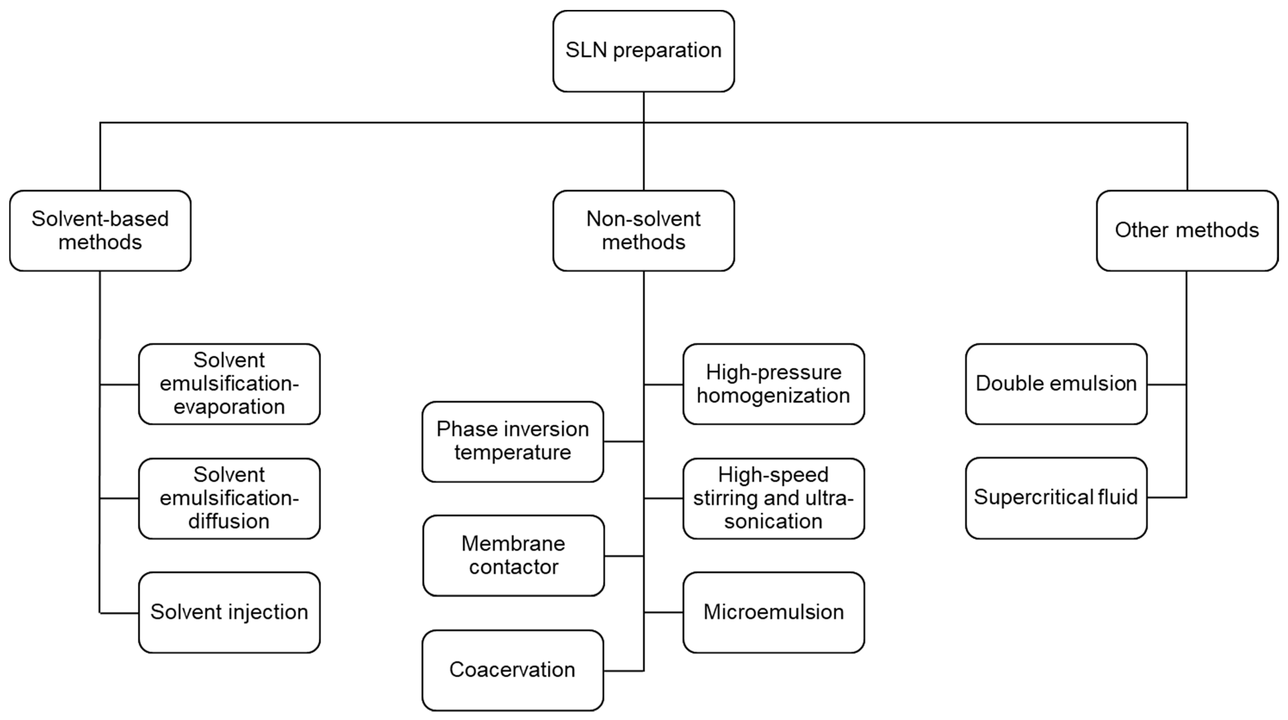
Table 2.
Physicochemical characterization of SLNs.
| Parameters | Characterization | References |
|---|---|---|
| Particle size | Dynamic light scattering and laser diffraction | [52] |
| Polydispersit index | Dynamic light scattering and laser diffraction | [52] |
| Zeta potential | Dynamic light scattering | [58,59] |
| Entrapment efficiency | Gel filtration chromatography | [63] |
| Dialysis | [64] | |
| Ultracentrifugation | [65,66] | |
| Filter membrane | [67] | |
| Crystallinity | Differential scanning calorimetry (DSC) and X-ray diffraction (XRD) | [35] |
| Shape and morphology | Scanning electron microscopy (SEM), transmission electron microscopy (TEM), and atomic force microscopy (AFM) | [52,55,70,71] |
Table 3.
Physicochemical characterization of SLNs.
| Method | Features | References |
|---|---|---|
| Solvent emulsification-evaporation | Suitable for highly thermo-labile drugs, no high temperatures and physical stress Toxic organic solvent removal | [91,92,93,94,95] |
| Solvent emulsification-diffusion | Suitable for highly thermo-labile drugs, does not require high temperatures and physical stress Toxic organic solvent removal, high water amount, and low SLN concentration | [101,102,103,104] |
| Solvent injection | Simple, fast production, no complicated instruments Toxic organic solvent removal | [67,108,109,110] |
| Hot high-pressure homogenization | Organic-solvent-free operation, short production time, and scale-up feasibility Unsuitable for heat-sensitive or hydrophilic drugs | [120,121,122,123,124] |
| Cold high-pressure homogenization | Suitable for water-soluble drugs to prevent drug loss Large particles and laborious processes | [126,129,130] |
| High-speed stirring and ultra-sonication | Organic-solvent-free operation and ease of implementation High surfactant amounts, exposal of drugs to high temperatures, and metal contamination | [138,139,140,141,142,143] |
| Microemulsion | Simple, reproducible, solvent-free, and feasible to scale up Large amount of surfactant and water | [149,150,151,152] |
| Phase inversion temperature | Based on non-ionic polyoxyethylated surfactants Solvent-free, little energy input Low stability of the nanoemulsion | [158,159,160] |
| Membrane contactor | Use of a specific membrane contactor Scale-up feasibility and particle-size controllability Sophisticated system and clogging risk of the membrane | [164] |
| Coacervation | Use of alkaline salts of fatty acids Straightforward and solvent-free Unsuitable for pH-sensitive drugs, only applicable to alkaline salt lipids | [168,169] |
| Double emulsion | Preparation of a water/oi/water double emulsion Suitable for hydrophilic drugs High drug loss and large particle size | [172,173,177,178] |
| Supercritical fluid | Uniform-particle-size distributions and high solvent-extraction efficiencies Use of organic solvents and expensive supercritical fluids | [180,183] |
Publisher’s Note: MDPI stays neutral with regard to jurisdictional claims in published maps and institutional affiliations. |
© 2022 by the authors. Licensee MDPI, Basel, Switzerland. This article is an open access article distributed under the terms and conditions of the Creative Commons Attribution (CC BY) license (https://creativecommons.org/licenses/by/4.0/).
Share and Cite
MDPI and ACS Style
Nguyen, T.-T.-L.; Duong, V.-A. Solid Lipid Nanoparticles. Encyclopedia 2022, 2, 952-973. https://doi.org/10.3390/encyclopedia2020063
AMA Style
Nguyen T-T-L, Duong V-A. Solid Lipid Nanoparticles. Encyclopedia. 2022; 2(2):952-973. https://doi.org/10.3390/encyclopedia2020063
Chicago/Turabian StyleNguyen, Thi-Thao-Linh, and Van-An Duong. 2022. "Solid Lipid Nanoparticles" Encyclopedia 2, no. 2: 952-973. https://doi.org/10.3390/encyclopedia2020063