Entry

Mismatch Repair Deficiency and Microsatellite Instability

Targos—A Discovery Life Sciences Company, Germaniastrasse 7, 34119 Kassel, Germany
*
Author to whom correspondence should be addressed.
Encyclopedia 2022, 2(3), 1559-1576; https://doi.org/10.3390/encyclopedia2030106
Submission received: 23 June 2022 / Revised: 21 August 2022 / Accepted: 26 August 2022 / Published: 31 August 2022
(This article belongs to the Section Biology & Life Sciences)

Definition

:
Mismatch repair deficiency (MMRd) is caused by the biallelic inactivation of an MMR gene, which can be attributed either to an inherited or an acquired pathway. MMRd is characterized by the inability of cells to repair spontaneous mutations in microsatellites that occur during replication. Microsatellites are repetitive nucleotide sequences composed of one to six base pairs. Mutations in microsatellites lead to deletions or insertions of sequence units that are designated as microsatellite instability (MSI). MMRd is diagnosed by immunochemistry and is characterized by loss of nuclear immunostaining for at least one of the four MMR proteins that are routinely examined, i.e., MSH2, MSH6, MLH1 and PMS2. Available tests for MSI are PCR and next generation sequencing. MMRd and MSI predispose to tumor initiation and progression, increase tumor mutational burden as well as tumor immunogenicity, facilitate the activation of the programmed cell death protein 1/programmed cell death ligand 1 (PD-1/PD-L1) immune checkpoint pathway and serve as prognostic and predictive biomarkers in solid tumors.

1. Introduction

This entry paper provides a comprehensive overview of the impact of mismatch repair deficiency (MMRd) and microsatellite instability (MSI) on tumor initiation and progression as well as the relevance of these alterations as diagnostic, prognostic and predictive biomarkers. In this regard, it highlights the gain in medical importance that both markers have received from the initial discovery of their causative role in Lynch syndrome until nowadays, where they influence prognostic and therapeutic decisions for Lynch syndrome-associated and sporadic tumors. It also provides guidance for the diagnostic evaluation of MMRd and MSI in different cancer types and explains the limitations of routinely available tests. Thereby, it identifies the immediate need for improved MSI detection across different cancer types and provides research guidance by summarizing recent scientific studies in this field. The development of standardized tests to detect MSI with high sensitivity and specificity in non-colorectal cancers will likely not only improve prognostic evaluation and treatment success of different tumor entities but may also help to identify additional therapeutic options.

2. History

In 1895, Warthin encountered a family with susceptibility to early age onset cancer. Approximately 32% of family members (n = 146) of this family (named as family “G”) developed cancer at a median age of 38 years; most frequently affected were the gastrointestinal tract and endometrium [1,2]. In the 1960s, Lynch continued to investigate this cancer family syndrome and detected an autosomal dominant inheritance [3,4]. Boland and Troncale [5] studied additional families with the same cancer family syndrome and they used the disease name “Lynch syndrome I” for families with exclusively colon cancer and the designation “Lynch syndrome II” for families with colonic cancer and additional extracolonic carcinomas [5]. Lynch et al. [6] discovered that Muir–Torre syndrome is a phenotypic variant of the latter. This family cancer syndrome was independently reported by Muir et al. in 1967 [7] and by Torre et al. in 1968 [8] and is characterized by sebaceous neoplasms together with carcinomas of the colon and less frequently of other organs [7,8]. In 1985, Lynch et al. introduced the disease name “hereditary nonpolyposis colorectal cancer” (HNPCC) as an umbrella term for “Lynch syndrome I and II” [9].
In 1993, a distinctive type of colorectal carcinoma with mutations in microsatellites as repetitive DNA sequences was described [10,11]. Microsatellite mutations occurred in 12% [10] and 28% [11] of examined colorectal carcinomas leading to microsatellite instability (MSI), which was initially named as a replication error (RER) [12]. The detection of these mutations in tumors, but not in normal tissues, supported their role as driver mutations for carcinogenesis [10]. A statistically significant correlation existed between the observed microsatellite alterations and a favorable prognosis [11]. Since patients with MSI tumors included those of younger age (<50 years), Ionov et al. [10] suspected an inherited nonpolyposis colon cancer syndrome [10]. In the same year, Peltomäki et al. [13] observed a genetic linkage of HNPCC syndrome to microsatellite markers on chromosome 2, and Aaltonen et al. [12] confirmed that HNPCC syndrome-associated cancers showed MSI. Notably, MSI was also detected in sporadic colon cancer [12].
An individual tumor cell could harbor more than 1000 mutations within microsatellites that accumulate during repeated runs of replication [10]. This led to the hypothesis that MSI developed secondarily to a mutation within a gene coding for a protein that corrects replication errors in repetitive sequences [10,11].
The MMR system is responsible for correcting errors of replication that most commonly involve repetitive nucleotide sequences [14]. This system was first characterized in E. coli [14]. The MMR system of E. coli is composed of the two homodimeric proteins MutS and MutL that recognize mismatches and activate excision repair, respectively [14]. Subsequently, investigations on nuclear extracts of HeLa cells revealed a human MMR system that shows remarkable homology to that of E. coli [15]. The human homolog of the DNA MMR gene MutS homolog 2 (MSH2) was cloned by Fishel et al. [16]. The human MSH2 gene is located on chromosome 2 close to a locus implicated in hereditary nonpolyposis colon cancer [16]. Subsequently, it was discovered that germline mutations in MSH2 are present in members of families with HNPCC [16,17]. Moreover, Douglas et al. [18] confirmed that germline mutations in the MSH2 were the cause of the inherited cancers in “family G”, which was the focus of the initial studies of Warthin and Lynch [1,2,3,4,6].
Subsequently, it was revealed that HNPCC can also be evoked by germline mutations in MutL homolog 1 (MLH1) [19,20], postmeiotic segregation increased 2 (PMS2) [21], MutS homolog 6 (MSH6) [22] and epithelial cell adhesion molecule (EpCAM) [23].
Moreover, microsatellite instable cancer also occurs in people without a genetic predisposition. Sporadic carcinomas with MSI are nearly exclusively caused by MLH1 promoter hypermethylation resulting in loss of gene expression [24].
Currently, the disease name Lynch syndrome has replaced the designation HNPCC for those cancers associated with inactivation of the MMR genes MSH2, MLH1, MSH6 and PMS2. Nowadays, the investigation of tumors for MMRd and/or MSI not only helps to identify patients with an inherited disease (Lynch syndrome, Muir–Torre syndrome), but also has prognostic and predictive relevance [25]. The detection of the molecular features of MMRd and MSI helps to select the most appropriate treatment option. Those tumors often show a favorable response to immune checkpoint inhibition [26,27].

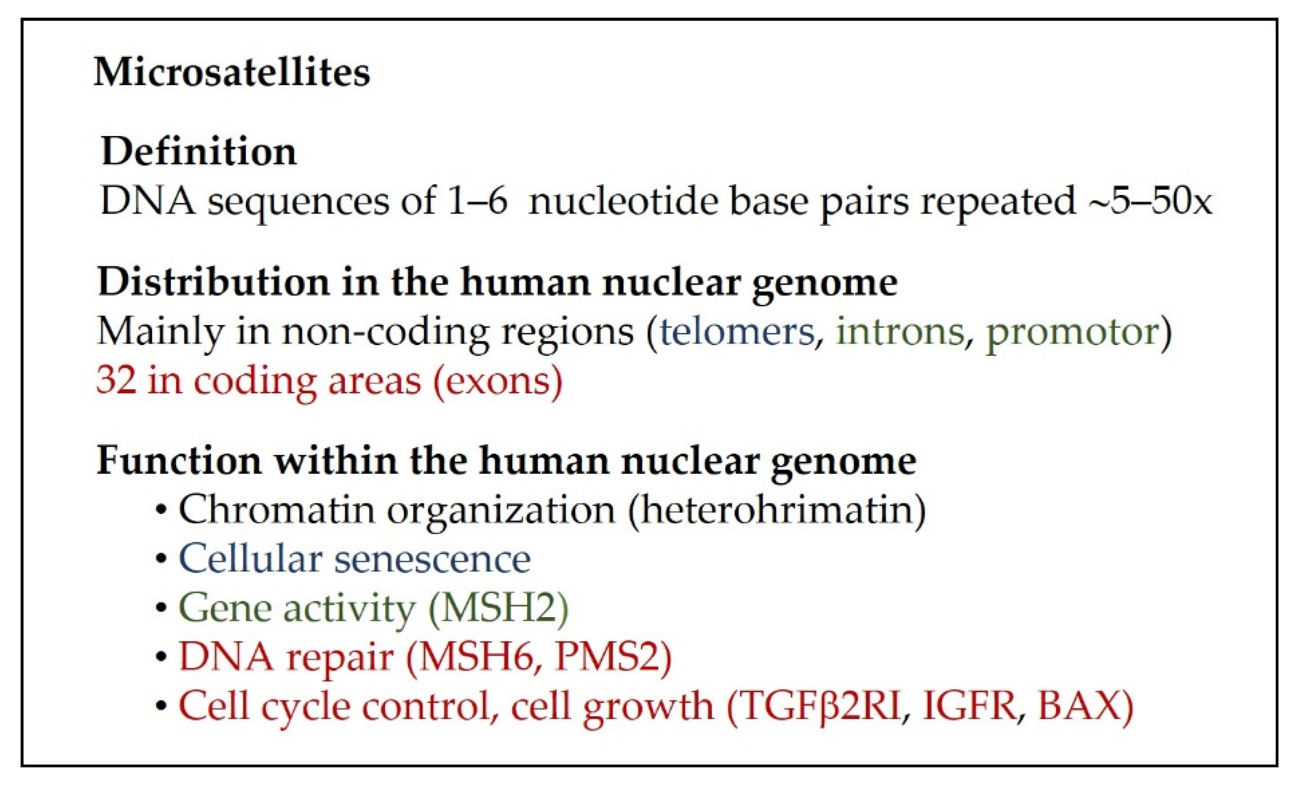
3. The Organization of the Human Nuclear Genome and Repetitive DNA Sequences

Notably, coding regions of the genome, which are named exons, occupy only 1.5% of the human nuclear genome [28]. Each exon is composed of a unique sequence of nucleotides and encodes for a particular protein [28]. The remaining DNA is referred to as non-coding DNA. This includes all DNA sequence areas outside the genes, but also introns and promoter regions [28]. About 50% of the non-coding DNA is composed of repetitive DNA sequences [29] that can be further classified into dispersed sequences, i.e., transposable elements and tandem repeats such as satellite DNA [28,30]. Satellite DNA covers about 3% of the human genome [30,31]. Based on the size of each repeat unit, satellite DNA can be further subdivided into macrosatellites, satellites as well as mini- and microsatellites [30,31] (Figure 1).
The name satellite DNA is derived from the result of the density gradient centrifugation of DNA [30,32]. Satellite DNA forms additional small bands above or below the main DNA band [30].
Microsatellites are defined as 1–6 base pairs that are repeated 5–50x [30,33,34]. The human genome contains thousands of microsatellite sequences. Most are localized in the non-coding areas outside the genes, whereas fewer are present within introns and promoter regions and only 32 are contained within exons, that mostly belong to tumor suppressor genes [35].
Microsatellites are involved in chromatin organization by heterochromatin formation and determine cellular senescence since they are an essential structural element of telomers [28,30,36]. Further, those localized in introns and promoter regions regulate gene transcription [36]. For example, intron 5 of the MSH2 gene contains the microsatellite marker BAT26, which regulates the transcription of exon 5 [34,37]. Genes with microsatellite sequences in exons control cellular DNA repair, proliferative activity, and apoptosis. Examples are bcl-2-associated X protein (Bax) [35,37], transforming growth factor β1 type II receptor (TGFRbetaII) [35,37], insulin-like growth factor type II (ILGFIIR) [35,37], PTEN (Phosphatase and TENsin homolog deleted on chromosome 10) [35,38] and MMR genes, e.g., MSH6 and PMS2 [34,35,38] (Figure 1).
Microsatellites display high pleomorphism and high heterozygosity [30,33,39]. Due to these features, microsatellite markers are used for paternity testing and in forensics [30]. In addition, during DNA replication, microsatellites show a high rate of spontaneous mutations since the DNA polymerase tends to slip at the repetitive sequences [33]. Spontaneous mutations in microsatellites are corrected by the MMR system [33].

4. The Human Mismatch Repair System

The MMR system ensures the stability of the genome during repeated cycles of replication [40,41,42]. In humans, it is mainly composed of the five MMR proteins, MSH2, MSH3, MSH6, PMS2 and MLH1 [36,40,41,42]. These form three heterodimeric complexes that are homolog to the E. coli MMR proteins MutS and MutL [40,41,42]. The MutS homologs consist of MutSα formed by MSH2/MSH6 and MutSβ composed of MSH2/MSH3 [40,41], whereas the MutL homolog MutLα is formed by MLH1/PMS2 [36,40,41,42] (Figure 2). In the cell, the MSH2/MSH6 complex is found 4x more frequent than the MSH2/MSH3 complex [36,40,41,42].
The functional complexes composed of MSH2/MSH6 and MSH2/MSH3 recognize mismatched DNA [36,40,41]. In this regard, MSH2/MSH6 strongly binds to base–base mispairs and insertion or deletion loops consisting of one to two extrahelical nucleotides [41,42], whereas MSH2/MSH3 preferential binds to insertion and deletions composed of 2–10 extrahelical nucleotides [36,41,42] (Figure 2). With low affinity, the MSH2/MSH6 complex can also recognize insertion and deletion (indel) lesions with more than two extrahelical nucleotides and the MSH2/MSH3 complex can also weakly bind to single nucleotide indel lesions [41,42]. As a consequence, the deficiency of MSH6 protein can be partially substituted by MSH2/MSH3 and the loss of MSH3 protein will be nearly completely replaced by the MSH2/MSH6 complex [41,42].
The binding to the damaged DNA initiates an ATP-dependent conformation change resulting in the formation of a sliding clamp [36,41]. The complex consisting of PMS2 and MLH1 recruits additional proteins responsible for the excision of the mismatched bases and adjacent DNA, resynthesis of a new daughter strand and the ligation of the repaired defect with the remaining DNA [36,40,41,42,43].
In addition to post-replicative MMR, the MMR system is also involved in meiotic and mitotic recombination events [41] and triplet-expansion that can lead to disease [41]. Further, it recognizes certain alterations of DNA bases such as alkylation or methylation and subsequently either mediates cell cycle arrest at the G2/M transition or apoptosis [40,41]. In B cells, the MMR system contributes to antibody diversification [40,41].
The presence of an intact MMR system is named as MMR proficiency (MMRp), whereas a defective MMR system leads to MMR deficiency (MMRd) [41].

5. Mismatch Repair Deficiency

The primary cause of MMRd is a biallelic genetic inactivation of an MMR gene [44]. This genetic change predisposes to cancer initiation and progression [44].
MMRd tumors can develop by two different pathways, i.e., the constitutional/hereditary and the acquired pathway [42,44].
The constitutional pathway occurs in carriers of Lynch syndrome (previously named as hereditary nonpolyposis colorectal cancer) [36,42,44]. Due to a germline mutation, affected people carry one mutated allele of a particular MMR gene in every cell in their body. They have an increased cancer risk since only one further mutation (somatic mutation) is required for tumor initiation, i.e., the mutation in the second allele of the affected MMR gene [44].
Interestingly, the carrier frequency of germline mutations within the general population is highest for PMS2 (1 in 714), followed by MSH6 (1 in 758), MLH1 (1 in 1946) and MSH2 (1 in 2841) [45]. However, people with germline mutations in MLH1 or MSH2 have a much higher lifetime cancer risk compared to carriers of MSH6 or PMS2 mutations [46] and may develop carcinomas already at an early adult age [47]. The following facts likely explain this discrepancy between the prevalence and penetrance: not all mutations confer MMRd and pathogenic mutations occur more often in MLH1 and MSH2 than in MSH6 and PMS2 [46]. MLH1 and MSH2 are obligatory components of MMR, whereas the functions of MSH6 and PMS2 can be partially substituted by MSH3 and MLH3, respectively [46]. A further rare cause for MMRd colorectal carcinoma is a germline mutation in the EpCAM gene leading to MSH2 hypermethylation that represses MSH2 transcription [23,34,48].
The acquired pathway is characterized by the sporadic consecutive inactivation of the two alleles of a particular MMR gene. The inactivation is nearly exclusively caused by hypermethylation of the MLH1 promoter with subsequent loss of MLH1 gene expression [44].
Tumors with MMRd and MSI frequently occur in the colon, rectum and endometrium, but can also be diagnosed in multiple other organs [34,44,49,50]. About 20–30% of endometrial carcinomas [51] and approximately 15% of colorectal carcinomas [52] display MMRd and high MSI. In regard to colorectal carcinomas with MMRd, about 20% are attributed to the constitutional pathway and 80% to the acquired pathway [46]. In comparison to patients with acquired pathogenesis, patients with inherited MMRd tumors usually present at a younger age [52] (Figure 3).
The mechanisms leading to MLH1 promoter hypermethylation, however, differ between colorectal carcinomas on the one hand and endometrial carcinoma [53], and likely also other solid tumors on the other hand.
In colorectal carcinomas, MLH1 promoter hypermethylation is promoted by distinct single nucleotide polymorphisms (SNPs), and it is initiated by a specific mutation in the v-raf murine sarcoma viral oncogene homolog B1 (BRAF) gene, where at position 600, the amino acid valine is substituted by glutamic acid (BRAF V600E) [53,54]. It is often associated with a high CpG island methylator (CIMP) phenotype and MSI-H status [55]. BRAF V600E increases methylation of the MLH1 promoter and CpG islands of other tumor suppressor protein genes by activating a specific transcriptional repressor and DNA (cytosine-5)-methyltransferase 3 beta [53,54]. MLH1 promoter methylation, however, is not only detected in neoplastic tissue with loss of MLH1 proteins, but also in adjacent normal tissue with retained MLH1 protein production [56]. Studies suggest that gene silencing is either associated with the methylation of a specific site within the promoter region [56] or with the overall extent of CpG promoter methylation [57].
In contrast, molecular alterations predisposing to MLH1 promoter hypermethylation of non-colorectal carcinomas are hitherto unknown [53]. Notably, in endometrial carcinomas, MLH1 gene silencing, loss of MLH1 protein expression and the consecutive MSI-H status develop if the MLH1 promoter methylation exceeds a certain threshold level [57,58,59,60]. This finding is supported by a study on endometrial carcinomas with subclonal MMRd due to loss of MLH1 and PMS2 protein expression, in which MLH1 promoter hypermethylation was detected not only in the tumor area with MMRd and MSI-H, but also in the adjacent neoplastic tissue [60].
Within endometrial carcinomas with MLH1 silencing, MLH1 promoters can either show homogenous dense methylation or heterogeneous methylation in more than 50% of the CpG islands [58,59]. The heterogeneous methylation pattern is likely explained by the presence of tumor cell subclones that accumulated additional MLH1 promoter methylation sites during tumorigenesis, whereas the tumor-initiating cell population likely harbors the least number of methylation sites [59]. Interestingly, MLH1 promoter hypermethylation is also detected in peritumoral normal endometrium, where it may even be associated with loss of MLH1 protein expression and/or MSI [61].
This confirms that MLH1 promoter hypermethylation is an early step in the development of sporadic MMRd carcinomas and that the transition of a premalignant change to cancer is influenced by additional external and internal factors, e.g., tumor cell replication time and presence of mutations within coding or non-coding regions.

6. The Diagnosis of Mismatch Repair Deficiency

The diagnosis of MMRd is performed by immunohistochemistry on formalin-fixed paraffin-embedded (FFPE) tissue. For the routine diagnostic evaluation, antibodies detecting MMR proteins of two functional heterodimers are available, i.e., MSH2 and MSH6, as well as PMS2 and MLH1 [48,62,63].

6.1. Immunohistochemical Features of Mismatch Repair Proficiency and Deficiency

An intact nuclear expression of an MMR protein occurs if both alleles of the corresponding gene are intact or only one allele is inactivated. Loss of nuclear immunostaining is observed after biallelic genetic inactivation of a particular MMR gene and after protein degradation of an unstable partner protein [36,62,63]. If loss of immunostaining for a particular MMR protein is observed, the presence of a reliable internal positive control should be examined. Cells that can serve as internal positive controls are stromal cells, tumor-infiltrating immune cells and the cells of the adjacent normal tissue [62,63].
A negative immunoreaction is characterized by complete loss of nuclear immunostaining [62,63]. In rare cases, immunolabeling for MLH1 protein may show a solely punctuate nuclear staining that corresponds to a negative immunohistochemical result [64,65,66]. In summary, MMRp is characterized by nuclear staining for all four MMR proteins. In comparison, MMRd is defined as the loss of nuclear staining for at least one of the examined MMR proteins in the presence of a retained internal positive control (Figure 4).

6.2. Molecular Mechanisms of Mismatch Repair Deficiency

The biallelic genetic inactivation of an MMR gene evokes its loss of protein expression. In case this MMR protein is associated with an unstable heterodimeric partner protein, this partner protein will be degraded [36,62,63,67]. MSH2 and MLH1 are stable proteins, whereas MSH6 and PMS2 are unstable proteins [36,62,63,67]. Thus, the absence of immunostaining for MSH2/MSH6 or MLH1/PMS2 is attributed to the biallelic genetic inactivation of MSH2 or MLH1 followed by degradation of the respective unstable partner protein, i.e., MSH6 or PMS2 [36,62,63,67]. In comparison, the biallelic genetic inactivation of MSH6 and PMS2 only leads to the loss of protein expression, but no degradation of the respective partner protein (MSH2 or MLH1) since this is stable [36,62,63,67] (Figure 5).

7. Microsatellite Instability

MSI is defined as an alteration in the number of repeat units of a particular microsatellite sequence that leads to contraction or expansion of microsatellite markers [34,37,41,68]. This definition applies to any change in the length of a particular microsatellite marker in the tumor tissue in comparison to the normal tissue [34]. It is detected in more than 90% of colorectal carcinomas from patients with Lynch syndrome and approximately 20% of sporadic colorectal carcinomas and develop secondary to MMRd [37]. During replication, the complementary and the template strands may dissociate followed by their incorrect reannealing [41]. The result is the presence of extrahelical nucleotides forming insertion or deletion loops [41,68]. These insertions and deletions, as well as base–base mismatches are normally corrected by the MMR system [41]. If MMRd exists, those spontaneous errors of replication remain unrepaired and are transmitted to daughter cells, thus, the sequel is MSI [36,41].

8. Diagnosis of Microsatellite Instability

The diagnostic evaluation of MSI is performed by multiplex polymerase chain reaction (PCR) followed by fragment length analysis or by next-generation sequencing (NGS) [34,37,69].
The basic principles for the evaluation of MSI were set in 1997 when Dietmaier et al. [37] characterized the sensitivity and specificity of 31 microsatellite markers in colorectal carcinomas and classified the results into three categories, i.e., microsatellite stability (MSS), and low-frequency and high-frequency microsatellite instability (MSI-L, MSI-H).
The comprehensive data of Dietmaier et al. [37] served as the foundation for the 1st and 2nd choice Bethesda panels (5 markers each) as well as the standardized result interpretation [37,70]. The 1st choice panel is composed of two mononucleotide markers (BAT25 and BAT26) as well as three dinucleotide markers (D5S346, D2S123 and D17S250). High-frequency MSI (MSI-H) is defined as the instability of two or more markers, and low-frequency MSI (MSI-L) as the instability of one of the five markers [70]. If an alteration in only a single marker is detected, the 2nd panel should be applied [52,70]. The 2nd choice Bethesda panel consists of one mononucleotide marker (BAT40), three dinucleotide markers (D10S197, D18S58, D18S69) and a tetranucleotide marker (Mycl1) [37,70]. This procedure aids the diagnosis of EMAST (“elevated microsatellite alterations at selected tetranucleotide repeats”) [63]. The causative agent of EMAST is oxidative stress mediated through elevated levels of interleukin 6 (IL6). The induced intracellular signal transduction with activation of STAT-3 evokes the shifting of MSH3 from the nucleus into the cytoplasm and subsequent functional loss of MSH3 [71,72].
A pentaplex panel of mononucleotide repeats (BAT-25, BAT-26, NR-21, NR-24 and MONO-27 a) was validated by Suraweera et al. [73]. This method was evaluated as sensitive and specific for the diagnosis of MSI-H in colon and gastric cancer [52]. It is also used by the multiplex fluorescent MSI analysis system of Promega®, newer versions include the two additional pentanucleotide repeat markers Penta C and Penta D [69,74].
Nowadays, diagnostic centers most frequently use PCR assays, which examine five to seven microsatellite markers for insertions or deletions [34,69,74]. Panels consist either of only mononucleotide repeat markers or a combination of mononucleotide repeat markers and higher nucleotide sequence markers [44,69]. Tests solely composed of mononucleotide markers can miss microsatellite instabilities resulting from mutations in higher nucleotide repeats.
For routine diagnostic examinations, deoxyribonucleic acid (DNA) is usually extracted from the FFPE tissue samples of tumor and normal control [69,74]. Amplicons are most commonly visualized by capillary electrophoresis, and microsatellite profiles of tumor tissue and normal tissue are compared [69]. Diagnostic for MSI are shifts in microsatellite repeat lengths as well as peak variations [69]. A turnaround time of 3 working days exists [69].
In addition, PCR-based automated systems are commercially distributed, e.g., the IdyllaTM MSI assay. This system uses a novel set of seven biomarkers (ACVR2A, BTBD7, DIDO1, MRE11, RYR3, SEC31A, SULF2) and examines FFPE tissue samples with a fully automated workflow including sample preparation, DNA amplification, melting curve analysis and data interpretation [74]. Notably, the IdyllaTM system does not require matched normal tissue [69,74]. The time requirements are short, i.e., approximately 5 min for manual preparation and about 150 min for complete sample analysis [74].
NGS-based computational MSI detection systems are also available [34] and may also be used for routine diagnostic examinations [69]. These are based on the comparison of microsatellite length between normal and tumor samples, or they calculate the indel burden in microsatellites [34]. The turnaround time constitutes about 7 working days [69].
PCR tests and NGS-based computational models were initially validated for colorectal carcinomas and are now also applied to diagnose microsatellite instability in endometrial carcinomas and other cancer types [34,49,63,69]. Thus, they have the highest sensitivity for colorectal carcinomas and a lower sensitivity for endometrial carcinomas and other cancer types [34,63,69].
Computational methods can be applied to analyze in detail MMRd evoked mutation patterns across different cancer types [75]. The subcategorization of mutation patterns will likely assist in identifying cancer type-specific mutation signatures, deciphering cancer type-associated patterns of carcinogenesis, predicting prognosis, and analyzing likely outcomes of different treatment modalities or even designing cancer type-specific therapies [75]. For this, the large publicly available repository of genomic data can be used [75].
For translational medicine research, an algorithm to predict MSI from the vast amount of publicly available gene expression data of colorectal cancer has been developed [76]. This model has been named “microsatellite instability absolute single sample predictor” and combines transcriptomics of cancer-related molecular pathways with those of the tumor microenvironment. It shows robustness and high predictive power for the identification of MSI in sporadic colorectal carcinoma [76].
Further, a residual learning convolutional neuronal network was trained to identify MSI status directly from digital images of hematoxylin-eosin (HE) stained cancer tissue [77]. The best predictive performance was obtained when identical tumor entities were used for training the algorithm and its application for MSI detection [77]. The correlation of the predicted MSI status with immunohistochemical results and transcriptomic data revealed that in gastric cancer, MSI status was associated with a lymphocyte gene expression signature, whereas in colorectal cancer, it was correlated with PD-L1 expression and an interferon γ signature [77]. The diagnostic use of this automatic MSI detector across different human tumors is currently still limited by its cancer-type restricted performance, which could possibly be resolved by its training on larger datasets and prospective validation [77]. In future, deep learning provided by convolutional neuronal networks is likely also to be used for molecular subclassification of cancer types from HE stained tissue sections, e.g., the identification of the MSI subtype and additional subtypes of gastric cancer [78].

9. Mismatch Repair Deficiency and Microsatellite Instability as Predictive Biomarkers

Tumors with MMRd and MSI-H develop more than 1000 mutations in microsatellites; this is diagnosed as hypermutation or mutator phenotype [36,44,49]. Thus, tumors with these molecular alterations tend to have a high tumor mutational burden (TMB) [49]. Because mutations in microsatellites are often frameshift mutations, tumor cells usually express neoantigens [36,50], that are named as frame shift-associated peptides [79]. Since these peptides are hitherto unknown to the immune system, they stimulate a strong anti-cancer immune response leading to tumor infiltration with numerous lymphocytes, including cytotoxic T cells [36,80] (Figure 6).
During a longer immune response, exhaustion of T cells develops [81,82]. This is characterized by the upregulation of the immune checkpoint molecule programmed cell death protein 1 (PD-1) on exhausted T cells [81,82,83]. Its ligand, programmed cell death ligand 1 (PD-L1), can be expressed on tumor-infiltrating macrophages [83,84] and lymphocytes [83,84] as well as tumor cells [82,84]. The binding between PD-1 and PD-L1 inhibits the anti-tumor immune response [81,82,84]. During an immune reaction, the activation of immune checkpoint cascades prevents an overwhelming immune response that may lead to tissue destruction and autoimmunity [81]. In the tumor microenvironment, however, the activation of the immune checkpoint molecules PD-1 and PD-L1 attenuates the anti-tumor immune response and may facilitate tumor progression [81]. Thus, the therapeutic blockage of PD-1 or PD-L1 by monoclonal antibodies is widely used as a treatment for those cancers with activation of this immune checkpoint cascade [82]. In 2018, James Allison and Tasuko Honjo received the Nobel Prize for Medicine for their investigations on immune checkpoint cascades.
Since tumors with MMRd and/or MSI-H commonly display the aforementioned features of high tumor mutational burden, high immunogenicity and infiltration with numerous immune cells followed by the activation of immune checkpoint cascades, they are optimal targets for immune checkpoint therapy [26,36,50,63,80] (Figure 7). In 2017, the US Food and Drug Administration (FDA) approved the anti PD-1 monoclonal antibody pembolizumab for solid tumors in adults or children that either display MMRd on immunostaining or MSI-H by PCR analysis and for which no other treatment options exist [85]. Notably, tumors with MMRd are resistant to treatment with certain cytotoxic drugs [26,42].
Tumors with mutations of the catalytic and proofreading domains of polymerase ε (POLE) and polymerase δ1 (POLD1) also show high TMB with the sequelae of increased neoantigen expression and a strong anti-cancer immune response [86]. Mutations in functional areas of their exonuclease domains (EDMs) cause a 10–100-fold increase in the mutation rate during DNA replication [86] and can be diagnosed in a variety of cancer types, including colorectal carcinomas, endometrial cancer, lung cancer and melanoma [86]. In general, mutations in POLD1-EDM are less frequently observed than POLE-EDM mutations [86]. Across different cancer types, Wang et al. [87] found mutation rates of 2.8% and 1.4% for POLE and POLD1, respectively. Solid tumors with POLE or POLD1 mutations accumulate missense mutations, have ultra-high mutation rates (>100 mutations per megabase genome) and are initially microsatellite stable [86]. In an early stage of tumorigenesis, POLE-EDM or POLD1-EDM mutations and MMRd are considered as independent molecular events [86]. During later stages of tumorigenesis, however, tumors may show the concurrent presence of a POLE-EDM or POLD1-EDM mutation and MMRd/MSI, since the loss of proofreading activity may lead to secondary mutations also in MMR genes [86].
Several studies suggest that high TMB predicts a favorable response to immune checkpoint inhibition that is independent of the tumor type and its MSI status [87,88,89,90]. In 2020, the FDA extended the approval of pembrolizumab in pediatric and adult patients for the treatment of unresectable or metastatic solid tumors that are diagnosed with TMB ≥ 10 mutations per megabase genome and have no other therapeutic options [91].

10. Concordance and Disconcordance of Test Results

The concordance rate between MMRd and MSI-H is 98% in colorectal carcinomas [60] and 94% in endometrial carcinomas [51,69]. The most frequent cause for disconcordance in endometrial carcinoma was MLH1 promoter hypermethylation in 1.6% of examined cases [51].
Reported reasons for detection of MSS or MSI-L in a tumor with immunohistochemical MMRd are summarized in Table 1. MLH1 promoter hypermethylation and consecutive loss of MLH1 and PMS2 immunostaining despite the molecular detection of MSS or MSI-L are reported nearly exclusively in non-colorectal cancer types, particularly endometrial carcinomas [51,69]. To confirm MSI-H in a tumor sample with an area of subclonal loss, it may be necessary to micro-dissect the area with loss of immunostaining and to analyze the microdissected tissue sample separately from the remaining tumor [51,92]. To ensure a valid PCR or NGS result, the tumor sample should contain at least 20% (colorectal carcinomas) or 40% (endometrial carcinoma) of cancer cells [51,63,69]. Tumors with a low proliferative activity, e.g., mesotheliomas may show MMRd and MSI-L [34]. Notably, MSH6 protein loss after neoadjuvant treatment (radiation or chemotherapy) is not a cause of MSI-H [34]. In carcinomas of other organs than the colon, MSI-H is likely underdiagnosed since the testing systems are not validated for these additional tumor entities [36,63,93]. The types and numbers of unstable microsatellite loci vary between different cancer types [36]. Further, endometrial carcinoma with MSI can present with only discrete length shift that may be under detected [69].
On the contrary, tumors can also display MSI-H despite the detection of immunostaining for all four MMR proteins. This has been described in association with a few cases that harbored mutations in the exonuclease domain of polymerase epsilon (POLE-EDM) [51]. In addition, it may be caused by missense mutations where an altered MMR protein is expressed that shows a functional loss, while the binding site for the antibody is still retained. This is reported in up to 30% of MLH1 mutations [94].
In order to overcome such discordances in routine diagnostics, different measures should be taken. First, quality assurance by using well-fixed tissue such as biopsies instead of resection specimens is advised [62,95]. Second, a test algorithm is recommended where immunohistochemistry is done first to determine the MMR status [63]. Third, in all cases where MMR status cannot reliably be assessed by immunohistochemistry, determination of MSI by PCR and/or NGS is needed (Table 2) [63]. Finally, regular participation in proficiency assessments such as those organized by the College of American Pathologists (CAP), United Kingdom National External Quality Assessment Service (UKNQAS), Nordic immunohistochemistry Quality Control (NordiqQC) or Quality Assurance in Pathology (QuIP) is required [63,96].

11. Testing Algorithm Applicable across Cancer Types

To identify cancer with MMRd and/or MSI-H, the primary use of an immunohistochemical testing algorithm is recommended since it represents a highly standardized, sensitive, and specific method for detection of MMRd across different cancer types, which is readily available, cost-effective and has a short turnaround time [63,69,93,97]. MMRd testing not only allows the recognition of carriers of Lynch syndrome, but it is also used for molecular subtyping of endometrial and gastric cancers as well as prognostic evaluations and treatment decisions [63,97,98]. Thus, it should be applied to all colorectal, gastroesophageal, and endometrial adenocarcinomas as well as additional tumors of the Lynch syndrome spectrum and all other advanced solid cancer types for which no alternative treatment options exist [63,85,97,98].
To further increase the cost-effectiveness of MMRd testing without loss of sensitivity or specificity, a two-step process can be used [63]. In an initial step, immunostaining for MSH6 and PMS2 is performed. This is already sufficient to characterize examined tumor samples as MMRp or MMRd [51,63]. Positive staining for both proteins is diagnostic for MMRp, whereas the loss of at least one protein is diagnostic for MMRd. The subsequent immunostaining for the respective binding partner allows a detailed characterization of the type of MMRd, i.e., isolated deficiency of PMS2 or MSH6, simultaneous loss of MSH2 and MSH6 or combined absence of MLH1 and PMS2 [63]. The latter should be followed by MLH1 promoter hypermethylation testing to distinguish between a Lynch syndrome-associated cancer and a sporadic acquired tumor [63]. Alternatively, colorectal carcinomas, but not other solid tumors, can be examined for the presence of BRAF V600E [63]. The indications that require PCR or NGS testing to determine MSI status are summarized in Table 2. In non-colorectal cancers, only an MSI-H test result is confirmatory, whereas the detection of MSI-L or even MSS does not completely rule out MSI-H status [51,63,69]. In these cases, immunohistochemistry has shown to be more sensitive than PCR-based tests [63,97].
In addition, TMB testing can be used as an alternative method to check for immune checkpoint therapy eligibility [91].

12. Conclusions and Prospects

Immune checkpoint therapy has been approved for cancers with MMRd and/or MSI-H, whereas the immunohistochemical diagnosis of MMRd and the PCR/NGS-based determination of MSI have been recognized as interchangeable and equally valid methods to identify those tumors [63]. The primary use of immunohistochemistry, however, would have the advantage that it directly identifies the absent MMR protein(s) and determines the distribution and extent of the MMR protein loss [63,69,93]. The former provides information on the genetic background and the latter gives guidance for the application (microdissection?) and interpretation of PCR/NGS testing [63]. The use of immunohistochemistry as the primary test will be even more important in non-colorectal cancer, since currently, PCR/NGS-based tests are not validated for those tumor entities [63,93]. The degree of MSI, however, is markedly influenced by the tumor type [34,69]. Cancer-specific MSI signatures exist that are evoked by tumor type-associated differences in gene expression patterns [75,76]. In addition to tumor internal factors (targeted microsatellite markers, proliferation rate, extent of nucleotide shifts) [34,69,76], the development of MSI is also influenced by external factors such as inflammation, commensal bacteria, and the exposure to DNA damaging agents [34]. Therefore, it has been suggested that MSI has likely to be regarded as a continuous phenotype [75].
Due to the importance of MSI as a diagnostic, prognostic and predictive biomarker, testing systems should be optimized for additional cancer types as well [34].
In summary, the diagnosis of MMRd and MSI-H in a carcinoma not only guides the evaluation for a possible familiar cancer syndrome (Lynch syndrome, Muir–Torre syndrome), but also steers prognostication and treatment selection. A detailed understanding of the differences and similarities of MMRd and MSI-H between different cancer types is an important prerequisite to ensuring precise diagnostic work-up across all cancer types.

Author Contributions

Conceptualization, S.S. and J.R.; investigation, S.S. and J.R.; writing—original draft preparation, S.S.; writing—review and editing, J.R.; visualization, S.S. All authors have read and agreed to the published version of the manuscript.

Funding

This research received no external funding.

Data Availability Statement

Not applicable.

Conflicts of Interest

The authors declare no conflict of interest.

References

  1. Warthin, A.S. Heredity with reference to carcinoma as shown by the study of the cases examined in the Pathological Laboratory of the University of Michigan, 1895–1912. Arch. Intern. Med. 1913, 12, 546–555. [Google Scholar] [CrossRef]
  2. Warthin, A.S. The further study of a cancer family. J. Cancer Res. 1925, 9, 279–286. [Google Scholar] [CrossRef]
  3. Lynch, H.T.; Krush, A.J.; Larsen, A.L. Heredity and multiple primary malignant neoplasms: Six cancer families. Am. J. Med. Sci. 1967, 254, 322–329. [Google Scholar] [CrossRef] [PubMed]
  4. Lynch, H.T.; Krush, A.J. Cancer family “G” revisited: 1895–1970. Cancer 1971, 27, 1505–1511. [Google Scholar] [CrossRef]
  5. Boland, C.R.; Troncale, F.J. Familial colonic cancer without antecedent polyposis. Ann. Intern. Med. 1984, 100, 700–701. [Google Scholar] [CrossRef] [PubMed]
  6. Lynch, H.T.; Lynch, P.M.; Pester, J.; Fusaro, R.M. The cancer family syndrome. Rare cutaneous phenotypic linkage of Torre’s syndrome. Arch. Intern. Med. 1981, 141, 607–611. [Google Scholar] [CrossRef]
  7. Muir, E.G.; Bell, A.J.; Barlow, K.A. Multiple primary carcinomata of the colon, duodenum, and larynx associated with kerato-acanthomata of the face. Br. J. Surg. 1967, 54, 191–195. [Google Scholar] [CrossRef]
  8. Torre, D. Multiple sebaceous tumors. Arch. Dermatol. 1968, 98, 549–551. [Google Scholar] [CrossRef]
  9. Lynch, H.T.; Kimberling, W.; Albano, W.A.; Lynch, J.F.; Biscone, K.; Schuelke, G.S.; Sandberg, A.A.; Lipkin, M.; Deschner, E.E.; Mikol, Y.B.; et al. Hereditary nonpolyposis colorectal cancer (Lynch syndromes I and II). I. Clinical description of resource. Cancer 1985, 56, 934–938. [Google Scholar] [CrossRef]
  10. Ionov, Y.; Peinado, M.A.; Malkhosyan, S.; Shibata, D.; Perucho, M. Ubiquitous somatic mutations in simple repeated sequences reveal a new mechanism for colonic carcinogenesis. Nature 1993, 363, 558–561. [Google Scholar] [CrossRef]
  11. Thibodeau, S.N.; Bren, G.; Schaid, D. Microsatellite instability in cancer of the proximal colon. Science 1993, 260, 816–819. [Google Scholar] [CrossRef] [PubMed]
  12. Aaltonen, L.A.; Peltomäki, P.; Leach, F.S.; Sistonen, P.; Pylkkänen, L.; Mecklin, J.P.; Järvinen, H.; Powell, S.M.; Jen, J.; Hamilton, S.R.; et al. Clues to the pathogenesis of familial colorectal cancer. Science 1993, 260, 812–816. [Google Scholar] [CrossRef]
  13. Peltomäki, P.; Aaltonen, L.A.; Sistonen, P.; Pylkkänen, L.; Mecklin, J.P.; Järvinen, H.; Green, J.S.; Jass, J.R.; Weber, J.L.; Leach, F.S.; et al. Genetic mapping of a locus predisposing to human colorectal cancer. Science 1993, 260, 810–812. [Google Scholar] [CrossRef]
  14. Modrich, P. DNA mismatch correction. Ann. Rev. Biochem. 1987, 56, 435–466. [Google Scholar] [CrossRef] [PubMed]
  15. Fang, W.H.; Modrich, P. Human strand-specific mismatch repair occurs by a bidirectional mechanism similar to that of the bacterial reaction. J. Biol. Chem. 1993, 268, 11838–11844. [Google Scholar] [CrossRef]
  16. Fishel, R.; Lescoe, M.K.; Rao, M.R.; Copeland, N.-G.; Jenkins, N.A.; Garbeer, J.; Kane, M.; Kolodner, R. The human mutator gene homolog MSH2 and its association with hereditary nonpolyposis colon cancer. Cell 1993, 75, 1027–1038. [Google Scholar] [CrossRef]
  17. Leach, F.S.; Nicolaides, N.C.; Papadopoulos, N.; Liu, B.; Jen, J.; Parsons, R.; Peltomäki, P.; Sistonen, P.; Aaltonen, L.A.; Nyström-Lahti, M.; et al. Mutations of a mutS homolog in hereditary nonpolyposis colorectal cancer. Cell 1993, 75, 1215–1225. [Google Scholar] [CrossRef]
  18. Douglas, J.A.; Gruber, S.B.; Meister, K.A.; Bonner, J.; Watson, P.; Krush, A.J.; Lynch, H.T. History and molecular genetics of Lynch syndrome in family G: A century later. J. Am. Med. Assoc. 2005, 294, 2195–2202. [Google Scholar] [CrossRef]
  19. Bronner, C.; Baker, S.; Morrison, P.; Warren, G.; Smith, L.G.; Lescoe, M.K.; Kane, M.; Earabino, C.; Lipford, J.; Lindblom, A.; et al. Mutation in the DNA mismatch repair gene homologue hMLH 1 is associated with hereditary non-polyposis colon cancer. Nature 1994, 368, 258–261. [Google Scholar] [CrossRef]
  20. Papadopoulos, N.; Nicolaides, N.C.; Wei, Y.F.; Ruben, S.M.; Carter, K.C.; Rosen, C.A.; Haseltine, W.A.; Fleischmann, R.D.; Fraser, C.M.; Adams, M.D.; et al. Mutation of a mutL homolog in hereditary colon cancer. Science 1994, 263, 1625–1629. [Google Scholar] [CrossRef]
  21. Nicolaides, N.C.; Papadopoulos, N.; Liu, B.; Wei, Y.F.; Carter, K.C.; Ruben, S.M.; Rosen, C.A.; Haseltine, W.A.; Fleischmann, R.D.; Fraser, C.; et al. Mutations of two PMS homologues in hereditary nonpolyposis colon cancer. Nature 1994, 371, 75–80. [Google Scholar] [CrossRef] [PubMed]
  22. Miyaki, M.; Konishi, M.; Tanaka, K.; Kikuchi-Yanoshita, R.; Muraoka, M.; Yasuno, M.; Igari, T.; Koike, M.; Chiba, M.; Mori, T. Germline mutation of MSH6 as the cause of hereditary nonpolyposis colorectal cancer. Nat. Genet. 1997, 17, 271–272. [Google Scholar] [CrossRef] [PubMed]
  23. Ligtenberg, M.J.; Kuiper, R.P.; Chan, T.L.; Goossens, M.; Hebeda, K.M.; Voorendt, M.; Lee, T.Y.; Bodmer, D.; Hoenselaar, E.; Hendriks-Cornelissen, S.J.; et al. Heritable somatic methylation and inactivation of MSH2 in families with Lynch syndrome due to deletion of the 3’ exons of TACSTD1. Nat. Genet. 2009, 41, 112–117. [Google Scholar] [CrossRef] [PubMed]
  24. Porkka, N.; Lahtinen, L.; Ahtiainen, M.; Böhm, J.P.; Kuopio, T.; Eldfors, S.; Mecklin, J.-P.; Seppälä, T.; Peltomäki, P. Epidemiological, clinical and molecular characterization of Lynch-like syndrome: A population-based study. Int. J. Cancer 2019, 145, 87–98. [Google Scholar] [CrossRef] [PubMed]
  25. Bateman, A.C. DNA mismatch repair proteins: Scientific update and practical guide. J. Clin. Pathol. 2021, 74, 264–268. [Google Scholar] [CrossRef]
  26. Le, D.T.; Uram, J.N.; Wang, H.; Bartlett, B.R.; Kemberling, H.; Eyring, A.D.; Skora, A.D.; Luber, B.S.; Azad, N.S.; Laheru, D.; et al. PD-1 blockade in tumors with mismatch-repair deficiency. N. Engl. J. Med. 2015, 372, 2509–2520. [Google Scholar] [CrossRef]
  27. Le, D.T.; Kim, T.W.; Van Cutsem, E.; Geva, R.; Jäger, D.; Hara, H.; Burge, M.; O’Neil, B.; Kavan, P.; Yoshino, T.; et al. Phase II open-label study of pembrolizumab in treatment-refractory, microsatellite instability-high/ mismatch repair-deficient metastatic colorectal cancer: Keynote-164. J. Clin. Oncol. 2020, 38, 11–19. [Google Scholar] [CrossRef]
  28. Pollard, T.D.; Earnshaw, W.C.; Lippincott-Schwartz, J.; Johnson, G. Chromosome organisation. Chapter 7. In Cell Biology, 3rd ed.; Pollard, T.D., Earnshaw, W.C., Lippincott-Schwartz, J., Johnson, G., Eds.; Elsevier: Amsterdam, The Netherlands, 2016; pp. 107–122. [Google Scholar]
  29. Nishibuchi, G.; Déjardin, J. The molecular basis of the organization of repetitive DNA-containing constitutive heterochromatin in mammals. Chromosome Res. 2017, 25, 77–87. [Google Scholar] [CrossRef]
  30. Thakur, J.; Packiaraj, J.; Henikoff, S. Sequence, chromatin, and evolution of satellite DNA. Int. J. Mol. Sci. 2021, 22, 4309. [Google Scholar] [CrossRef]
  31. Dumbovic, G.; Forcales, S.-V.; Perucho, M. Emerging roles of microsatellite repeats in genome organization and disease development. Epigenetics 2017, 12, 515–526. [Google Scholar] [CrossRef]
  32. Cooney, C.A.; Matthews, H.R. The isolation of satellite DNA by density gradient centrifugation. Methods Mol. Biol. 1985, 2, 21–29. [Google Scholar] [CrossRef]
  33. Schlötterer, C. Genome evolution: Are microsatellites really simple sequences? Curr. Biol. 1998, 8, R132–R134. [Google Scholar] [CrossRef]
  34. Shia, J. The diversity of tumours with microsatellite instability: Molecular mechanisms and impact upon microsatellite instability testing and mismatch repair protein immunohistochemistry. Histopathology 2021, 78, 485–497. [Google Scholar] [CrossRef] [PubMed]
  35. Boland, C.R.; Koi, M.; Chang, D.K.; Carethers, J.M. The biochemical basis of microsatellite instability and abnormal immunohistochemistry and clinical behavior in Lynch syndrome: From bench to bedside. Fam. Cancer 2008, 7, 41–52. [Google Scholar] [CrossRef]
  36. Pećina-Šlaus, N.; Kafka, A.; Salamon, I.; Bukovac, A. Mismatch repair pathway, genome stability and cancer. Front. Mol. Biosci. 2020, 7, 122. [Google Scholar] [CrossRef] [PubMed]
  37. Dietmaier, W.; Wallinger, S.; Bocker, T.; Kullmann, F.; Fishel, R.; Rüschoff, J. Diagnostic microsatellite instability: Definition and correlation with mismatch repair protein expression. Cancer Res. 1997, 57, 4749–4756. [Google Scholar]
  38. Guanti, G.; Resta, N.; Simone, C.; Cariola, F.; Demma, I.; Fiorente, P.; Gentile, M. Involvement of PTEN mutations in the genetic pathways of colorectal cancerogenesis. Hum. Mol. Genet. 2000, 9, 283–287. [Google Scholar] [CrossRef] [PubMed]
  39. Park, M.H.; Kim, K.S.; Lee, H.J.; Cho, Y.-M.; Park, K.-S.; Shin, D.-J.; Jang, Y.; Jung, J.; Kim, H.-L.; Oh, B.; et al. Allelic frequencies and heterozygosities of microsatellite markers covering the whole genome in the Korean. J. Hum. Genet. 2008, 53, 254–266. [Google Scholar] [CrossRef]
  40. Jun, S.H.; Kim, T.G.; Ban, C. DNA mismatch repair system. Classical and fresh roles. FEBS J. 2006, 273, 1609–1619. [Google Scholar] [CrossRef]
  41. Jiricny, J. The multifaceted mismatch-repair system. Nat. Rev. Mol. Cell Biol. 2006, 7, 335–346. [Google Scholar] [CrossRef]
  42. Modrich, P. Mechanisms in eukaryotic mismatch repair. J. Biol. Chem. 2006, 281, 30305–30309. [Google Scholar] [CrossRef] [PubMed] [Green Version]
  43. Gradia, S.; Subramanian, D.; Wilson, T.; Acharya, S.; Makhov, A.; Griffith, J.; Fishel, R. hMSH2-hMSH6 forms a hydrolysis-independent sliding clamp on mismatched DNA. Mol. Cell 1999, 3, 255–261. [Google Scholar] [CrossRef]
  44. Imai, K.; Yamamoto, H. Carcinogenesis, and microsatellite instability: The interrelationship between genetics and epigenetics. Carcinogenesis 2008, 29, 673–680. [Google Scholar] [CrossRef]
  45. Win, A.K.; Jenkins, M.A.; Dowty, J.G.; Antoniou, A.C.; Lee, A.; Giles, G.G.; Buchanan, D.D.; Clendenning, M.; Rosty, C.; Ahnen, D.J.; et al. Prevalence and penetrance of major genes and polygenes for colorectal cancer. Cancer Epidemiol. Biomarkers Prev. 2017, 26, 404–412. [Google Scholar] [CrossRef] [PubMed]
  46. Peltomäki, P. Update on Lynch syndrome genomics. Fam. Cancer 2016, 15, 385–393. [Google Scholar] [CrossRef]
  47. Dominguez-Valentin, M.; Sampson, J.R.; Seppälä, T.T.; Ten Broeke, S.W.; Plazzer, J.P.; Nakken, S.; Engel, C.; Aretz, S.; Jenkins, M.A.; Sunde, L.; et al. Cancer risks by gene, age, and gender in 6350 carriers of pathogenic mismatch repair variants: Findings from the prospective Lynch syndrome database. Genet. Med. 2020, 22, 15–25. [Google Scholar] [CrossRef]
  48. Kurnit, K.C.; Westin, S.N.; Coleman, R.L. Microsatellite instability in endometrial cancer: New purpose for an old test. Cancer 2019, 125, 2154–2163. [Google Scholar] [CrossRef] [PubMed]
  49. Bonneville, R.; Krook, M.A.; Kautto, E.A.; Miya, J.; Wing, M.R.; Chen, H.-Z.; Reeser, J.W.; Yu, L.; Roychowdhury, S. Landscape of microsatellite instability across 39 cancer types. JCO Precis. Oncol. 2017, 1, 1–15. [Google Scholar] [CrossRef]
  50. Le, D.T.; Durham, J.N.; Smith, K.N.; Wang, H.; Bartlett, B.R.; Aulakh, L.K.; Lu, S.; Kemberling, H.; Wilt, C.; Luber, B.S.; et al. Mismatch repair deficiency predicts response of solid tumors to PD-1 blockade. Science 2017, 357, 409–413. [Google Scholar] [CrossRef]
  51. Stelloo, E.; Jansen, A.M.L.; Osse, E.M.; Nout, R.A.; Creutzberg, C.L.; Ruano, D.; Church, D.N.; Morreau, H.; Smit, V.T.H.B.M.; van Wezel, T.; et al. Practical guidance for mismatch repair-deficiency testing in endometrial cancer. Ann. Oncol. 2017, 28, 96–102. [Google Scholar] [CrossRef]
  52. Umar, A.; Boland, C.R.; Terdiman, J.P.; Syngal, S.; de la Chapelle, A.; Rüschoff, J.; Fishel, R.; Lindor, N.M.; Burgart, L.J.; Hamelin, R.; et al. Revised bethesda guidelines for hereditary nonpolyposis colorectal cancer (Lynch Syndrome) and microsatellite instability. J. Natl. Cancer Inst. 2004, 96, 261–268. [Google Scholar] [CrossRef]
  53. Russell, H.; Kedzierska, K.; Buchanan, D.D.; Thomas, R.; Tham, E.; Mints, M.; Keränen, A.; Giles, G.G.; Southey, M.C.; Milne, R.L.; et al. The MLH1 polymorphism rs1800734 and risk of endometrial cancer with microsatellite instability. Clin. Epigenetics 2020, 12, 102. [Google Scholar] [CrossRef] [PubMed]
  54. Fang, M.; Ou, J.; Hutchinson, L.; Green, M.R. The BRAF oncoprotein functions through the transcriptional repressor MAFG to mediate the CpG Island Methylator phenotype. Mol. Cell 2014, 55, 904–915. [Google Scholar] [CrossRef] [PubMed]
  55. Chang, S.C.; Li, A.F.; Lin, P.C.; Lin, C.C.; Lin, H.H.; Huang, S.C.; Lin, C.-H.; Liang, W.-Y.; Chen, W.-S.; Jiang, J.-K.; et al. Clinicopathological and molecular profiles of sporadic microsatellite unstable colorectal cancer with or without the CpG island methylator phenotype (CIMP). Cancers 2020, 12, 3487. [Google Scholar] [CrossRef] [PubMed]
  56. Deng, G.; Peng, E.; Gum, J.; Terdiman, J.; Sleisenger, M.; Kim, Y.S. Methylation of hMLH1 promoter correlates with the gene silencing with a region-specific manner in colorectal cancer. Br. J. Cancer. 2002, 86, 574–579. [Google Scholar] [CrossRef] [PubMed]
  57. Strazzullo, M.; Cossu, A.; Baldinu, P.; Colombino, M.; Satta, M.P.; Tanda, F.; De Bonis, M.L.; Cerase, A.; D’Urso, M.; D’Esposito, M.; et al. High-resolution methylation analysis of the hMLH1 promoter in sporadic endometrial and colorectal carcinomas. Cancer 2003, 98, 1540–1546. [Google Scholar] [CrossRef]
  58. Helmle, K.E.; Otto, C.J.; Constantinescu, G.; Honore, L.H.; Andrew, S.E. Variable MLH1 promoter methylation patterns in endometrial carcinomas of endometrioid subtype lacking DNA mismatch repair. Int. J. Gynecol. Cancer 2005, 15, 1089–1096. [Google Scholar] [CrossRef] [PubMed]
  59. Varley, K.E.; Mutch, D.G.; Edmonston, T.B.; Goodfellow, P.J.; Mitra, R.D. Intra-tumor heterogeneity of MLH1 promoter methylation revealed by deep single molecule bisulfite sequencing. Nucleic Acids Res. 2009, 37, 4603–4612. [Google Scholar] [CrossRef]
  60. Watkins, J.C.; Nucci, M.R.; Ritterhouse, L.L.; Howitt, B.E.; Sholl, L.M. Unusual mismatch repair immunohistochemical patterns in endometrial carcinoma. Am. J. Surg. Pathol. 2016, 40, 909–916. [Google Scholar] [CrossRef]
  61. Kanaya, T.; Kyo, S.; Maida, Y.; Yatabe, N.; Tanaka, M.; Nakamura, M.; Inoue, M. Frequent hypermethylation of MLH1 promoter in normal endometrium of patients with endometrial cancers. Oncogene 2003, 22, 2352–2360. [Google Scholar] [CrossRef]
  62. Fadhil, W.; Ilyas, M. Immunostaining for mismatch repair (MMR) protein expression in colorectal cancer is better and easier to interpret when performed on diagnostic biopsies. Histopathology 2012, 60, 653–674. [Google Scholar] [CrossRef]
  63. Rüschoff, J.; Baretton, G.; Bläker, H.; Dietmaier, W.; Dietel, M.; Hartmann, A.; Horn, L.-C.; Jöhrens, K.; Kirchner, T.; Knüchel, R.; et al. MSI-Testung: Was ist neu? Was ist zu beachten? Pathologe 2021, 42 (Suppl. S1), 110–118. (In German) [Google Scholar] [CrossRef] [PubMed]
  64. Niu, B.T.; Hammond, R.F.L.; Leen, S.L.S.; Faruqi, A.Z.; Trevisan, G.; Gilks, C.B.; Singh, N. Artefactual punctate MLH1 staining can lead to erroneous reporting of isolated PMS2 loss. Histopathology 2018, 73, 703–705. [Google Scholar] [CrossRef] [PubMed]
  65. Loughrey, M.B.; Dunne, P.D.; Coleman, H.G.; McQuaid, S.; James, J.A. Punctate MLH1 mismatch repair immunostaining in colorectal cancer. Histopathology 2019, 74, 795–797. [Google Scholar] [CrossRef]
  66. Zhang, Q.; Young, G.Q.; Yang, Z. Pure discrete punctate nuclear staining pattern for MLH1 protein does not represent intact nuclear expression. Int. J. Surg. Pathol. 2020, 28, 146–152. [Google Scholar] [CrossRef]
  67. Edelbrock, M.A.; Kaliyaperumal, S.; Williams, K.J. Structural, molecular, and cellular functions of MSH2 and MSH6 during DNA mismatch repair, damage signaling and other noncanonical activities. Mutat. Res. 2013, 743–744, 53–66. [Google Scholar] [CrossRef]
  68. Kunkel, T. Slippery DNA, and diseases. Nature 1993, 365, 207–208. [Google Scholar] [CrossRef]
  69. Siemanowski, J.; Schömig-Markiefka, B.; Buhl, T.; Haak, A.; Siebolts, U.; Dietmaier, W.; Arens, N.; Pauly, N.; Ataseven, B.; Büttner, R.; et al. Difficulties of microsatellite instability testing in endometrial cancer-limitations and advantages of four different PCR-based approaches. Cancers 2021, 13, 1268. [Google Scholar] [CrossRef] [PubMed]
  70. Boland, C.R.; Thibodeau, S.N.; Hamilton, S.R.; Sidransky, D.; Eshleman, J.R.; Burt, R.W.; Meltzer, S.J.; Rodriguez-Bigas, M.A.; Fodde, R.; Ranzani, G.N.; et al. A National Cancer Institute Workshop on microsatellite instability for cancer detection and familial predisposition: Development of international criteria for the determination of microsatellite instability in colorectal cancer. Cancer Res. 1998, 58, 5248–5257. [Google Scholar] [PubMed]
  71. Carethers, J.M.; Koi, M.; Tseng-Rogenski, S.S. EMAST is a form of microsatellite instability that is initiated by inflammation and modulates colorectal cancer progression. Genes 2015, 6, 185–205. [Google Scholar] [CrossRef] [PubMed]
  72. Tseng-Rogenski, S.S.; Hamaya, Y.; Choi, D.Y.; Carethers, J.M. Interleukin 6 alters localization of hMSH3, leading to DNA mismatch repair defects in colorectal cancer cells. Gastroenterology 2015, 148, 579–589. [Google Scholar] [CrossRef] [PubMed] [Green Version]
  73. Suraweera, N.; Duval, A.; Reperant, M.; Vaury, C.; Furlan, D.; Leroy, K.; Seruca, R.; Iacopetta, B.; Hamelin, R. Evaluation of tumor microsatellite instability using five quasimonomorphic mononucleotide repeats and pentaplex PCR. Gastroenterology 2002, 123, 1804–1811. [Google Scholar] [CrossRef] [PubMed]
  74. Velasco, A.; Tokat, F.; Bonde, J.; Trim, N.; Bauer, E.; Meeney, A.; de Leng, W.; Chong, G.; Dalstein, V.; Kis, L.L.; et al. Multi-center real-world comparison of the fully automated Idylla™ microsatellite instability assay with routine molecular methods and immunohistochemistry on formalin-fixed paraffin-embedded tissue of colorectal cancer. Virchows Arch. 2021, 478, 851–863. [Google Scholar] [CrossRef]
  75. Mas-Ponte, D.; McCullough, M.; Supek, F. Spectrum of DNA mismatch repair failures viewed through the lens of cancer genomics and implications for therapy. Clin. Sci. 2022, 136, 383–404. [Google Scholar] [CrossRef]
  76. Seo, M.-K.; Kang, H.; Kim, S. Tumor microenvironment-aware, single-transcriptome prediction of microsatellite instability in colorectal cancer using meta-analysis. Sci. Rep. 2022, 12, 6283. [Google Scholar] [CrossRef] [PubMed]
  77. Kather, J.N.; Pearson, A.T.; Halama, N.; Jäger, D.; Krause, J.; Loosen, S.H.; Marx, A.; Boor, P.; Tacke, F.; Neumann, U.P.; et al. Deep learning can predict microsatellite instability directly from histology in gastrointestinal cancer. Nat. Med. 2019, 25, 1054–1056. [Google Scholar] [CrossRef]
  78. Flinner, N.; Gretser, S.; Quaas, A.; Bankov, K.; Stoll, A.; Heckmann, L.E.; Mayer, R.S.; Doering, C.; Demes, M.C.; Buettner, R.; et al. Deep learning based on hematoxylin-eosin staining outperforms immunohistochemistry in predicting molecular subtypes of gastric adenocarcinoma. J. Pathol. 2022, 257, 218–226. [Google Scholar] [CrossRef] [PubMed]
  79. Ballhausen, A.; Przybilla, M.J.; Jendrusch, M.; Haupt, S.; Pfaffendorf, E.; Seidler, F.; Witt, J.; Sanchez, A.H.; Urban, K.; Draxlbauer, M.; et al. The shared frameshift mutation landscape of microsatellite-unstable cancers suggests immunoediting during tumor evolution. Nat. Commun. 2020, 11, 4740. [Google Scholar] [CrossRef]
  80. Yamashita, H.; Nakayama, K.; Ishikawa, M.; Nakamura, K.; Ishibashi, T.; Sanuki, K.; Ono, R.; Sasamori, H.; Minamoto, T.; Iida, K.; et al. Microsatellite instability is a biomarker for immune checkpoint inhibitors in endometrial cancer. Oncotarget 2017, 9, 5652–5664. [Google Scholar] [CrossRef] [PubMed]
  81. Chen, D.S.; Irving, B.A.; Hodi, F.S. Molecular pathways: Next-generation immunotherapy—Inhibiting programmed death-ligand 1 and programmed death-1. Clin. Cancer Res. 2012, 18, 6580–6587. [Google Scholar] [CrossRef] [PubMed]
  82. Galon, J.; Bruni, D. Approaches to treat immune hot, altered and cold tumours with combination immunotherapies. Nat. Rev. Drug Discov. 2019, 18, 197–218. [Google Scholar] [CrossRef]
  83. Wang, Q.; Bardhan, K.; Boussiotis, V.A.; Patsoukis, N. The PD-1 interactome. Adv. Biol. 2021, 5, e2100758. [Google Scholar] [CrossRef]
  84. Liu, S.; Gӧnen, M.; Stadler, Z.K.; Weiser, M.R.; Hechtman, J.F.; Vakiani, E.; Wang, T.; Vyas, M.; Joneja, U.; Al-Bayati, M.; et al. Cellular localization of PD-L1 expression in mismatch-repair-deficient and proficient colorectal carcinomas. Mod. Pathol. 2019, 32, 110–121. [Google Scholar] [CrossRef] [PubMed]
  85. Marcus, L.; Lemery, S.J.; Keegan, P.; Pazdur, R. FDA approval summary: Pembrolizumab for the treatment of microsatellite instability-high solid tumors. Clin. Cancer Res. 2019, 25, 3753–3758. [Google Scholar] [CrossRef]
  86. Ma, X.; Dong, L.; Liu, X.; Ou, K.; Yang, L. POLE/POLD1 mutation and tumor immunotherapy. J. Exp. Clin. Cancer Res. 2022, 41, 216. [Google Scholar] [CrossRef] [PubMed]
  87. Wang, F.; Zhao, Q.; Wang, Y.N.; Jin, Y.; He, M.M.; Liu, Z.X.; Xu, R.H. Evaluation of POLE and POLD1 mutations as biomarkers for immunotherapy Outcomes across multiple cancer types. JAMA Oncol. 2019, 5, 1504–1506. [Google Scholar] [CrossRef]
  88. Goodman, A.M.; Sokol, E.S.; Frampton, G.M.; Lippman, S.M.; Kurzrock, R. Microsatellite-stable tumors with high mutational burden benefit from immunotherapy. Cancer Immunol. Res. 2019, 7, 1570–1573. [Google Scholar] [CrossRef] [PubMed]
  89. Samstein, R.M.; Lee, C.H.; Shoushtari, A.N.; Hellmann, M.D.; Shen, R.; Janjigian, Y.Y.; Barron, D.A.; Zehir, A.; Jordan, E.J.; Omuro, A.; et al. Tumor mutational load predicts survival after immunotherapy across multiple cancer types. Nat. Genet. 2019, 51, 202–206. [Google Scholar] [CrossRef]
  90. Marabelle, A.; Fakih, M.; Lopez, J.; Shah, M.; Shapira-Frommer, R.; Nakagawa, K.; Chung, H.C.; Kindler, H.L.; Lopez-Martin, J.A.; Miller, W.H., Jr.; et al. Association of tumour mutational burden with outcomes in patients with advanced solid tumours treated with pembrolizumab: Prospective biomarker analysis of the multicohort, open-label, phase 2 KEYNOTE-158 study. Lancet Oncol. 2020, 21, 1353–1365. [Google Scholar] [CrossRef]
  91. Marcus, L.; Fashoyin-Aje, L.A.; Donoghue, M.; Yuan, M.; Rodriguez, L.; Gallagher, P.S.; Philip, R.; Ghosh, S.; Theoret, M.R.; Beaver, J.A.; et al. FDA approval summary: Pembrolizumab for the treatment of tumor mutational burden-high solid tumors. Clin. Cancer Res. 2021, 27, 4685–4689. [Google Scholar] [CrossRef]
  92. Pai, R.K.; Plesec, T.P.; Abdul-Karim, F.W.; Yang, B.; Marquard, J.; Shadrach, B.; Roma, A.R. Abrupt loss of MLH1 and PMS2 expression in endometrial carcinoma: Molecular and morphologic analysis of 6 cases. Am. J. Surg. Pathol. 2015, 39, 993–999. [Google Scholar] [CrossRef]
  93. Jaffrelot, M.; Farés, N.; Brunac, A.C.; Laurenty, A.P.; Danjoux, M.; Grand, D.; Icher, S.; Meilleroux, J.; Mery, E.; Buscail, E.; et al. An unusual phenotype occurs in 15% of mismatch repair-deficient tumors and is associated with non-colorectal cancers and genetic syndromes. Mod. Pathol. 2022, 35, 427–437. [Google Scholar] [CrossRef] [PubMed]
  94. Evrard, C.; Tachon, G.; Randrian, V.; Karayan-Tapon, L.; Tougeron, D. Microsatellite instability: Diagnosis, heterogeneity, discordance, and clinical impact in colorectal cancer. Cancers 2019, 11, 1567. [Google Scholar] [CrossRef] [PubMed]
  95. Kumarasinghe, A.P.; de Boer, B.; Bateman, A.C.; Kumarasinghe, M.P. DNA mismatch repair enzyme immunohistochemistry in colorectal cancer: A comparison of biopsy and resection material. Pathology 2010, 42, 414–420. [Google Scholar] [CrossRef] [PubMed]
  96. Jöhrens, K.; Dietmaier, W.; Utpatel, K.; Dietel, M.; Rüschoff, J.; Fischer, J. Quality assurance in dMMR and MSI diagnostics. Pathologe 2021, 42, 405–413. (In German) [Google Scholar] [CrossRef] [PubMed]
  97. Luchini, C.; Bibeau, F.; Ligtenberg, M.J.L.; Singh, N.; Nottegar, A.; Bosse, T.; Miller, R.; Riaz, N.; Douillard, J.-Y.; Andre, F.; et al. ESMO recommendations on microsatellite instability testing for immunotherapy in cancer, and its relationship with PD-1/PD-L1 expression and tumour mutational burden: A systematic review-based approach. Ann. Oncol. 2019, 30, 1232–1243. [Google Scholar] [CrossRef]
  98. Ramos, M.F.K.P.K.; Pereira, M.A.; Amorim, L.C.; de Mello, E.S.; Faraj, S.F.; Ribeiro, U.; Hoff, P.M.G.; Cecconello, I.; de Castria, T.B. Gastric cancer molecular classification and adjuvant therapy: Is there a different benefit according to the subtype? J. Surg. Oncol. 2020, 121, 804–813. [Google Scholar] [CrossRef]
Figure 1. Definition of microsatellites and their functions within the human nuclear genome.
Encyclopedia 02 00106 g001
Figure 2. Functional complexes formed by the human MMR proteins and their functions.
Encyclopedia 02 00106 g002
Figure 3. Depicted are the two different pathways for mismatch repair deficiency, i.e., hereditary (a) and acquired (b). S. = Syndrome; CRC = colorectal carcinoma; EC = endometrial carcinoma; PHM = promoter hypermethylation; y = year.
Encyclopedia 02 00106 g003
Figure 4. Immunohistochemistry for the detection of MMR proficiency or MMR deficiency. Exemplarily depicted is immunostaining for PMS2 on colon tissue (a) with an adenocarcinoma (b). Normal colon tissue (a) with positive nuclear staining in crypt enterocytes (black arrowheads) and infiltrating lymphocytes (blue arrowhead) as well as some fibrocytes/fibroblasts (not labeled); Colon adenocarcinoma (b) with immunonegative tumor cells (orange arrows) and positive lymphocytes (blue arrowheads) as well as some positive stromal cells (not labeled). Tissue kindly provided by Pathology Nordhessen, Kassel, Germany.
Encyclopedia 02 00106 g004
Figure 5. Illustrated are the main molecular mechanisms of mismatch repair deficiency, which cause negative immunostaining for MSH2/MSH6 (a), MSH6 (b), MLH1/PMS2 (c) and PMS2 (d).
Encyclopedia 02 00106 g005
Figure 6. Summarized are the sequelae of mismatch repair (MMR) deficiency and microsatellite instability. FSPs = frameshift-associated peptides.
Encyclopedia 02 00106 g006
Figure 7. Chronic immune stimulation leads to the exhaustion of lymphocytes that upregulate the immune checkpoint molecule PD-1. Its binding to PD-L1, expressed on tumor cells (depicted), lymphocytes and macrophages, downregulates the anti-cancer immune response (a). Blocking of PD-1 (as illustrated) or PD-L1 by monoclonal antibodies restores the anti-cancer immune response (b). This is the mechanism of the immune checkpoint therapy.
Encyclopedia 02 00106 g007
Table 1. Most frequent reasons for discordance between testing for MMRd and MSI.
A. MSS * or MSI-L * despite MMRd #
• MLH1 promoter hypermethylation a
• Subclonal loss of a MMR protein a,b
• Low amount of tumor cells a,c,d
• Low proliferation rate of tumor cells e
• Loss of MSH6 after neoadjuvant treatment a
• Non-colorectal tumor c–g
B. MMRp # despite MSI-H *
• (Non-truncating) missense mutations h
• In a few cases of POLE mutated tumors a
• Misinterpretation of IHC (e.g., punctate stain of MLH1) i–k
* MSS = microsatellite stability based on polymerase chain reaction (PCR) or next generation sequencing (NGS); MSI-L = low-frequency microsatellite instability based on PCR or NGS; MSI-H = high-frequency microsatellite instability based on PCR or NGS; # MMRd = mismatch repair deficiency based on immunohistochemistry (IHC); MMRp = mismatch repair proficiency based on immunohistochemistry (IHC). a Stelloo et al. [51]; b Pai et al. [92]; c Rüschoff et al. [63]; d Siemanowski et al. [69]; e Shia [34]; f Pecina-Slaus et al. [36]; g Jaffrelot et al. [93]; h Evrard et al. [94]; i Niu et al. [64]; j Loughery et al. [65]; k Zhang et al. [66].
Table 2. Indications for PCR or NGS testing to determine MSI status.
Immunostaining Patterns That Require Follow-Up by PCR or NGS Testing
  • Isolated loss of MSH6 or PMS2 a
  • Heterogenous loss of MMR (unusual staining pattens, e.g., subclonal loss *) a,b,c
  • Reduced staining intensity of tumor cells compared to the internal positive control, e.g., normal tissue d
  • Retained staining for all 4 MMR proteins, but fulfilled Bethesda guidelines for Lynch syndrome (<60 years, family history) d
* Separate testing of the area with clonal loss of MMRp may be required. a Jaffrelot et al. [93]; b Stelloo et al. [51]; c Pai et al. [92]; d Rüschoff et al. [63].
Publisher’s Note: MDPI stays neutral with regard to jurisdictional claims in published maps and institutional affiliations.

