Review

Dysfunctional K+ Homeostasis as a Driver for Brain Inflammation

1
Reading School of Pharmacy, University of Reading, Reading RG6 6UR, UK
2
Division of Neuroscience, Faculty of Science, Izmir Institute of Technology, 35430 Izmir, Turkey
*
Author to whom correspondence should be addressed.
Encyclopedia 2024, 4(4), 1681-1699; https://doi.org/10.3390/encyclopedia4040110
Submission received: 1 October 2024 / Revised: 6 November 2024 / Accepted: 12 November 2024 / Published: 18 November 2024
(This article belongs to the Section Medicine & Pharmacology)

Abstract

:
The central nervous system (CNS) relies on precise regulation of potassium ion (K+) concentrations to maintain physiology. This regulation involves complex cellular and molecular mechanisms that work in concert to regulate both intracellular and extracellular K+ levels. Inflammation, a key physiological response, encompasses a series of cell-specific events leading to inflammasome activation. Perturbations in K+-sensitive processes can result in either chronic or uncontrolled inflammation, highlighting the intricate relationship between K+ homeostasis and inflammatory signalling. This review explores molecular targets that influence K+ homeostasis and have been implicated in inflammatory cascades, offering potential therapeutic avenues for managing inflammation. We examine both cell-specific and common molecular targets across different cell types, providing a comprehensive overview of the interplay between K+ regulation and inflammation in the CNS. By elucidating these mechanisms, we identify leads for drug discovery programmes aimed at modulating inflammatory responses. Additionally, we highlight potential consequences of targeting individual molecular entities for therapeutic purposes, emphasizing the need for a nuanced approach in developing anti-inflammatory strategies. This review considers current knowledge on K+-sensitive inflammatory processes within the CNS, offering critical insights into the molecular underpinnings of inflammation and potential therapeutic interventions. Our findings underscore the importance of considering K+ homeostasis in the development of targeted therapies for inflammatory conditions within the CNS.

1. Introduction

Potassium ions (K+) are central to cell physiology, playing an important role in cell electrophysiology, especially in preserving resting membrane potential and in producing action potentials in the nervous system and heart [1]. K+ is actively transported into cells by sodium potassium adenosine triphosphatase (Na, K-ATPase; Na+ pump), which maintains intracellular K+ at least 30-fold greater than extracellular K+. Intracellular K+ concentration ([K+]i) is normally 150 mM, while the extracellular concentration can range from 3.5 to 5.0 mM depending on the physiological setting [2].
K+ is a key player in neuronal excitability and signalling, playing an essential role in various physiological processes within the central nervous system. However, emerging evidence suggests that disturbances in K+ homeostasis can have profound implications for inflammatory processes and contribute to the pathogenesis of neurological disorders characterised by chronic inflammation [3,4].
Inflammation within the brain is a complex cascade of immune responses involving distinct cellular and molecular entities [5,6,7]. Inflammation exists as a double-edged sword aiming to protect against injury and infection, however dysregulated or persistent inflammation can induce detrimental effects on neuronal function and contribute to the development and progression of neuroinflammatory disorders.
Studies have demonstrated the intricate interplay between K+ homeostasis and inflammation within the brain [8,9,10]. Disruptions in K+ dynamics, whether due to impaired ion channel function, altered transporter activity, or dysregulated extracellular K+ levels, have been implicated in triggering and perpetuating inflammatory processes. Conversely, inflammatory mediators released during inflammation can influence potassium ion channels and transporters, leading to imbalances in K+ homeostasis.
This article aims to explore the complex relationship between potassium ion homeostasis and inflammation in the brain. It seeks to highlight the compendium of molecular mechanisms proposed to underly the crosstalk between these two fundamental processes and shed light on their implications for inflammatory disorders. By elucidating the intricate regulatory cellular and molecular networks involved, it provides a comprehensive understanding of how disruptions in brain K+ homeostasis contribute to inflammation and strategies to modify this.
In conclusion, understanding the intricate interplay between potassium ion homeostasis and inflammation in the brain is pivotal for unravelling the underlying mechanisms of inflammatory disorders. This review aims to contribute to the growing body of knowledge surrounding the complex relationship between K+ dynamics and inflammatory signalling in the brain with the goal of identifying potential therapeutic strategies for modulating K+ homeostasis and ameliorating inflammatory cascades. To accomplish these objectives, this research paper will critically analyse and review the existing literature, incorporating data from in vitro and in vivo studies, animal models, and preclinical investigations. Shedding light on this intricate relationship could open new avenues for targeted interventions in the treatment of brain diseases, where inflammation is known to play a role.

2. Potassium Homeostasis and Brain Inflammation

K+ homoeostasis is a target for therapeutic intervention for a range of diseases [11,12,13]; this opens up a range of molecular targets that could provide therapeutic options to restore K+ levels to physiological levels. This restoration refers to extracellular and intracellular levels, with a growing awareness of compartment specific K+ levels and their homeostatic mechanisms. The cellular entities that make up the human brain express a repertoire of K+ permeant protein, some that are cell specific while others are expressed in multiple cell types. There is a growing appreciation of not only cell specific variations but also regional specific variations in protein expression and this is something to consider when contrasting K+ pathways between cell types.

2.1. Microglia

Microglia, the resident immune cells within the central nervous system (CNS), play a crucial role in maintaining cerebral homeostasis. The expression of ion channels and receptors in microglia is tightly regulated through multiple mechanisms, allowing these cells to dynamically adjust their proteomic landscape [14,15]. This regulated adaptability enables microglia to contribute to essential CNS functions, including immune responses, synaptic pruning, and the development of neuronal network circuitry [16]. Notably, changes in these functional attributes have been implicated in the onset and progression of various neurological disorders, where inflammation appears to be a central modulator [17]. Extensive studies underscore the role of microglial inflammatory responses in CNS diseases [18,19]. For instance, considerable evidence from Alzheimer’s disease (AD) research reveals that microglial inflammation is a critical factor in disease onset and progression, thus establishing it as a valuable target for investigating disease mechanisms [20]. As a result, the importance of microglial ionic and inflammatory homeostasis in neurodegenerative disease, immunity, and CNS defence mechanisms has become increasingly apparent.
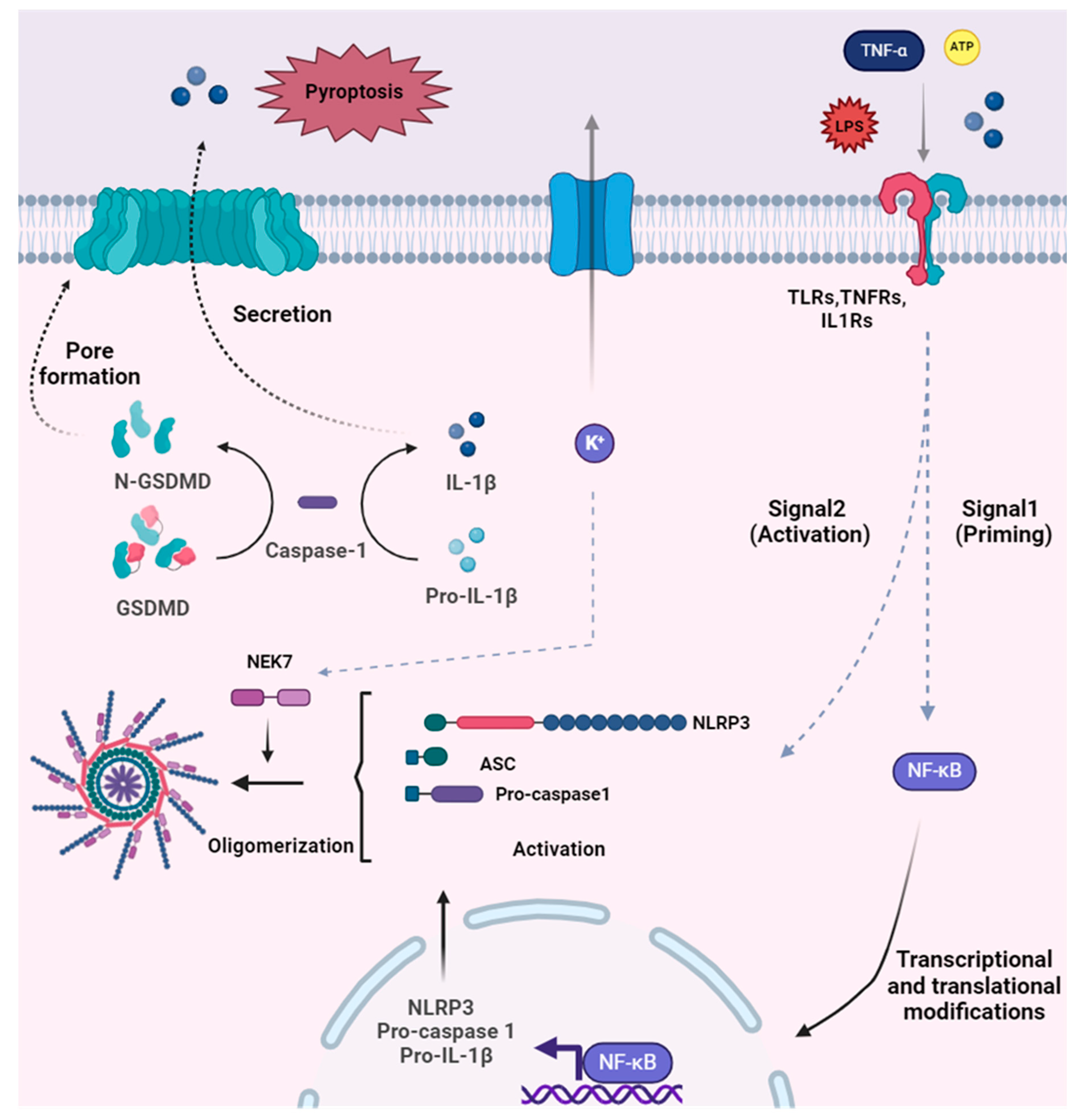
The regulation of microglial inflammatory responses primarily involves protein complexes known as inflammasomes [21]. These inflammasomes (Figure 1) are activated through specialised receptors called pathogen recognition receptors, such as toll-like receptors (TLRs), which initiate signalling pathways upon binding to damage-associated and/or pathogen-associated molecular patterns (DAMPs and PAMPs).
Emerging research highlights the significant role of nucleotide-binding oligomerization domain, leucine-rich repeat (LRR), and pyrin domain-containing protein 3 (NLRP3) in mediating microglial inflammatory responses [22]. The NLRP3–inflammasome complex, composed of NLRP3, apoptosis-associated speck-like protein containing a CARD (ASC), and procaspase 1 enzyme, plays a crucial role in this process. Activation of the NLRP3 inflammasome involves the autocatalytic activity of procaspase 1, leading to its cleavage and formation of active caspase 1 (see Figure 2) [23]. Active caspase 1, in turn, triggers the activation of interleukin 1β and 18 (pro-IL-1β and pro-IL-18) by cleaving their inactive zymogen forms, resulting in the production of IL-1β and IL-18. Additionally, caspase 1 cleaves gasdermin D (GSDMD), leading to the formation of an oligomeric pore from the cleaved GSDMD molecule [24], thereby facilitating the secretion of IL-1β and IL-18 from the cell [25].
Furthermore, the involvement of NIMA-related kinase 7 (NEK7), a serine-threonine kinase, has been implicated in NLRP3 inflammasome activation and oligomerization [26,27]. NEK7 acts as an essential binding partner for the ASC adaptor protein and caspase 1. It also appears to play a critical role downstream of K+ efflux in the oligomerization and activation of the NLRP3 inflammasome [27,28]. This underscores the significance of microglial K+ homeostasis, which relies on the coordinated regulation of diverse molecular targets involved in facilitating K+ fluxes.

2.1.1. Microglial Voltage-Gated Potassium Channels in Inflammation

Voltage-gated potassium (Kv) channels are found throughout the CNS, however expression and functionality within microglia is often overlooked. A range of studies have highlighted roles that Kv channels have in regulating microglial-driven inflammatory responses [29,30,31,32].
The Kv1 subfamily has been widely reported to modulate inflammatory responses and is now a target for drug discovery programmes. The Kv1.1 and Kv1.2 members of the Kv1 family have been implicated at both the mRNA and protein level with in vivo and in vitro data reporting microglia specific upregulation following lipopolysaccharide (LPS) administration and hypoxia exposure [32,33]. Both stimuli initiated an increase in cytokine release from microglia typical of a proinflammatory response (TNF-α and IL-1β). Pharmacological blockade of Kv1.1 and Kv1.2 channels (with dexamethasone and rTi-tyustoxin-Ka (TsTx), respectively) was shown to suppress cytokine secretion, suggesting that these channels exert a pivotal influence on microglia inflammatory responses.
A wealth of in vitro and in vivo evidence indicates microglia protein and gene expression of Kv1.3 [30,34,35,36,37,38,39,40,41,42]. These channels have been implicated in inflammatory responses triggered by LPS. Multiple approaches using selective blockade of the Kv1.3 channel or genetic deletion of the channel resulted in reducing microglia secretion of a panel of proinflammatory cytokines [29,30,43,44]. Kv1.3 channel is shown to be related and implicated in membrane depolarization of microglia [45]. Kv1.3 channel can also impact Ca2+ signalling via changing P2X4 receptor permeability to Ca2+ ions by changing the driving force of Ca2+ [43].
In the context of disease-associated inflammation, a range of models have been used. AD is characterised by long-term inflammation and so there is a growing interest in microglia physiology and their contribution to the pathological timeline of AD [46]. Kv1.3 channel functional and biophysical changes in AD-related pathology has been explored in detail [30,47,48]. For instance, Rangaraju et al. (2015) showed upregulation of Kv1.3 protein predominantly in microglia in the frontal cortex from human AD brains, when compared to age-matched controls [47]. Additionally, Ramesha et al. (2021) demonstrated that Kv1.3 protein can influence the phagocytic capabilities of microglia. Using the 5xFAD model, selective blockage of Kv1.3 resulted in a reduced amyloid burden and associated proinflammatory markers (IL-1β) because of dysfunctional phagocytosis [45]. Using a similar mouse model, Maezewa et al. (2018) highlighted a disease-specific spatial expression pattern for microglia Kv1.3. 5xFAD mice at 4, 6, and 10 months old, which showed an increase in current density of Kv1.3 channel compared to wild type litter mates, and this was predominantly in the proximity of the developing amyloid plaques; however, the 15 months of age group of mice did not show difference comparing age-matched controls [30]. Also, Aβ oligomers were shown to induce Kv1.3 gene and protein expression of Kv1.3. To explore the effects of inhibiting Kv1.3 on AD pathology, PAP1 was administered in different models of AD and a reduction in amyloid burden was reported alongside improved electrophysiological correlates of memory. Furthermore, blockade of the Kv1.3 channel led to a reduction in proinflammatory markers, namely TNF-α secretion and NFκB activation [30].
Ageing also contributes to a change in the inflammatory status; increases in proinflammatory cytokines (TNF-α and IL-6) and chemokines (CCL2 and CXCL1) are reported in mice age-dependent brain slices [34]. Following 24 h of LPS stimulation (1 µg/mL), the differences in cytokine and chemokine secretion between young and aged mice were no longer significant. This study has also linked Kv1.3 to these age-related changes. Here selective pharmacological Kv1.3 channel inhibition of microglia caused reduction in IL-6 levels in both young (2–3 months) and old (21–24 months) preparations without affecting the release of CCL2 and CXCL1 [21]. In addition, evidence indicated Kv1.3 control of cytokine signalling while P2Y12 receptor signalling mediated changes in chemokines.
In two different animal models of Parkinson’s disease (PD), pharmacological inhibition of microglial Kv1.3 suppressed cytokine secretion in rodent brain slices [49]. Their functional studies of Kv1.3 channel activity indicated that primary microglia exposed to exogenous α-synuclein aggregates were shown to have increase channel activity, also mRNA expression levels of Kv1.3 were elevated after exogenous α-synuclein treatment. This Kv1.3 protein upregulation was also evident in post-mortem tissue from human PD brains [49].
A study examining epileptic-induced inflammation indicated modulation of microglia Kv1.3 protein expression post epilepsy induction, and that inhibition of the channel (PAP1) suppressed microglia contribution to brain inflammation in vivo and in vitro [50]. They also demonstrated that the inflammatory response of BV2 cells following LPS stimulation is through downstream targeting of Ca2+/NFκB pathways. It was shown that LPS stimulation activated NFκB pathway and increased intracellular Ca2+ concentration (Ca2+ influx) Kv1.3 inhibition via PAP1 alleviated proinflammatory response, and suppressed NFκB signalling and Ca2+ influx.
Kv1.3 has also been linked to immunological responses to cancer therapy. Peng et al. [51] demonstrated that in radiation-induced brain injury both microglia Kv1.3 mRNA and protein expression was shown to increase following radiation therapy. Importantly, pharmacological blockade of the channel (with ShK-170) at the time of radiation injury was indicated to mitigate radiation-induced inflammatory response in microglia [51].
While Kv1.3 protein has been implicated, this may involve Kv1.3 homomers or Kv1.3/Kv1.5 heteromeric channels. Kv1.5 protein has also been suggested to regulate microglial inflammation. OX42CD11b positive microglial cells were shown to increase Kv1.5 channel protein expression after in vivo LPS stimulation [52]. On the other hand, they showed no changes in Kv1.3 protein expression following the stimulation in contrast to other studies [41].
Current research is actively exploring pharmacological agents that specifically inhibit Kv1.3 activity [53,54,55]. A range of clinical trials have focused on identifying effective Kv1.3 channel blockers and assessing their potential benefits in various inflammatory and autoimmune conditions. One notable compound, dalazatide (ShK-186), an analogue of the toxin from the sea anemone Stichodactyla helianthus, has shown efficacy in inhibiting Kv1.3 channels. Dalazatide’s success in managing autoimmune diseases like psoriasis has highlighted its potential broader application in CNS disorders where inflammation is a critical factor [56].
Dalazatide functions by selectively blocking Kv1.3 channels, thereby reducing the activation and proliferation of inflammatory cells [56,57]. In preclinical studies, dalazatide has been shown to downregulate microglial activation, decrease proinflammatory cytokine release, and promote neuroprotection. These findings support the hypothesis that targeting Kv1.3 channels could provide therapeutic benefits in diseases characterised by chronic CNS inflammation, such as multiple sclerosis, Alzheimer’s disease, and Parkinson’s disease. Further research into Kv1.3 inhibitors, including safety and long-term efficacy studies, is necessary to determine their clinical utility in managing neuroinflammatory conditions and advancing CNS disease therapeutics.

2.1.2. Microglial 2KP Channels in Inflammation

Tandem pore domain halothane-inhibited potassium channel 1 (THIK-1) has been suggested to facilitate K+ efflux in the activation of microglia NLRP3 inflammasome. Microglia with THIK-1 knocked out have a different morphology to wild type controls. These changes support a reduction in the ability of microglia to respond to their microenvironment without functional THIK-1 channels [58]. Pharmacological blockade and genetic modification of THIK-1 reduced the release of IL-1β post LPS and ATP stimulation in rodent microglia from hippocampal slices [58]. Similarly, another study showed IL-1β secretion from microglia in THIK1 KO mice and diminished activation of NLRP3 inflammasome regarding ATP stimulation via P2X7 receptor [59]. Pharmacological inhibition of THIK-1 (selective inhibitor C101248) was also shown to restrict NLRP3-related secretion of IL-1β in adult mice microglia following LPS and ATP treatment [60]. This THIK-1 dependent secretion was age dependent with C101248, having no effect in neonatal mouse microglia. There is also evidence for K+ independent THIK-1 activation of NLPR3 and a suggestion that selective targeting of THIK-1 could be a human microglia specific strategy for restoring a physiological inflammation status [60,61].

2.1.3. Microglial Calcium-Activated Potassium Channels in Inflammation

Kca3.1 is a small- and intermediate-conductance Ca2+-activated K+ channel, and has been proposed to regulate a host of microglial inflammatory responses. Studies indicate the importance of KCa3.1 channel in cell migration and Ca2+ regulation in rat microglia [62,63]. These are important cellular events/responses in microglia to suppress inflammation. Also, Kaushal et al. showed that a selective blocker of Kca3.1, TRAM-34, was used for blocking Kca3.1 in LPS-stimulated rodent microglia and caused a significant decrease in the inflammatory response, identified by inducible nitric oxide synthase (iNOS) induction and NO release. Furthermore, Kca3.1 blocking suppressed phosphorylation of mitogen-activated protein kinase (p38MAPK) in microglia, without influencing NFκB, which indicates a divergence in the inflammatory response based on the potassium channels (Kv1.3 vs. Kca3.1) [64]. This attenuation of proinflammatory signalling led to a reduction in retinal ganglion cytotoxicity [63]. Furthermore, Kca3.1 mRNA and protein expression has been reported to increase following LPS stimulation in primary murine microglia [65]. Another study indicated that a cytokine, IL4, that is an important modulator of inflammation can induce gene expression of KCNN4, which encodes for the Kca3.1 protein, and lead to an enhancement of the Kca3.1 current in rat microglia [62].

2.1.4. Microglial Inward Rectifying Potassium Channels in Inflammation

Potassium inward rectifying K+ channels (Kir) have a critical role to play in microglial physiology. For instance, Kir2.1 channel regulates depolarisation of microglia membranes and is responsible for Ca2+ signalling through Ca2+ store loading via inward rectifying Ca2+-release-activated-Ca2+-channels, Ca2+ permeable cation channels [29,66].
As we discussed above in the section about Kv1.3 channel, Maezawa et al. (2018) also demonstrated Kir2.1 channel activity in microglia in different age group 5xFAD mice [30]. Kir2.1 exhibited a consistently high current density (pA/pF) at 4, 6, and 10 months of age, similar to what was observed in Aβ oligomer-treated primary microglia isolated from newborn mice. Kir2.1 was also suggested to be one promising target for neurotrophic pain by regulating microglial proliferation and morphology [67]. Additionally, the ATP-sensitive K+ channel Kir6.1 is also known to be highly expressed in the anti-inflammatory phenotype of BV-2 cells and involved in altering microglial inflammatory phenotypes in two mouse models of PD [68]. Here, genetic deficiency of Kir6.1 (Kir6.1 +/−) led to an increase in neuronal loss in the MTPT model of PD. This neuronal loss was attributed to microglia-mediated increases in TNF-α, IL-1B, and iNOS. This indicates that targeting of Kir6.1 channels can modulate the secretion and/or the production of a panel of proinflammatory mediators [69]. However, a disease-specific approach may be required with evidence of Kir6.1 plasticity. In a cellular model of diabetes, pinacidil, a Kir6.1 opener led to increased levels of Kir6.1 protein and attenuated the offset of the observed diabetes-induced inflammatory responses [70]. Therefore, currently it is unclear if inhibition or activation of Kir6.1 would be a beneficial approach to tackling inflammation, or a disease-specific approach may be required.
From a therapeutic perspective, SB216763 is a selective inhibitor of glycogen synthase kinase 3 beta (GSK-3β), a key enzyme involved in multiple cellular pathways, including cell proliferation, differentiation, and apoptosis [71]. Recent studies have indicated its therapeutic potential in cardiac contexts, particularly for enhancing the expression of Kir2.1, known for maintaining resting membrane potential and cellular excitability in cardiomyocytes [72,73,74]. In myocardial infarction models, SB216763’s upregulation of Kir2.1 has been associated with improved cellular survival and functional recovery of cardiac tissue, suggesting a promising role for GSK-3β inhibition in heart disease management [75]. Additionally, amlodipine, a widely used antihypertensive agent, has shown efficacy in restoring Kir2.1 channel function, an effect that may extend beyond cardiovascular health. Specifically, research indicates that amlodipine’s modulation of Kir2.1 could be beneficial in treating neurovascular disorders, such as vascular dementia, where dysregulated potassium channel function contributes to disease pathology [76]. Together, these findings support the potential of targeting Kir2.1 channels through distinct mechanisms—as with SB216763 in myocardial contexts and amlodipine in neurovascular disease—highlighting novel therapeutic avenues for conditions involving Kir2.1 dysregulation. The impact of these drugs on the immune system has not been fully resolved and needs to be addressed ahead of any adoption for therapeutic purposes with a primary inflammatory component.

2.1.5. Microglial ATP-Gated Cation Channels in Inflammation

Many channel families and receptors are revealed to have a part regulatory effect on microglial functions. Specific receptors that are stimulated by their agonists like ATP also play a critical role in K+ homeostasis in microglia during inflammation. For instance, purinergic receptors (e.g., P2X4, P2X7) can regulate K+ efflux, while in the meantime can also induce Ca2+ flux [77,78].
Given there is the fact of the importance of NLRP3 inflammasomes in inflammation, P2X7 and P2X4 were demonstrated to be a critical mediator of NLRP3 inflammasome activation [79,80,81,82]. Accumulated research has also suggested this critical role in microglial inflammation. The addition of cuprizone to mouse diet for 3 and 6 weeks was shown to cause overexpression in protein and mRNA expression of P2X7 receptors and stimulate the proinflammatory response of microglia/macrophage [83]. Moreover, they showed an increase in gene expression of NLRP3 inflammasome-related compounds following cuprizone exposure (NLRP3, ASC, IL1b), but not active caspase1 and IL18 expressions. P2X4 receptors and NLRP3 inflammasomes in traumatic brain injury (TBI) and perioperative neurocognitive disorder (PND) models were shown to have increase protein expression in microglia [84,85]. Furthermore, inhibition of the receptor (with 5-BDBD) reduced NLRP3 inflammasome activation and secretion of cytokines (IL-1β, IL-6, and TNF-α) [84,85]. This research demonstrates that P2X4 and P2X7 predominantly contribute to the inflammatory response of microglia across various pathological conditions and can be critical targets to regulate microglial inflammation.

2.1.6. Microglial Transporters in Inflammation

Ionic transporters such as K+-Cl co-transporter (KCC) or Na+-K+-Cl co-transporter NKCC1 are also prominent contributors to microglial K+ homeostasis and physiology [86,87]. Recent studies have demonstrated that NKCC1 influences microglial responses to inflammation [88,89]. Toth et al. revealed that microglial NKCC1 deletion and intracortical inhibition via NKCC1 selective inhibitor bumetanide increased IL-1β release. On the other hand, they also showed a reduction in IL-1β release via systemic bumetanide administration in mice [65]. Tessier et al. recently demonstrated that bumetanide treatment induces IL-6 release in microglia of mice [89]. There are several ongoing clinical trials of bumetanide in neurodevelopmental disorders, such as autism spectrum disorder (ASD) [90]. Bumetanide is shown to inhibit excitatory actions of GABAergic neurotransmitter via blocking Cl transport in ASD brains [91]. Preclinical research using an APOE4 model of AD shows that bumetanide is a promising therapeutic agent for neurodegenerative diseases [92,93].
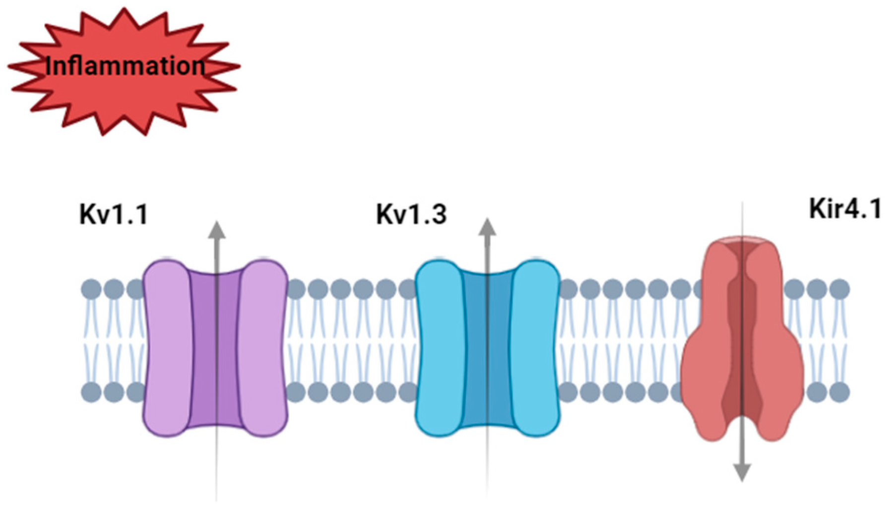
Microglia K+ homeostasis is critical to regulating the dynamic responses of these cells to a wide range of inflammatory stimuli. The impact of dysfunction of microglia K+ homeostasis is evident in a host of neurological diseases. Furthermore, there is an array of molecular targets that influence K+ flux that have been implicated as potential therapeutic targets to attenuate microglia driven inflammation (see Figure 3).

2.2. Neurons

Recent studies demonstrated that neurons are also capable of mounting inflammatory response and induce NLRP1, NLRP3, and AIM2 inflammasomes in neurodevelopment and neurodegenerative diseases [94,95,96,97,98,99]. However, the interplay between neuronal K+ homeostasis and brain inflammation remains to be resolved. Several studies have implicated that neuronal potassium channels are regulated by mediators of inflammation. Here, we will discuss how neuronal potassium channels respond to inflammatory conditions.
Neuronal K+ homeostasis is regulated by a variety of channel families, and diversity within these families supports a large compendium of molecular targets that inflammation could modulate [100,101,102,103]. These homeostatic proteins play a pivotal role in neuronal physiology, however their impact on inflammation is less well defined. It is evident from in vivo and in vitro studies that LPS-induced inflammation leads to elevated neuronal excitability [104,105]. For instance, modulation of K+ permeant channels via neuronal Kv7/M channels was demonstrated to regulate neuronal communication through the application of LPS [106].
TWIK-related acid-sensitive K+ (TASK) channels are members of the two-pore domain potassium (K2P) channel family which are sensitive to acidosis, which is known to be a marker of inflammation [107]. Two TASK channel subtypes, named TASK-1 and TASK-3, have been implicated in both human and mouse models of inflammation [108]. TASK-1 and TASK-3 protein and mRNA expressions were reduced following inflammatory stimulation in experimental autoimmune encephalomyelitis (EAE) rats [81]. They also showed downregulation of TASK-1 and TASK-3 protein expression in human multiple sclerosis (MS) patients. TASK-3 channel activation was also demonstrated to have a mitigating effect on pain in acute and chronic pain models in mice [109]. Inflammation is also known to be a part of acute and chronic pain. Research has indicated changes in neuronal potassium current in different neuron types during acute and chronic inflammation-related pain [110,111], and a reduction in pain threshold and increase in activity of nociceptive neurons [111].
As we discussed earlier, pannexin 1 (Panx1) and P2X7 were known to have a prominent role in inflammation, particularly NLRP3 inflammasome activation [112,113,114]. These channels are also shown to be expressed by neurons. Silvermann et al. (2019) showed Panx1 channels and an ATP-gated cation channel type P2X7 receptor’s involvement in the inflammatory response in rat cortical neurons [115]. According to the results, exposure to high extracellular potassium concentration caused NLRP1 inflammasome activation, caspase 1 activation, and IL-1β secretion in rat cortical neurons via Panx1 through P2X7 and pharmacological inhibition of Panx1-attenuated IL-1β release. These results suggest that neuronal inflammatory response is associated with Panx1–P2X7 complexes.
Growing evidence suggests that K+ sensitive molecular targets can contribute to inflammatory signalling in the brain (see Figure 4). It remains to be seen that the targeting of these proteins may provide relief to ongoing inflammation or prevent inflammation from becoming detrimental to brain physiology.

2.3. Astrocytes

Astrocytes play key roles in synaptic homeostasis, neuronal health, blood–brain barrier integrity and immune response [116]. Research into their role in inflammation has highlighted their potent functionality of releasing inflammatory cytokines/chemokines in the inflammatory status of the CNS [117]. Extensive research underscores the significant role of astrocytes in inflammasome-related inflammation [118,119]. Activation of the NLRP3 inflammasome in astrocytes has been documented across various disease conditions, including human post-mortem tissue from amyotrophic lateral sclerosis (ALS) patients, as well as in the superoxide dismutase 1 (SOD1) mutant mouse model [120]. Additionally, NLRP3 activation in astrocytes has been observed in brain metastatic tumours [121], ischemia [122], and in primary mouse astrocytes following exposure to inflammatory stimuli such as lipopolysaccharide (LPS) and adenosine triphosphate (ATP) [123]. Astrocytes also express other types of inflammasomes, including NLRP2 [124], NLRP6 [125], NLRC4 [126,127], and AIM2 [128] across diverse pathophysiological conditions such as multiple sclerosis, traumatic brain injury, and depression. However, the role of potassium (K⁺) in astrocytic inflammasome activation remains inadequately characterised. While several studies have examined the impact of astrocytic K⁺ buffering on modulating astrocyte phenotype in response to inflammatory stimuli, more research is needed to clarify the specific mechanisms by which K⁺ may influence inflammasome activation in astrocytes. This section discusses the role of these potassium channels in neuroinflammation, emphasising their potential implications in astrocytic function and pathology.

2.3.1. Astrocytic Voltage-Gated Potassium Channels in Inflammation

Voltage-gated potassium (Kv) channel family members Kv1.1 and Kv1.3, previously identified in microglia and modulated by LPS stimulation (see above), have also been detected in astrocytes. The expression patterns of these voltage-gated potassium channels are region specific and dependent on the preparation use to examine astrocyte protein expression [129]. Studies using an astroglia cell line indicated that both Kv1.1 and Kv1.3 protein expression patterns are sensitive to LPS exposure, but the semi-quantification data are open to alternative interpretations [129]. Therefore, robust data are not available to support LPS modulation of the channels in astrocyte subpopulations; this may highlight cell specific differences in the modulation of Kv channels via LPS. Presence of the Kv1.3 channel in astrocytes has been implicated in disease progression in EAE rats. In contrast to microglia Kv1.3 protein expression, GFAP positive astrocytic protein expression developed only at the peak of clinical presentation in the animals. Furthermore, pharmacological blockade of Kv1.3 in an astrocyte cell line revealed a central role in regulating the gene expression of the proinflammatory markers TNF-α and CCL2 [130]. These studies indicate that alongside microglia Kv1.3, regulation of the astrocyte isoform may also open therapeutic options to suppress a proinflammatory surge.

2.3.2. Astrocytic Inward Rectifying Potassium Channels in Inflammation

Kir4.1, the inward rectifying K+ channel, is predominantly expressed in astrocytes and has been implicated in a range of pathologies linked to changes in inflammatory status [131,132]. Human evidence implicates the channel in multiple sclerosis progression, with autoantibodies against Kir4.1 detected in sera of a subpopulation of individuals living with MS [133]. The relative contribution of this channel to the human condition needs further evaluation, but preclinical research to date has furthered our understanding of the interplay between inflammatory mediators and the Kir4.1 channel.
Studies looking at epilepsy have reported that post induction of status epilepticus in rodents Kir4.1 channel mRNA expression is decreased compared to controls (at 24 h post induction), but these levels are restored to baseline after 1 week. Interestingly, these changes in Kir4.1 expression were aligned to changes in IL-1β mRNA, where an increase in IL-1β corresponded with a decrease in Kir4.1 mRNA and protein expressions [131]. Another study indicated that Kir4.1 channel expression changes can regulate inflammatory response of astrocytes via NLRP3 inflammasome activation. LPS administration induced an increase in hippocampal levels of Kir4.1 protein 2 h after administration and this was maintained until the end point of analysis (24 h after LPS injection). shRNA directed knockdown of Kir4.1 expression levels suppressed LPS-induced NLRP3 inflammasome activation, indicating a role for astrocytic Kir4.1 in orchestrating NLPR3 responsiveness to LPS [132]. Preclinical research demonstrated that in Kir4.1 channel targeting via Kir4.1 selective blocker fluoxetine, that is also known as an antidepressant, and Kir4.1 knockdown in astrocytes, result in an increase in mRNA and protein expression of brain-derived neurotrophic factor (BDNF) [134]. In previous research on antidepressant agents, fluoxetine [135] and the tricyclic antidepressant (TCA) nortriptyline have been identified as blockers of Kir4.1 channel [136] in epilepsy and depression [137,138,139], indicating that Kir4.1 channel is a potent target for therapeutic interventions of CNS diseases, particularly depression.
Another member of the K+ inward rectifier family, Kir6.1, has also been reported to mediate inflammatory responses in a disease setting. In murine astrocytes from an animal model of Parkinson’s disease, the Kir6.1 channel has been linked to a critical role in initiating proinflammatory signalling that influences neuronal health [133]. Knock out of the Kir6.1 channel in astrocytes led to activation of caspase 1 and proinflammatory cytokines TNF-α and IL-1β release, and resulted in degeneration of dopaminergic neurons in a PD mouse model [133].
ATP-sensitive potassium (K-ATP) channel subunit Kir6.1 was also suggested to have a critical role regarding its subcellular localisation of the channel in mitochondria and endoplasmic reticulum membrane and Ca2+ influx [140]. In a stroke model, Kir6.1 knock out mice were shown to induce inflammatory response (TNF-α and IL-1β secretion) and endoplasmic reticulum stress [141]. Dong et al. (2016) investigated that oxygen–glucose deprivation (OGD) can cause endoplasmic stress (ER) and inflammation in astrocytes [142]. They showed that an ATP sensitive potassium channel opener nicorandil can mitigate the detrimental effects of OGD in astrocytes, such as reducing TNF-α and IL-1β release [142].

2.3.3. Astrocytic Calcium-Activated Potassium Channels in Inflammation

Kca3.1 was shown to have critical role in astrogliosis in AD and stroke models and is another important player in the inflammatory response of astrocytes. Bouhy et al. (2011) showed that Kca3.1 channels can increase TNF-α and IL-1β release in active astrocytes, and selectively blocking the channel can reduce inflammation in mice following spinal brain injury [65]. LPS stimulation to astrocytes was also shown to trigger NFκB signalling linked to astrogliosis-mediated neuroinflammation in primary astrocyte cell cultures [143]. They also showed that Kca3.1 channel can regulate neuronal loss and memory deficits in a mouse model of neuroinflammation following LPS stimulation [143].

2.3.4. Other Astrocyte K+ Homeostatic Molecular Targets

Only one channel within this family has been identified by Cong et al. (2023), who demonstrated that blocking of the TWIK-related potassium channel 1 (TREK1 channel) can suppress activation of reactive astrocytes via NFκB signalling that is a prominent pathway for expression of many inflammatory genes, such as IL-1β, IL-18, caspase1 [144].
Silverman and his colleagues (2009) suggested that astrocyte-related inflammasome activation is triggered via low K+ concentration regulated by pannexin 1 channels [115]. They showed that pharmacological inhibition of pannexin 1 is related to suppression of inflammasome activation and blocking the channel reduced caspase 1 recruitment.
Astrocyte control of brain K+ homeostasis is cited as a key function, and there is a repertoire of molecular targets to support efficient K+ flux (see Figure 5). As a consequence, they are uniquely placed to modulate K+ levels in response to inflammatory stimuli; this seems parallel with microglia actions to some extent. There are still unknown mechanisms by which K+ channels can modulate disease-specific responses in astrocytes, which will open up novel avenues for drug discovery programmes.

2.4. Oligodendrocytes

Oligodendrocytes (OLs) have been shown to contribute to inflammation and tissue homeostasis [145,146], particularly in MS which is associated with progressive inflammatory demyelination. Mature OLs have also been demonstrated to be implicated in AD pathology; NLRP3 inflammasome activation in OLs is shown to be associated with metabolic stress in AD mice, and modulating NLRP3 inflammasome activation can alleviate axonal degeneration and demyelination [147].
Although we have a limited amount of knowledge about the inflammatory response of OLs, there is not enough data investigating the relation between the inflammatory response of OLs and potassium channels of oligodendrocytes. However, recently, exposure of OLs to inflammation has been investigated, and existing data suggest that OLs are strongly responsive to neuroinflammation.
As we discussed previously, there is an increase in Kv1.1 and Kv1.3 channel protein expression in astrocytes after LPS stimulation; the same study showed that oligodendrocytes also increase in Kv1.1 and Kv1.3 expression in cuprizone conditions in mice [148]. Cuprizone is a copper chelator agent that was previously shown to cause OLs loss and demyelination [149]. Other study indicated that OLs are affected by neuroinflammation heavily in rats in cuprizone conditions [150]. These data suggest that Kv channels are sensitive to inflammation in oligodendrocytes.
Kir4.1 channels in oligodendrocytes have been previously investigated in inflammatory demyelination research. Kir4.1 channel expression is shown to be downregulated in human MS patients and mice with EAE [151]. BK channel activity in OLs was also discussed to be a promising therapeutic target by reducing the excitotoxic effect on oligodendrocytes [152].
A comprehensive picture of oligodendrocytes contribution to K+ homeostasis is emerging and so too is there a contribution to inflammatory signalling. A range of molecular targets have been proposed to influence inflammatory biomarkers (see Figure 6), but further research is required to provide more robust data in this emergent area of inflammatory research.

3. Conclusions and Prospects

In conclusion, cell-specific changes in the CNS are widely reported in models of disease with a known inflammatory component. This review has focussed on not only cell specific changes, but examined the cell specific changes critical for K+ homeostasis. Glia cells are predominantly involved here, but neurons are also linked via know molecular targets. There are common targets across all cell types, namely the Kv1 subfamily, and there is extensive research looking at targeting channels from this family to offset inflammation in both central and peripheral conditions. Therefore, it is likely that drug molecules will progress into clinical trials in the near future; what remains to be resolved is the temporal profile of the modulation of K+ homeostasis and its contribution to a long-term therapeutic strategy.

Funding

This research received no external funding.

Conflicts of Interest

The authors declare no conflicts of interest.

References

  1. Kettritz, R.; Loffing, J. Potassium homeostasis—Physiology and pharmacology in a clinical context. Pharmacol. Ther. 2023, 249, 108489. [Google Scholar] [CrossRef] [PubMed]
  2. Mcdonough, A.A.; Fenton, R.A. Potassium homeostasis: Sensors, mediators, and targets. Pflügers Arch.-Eur. J. Physiol. 2022, 474, 853–867. [Google Scholar] [CrossRef] [PubMed]
  3. Schmaul, S.; Hanuscheck, N.; Bittner, S. Astrocytic potassium and calcium channels as integrators of the inflammatory and ischemic CNS microenvironment. Biol. Chem. 2021, 402, 1519–1530. [Google Scholar] [CrossRef] [PubMed]
  4. Sahranavard, T.; Carbone, F.; Montecucco, F.; Xu, S.; Al-Rasadi, K.; Jamialahmadi, T.; Sahebkar, A. The role of potassium in atherosclerosis. Eur. J. Clin. Investig. 2021, 51, e13454. [Google Scholar] [CrossRef] [PubMed]
  5. Dantzer, R.; O’Connor, J.C.; Freund, G.G.; Johnson, R.W.; Kelley, K.W. From inflammation to sickness and depression: When the immune system subjugates the brain. Nat. Rev. Neurosci. 2008, 9, 46–56. [Google Scholar] [CrossRef]
  6. Galea, I. The blood–brain barrier in systemic infection and inflammation. Cell. Mol. Immunol. 2021, 18, 2489–2501. [Google Scholar] [CrossRef]
  7. Zhang, W.; Xiao, D.; Mao, Q.; Xia, H. Role of neuroinflammation in neurodegeneration development. Signal Transduct. Target. Ther. 2023, 8, 267. [Google Scholar] [CrossRef]
  8. Fujikawa, D.G.; Kim, J.S.; Daniels, A.H.; Alcaraz, A.F.; Sohn, T.B. In vivo elevation of extracellular potassium in the rat amygdala increases extracellular glutamate and aspartate and damages neurons. Neuroscience 1996, 74, 695–706. [Google Scholar] [CrossRef]
  9. Villa, C.; Suphesiz, H.; Combi, R.; Akyuz, E. Potassium channels in the neuronal homeostasis and neurodegenerative pathways underlying Alzheimer’s disease: An update. Mech. Ageing Dev. 2020, 185, 111197. [Google Scholar] [CrossRef]
  10. Ding, F.; Sun, Q.; Long, C.; Rasmussen, R.N.; Peng, S.; Xu, Q.; Kang, N.; Song, W.; Weikop, P.; Goldman, S.A.; et al. Dysregulation of extracellular potassium distinguishes healthy ageing from neurodegeneration. Brain 2024, 147, 1726–1739. [Google Scholar] [CrossRef]
  11. Beeton, C.; Wulff, H.; Standifer, N.E.; Azam, P.; Mullen, K.M.; Pennington, M.W.; Kolski-Andreaco, A.; Wei, E.; Grino, A.; Counts, D.R.; et al. Kv1.3 channels are a therapeutic target for T cell-mediated autoimmune diseases. Proc. Natl. Acad. Sci. USA 2006, 103, 17414–17419. [Google Scholar] [CrossRef] [PubMed]
  12. Canas, C.A.; Castano-Valencia, S.; Castro-Herrera, F. Pharmacological blockade of KV1.3 channel as a promising treatment in autoimmune diseases. J. Transl. Autoimmun. 2022, 5, 100146. [Google Scholar] [CrossRef] [PubMed]
  13. Chen, X.; Feng, Y.; Quinn, R.J.; Pountney, D.L.; Richardson, D.R.; Mellick, G.D.; Ma, L. Potassium Channels in Parkinson’s Disease: Potential Roles in Its Pathogenesis and Innovative Molecular Targets for Treatment. Pharmacol. Rev. 2023, 75, 758–788. [Google Scholar] [CrossRef]
  14. Luo, Y.; Huang, L.; Liao, P.; Jiang, R. Contribution of Neuronal and Glial Two-Pore-Domain Potassium Channels in Health and Neurological Disorders. Neural Plasticity 2021, 2021, 1–12. [Google Scholar] [CrossRef] [PubMed]
  15. Saxena, S.; Kruys, V.; Vamecq, J.; Maze, M. The Role of Microglia in Perioperative Neuroinflammation and Neurocognitive Disorders. Front. Aging Neurosci. 2021, 13, 671499. [Google Scholar] [CrossRef] [PubMed]
  16. York, E.M.; Bernier, L.-P.; Macvicar, B.A. Microglial modulation of neuronal activity in the healthy brain. Dev. Neurobiol. 2018, 78, 593–603. [Google Scholar] [CrossRef]
  17. Colonna, M.; Butovsky, O. Microglia Function in the Central Nervous System During Health and Neurodegeneration. Annu. Rev. Immunol. 2017, 35, 441–468. [Google Scholar] [CrossRef]
  18. Muzio, L.; Viotti, A.; Martino, G. Microglia in Neuroinflammation and Neurodegeneration: From Understanding to Therapy. Front. Neurosci. 2021, 15, 742065. [Google Scholar] [CrossRef]
  19. Heneka, M.T.; Kummer, M.P.; Latz, E. Innate immune activation in neurodegenerative disease. Nat. Rev. Immunol. 2014, 14, 463–477. [Google Scholar] [CrossRef]
  20. Venegas, C.; Kumar, S.; Franklin, B.S.; Dierkes, T.; Brinkschulte, R.; Tejera, D.; Vieira-Saecker, A.; Schwartz, S.; Santarelli, F.; Kummer, M.P.; et al. Microglia-derived ASC specks cross-seed amyloid-β in Alzheimer’s disease. Nature 2017, 552, 355–361. [Google Scholar] [CrossRef]
  21. Guo, H.; Callaway, J.B.; Ting, J.P. Inflammasomes: Mechanism of action, role in disease, and therapeutics. Nat. Med. 2015, 21, 677–687. [Google Scholar] [CrossRef] [PubMed]
  22. Martinon, F.; Tschopp, J. Inflammatory caspases and inflammasomes: Master switches of inflammation. Cell Death Differ. 2007, 14, 10–22. [Google Scholar] [CrossRef] [PubMed]
  23. Jo, E.-K.; Kim, J.K.; Shin, D.-M.; Sasakawa, C. Molecular mechanisms regulating NLRP3 inflammasome activation. Cell. Mol. Immunol. 2016, 13, 148–159. [Google Scholar] [CrossRef] [PubMed]
  24. He, W.-T.; Wan, H.; Hu, L.; Chen, P.; Wang, X.; Huang, Z.; Yang, Z.-H.; Zhong, C.-Q.; Han, J. Gasdermin D is an executor of pyroptosis and required for interleukin-1β secretion. Cell Res. 2015, 25, 1285–1298. [Google Scholar] [CrossRef]
  25. Nizami, S.; Hall-Roberts, H.; Warrier, S.; Cowley, S.A.; Di Daniel, E. Microglial inflammation and phagocytosis in Alzheimer’s disease: Potential therapeutic targets. Br. J. Pharmacol. 2019, 176, 3515–3532. [Google Scholar] [CrossRef]
  26. Schmid-Burgk, J.L.; Chauhan, D.; Schmidt, T.; Ebert, T.S.; Reinhardt, J.; Endl, E.; Hornung, V. A Genome-wide CRISPR (Clustered Regularly Interspaced Short Palindromic Repeats) Screen Identifies NEK7 as an Essential Component of NLRP3 Inflammasome Activation. J. Biol. Chem. 2016, 291, 103–109. [Google Scholar] [CrossRef]
  27. Shi, H.; Wang, Y.; Li, X.; Zhan, X.; Tang, M.; Fina, M.; Su, L.; Pratt, D.; Bu, C.H.; Hildebrand, S.; et al. NLRP3 activation and mitosis are mutually exclusive events coordinated by NEK7, a new inflammasome component. Nat. Immunol. 2016, 17, 250–258. [Google Scholar] [CrossRef]
  28. He, Y.; Zeng, M.Y.; Yang, D.; Motro, B.; Nunez, G. NEK7 is an essential mediator of NLRP3 activation downstream of potassium efflux. Nature 2016, 530, 354–357. [Google Scholar] [CrossRef]
  29. Nguyen, H.M.; Grossinger, E.M.; Horiuchi, M.; Davis, K.W.; Jin, L.W.; Maezawa, I.; Wulff, H. Differential Kv1.3, KCa3.1, and Kir2.1 expression in “classically” and “alternatively” activated microglia. Glia 2017, 65, 106–121. [Google Scholar] [CrossRef]
  30. Maezawa, I.; Nguyen, H.M.; Di Lucente, J.; Jenkins, D.P.; Singh, V.; Hilt, S.; Kim, K.; Rangaraju, S.; Levey, A.I.; Wulff, H.; et al. Kv1.3 inhibition as a potential microglia-targeted therapy for Alzheimer’s disease: Preclinical proof of concept. Brain 2018, 141, 596–612. [Google Scholar] [CrossRef]
  31. Rangaraju, S.; Raza, S.A.; Pennati, A.; Deng, Q.; Dammer, E.B.; Duong, D.; Pennington, M.W.; Tansey, M.G.; Lah, J.J.; Betarbet, R.; et al. A systems pharmacology-based approach to identify novel Kv1.3 channel-dependent mechanisms in microglial activation. J. Neuroinflamm. 2017, 14, 128. [Google Scholar] [CrossRef] [PubMed]
  32. Wu, C.Y.; Kaur, C.; Sivakumar, V.; Lu, J.; Ling, E.A. Kv1.1 expression in microglia regulates production and release of proinflammatory cytokines, endothelins and nitric oxide. Neuroscience 2009, 158, 1500–1508. [Google Scholar] [CrossRef] [PubMed]
  33. Li, F.; Lu, J.; Wu, C.Y.; Kaur, C.; Sivakumar, V.; Sun, J.; Li, S.; Ling, E.A. Expression of Kv1.2 in microglia and its putative roles in modulating production of proinflammatory cytokines and reactive oxygen species. J. Neurochem. 2008, 106, 2093–2105. [Google Scholar] [CrossRef] [PubMed]
  34. Charolidi, N.; Schilling, T.; Eder, C. Microglial Kv1.3 Channels and P2Y12 Receptors Differentially Regulate Cytokine and Chemokine Release from Brain Slices of Young Adult and Aged Mice. PLoS ONE 2015, 10, e0128463. [Google Scholar] [CrossRef]
  35. Fordyce, C.B. Microglia Kv1.3 Channels Contribute to Their Ability to Kill Neurons. J. Neurosci. 2005, 25, 7139–7149. [Google Scholar] [CrossRef]
  36. Khanna, R.; Roy, L.; Zhu, X.; Schlichter, L.C. K+ channels and the microglial respiratory burst. Am. J. Physiol.-Cell Physiol. 2001, 280, C796–C806. [Google Scholar] [CrossRef]
  37. Kotecha, S.A.; Schlichter, L.C. A Kv1.5 to Kv1.3 switch in endogenous hippocampal microglia and a role in proliferation. J. Neurosci. 1999, 19, 10680–10693. [Google Scholar] [CrossRef]
  38. Nguyen, H.M.; Blomster, L.V.; Christophersen, P.; Wulff, H. Potassium channel expression and function in microglia: Plasticity and possible species variations. Channels 2017, 11, 305–315. [Google Scholar] [CrossRef]
  39. Schilling, T.; Eder, C. Effects of kinase inhibitors on TGF-β induced upregulation of Kv1.3 K+ channels in brain macrophages. Pflügers Arch. Eur. J. Physiol. 2003, 447, 312–315. [Google Scholar] [CrossRef]
  40. Kust, B.M.; Biber, K.; van Calker, D.; Gebicke-Haerter, P.J. Regulation of K+ channel mRNA expression by stimulation of adenosine A2a-receptors in cultured rat microglia. Glia 1999, 25, 120–130. [Google Scholar] [CrossRef]
  41. Schilling, T.; Eder, C. Amyloid-β-induced reactive oxygen species production and priming are differentially regulated by ion channels in microglia. J. Cell. Physiol. 2011, 226, 3295–3302. [Google Scholar] [CrossRef] [PubMed]
  42. Di Lucente, J.; Nguyen, H.M.; Wulff, H.; Jin, L.W.; Maezawa, I. The voltage-gated potassium channel Kv1.3 is required for microglial pro-inflammatory activation in vivo. Glia 2018, 66, 1881–1895. [Google Scholar] [CrossRef] [PubMed]
  43. Nguyen, H.M.; di Lucente, J.; Chen, Y.J.; Cui, Y.; Ibrahim, R.H.; Pennington, M.W.; Jin, L.W.; Maezawa, I.; Wulff, H. Biophysical basis for Kv1.3 regulation of membrane potential changes induced by P2X4-mediated calcium entry in microglia. Glia 2020, 68, 2377–2394. [Google Scholar] [CrossRef] [PubMed]
  44. Bowen, C.A.; Nguyen, H.M.; Lin, Y.; Bagchi, P.; Natu, A.; Espinosa-Garcia, C.; Werner, E.; Kumari, R.; Brandelli, A.D.; Kumar, P.; et al. Proximity Labeling Proteomics Reveals Kv1.3 Potassium Channel Immune Interactors in Microglia. Mol. Cell. Proteom. 2024, 23, 100809. [Google Scholar] [CrossRef]
  45. Ramesha, S.; Rayaprolu, S.; Bowen, C.A.; Giver, C.R.; Bitarafan, S.; Nguyen, H.M.; Gao, T.; Chen, M.J.; Nwabueze, N.; Dammer, E.B.; et al. Unique molecular characteristics and microglial origin of Kv1.3 channel-positive brain myeloid cells in Alzheimer’s disease. Proc. Natl. Acad. Sci. USA 2021, 118, e2013545118. [Google Scholar] [CrossRef]
  46. Kinney, J.W.; Bemiller, S.M.; Murtishaw, A.S.; Leisgang, A.M.; Salazar, A.M.; Lamb, B.T. Inflammation as a central mechanism in Alzheimer’s disease. Alzheimers Dement. Transl. Res. Clin. Interv. 2018, 4, 575–590. [Google Scholar] [CrossRef]
  47. Rangaraju, S.; Gearing, M.; Jin, L.W.; Levey, A. Potassium channel Kv1.3 is highly expressed by microglia in human Alzheimer’s disease. J. Alzheimers Dis. 2015, 44, 797–808. [Google Scholar] [CrossRef]
  48. Chung, S.; Lee, J.; Joe, E.H.; Uhm, D.Y. Beta-amyloid peptide induces the expression of voltage dependent outward rectifying K+ channels in rat microglia. Neurosci. Lett. 2001, 300, 67–70. [Google Scholar] [CrossRef]
  49. Sarkar, S.; Nguyen, H.M.; Malovic, E.; Luo, J.; Langley, M.; Palanisamy, B.N.; Singh, N.; Manne, S.; Neal, M.; Gabrielle, M.; et al. Kv1.3 modulates neuroinflammation and neurodegeneration in Parkinson’s disease. J. Clin. Investig. 2020, 130, 4195–4212. [Google Scholar] [CrossRef]
  50. Zhang, X.; Liang, P.; Zhang, Y.; Wu, Y.; Song, Y.; Wang, X.; Chen, T.; Peng, B.; Liu, W.; Yin, J.; et al. Blockade of Kv1.3 Potassium Channel Inhibits Microglia-Mediated Neuroinflammation in Epilepsy. Int. J. Mol. Sci. 2022, 23, 14693. [Google Scholar] [CrossRef]
  51. Peng, Y.; Lu, K.; Li, Z.; Zhao, Y.; Wang, Y.; Hu, B.; Xu, P.; Shi, X.; Zhou, B.; Pennington, M.; et al. Blockade of Kv1.3 channels ameliorates radiation-induced brain injury. Neuro-Oncology 2014, 16, 528–539. [Google Scholar] [CrossRef] [PubMed]
  52. Jou, I.; Pyo, H.; Chung, S.; Jung, S.Y.; Gwag, B.J.; Joe, E.H. Expression of Kv1.5 K+ channels in activated microglia in vivo. Glia 1998, 24, 408–414. [Google Scholar] [CrossRef]
  53. Beeton, C.; Pennington, M.W.; Wulff, H.; Singh, S.; Nugent, D.; Crossley, G.; Khaytin, I.; Calabresi, P.A.; Chen, C.-Y.; Gutman, G.A.; et al. Targeting Effector Memory T Cells with a Selective Peptide Inhibitor of Kv1.3 Channels for Therapy of Autoimmune Diseases. Mol. Pharmacol. 2005, 67, 1369–1381. [Google Scholar] [CrossRef] [PubMed]
  54. Kundu-Raychaudhuri, S.; Chen, Y.-J.; Wulff, H.; Raychaudhuri, S.P. Kv1.3 in psoriatic disease: PAP-1, a small molecule inhibitor of Kv1.3 is effective in the SCID mouse psoriasis—Xenograft model. J. Autoimmun. 2014, 55, 63–72. [Google Scholar] [CrossRef] [PubMed]
  55. Varga, Z.; Tajti, G.; Panyi, G. The Kv1.3 K+ channel in the immune system and its “precision pharmacology” using peptide toxins. Biol. Futur. 2021, 72, 75–83. [Google Scholar] [CrossRef] [PubMed]
  56. Tarcha, E.J.; Olsen, C.M.; Probst, P.; Peckham, D.; Muñoz-Elías, E.J.; Kruger, J.G.; Iadonato, S.P. Safety and pharmacodynamics of dalazatide, a Kv1.3 channel inhibitor, in the treatment of plaque psoriasis: A randomized phase 1b trial. PLoS ONE 2017, 12, e0180762. [Google Scholar] [CrossRef]
  57. Tarcha, E.J.; Chi, V.; Muñoz-Elías, E.J.; Bailey, D.; Londono, L.M.; Upadhyay, S.K.; Norton, K.; Banks, A.; Tjong, I.; Nguyen, H.; et al. Durable Pharmacological Responses from the Peptide ShK-186, a Specific Kv1.3 Channel Inhibitor That Suppresses T Cell Mediators of Autoimmune Disease. J. Pharmacol. Exp. Ther. 2012, 342, 642–653. [Google Scholar] [CrossRef]
  58. Madry, C.; Kyrargyri, V.; Arancibia-Carcamo, I.L.; Jolivet, R.; Kohsaka, S.; Bryan, R.M.; Attwell, D. Microglial Ramification, Surveillance, and Interleukin-1β Release Are Regulated by the Two-Pore Domain K+ Channel THIK-1. Neuron 2018, 97, 299–312.e296. [Google Scholar] [CrossRef]
  59. Drinkall, S.; Lawrence, C.B.; Ossola, B.; Russell, S.; Bender, C.; Brice, N.B.; Dawson, L.A.; Harte, M.; Brough, D. The two pore potassium channel THIK-1 regulates NLRP3 inflammasome activation. Glia 2022, 70, 1301–1316. [Google Scholar] [CrossRef]
  60. Ossola, B.; Rifat, A.; Rowland, A.; Hunter, H.; Drinkall, S.; Bender, C.; Hamlischer, M.; Teall, M.; Burley, R.; Barker, D.F.; et al. Characterisation of C101248: A novel selective THIK-1 channel inhibitor for the modulation of microglial NLRP3-inflammasome. Neuropharmacology 2023, 224, 109330. [Google Scholar] [CrossRef]
  61. Rifat, A.; Ossola, B.; Bürli, R.W.; Dawson, L.A.; Brice, N.L.; Rowland, A.; Lizio, M.; Xu, X.; Page, K.; Fidzinski, P.; et al. Differential contribution of THIK-1 K+ channels and P2X7 receptors to ATP-mediated neuroinflammation by human microglia. J. Neuroinflamm. 2024, 21, 58. [Google Scholar] [CrossRef] [PubMed]
  62. Ferreira, R.; Lively, S.; Schlichter, L.C. IL-4 type 1 receptor signaling up-regulates KCNN4 expression, and increases the KCa3.1 current and its contribution to migration of alternative-activated microglia. Front. Cell. Neurosci. 2014, 8, 183. [Google Scholar] [CrossRef] [PubMed]
  63. Ferreira, R.; Schlichter, L.C. Selective Activation of KCa3.1 and CRAC Channels by P2Y2 Receptors Promotes Ca2+ Signaling, Store Refilling and Migration of Rat Microglial Cells. PLoS ONE 2013, 8, e62345. [Google Scholar] [CrossRef] [PubMed]
  64. Kaushal, V.; Koeberle, P.D.; Wang, Y.; Schlichter, L.C. The Ca2+-Activated K+ Channel KCNN4/KCa3.1 Contributes to Microglia Activation and Nitric Oxide-Dependent Neurodegeneration. J. Neurosci. 2007, 27, 234–244. [Google Scholar] [CrossRef] [PubMed]
  65. Bouhy, D.; Ghasemlou, N.; Lively, S.; Redensek, A.; Rathore, K.I.; Schlichter, L.C.; David, S. Inhibition of the Ca2+-Dependent K+Channel, KCNN4/KCa3.1, Improves Tissue Protection and Locomotor Recovery after Spinal Cord Injury. J. Neurosci. 2011, 31, 16298–16308. [Google Scholar] [CrossRef]
  66. Lam, D.; Schlichter, L.C. Expression and contributions of the Kir2.1 inward-rectifier K+ channel to proliferation, migration and chemotaxis of microglia in unstimulated and anti-inflammatory states. Front. Cell. Neurosci. 2015, 9, 185. [Google Scholar] [CrossRef]
  67. Gattlen, C.; Deftu, A.F.; Tonello, R.; Ling, Y.; Berta, T.; Ristoiu, V.; Suter, M.R. The inhibition of Kir2.1 potassium channels depolarizes spinal microglial cells, reduces their proliferation, and attenuates neuropathic pain. Glia 2020, 68, 2119–2135. [Google Scholar] [CrossRef]
  68. Du, R.-H.; Sun, H.-B.; Hu, Z.-L.; Lu, M.; Ding, J.-H.; Hu, G. Kir6.1/K-ATP channel modulates microglia phenotypes: Implication in Parkinson’s disease. Cell Death Dis. 2018, 9, 404. [Google Scholar] [CrossRef]
  69. Tang, Z.; Shao, X.; Wu, J.; Chen, H.; Zhang, A.; Xu, F.; Ping, H.; Li, S.; Liu, C.; Li, Y.; et al. Naloxone Protects against Lipopolysaccharide-Induced Neuroinflammation and Microglial Activation via Inhibiting ATP-Sensitive Potassium Channel. Comput. Math. Methods Med. 2021, 2021, 1–9. [Google Scholar] [CrossRef]
  70. Li, H.; Chen, D.; Sun, W.; Chen, J.; Luo, C.; Xu, H.; Ma, J.H.; Tang, S. KATP Opener Attenuates Diabetic-Induced Muller Gliosis and Inflammation by Modulating Kir6.1 in Microglia. Investig. Ophthalmol. Vis. Sci. 2021, 62, 3. [Google Scholar] [CrossRef]
  71. Shu, Q.; Liu, J.; Liu, X.; Zhao, S.; Li, H.; Tan, Y.; Xu, J. GABABR/GSK-3β/NF-κB signaling pathway regulates the proliferation of colorectal cancer cells. Cancer Med. 2016, 5, 1259–1267. [Google Scholar] [CrossRef] [PubMed]
  72. Zuo, D.; Chen, K.; Zhou, M.; Liu, Z.; Chen, H. Kir2.1 and K2P1 channels reconstitute two levels of resting membrane potential in cardiomyocytes. J. Physiol. 2017, 595, 5129–5142. [Google Scholar] [CrossRef] [PubMed]
  73. Reilly, L.; Eckhardt, L.L. Cardiac potassium inward rectifier Kir2: Review of structure, regulation, pharmacology, and arrhythmogenesis. Heart Rhythm. 2021, 18, 1423–1434. [Google Scholar] [CrossRef] [PubMed]
  74. Li, E.; Van Der Heyden, M.A.G. The network of cardiac KIR2.1: Its function, cellular regulation, electrical signaling, diseases and new drug avenues. Naunyn-Schmiedebergs Arch. Pharmacol. 2024, 397, 6369–6389. [Google Scholar] [CrossRef] [PubMed]
  75. Chang, C.; Wang, S.-H.; Xu, L.-N.; Su, X.-L.; Zeng, Y.-F.; Wang, P.; Zhang, L.-R.; Han, S.-N. Glycogen synthase kinase 3 beta inhibitor SB216763 improves Kir2.1 expression after myocardia infraction in rats. J. Interv. Card. Electrophysiol. 2022, 63, 239–248. [Google Scholar] [CrossRef]
  76. Koide, M.; Harraz, O.F.; Dabertrand, F.; Longden, T.A.; Ferris, H.R.; Wellman, G.C.; Hill-Eubanks, D.C.; Greenstein, A.S.; Nelson, M.T. Differential restoration of functional hyperemia by antihypertensive drug classes in hypertension-related cerebral small vessel disease. J. Clin. Investig. 2021, 131(18), e149029. [Google Scholar] [CrossRef]
  77. Barbera-Cremades, M.; Gomez, A.I.; Baroja-Mazo, A.; Martinez-Alarcon, L.; Martinez, C.M.; de Torre-Minguela, C.; Pelegrin, P. P2X7 Receptor Induces Tumor Necrosis Factor-alpha Converting Enzyme Activation and Release to Boost TNF-alpha Production. Front. Immunol. 2017, 8, 862. [Google Scholar] [CrossRef]
  78. Xu, Z.; Chen, Z.M.; Wu, X.; Zhang, L.; Cao, Y.; Zhou, P. Distinct Molecular Mechanisms Underlying Potassium Efflux for NLRP3 Inflammasome Activation. Front. Immunol. 2020, 11, 609441. [Google Scholar] [CrossRef]
  79. Burm, S.M.; Zuiderwijk-Sick, E.A.; Weert, P.M.; Bajramovic, J.J. ATP-induced IL-1beta secretion is selectively impaired in microglia as compared to hematopoietic macrophages. Glia 2016, 64, 2231–2246. [Google Scholar] [CrossRef]
  80. Ronning, K.E.; Déchelle-Marquet, P.-A.; Che, Y.; Guillonneau, X.; Sennlaub, F.; Delarasse, C. The P2X7 Receptor, a Multifaceted Receptor in Alzheimer’s Disease. Int. J. Mol. Sci. 2023, 24, 11747. [Google Scholar] [CrossRef]
  81. Chen, K.; Zhang, J.; Zhang, W.; Zhang, J.; Yang, J.; Li, K.; He, Y. ATP-P2X4 signaling mediates NLRP3 inflammasome activation: A novel pathway of diabetic nephropathy. Int. J. Biochem. Cell Biol. 2013, 45, 932–943. [Google Scholar] [CrossRef] [PubMed]
  82. Kanellopoulos, J.M.; Almeida-Da-Silva, C.L.C.; Rüütel Boudinot, S.; Ojcius, D.M. Structural and Functional Features of the P2X4 Receptor: An Immunological Perspective. Front. Immunol. 2021, 12, 645834. [Google Scholar] [CrossRef] [PubMed]
  83. Bernal-Chico, A.; Manterola, A.; Cipriani, R.; Katona, I.; Matute, C.; Mato, S. P2x7 receptors control demyelination and inflammation in the cuprizone model. Brain Behav. Immun. Health 2020, 4, 100062. [Google Scholar] [CrossRef]
  84. Yuan, H.; Lu, B.; Ji, Y.; Meng, B.; Wang, R.; Sun, D.; Liu, R.; Zhai, X.; Li, X.; Qin, J.; et al. Role of P2X4/NLRP3 Pathway-Mediated Neuroinflammation in Perioperative Neurocognitive Disorders. Mediat. Inflamm. 2022, 2022, 6355805. [Google Scholar] [CrossRef]
  85. He, W.; Wang, Q.; Sha, W.; Wang, L.; Li, D.; Chen, G. P2X4 Inhibition reduces microglia inflammation and apoptosis by NLRP3 and improves nervous system defects in rat brain trauma model. J. Clin. Neurosci. 2022, 99, 224–232. [Google Scholar] [CrossRef]
  86. Schilling, T.; Stock, C.; Schwab, A.; Eder, C. Functional importance of Ca2+-activated K+ channels for lysophosphatidic acid-induced microglial migration. Eur. J. Neurosci. 2004, 19, 1469–1474. [Google Scholar] [CrossRef]
  87. Annunziato, L.; Boscia, F.; Pignataro, G. Ionic transporter activity in astrocytes, microglia, and oligodendrocytes during brain ischemia. J. Cereb. Blood Flow Metab. 2013, 33, 969–982. [Google Scholar] [CrossRef]
  88. Tóth, K.; Lénárt, N.; Berki, P.; Fekete, R.; Szabadits, E.; Pósfai, B.; Cserép, C.; Alatshan, A.; Benkő, S.; Kiss, D.; et al. The NKCC1 ion transporter modulates microglial phenotype and inflammatory response to brain injury in a cell-autonomous manner. PLoS Biol. 2022, 20, e3001526. [Google Scholar] [CrossRef]
  89. Tessier, M.; Saez Garcia, M.; Goubert, E.; Blasco, E.; Consumi, A.; Dehapiot, B.; Tian, L.; Molinari, F.; Laurin, J.; Guillemot, F.; et al. Bumetanide induces post-traumatic microglia-interneuron contact to promote neurogenesis and recovery. Brain 2023, 146, 4247–4261. [Google Scholar] [CrossRef]
  90. Georgoula, C.; Ferrin, M.; Pietraszczyk-Kedziora, B.; Hervas, A.; Marret, S.; Oliveira, G.; Rosier, A.; Crutel, V.; Besse, E.; Severo, C.A.; et al. A Phase III Study of Bumetanide Oral Liquid Formulation for the Treatment of Children and Adolescents Aged Between 7 and 17 Years with Autism Spectrum Disorder (SIGN 1 Trial): Participant Baseline Characteristics. Child Psychiatry Hum. Dev. 2023, 54, 1360–1372. [Google Scholar] [CrossRef]
  91. Wang, T.; Shan, L.; Miao, C.; Xu, Z.; Jia, F. Treatment Effect of Bumetanide in Children With Autism Spectrum Disorder: A Systematic Review and Meta-Analysis. Front. Psychiatry 2021, 12, 751575. [Google Scholar] [CrossRef] [PubMed]
  92. Taubes, A.; Nova, P.; Zalocusky, K.A.; Kosti, I.; Bicak, M.; Zilberter, M.Y.; Hao, Y.; Yoon, S.Y.; Oskotsky, T.; Pineda, S.; et al. Experimental and real-world evidence supporting the computational repurposing of bumetanide for APOE4-related Alzheimer’s disease. Nat. Aging 2021, 1, 932–947. [Google Scholar] [CrossRef] [PubMed]
  93. Boyarko, B.; Podvin, S.; Greenberg, B.; Momper, J.D.; Huang, Y.; Gerwick, W.H.; Bang, A.G.; Quinti, L.; Griciuc, A.; Kim, D.Y.; et al. Evaluation of bumetanide as a potential therapeutic agent for Alzheimer’s disease. Front. Pharmacol. 2023, 14, 1190402. [Google Scholar] [CrossRef] [PubMed]
  94. Panicker, N.; Kam, T.-I.; Wang, H.; Neifert, S.; Chou, S.-C.; Kumar, M.; Brahmachari, S.; Jhaldiyal, A.; Hinkle, J.T.; Akkentli, F.; et al. Neuronal NLRP3 is a parkin substrate that drives neurodegeneration in Parkinson’s disease. Neuron 2022, 110, 2422–2437.e2429. [Google Scholar] [CrossRef] [PubMed]
  95. Tan, M.-S.; Tan, L.; Jiang, T.; Zhu, X.-C.; Wang, H.-F.; Jia, C.-D.; Yu, J.-T. Amyloid-β induces NLRP1-dependent neuronal pyroptosis in models of Alzheimer’s disease. Cell Death Dis. 2014, 5, e1382. [Google Scholar] [CrossRef]
  96. Wang, W.; Nguyen, L.T.T.; Burlak, C.; Chegini, F.; Guo, F.; Chataway, T.; Ju, S.; Fisher, O.S.; Miller, D.W.; Datta, D.; et al. Caspase-1 causes truncation and aggregation of the Parkinson’s disease-associated protein α-synuclein. Proc. Natl. Acad. Sci. USA 2016, 113, 9587–9592. [Google Scholar] [CrossRef]
  97. Kaushal, V.; Dye, R.; Pakavathkumar, P.; Foveau, B.; Flores, J.; Hyman, B.; Ghetti, B.; Koller, B.H.; Leblanc, A.C. Neuronal NLRP1 inflammasome activation of Caspase-1 coordinately regulates inflammatory interleukin-1-beta production and axonal degeneration-associated Caspase-6 activation. Cell Death Differ. 2015, 22, 1676–1686. [Google Scholar] [CrossRef]
  98. Lammert, C.R.; Frost, E.L.; Bellinger, C.E.; Bolte, A.C.; McKee, C.A.; Hurt, M.E.; Paysour, M.J.; Ennerfelt, H.E.; Lukens, J.R. AIM2 inflammasome surveillance of DNA damage shapes neurodevelopment. Nature 2020, 580, 647–652. [Google Scholar] [CrossRef]
  99. Fann, D.Y.-W.; Lim, Y.-A.; Cheng, Y.-L.; Lok, K.-Z.; Chunduri, P.; Baik, S.-H.; Drummond, G.R.; Dheen, S.T.; Sobey, C.G.; Jo, D.-G.; et al. Evidence that NF-κB and MAPK Signaling Promotes NLRP Inflammasome Activation in Neurons Following Ischemic Stroke. Mol. Neurobiol. 2018, 55, 1082–1096. [Google Scholar] [CrossRef]
  100. Noh, W.; Pak, S.; Choi, G.; Yang, S.; Yang, S. Transient Potassium Channels: Therapeutic Targets for Brain Disorders. Front. Cell. Neurosci. 2019, 13, 265. [Google Scholar] [CrossRef]
  101. Goldstein, S.A.N.; Bayliss, D.A.; Kim, D.; Lesage, F.; Plant, L.D.; Rajan, S. International Union of Pharmacology. LV. Nomenclature and Molecular Relationships of Two-P Potassium Channels. Pharmacol. Rev. 2005, 57, 527–540. [Google Scholar] [CrossRef] [PubMed]
  102. Gutman, G.A.; Chandy, K.G.; Grissmer, S.; Lazdunski, M.; Mckinnon, D.; Pardo, L.A.; Robertson, G.A.; Rudy, B.; Sanguinetti, M.C.; Stühmer, W.; et al. International Union of Pharmacology. LIII. Nomenclature and Molecular Relationships of Voltage-Gated Potassium Channels. Pharmacol. Rev. 2005, 57, 473–508. [Google Scholar] [CrossRef] [PubMed]
  103. Kubo, Y.; Adelman, J.P.; Clapham, D.E.; Jan, L.Y.; Karschin, A.; Kurachi, Y.; Lazdunski, M.; Nichols, C.G.; Seino, S.; Vandenberg, C.A. International Union of Pharmacology. LIV. Nomenclature and Molecular Relationships of Inwardly Rectifying Potassium Channels. Pharmacol. Rev. 2005, 57, 509–526. [Google Scholar] [CrossRef] [PubMed]
  104. Pascual, O.; Ben Achour, S.; Rostaing, P.; Triller, A.; Bessis, A. Microglia activation triggers astrocyte-mediated modulation of excitatory neurotransmission. Proc. Natl. Acad. Sci. USA 2012, 109, E197–E205. [Google Scholar] [CrossRef]
  105. Gao, F.; Liu, Z.; Ren, W.; Jiang, W. Acute lipopolysaccharide exposure facilitates epileptiform activity via enhanced excitatory synaptic transmission and neuronal excitability in vitro. Neuropsychiatr. Dis. Treat. 2014, 10, 1489–1495. [Google Scholar] [CrossRef]
  106. Tzour, A.; Leibovich, H.; Barkai, O.; Biala, Y.; Lev, S.; Yaari, Y.; Binshtok, A.M. KV7/M channels as targets for lipopolysaccharide-induced inflammatory neuronal hyperexcitability. J. Physiol. 2017, 595, 713–738. [Google Scholar] [CrossRef]
  107. Hung, C.H.; Chin, Y.; Fong, Y.O.; Lee, C.H.; Han, D.S.; Lin, J.H.; Sun, W.H.; Chen, C.C. Acidosis-related pain and its receptors as targets for chronic pain. Pharmacol. Ther. 2023, 247, 108444. [Google Scholar] [CrossRef]
  108. Meuth, S.G.; Kanyshkov, T.; Melzer, N.; Bittner, S.; Kieseier, B.C.; Budde, T.; Wiendl, H. Altered neuronal expression of TASK1 and TASK3 potassium channels in rodent and human autoimmune CNS inflammation. Neurosci. Lett. 2008, 446, 133–138. [Google Scholar] [CrossRef]
  109. Liao, P.; Qiu, Y.; Mo, Y.; Fu, J.; Song, Z.; Huang, L.; Bai, S.; Wang, Y.; Zhu, J.J.; Tian, F.; et al. Selective activation of TWIK-related acid-sensitive K+ 3 subunit-containing channels is analgesic in rodent models. Sci. Transl. Med. 2019, 11, eaaw8434. [Google Scholar] [CrossRef]
  110. Harris, B.M.; Hughes, D.I.; Bolton, P.S.; Tadros, M.A.; Callister, R.J.; Graham, B.A. Contrasting Alterations to Synaptic and Intrinsic Properties in Upper-Cervical Superficial Dorsal Horn Neurons following Acute Neck Muscle Inflammation. Mol. Pain 2014, 10, 25. [Google Scholar] [CrossRef]
  111. Biet, M.; Dansereau, M.A.; Sarret, P.; Dumaine, R. The neuronal potassium current IA is a potential target for pain during chronic inflammation. Physiol. Rep. 2021, 9, e14975. [Google Scholar] [CrossRef] [PubMed]
  112. Pelegrin, P.; Surprenant, A. Pannexin-1 mediates large pore formation and interleukin-1β release by the ATP-gated P2X7 receptor. EMBO J. 2006, 25, 5071–5082. [Google Scholar] [CrossRef] [PubMed]
  113. Kanneganti, T.-D.; Lamkanfi, M.; Kim, Y.-G.; Chen, G.; Park, J.-H.; Franchi, L.; Vandenabeele, P.; Núñez, G. Pannexin-1-Mediated Recognition of Bacterial Molecules Activates the Cryopyrin Inflammasome Independent of Toll-like Receptor Signaling. Immunity 2007, 26, 433–443. [Google Scholar] [CrossRef] [PubMed]
  114. Purohit, R.; Bera, A.K. Pannexin 1 plays a pro-survival role by attenuating P2X7 receptor-mediated Ca2+ influx. Cell Calcium 2021, 99, 102458. [Google Scholar] [CrossRef]
  115. Silverman, W.R.; De Rivero Vaccari, J.P.; Locovei, S.; Qiu, F.; Carlsson, S.K.; Scemes, E.; Keane, R.W.; Dahl, G. The Pannexin 1 Channel Activates the Inflammasome in Neurons and Astrocytes. J. Biol. Chem. 2009, 284, 18143–18151. [Google Scholar] [CrossRef]
  116. Verkhratsky, A.; Pivoriunas, A. Astroglia support, regulate and reinforce brain barriers. Neurobiol. Dis. 2023, 179, 106054. [Google Scholar] [CrossRef]
  117. Giovannoni, F.; Quintana, F.J. The Role of Astrocytes in CNS Inflammation. Trends Immunol. 2020, 41, 805–819. [Google Scholar] [CrossRef]
  118. Mata-Martínez, E.; Díaz-Muñoz, M.; Vázquez-Cuevas, F.G. Glial Cells and Brain Diseases: Inflammasomes as Relevant Pathological Entities. Front. Cell. Neurosci. 2022, 16, 929529. [Google Scholar] [CrossRef]
  119. Lénárt, N.; Brough, D.; Dénes, Á. Inflammasomes link vascular disease with neuroinflammation and brain disorders. J. Cereb. Blood Flow Metab. 2016, 36, 1668–1685. [Google Scholar] [CrossRef]
  120. Johann, S.; Heitzer, M.; Kanagaratnam, M.; Goswami, A.; Rizo, T.; Weis, J.; Troost, D.; Beyer, C. NLRP3 inflammasome is expressed by astrocytes in the SOD1 mouse model of ALS and in human sporadic ALS patients. Glia 2015, 63, 2260–2273. [Google Scholar] [CrossRef]
  121. Mészáros, Á.; Molnár, K.; Fazakas, C.; Nógrádi, B.; Lüvi, A.; Dudás, T.; Tiszlavicz, L.; Farkas, A.E.; Krizbai, I.A.; Wilhelm, I. Inflammasome activation in peritumoral astrocytes is a key player in breast cancer brain metastasis development. Acta Neuropathol. Commun. 2023, 11, 155. [Google Scholar] [CrossRef] [PubMed]
  122. Wong, L.J.; Lee, B.W.L.; Sng, Y.J.; Poh, L.; Rajeev, V.; Selvaraji, S.; Drummond, G.R.; Sobey, C.G.; Arumugam, T.V.; Fann, D.Y. Inflammasome Activation Mediates Apoptotic and Pyroptotic Death in Astrocytes Under Ischemic Conditions. NeuroMol. Med. 2023, 25, 533–544. [Google Scholar] [CrossRef] [PubMed]
  123. Zhu, J.; Hu, Z.; Han, X.; Wang, D.; Jiang, Q.; Ding, J.; Xiao, M.; Wang, C.; Lu, M.; Hu, G. Dopamine D2 receptor restricts astrocytic NLRP3 inflammasome activation via enhancing the interaction of β-arrestin2 and NLRP3. Cell Death Differ. 2018, 25, 2037–2049. [Google Scholar] [CrossRef] [PubMed]
  124. Zhang, Q.; Sun, Y.; He, Z.; Xu, Y.; Li, X.; Ding, J.; Lu, M.; Hu, G. Kynurenine regulates NLRP2 inflammasome in astrocytes and its implications in depression. Brain Behav. Immun. 2020, 88, 471–481. [Google Scholar] [CrossRef] [PubMed]
  125. Zhang, J.; Jiang, N.; Zhang, L.; Meng, C.; Zhao, J.; Wu, J. NLRP6 expressed in astrocytes aggravates neurons injury after OGD/R through activating the inflammasome and inducing pyroptosis. Int. Immunopharmacol. 2020, 80, 106183. [Google Scholar] [CrossRef]
  126. Freeman, L.; Guo, H.; David, C.N.; Brickey, W.J.; Jha, S.; Ting, J.P.-Y. NLR members NLRC4 and NLRP3 mediate sterile inflammasome activation in microglia and astrocytes. J. Exp. Med. 2017, 214, 1351–1370. [Google Scholar] [CrossRef]
  127. Sim, J.; Ahn, J.W.; Park, J.; Kim, Y.J.; Jeong, J.-Y.; Lee, J.M.; Cho, K.; Ahn, H.J.; Sung, K.S.; Moon, J.-S.; et al. Non-canonical NLRC4 inflammasomes in astrocytes contribute to glioma malignancy. Inflamm. Res. 2023, 72, 813–827. [Google Scholar] [CrossRef]
  128. Barclay, W.E.; Aggarwal, N.; Deerhake, M.E.; Inoue, M.; Nonaka, T.; Nozaki, K.; Luzum, N.A.; Miao, E.A.; Shinohara, M.L. The AIM2 inflammasome is activated in astrocytes during the late phase of EAE. JCI Insight 2022, 7, e155563. [Google Scholar] [CrossRef]
  129. Mcneill, J.; Rudyk, C.; Hildebrand, M.E.; Salmaso, N. Ion Channels and Electrophysiological Properties of Astrocytes: Implications for Emergent Stimulation Technologies. Front. Cell. Neurosci. 2021, 15, 644126. [Google Scholar] [CrossRef]
  130. Bozic, I.; Tesovic, K.; Laketa, D.; Adzic, M.; Jakovljevic, M.; Bjelobaba, I.; Savic, D.; Nedeljkovic, N.; Pekovic, S.; Lavrnja, I. Voltage Gated Potassium Channel Kv1.3 Is Upregulated on Activated Astrocytes in Experimental Autoimmune Encephalomyelitis. Neurochem. Res. 2018, 43, 1020–1034. [Google Scholar] [CrossRef]
  131. Zurolo, E.; De Groot, M.; Iyer, A.; Anink, J.; Van Vliet, E.A.; Heimans, J.J.; Reijneveld, J.C.; Gorter, J.A.; Aronica, E. Regulation of Kir4.1 expression in astrocytes and astrocytic tumors: A role for interleukin-1 β. J. Neuroinflamm. 2012, 9, 280. [Google Scholar] [CrossRef] [PubMed]
  132. Song, Z.; Bian, Z.; Zhang, Z.; Wang, X.; Zhu, A.; Zhu, G. Astrocytic Kir4.1 regulates NMDAR/calpain signaling axis in lipopolysaccharide-induced depression-like behaviors in mice. Toxicol. Appl. Pharmacol. 2021, 429, 115711. [Google Scholar] [CrossRef] [PubMed]
  133. Srivastava, R.; Aslam, M.; Kalluri, S.R.; Schirmer, L.; Buck, D.; Tackenberg, B.; Rothhammer, V.; Chan, A.; Gold, R.; Berthele, A.; et al. Potassium Channel KIR4.1 as an Immune Target in Multiple Sclerosis. N. Engl. J. Med. 2012, 367, 115–123. [Google Scholar] [CrossRef] [PubMed]
  134. Kinboshi, M.; Mukai, T.; Nagao, Y.; Matsuba, Y.; Tsuji, Y.; Tanaka, S.; Tokudome, K.; Shimizu, S.; Ito, H.; Ikeda, A.; et al. Inhibition of Inwardly Rectifying Potassium (Kir) 4.1 Channels Facilitates Brain-Derived Neurotrophic Factor (BDNF) Expression in Astrocytes. Front. Mol. Neurosci. 2017, 10, 408. [Google Scholar] [CrossRef]
  135. Ohno, Y.; Hibino, H.; Lossin, C.; Inanobe, A.; Kurachi, Y. Inhibition of astroglial Kir4.1 channels by selective serotonin reuptake inhibitors. Brain Res. 2007, 1178, 44–51. [Google Scholar] [CrossRef]
  136. Su, S.; Ohno, Y.; Lossin, C.; Hibino, H.; Inanobe, A.; Kurachi, Y. Inhibition of astroglial inwardly rectifying Kir4.1 channels by a tricyclic antidepressant, nortriptyline. J. Pharmacol. Exp. Ther. 2007, 320, 573–580. [Google Scholar] [CrossRef]
  137. Ohno, Y. Astrocytic Kir4.1 potassium channels as a novel therapeutic target for epilepsy and mood disorders. Neural Regen. Res. 2018, 13, 651–652. [Google Scholar] [CrossRef]
  138. Ohno, Y.; Kinboshi, M.; Shimizu, S. Inwardly Rectifying Potassium Channel Kir4.1 as a Novel Modulator of BDNF Expression in Astrocytes. Int. J. Mol. Sci. 2018, 19, 3313. [Google Scholar] [CrossRef]
  139. Li, X.-T. The involvement of K+ channels in depression and pharmacological effects of antidepressants on these channels. Transl. Psychiatry 2024, 14, 411. [Google Scholar] [CrossRef]
  140. Hu, Z.L.; Sun, T.; Lu, M.; Ding, J.H.; Du, R.H.; Hu, G. Kir6.1/K-ATP channel on astrocytes protects against dopaminergic neurodegeneration in the MPTP mouse model of Parkinson’s disease via promoting mitophagy. Brain Behav. Immun. 2019, 81, 509–522. [Google Scholar] [CrossRef]
  141. Ng, K.-E.; Schwarzer, S.; Duchen, M.R.; Tinker, A. The Intracellular Localization and Function of the ATP-Sensitive K+ Channel Subunit Kir6.1. J. Membr. Biol. 2010, 234, 137–147. [Google Scholar] [CrossRef] [PubMed]
  142. Dong, Y.-F.; Chen, Z.-Z.; Zhao, Z.; Yang, D.-D.; Yan, H.; Ji, J.; Sun, X.-L. Potential role of microRNA-7 in the anti-neuroinflammation effects of nicorandil in astrocytes induced by oxygen-glucose deprivation. J. Neuroinflamm. 2016, 13, 60. [Google Scholar] [CrossRef] [PubMed]
  143. Wei, T.; Wang, Y.; Xu, W.; Liu, Y.; Chen, H.; Yu, Z. KCa3.1 deficiency attenuates neuroinflammation by regulating an astrocyte phenotype switch involving the PI3K/AKT/GSK3beta pathway. Neurobiol. Dis. 2019, 132, 104588. [Google Scholar] [CrossRef] [PubMed]
  144. Cong, T.; Sun, Y.; Zhou, Y.; Wu, H.; Li, L.; Chu, Z.; Chen, X.; Li, J.; Zhao, D.; Wang, Y.; et al. Blocking Two-Pore Domain Potassium Channel TREK-1 Inhibits the Activation of A1-Like Reactive Astrocyte Through the NF-κB Signaling Pathway in a Rat Model of Major Depressive Disorder. Neurochem. Res. 2023, 48, 1737–1754. [Google Scholar] [CrossRef]
  145. Lawrence, T. The Nuclear Factor NF-κB Pathway in Inflammation. Cold Spring Harb. Perspect. Biol. 2009, 1, a001651. [Google Scholar] [CrossRef]
  146. Boccazzi, M.; Van Steenwinckel, J.; Schang, A.-L.; Faivre, V.; Le Charpentier, T.; Bokobza, C.; Csaba, Z.; Verderio, C.; Fumagalli, M.; Mani, S.; et al. The immune-inflammatory response of oligodendrocytes in a murine model of preterm white matter injury: The role of TLR3 activation. Cell Death Dis. 2021, 12, 166. [Google Scholar] [CrossRef]
  147. Boccazzi, M.S.R.; Raffaele, S.; Fumagalli, M. Not only myelination: The immuneinflammatory functions of oligodendrocytes. Neural Regen. Res. 2022, 17, 2661–2663. [Google Scholar]
  148. Zhang, X.; Wang, R.; Hu, D.; Sun, X.; Fujioka, H.; Lundberg, K.; Chan, E.R.; Wang, Q.; Xu, R.; Flanagan, M.E.; et al. Oligodendroglial glycolytic stress triggers inflammasome activation and neuropathology in Alzheimer’s disease. Sci. Adv. 2020, 6, eabb8680. [Google Scholar] [CrossRef]
  149. ELBini, I.; Neili, N.-E. Potassium channels at the crossroads of neuroinflammation and myelination in experimental models of multiple sclerosis. Biochem. Biophys. Res. Commun. 2023, 653, 140–146. [Google Scholar] [CrossRef]
  150. Xuan, Y.; Yan, G.; Wu, R.; Huang, Q.; Li, X.; Xu, H. The cuprizone-induced changes in (1)H-MRS metabolites and oxidative parameters in C57BL/6 mouse brain: Effects of quetiapine. Neurochem. Int. 2015, 90, 185–192. [Google Scholar] [CrossRef]
  151. Pasquini, L.A.; Calatayud, C.A.; Bertone Uña, A.L.; Millet, V.; Pasquini, J.M.; Soto, E.F. The Neurotoxic Effect of Cuprizone on Oligodendrocytes Depends on the Presence of Pro-inflammatory Cytokines Secreted by Microglia. Neurochem. Res. 2007, 32, 279–292. [Google Scholar] [CrossRef]
  152. Kapell, H.; Fazio, L.; Dyckow, J.; Schwarz, S.; Cruz-Herranz, A.; Mayer, C.; Campos, J.; D’Este, E.; Möbius, W.; Cordano, C.; et al. Neuron-oligodendrocyte potassium shuttling at nodes of Ranvier protects against inflammatory demyelination. J. Clin. Investig. 2023, 133, e164223. [Google Scholar] [CrossRef]
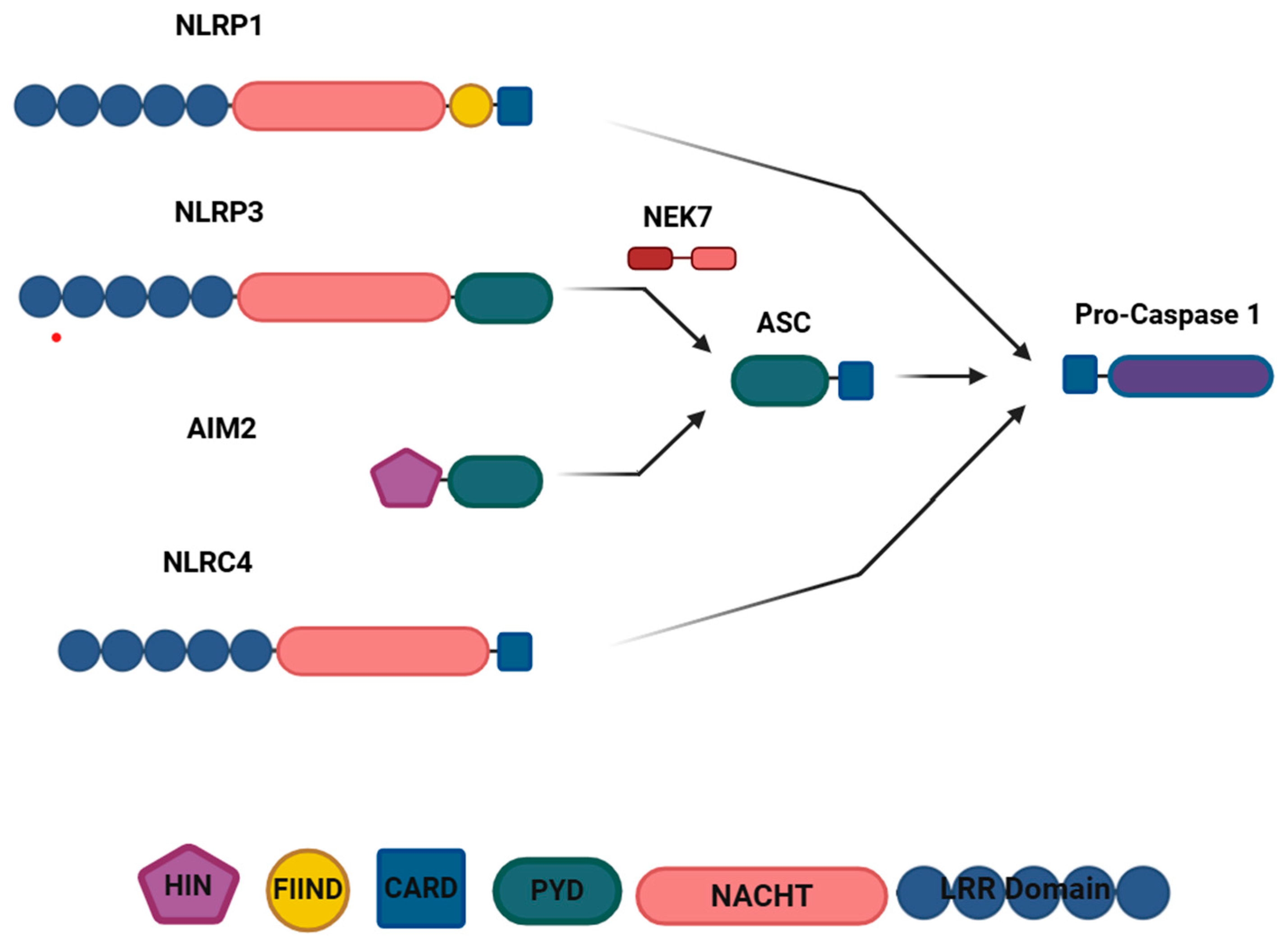
Figure 1. Components of inflammasomes. Inflammasomes are comprised of the three protein subunits to conduct assembly and activation. Sensor protein, inflammasomes are named after their sensor protein; adopter protein, may not universally take part in every inflammasome structure and they mainly play intermediatory role between sensor and effector (caspase-1) proteins via their homolog domains (PYD and CARD domains). NLRP1 inflammasome consists of leucine rich (LRR) domain, NACHT domain, FIIND domain, and CARD domain. Similarly, the NLRP3 inflammasome contains the LRR, NACHT, and CARD domains. In contrast, the AIM2 inflammasome consists of only two domains: the HIN and PYD domains. NLRC4 inflammasomes share similarities with NLRP1, featuring the LRR, NACHT, and CARD domains. NLRP3 and AIM2 inflammasomes, which include the PYD domain, require an additional component: the adaptor protein ASC. ASC, possessing both the PYD domain for binding to the PYD domain of the sensor protein and the CARD domain for facilitating binding to CARD domain of pro-caspase 1, complements these inflammasomes. On the other hand, NLRP1 and NLRC4 inflammasomes possess the CARD domain, which directly interacts with the CARD domain of pro-caspase 1. Additionally, the NLRP3 inflammasome is associated with the kinase NEK7, which has sensitivity to intracellular K concentration, regulates conformational changes of NLRP3 inflammasome and oligomerisation. This figure was created in Biorender.
Encyclopedia 04 00110 g001
Figure 2. Activation mechanism of NLRP3 inflammasome. NLRP3 inflammasome activation is triggered by molecular patterns, PAMPs (e.g., LPS, TNF-α) or DAMPS (e.g., ATP, IL1β) via pattern recognition receptors (PRRs, e.g., TLRs, TNFRs, IL1Rs). Binding PAMPs/DAMPs to a receptor initiates two signalling pathways, signal 1 (priming), and signal 2 (activation/assembly). Signal 1 regulates transcriptional and translational modifications of protein components (NLRP3, caspase 1, pro-IL1β) via NFκB signalling pathway. Signal 2 mainly triggers assembly and activation of the inflammasome. Also, a kinase called NEK7 takes part in the inflammasome complex structure and regulates conformational changes of NLRP3 inflammasome. This binding causes oligomerisation of the inflammasome. Activation of NLRP3 inflammasome leads to recruitment of caspase 1 through triggering autocatalytic activity of the pro-caspase 1 and cleavage of itself. Active caspase 1 can cleave pro-IL1β and pro-IL18 and reveals active IL1β and IL18. Caspase 1 also can cleave GSDRMND protein that is able to form a pore with its revealed N-terminus and secrete IL1β and IL18 to extracellular membrane. This pore formation and release of IL1β and IL18 result in an inflammation-mediated cell death, pyroptosis. This figure was created in Biorender.
Encyclopedia 04 00110 g002
Figure 3. A schematic representation of the microglia molecular targets that have been linked to inflammation. The strongest evidence presented relates to the Kv1 subtypes that are described within the text. The Kir subtypes are linked through preclinical datasets and may contribute to a change in the inflammatory status of the CNS through development stages. Limited evidence is presented for the THIK-1 channel and NKCC1. This figure was created in Biorender.
Encyclopedia 04 00110 g003
Figure 4. A schematic representation of the neuronal molecular targets that have been linked to inflammation. This figure was created in Biorender.
Encyclopedia 04 00110 g004
Figure 5. A schematic representation of the astrocytic molecular targets that have been linked to inflammation. This figure was created in Biorender.
Encyclopedia 04 00110 g005
Figure 6. A schematic representation of the oligodendrocytic molecular targets that have been linked to inflammation. This figure was created in Biorender.
Encyclopedia 04 00110 g006
Disclaimer/Publisher’s Note: The statements, opinions and data contained in all publications are solely those of the individual author(s) and contributor(s) and not of MDPI and/or the editor(s). MDPI and/or the editor(s) disclaim responsibility for any injury to people or property resulting from any ideas, methods, instructions or products referred to in the content.