Brief Report

Manual Reduction for Subacute Osteoporotic Burst and Severe Compression Thoracolumbar Fractures

Yang Ming Hospital, No 252, Wufong N. Road, Chiayi 60054, Taiwan
*
Author to whom correspondence should be addressed.
BioMed 2024, 4(2), 136-145; https://doi.org/10.3390/biomed4020011
Submission received: 6 March 2024 / Revised: 28 April 2024 / Accepted: 8 May 2024 / Published: 24 May 2024

Abstract

:
The objective of this study was to retrospectively assess the impact of manual reduction (MR) on patients with subacute osteoporotic thoracolumbar burst fractures and severe compression fractures (OTLBFSCFs). From January 2016 to May 2020, 101 cases of OTLBFSCFs were reviewed, comprising 73 women and 28 men, with an average age of 77.4 ± 8.5 years. Preoperative radiographs, CT or MRI scans, intraoperative C-arm fluoroscopic images, and postoperative X-ray films were utilized to evaluate spinal radiographic parameters. Initially, all patients underwent 3 min of prone positioning as posture reduction (PR), followed by 1–3 sessions of six-member MR to approximate anatomical reduction of the fracture. The average preoperative anterior body height ratio (ABH%) and lateral Cobb angle (LCA) were 38.8% ± 6.2% and 22.6° ± 4.2°, respectively. Post-PR, the average ABH% and LCA were 50.5% ± 8.0% and 14.7° ± 2.7°, respectively. Following MR, the average ABH% and LCA were 99.6% ± 2.4% and 0.4° ± 2.4°, respectively. PR achieved an ABH% correction of 11.7%, while MR achieved 49.1%. LCA restoration was 7.9° with PR and 14.3° with MR. It was deduced that MR proved to be safe and efficacious for subacute OTLBFSCFs and could potentially alter the approach to subsequent surgeries.

1. Introduction

In the initial stages of acute osteoporotic compression fractures, approximately 70% of individuals may not exhibit spinal deformities [1,2]. However, without treatment, particularly in cases of severe osteoporosis, spinal deformities will develop over time, regardless of the presence of symptoms. With prolonged duration, the bones will fuse at a kyphotic angle, making it impossible to correct the kyphosis through manual reduction. Therefore, this article refers to cases of subacute vertebral fractures with associated deformities without fusion. In such instances, manual reduction can be utilized to address the deformity.
Manual reduction is an effective method for correcting deformities associated with acute spinal trauma. Biomechanical studies involving cadaver burst fractures have demonstrated that traction force applied to the fractured vertebra can reduce displaced bony fragments within the spinal canal, thereby restoring vertebral height. Additionally, extension force can effectively address kyphotic angles [3,4]. While posture reduction for thoracolumbar fractures is regarded as safe and effective [5], it is not without limitations. The treatment for thoracolumbar vertebral fractures without surgery includes reposition without anesthesia and a plaster corset for 4 months, which has evolved from the one used in the past century by Sir R. Watson-Jones and Lorenz Böhler, to recent advancements by Tropiano [6]. However, this method is only suitable for non-surgical cases and is not applicable to open surgery. In 2005, Li et al. [7] reported a successful six-member manual reduction technique for the surgery of high-energy burst fractures. This approach achieved close to a 100% reduction in acute fractures. The prone position facilitated precise manual force application and subsequent surgical intervention. Subsequently, in 2007, Li et al. [8] applied the same six-member manual reduction method to acute osteoporotic compression fractures, yielding comparable results. Recently, Carlo et al. in 2018 [9] and Li et al. in 2021 [10] confirmed the effects of manual reduction on acute spinal fracture, which was quite different from intraoperative manual reduction [11]. While most studies in the literature on manual reduction focus on acute spinal fractures, there is limited information on subacute fractures with deformities. This retrospective study aims to evaluate the outcomes of a six-member manual reduction technique for single subacute osteoporotic thoracolumbar burst and severe compression fractures with kyphosis, comparing its efficacy with that of posture reduction to deepen our understanding of posture and manual reduction mechanisms. The hypothesis posits that posture reduction alone may be insufficient for subacute osteoporotic thoracolumbar burst or severe compression fractures, and that six-member manual reduction can effectively correct anterior vertebral body height and kyphosis.

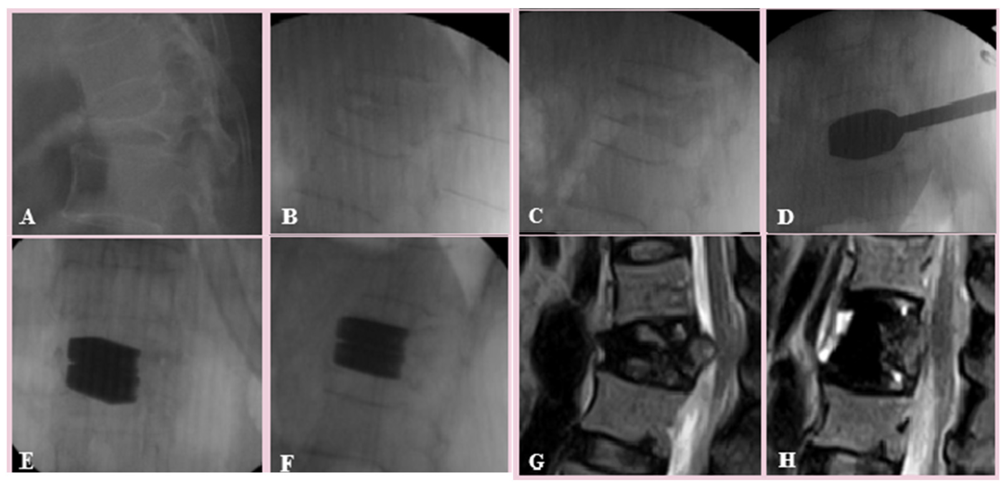
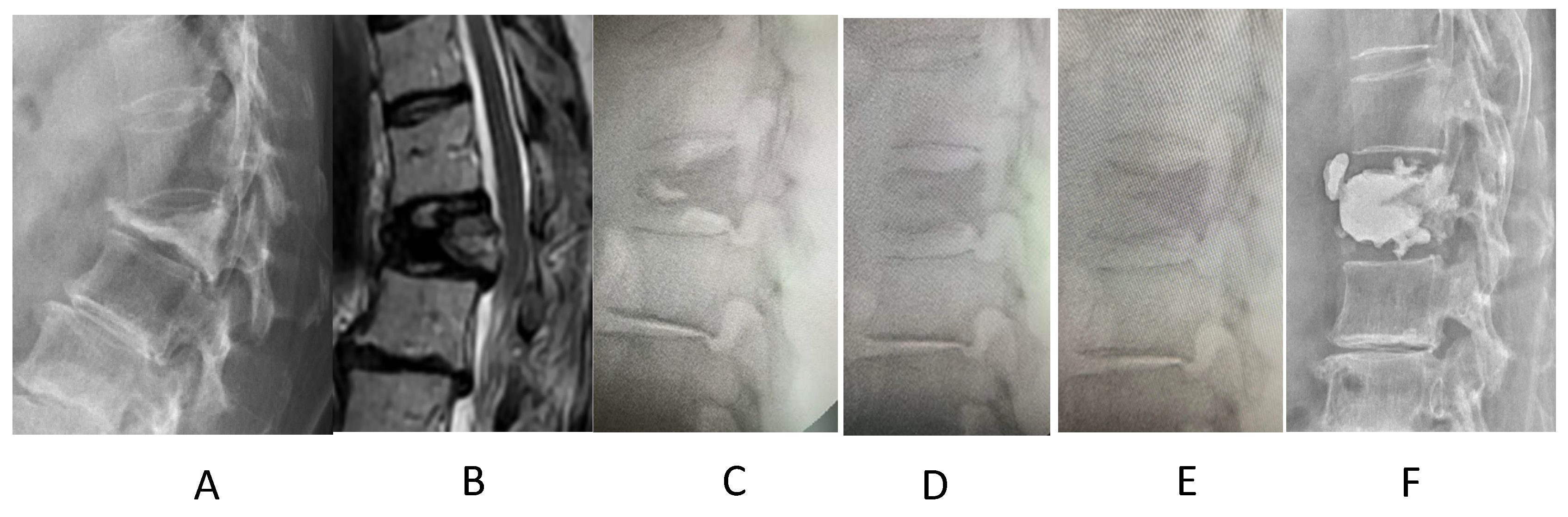
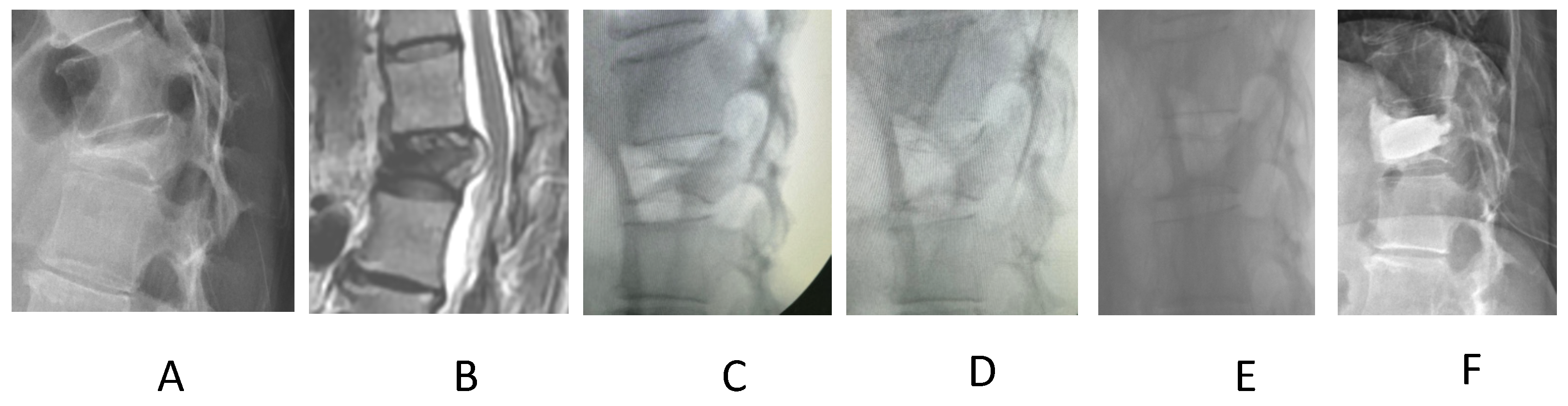
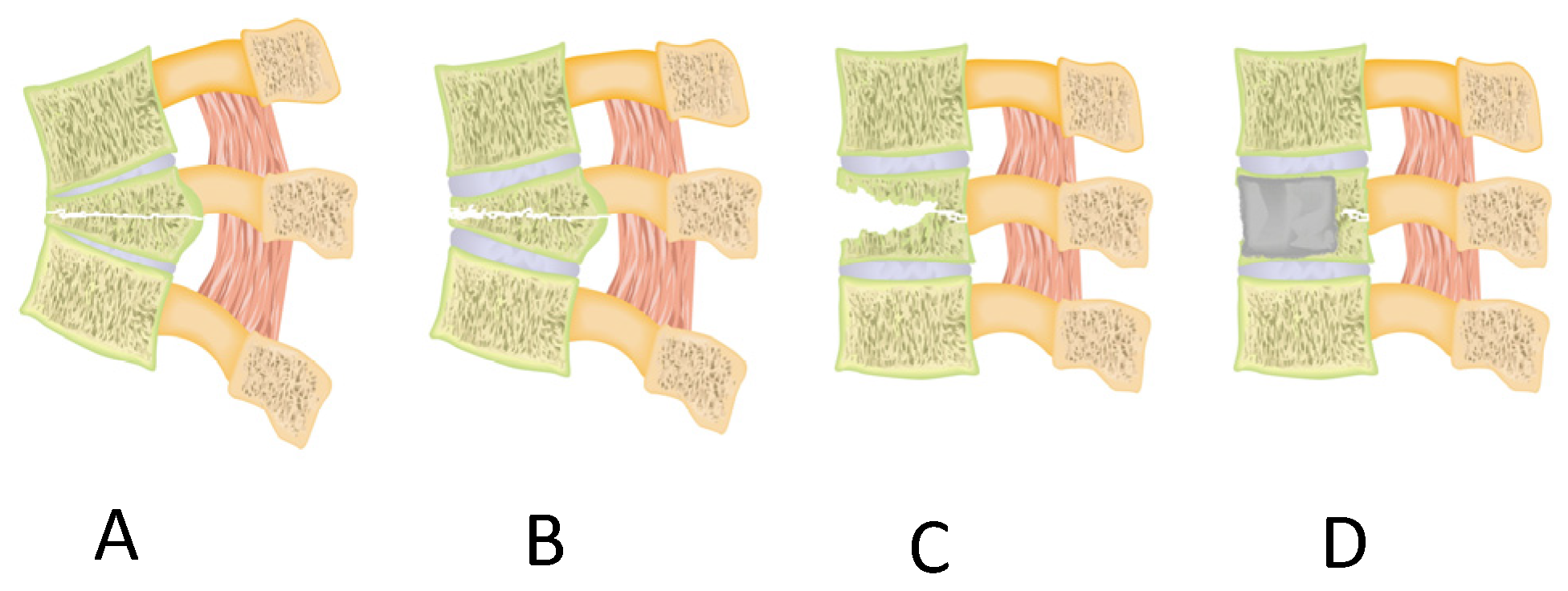
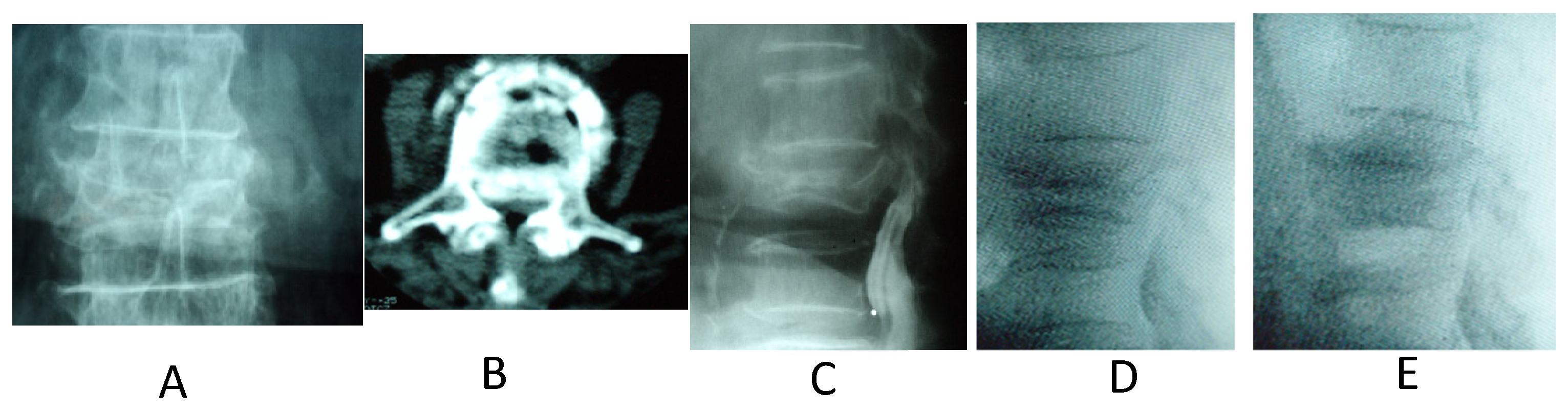
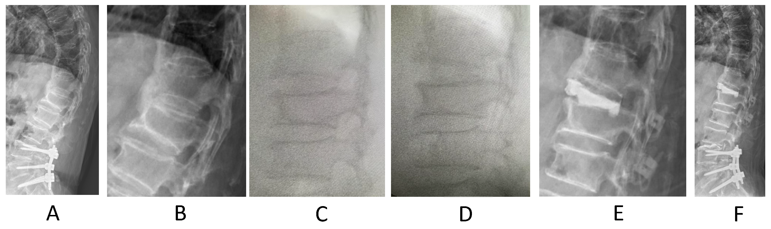
2. Materials and Methods
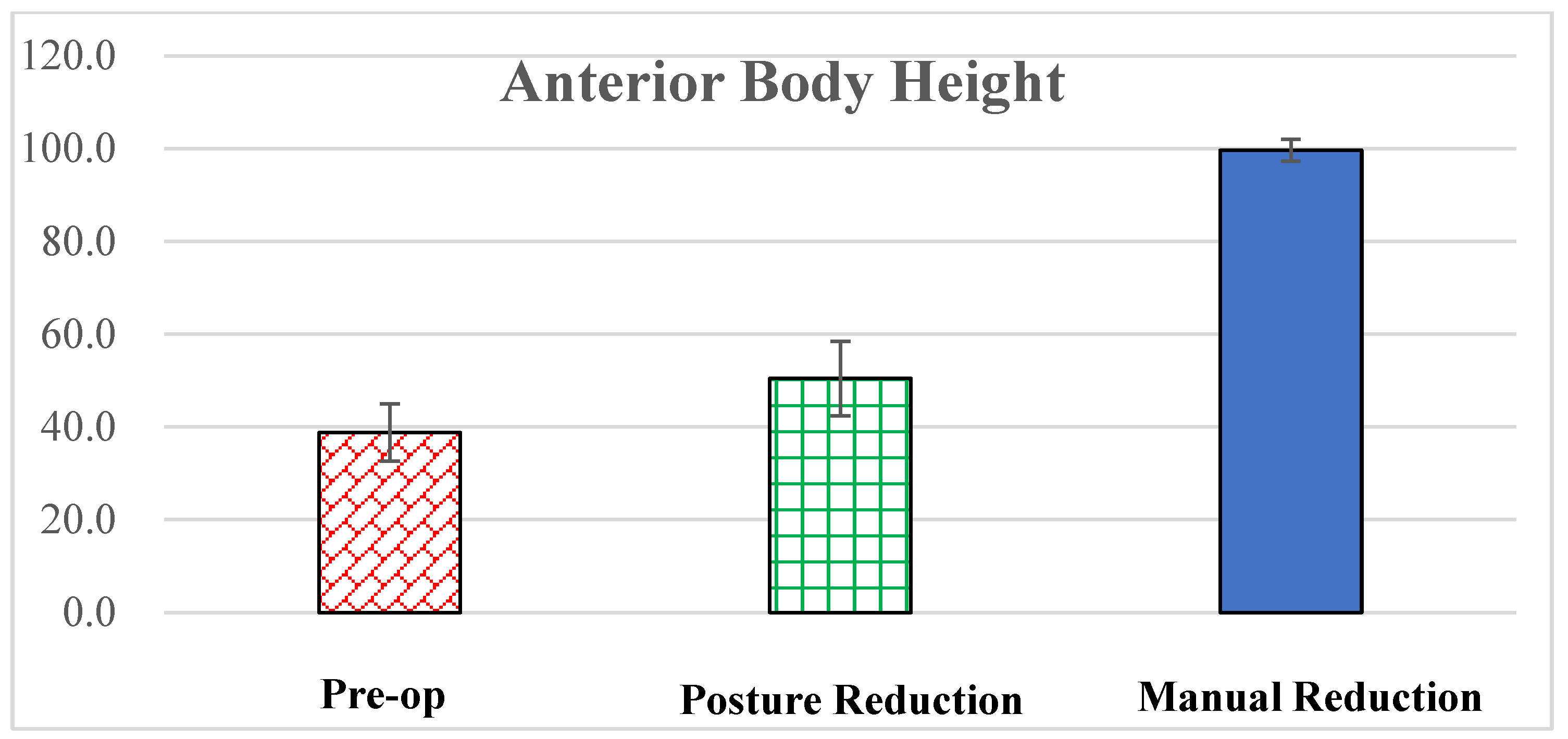
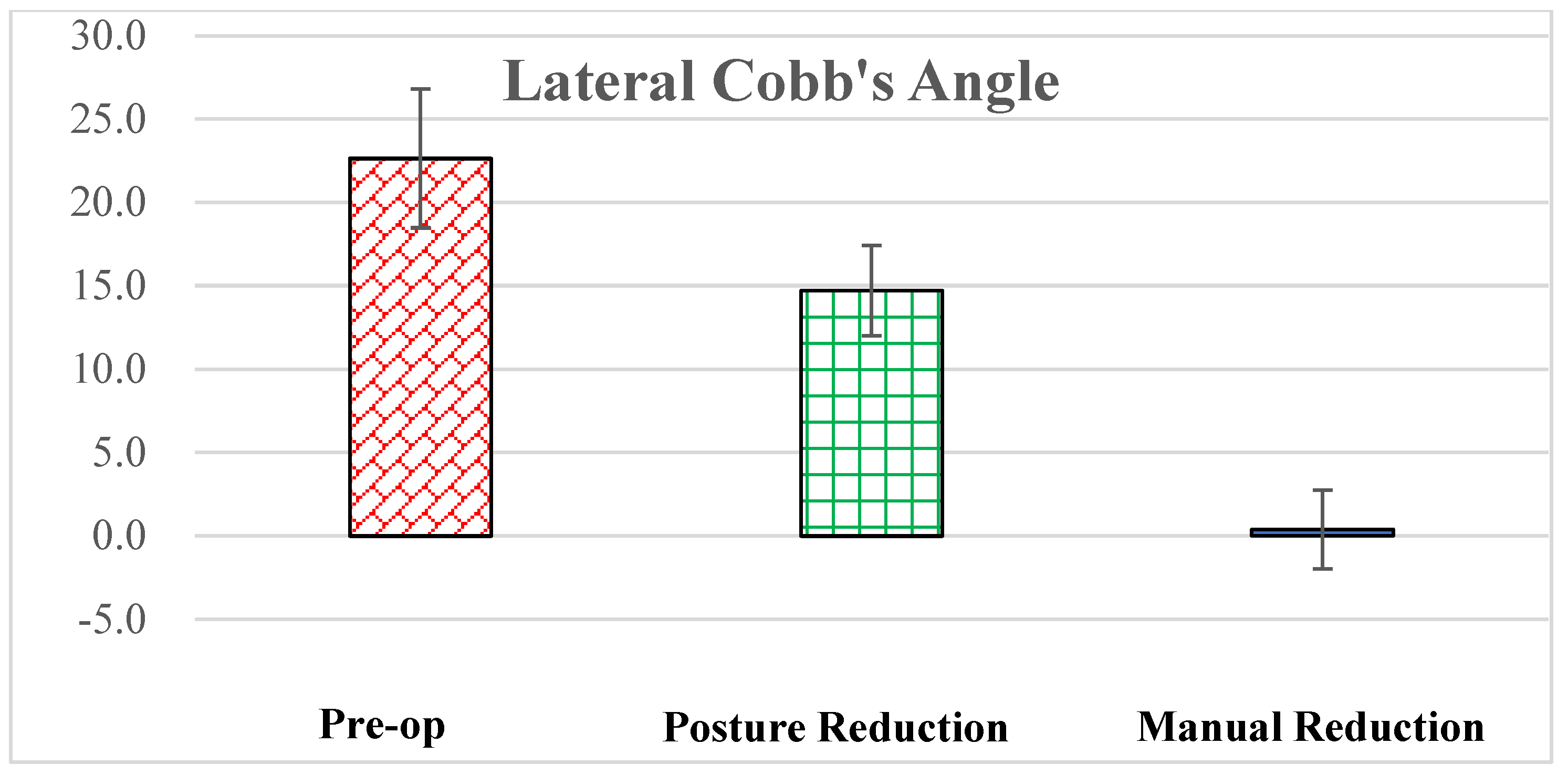
The St Martin De Porres Hospital, Chia-Yi, Taiwan ethics review board (IRB 24C-001) approved this retrospective study.
Between January 2016 and May 2020, four hundred and ninety-seven consecutive patients diagnosed with a single subacute vertebral fracture were treated at our institute. The inclusion criteria for this study were patients with a single subacute burst (AO/Type A3) or severe (AO/Type A2) compression (loss of more than half of the anterior vertebral body height) thoracolumbar fracture (T10-L2) who underwent surgery or vertebroplasty. Subacute osteoporotic vertebral fracture was defined as the trauma–treatment interval between 2 and 8 weeks [12]. The exclusion criteria were as follows: polytrauma, spinal deformity, previous vertebral fracture in the same or adjacent vertebrae above and below the fracture, and previous spinal surgery. Finally, 101 of 497 patients were enrolled in this retrospective study. There were 73 women and 28 men, ranging from 61 to 94 years of age (average, 77.4 ± 8.5 years) at surgery. The mechanisms of injury included falling accidents in 54 patients, traffic accidents in 13, and minor traumas in the remaining 34. The demographic data are summarized in Table 1. Supine anteroposterior and lateral roentgenograms of the thoracic and lumbar spine were obtained on admission for all patients. Imaging assessment was performed using computed tomography and/or MRI before the surgery. C-arm fluoroscopy (Radius R-9 AFG, Intermedical s.r.l. Grassobbio-BG-Italy) was used to monitor posture reduction, manual reduction, and subsequent surgery.
Procedures of 6-member manual reduction [13]
Patients with spinal fractures were anesthetized and positioned prone for 3 min, defined as posture reduction. Before manual reduction, the kyphotic apex was located and evaluated using C-arm fluoroscopy. The manual reduction procedure was carried out by 6 team members: one anesthetist to manage the patient’s head, monitor vital signs and, especially, to manage the endotracheal tube; two assistants to support the patient’s shoulders (one on each side) and provide traction and elevation of the trunk; two assistants to hold and draw the patient’s legs to prevent upward motion during traction; and a surgeon to position his dominant hand at the apex region of the kyphotic deformity (Figure 1). While treating lumbar fracture, manual reduction began with gentle traction and elevation of the pelvis and lower limbs by the leg-holding assistants. While dealing with thoracic fracture, manual reduction commenced with tender cephalic pulling and elevation of the trunk by the shoulder-holding assistants. The operator compressed the spine against the trunk motion to induce realignment of the fractured deformity. Following manual reduction, C-arm fluoroscopy was used again to confirm deformity correction and spinal canal decompression. Manual reduction can be repeated as often as necessary until anatomical reduction is achieved.
The lateral Cobb’s angle, representing the kyphosis angle, was measured from the superior margin of the vertebral body above the fracture to the inferior margin of the vertebral body below the fracture level. Predicted anterior vertebral body heights were estimated using mean heights of adjacent upper and lower segments. The anterior body height ratio (%) was determined by dividing measured body height by predicted body height. Kyphosis angle and anterior body height were assessed on neutral thoracolumbar radiographs pre- and post-surgery. Fluoroscopic images were evaluated to assess the effects of posture reduction and manual reduction. All digitization processes and measurements were conducted using EBM-viewer software (EBM Technologies Inc., Taipei, Taiwan) with an accuracy of ±0.1 mm. Initially, radiographic measurements were performed by a graduate student and then rechecked by surgeons. Canal encroachment was not measured due to the absence of intraoperative computed tomography images before and after manual reduction.
The quantitative variables were described as mean ± standard deviation. Preoperative, post-posture reduction, post-manual reduction, and postoperative lateral Cobb’s angle and anterior body height Cobb’s angle and anterior body height were compared through a paired t-test; the statistical significance (p < 0.05) of these differences was calculated. The statistical analysis was performed with the Excel (Microsoft 365, 2023 version, Redmond, WA, USA).

3. Results

From January 2016 to May 2020, a total of 101 cases of subacute osteoporotic thoracolumbar burst and severe compression fractures were examined in this study. Manual reduction effectively restored spinal column height and corrected kyphosis in these subacute cases (Figure 2, Figure 3 and Figure 4). The preoperative average anterior body height ratio and lateral Cobb’s angle measured 38.8% ± 6.2% and 22.6° ± 4.2°, respectively. Following posture reduction, the average anterior body height ratio and lateral Cobb angle increased to 50.5% ± 8.0% and decreased to 14.7° ± 2.7°, respectively. After manual reduction, the average anterior body height ratio and lateral Cobb angle improved to 99.6% ± 2.4% and reduced to 0.4° ± 2.4°, respectively (Figure 5 and Figure 6). Statistical analysis revealed significant differences in the anterior body height ratio and lateral Cobb’s angle among the preoperative, post-posture reduction, and post-manual reduction stages. The correction in anterior body height amounted to 11.7% through posture reduction and 49.1% through manual reduction. Similarly, the restoration in lateral Cobb’s angle was 7.9° through posture reduction and 14.3° through manual reduction. Posture reduction contributed to 35.6% of the total angle correction and 19.2% of the anterior body height correction, while manual reduction accounted for 64.4% of the total kyphosis correction and 80.8% of the total anterior body height correction.
There were no complications due to MR, that is, no spinal cord injury, hemothorax, pneumothorax, or dislodgement of the endotracheal tube.

4. Discussion

Six-member manual reduction for subacute osteoporotic thoracolumbar burst fractures and severe compression fractures is effectual and protected. Manual reduction is commonly used to treat limb fractures or dislocations [14]. Cervical traction is often used for neck injury [15]. The six-member manual reduction was created from biomechanical knowledge [3,4,16,17] and hyperextension posture reduction [18]. In acute thoracolumbar fractures, manual reduction can lead to anatomical correction of the spinal construct. Reduction of acute spinal fractures was technically easier with six-member manual reduction than with instrumentation reduction [19,20,21]. Non-union osteoporotic fractures [22,23,24,25], can be treated with six-member manual reduction and body reconstruction [22]. Vertebral body height can be increased restrictedly by vertebroplasty and kyphoplasty [19,26,27,28]. Based on this retrospective study, manual reduction can lead to anatomic reduction of subacute thoracolumbar fractures without complications.
Compared to posture reduction, manual reduction is more important for the restoration of bony structures. While the patients were turned in the prone position, the unfused disc was relatively easily opened. (Figure 2, Figure 3, Figure 4 and Figure 7) This is why the lateral Cobb’s angle improved significantly (35.6% of total correction) only by postural reduction. However, the broken bony structures were partially restored by postural reduction, with only 19.2% of the total correction of the anterior body height in this study. Further manual reduction with greater extension force and hyperextension is needed to fully restore body height, accounting for 80.8% of the total correction. The mechanism is due to prone position traction potentially leading to a loss in surrounding muscles and anterior and posterior longitudinal ligaments around the fractured vertebrae to recover the body height [9]. Furthermore, manual reduction can be reiterated, and the force amount and direction can be adjusted to ensure anatomical restoration. Therefore, manual reduction is important before skin incision for spinal realignment and cord decompression.
In cases of secondary body collapse resulting from active tuberculosis, multiple myeloma, or osteolytic metastasis, manual reduction can offer a feasible solution. This discovery holds potential for shaping future treatment protocols. Classically, body collapse accompanied by cord compression from active tuberculosis [29] or metastasis [30,31] has been addressed through anterior decompression. However, manual reduction has demonstrated efficacy in reducing kyphosis and decompressing the spinal cord, and it may confirm intraoperative myelography (Figure 8). The traditional procedures of cord-decompression and instrumentation reduction may be replaced by manual reduction. The following operation may focus on debridement and fixation in tuberculosis-related spine collapse complemented by tuberculosis medical treatment. In cases of secondary body collapse due to spinal metastasis, the next procedures may focus on stabilizing the vertebral body and subsequent chemotherapy or radiotherapy. Manual reduction has the potential to alter the traditional approach and subsequent surgical interventions.
Manual reduction can be used to reduce the most unstable spinal deformities, including listhesis, scoliosis, and kyphosis [13,32,33]. Advantages include indirect decompression of the spinal cord and nerve roots, avoidance of procedures to reduce deformities by instrumentation, shortening the operation time, and decreasing blood loss. Manual reductions may be helpful in certain situations. As in proximal junctional kyphosis after long instrumentation (Figure 9), the restoration of spinal alignment is a significant issue that is technically demanding and time-consuming. With the advantage of manual reduction before skin incision, alignment can be easily restored without any blood loss. Subsequent operations can include other options, depending on the medical conditions. In other common situations, such as rekyphosis after vertebroplasty, manual reduction can also have a significant effect on kyphosis correction by inducing an iatrogenic open-wedge fracture in the osteoporotic spine. (Figure 10) Then, a simple vertebroplasty can be performed to fix the spine alignment. Manual reduction can also correct osteoporotic anterior ankylosing kyphosis by breaking the bone bridge. Percutaneous cement discoplasty can maintain the restoration (Figure 11). Compared with correcting osteotomy with long posterior instrumentation to restore spinal alignment, manual reduction with subsequent procedures is more suitable for elderly patients. At our institute, manual reduction is a routine procedure for spinal surgery.
A few limitations of this study should be mentioned when interpreting the data. Firstly, the retrospective design might have a selection bias, which impacts the strength of the data. Secondly, this study involved only one institute. Thirdly, cooperation between team members may not be technically consistent. Further multicenter prospective studies involving a larger sample size would be better.

5. Conclusions

Manual reduction can safely and successfully reduce subacute thoracolumbar burst and severe compression fractures. Posture reduction contributed to 35.6% of the total angle correction and 19.2% of the anterior body height correction, while manual reduction accounted for 64.4% of the total kyphosis correction and 80.8% of the total anterior body height correction. Manual reduction may affect the options for surgery or vertebroplasty to avoid operative decompression and instrumentation reduction.

Author Contributions

K.-C.L. performed the surgery, organized the structure of this study, and coordinated the drafting of the manuscript. C.-H.H., T.-H.L. and C.-H.C. participated in the surgery and data collection. All authors have read and agreed to the published version of the manuscript.

Funding

This research received no external funding.

Institutional Review Board Statement

This study was conducted in accordance with the Declaration of Helsinki and approved by the Ethics Committee of St. Martin De Porres Hospital, Chia-Yi, Taiwan (approval number: IRB 24C-001).

Informed Consent Statement

Not applicable.

Data Availability Statement

The data presented in this study are available on request from the corresponding author due to ethical reason.

Conflicts of Interest

The authors declare no conflicts of interest.

References

  1. Buchbinder, R.; Johnston, R.V.; Rischin, K.J.; Homik, J.; Jones, C.A.; Golmohammadi, K.; Kallmes, D.F. Percutaneous vertebroplasty for osteoporotic vertebral compression fracture. Cochrane Database Syst. Rev. 2018, 4, CD006349, Update in Cochrane Database Syst. Rev. 2018, 11, CD006349. [Google Scholar] [CrossRef] [PubMed] [PubMed Central]
  2. Zuo, X.H.; Zhu, X.P.; Bao, H.G.; Xu, C.J.; Chen, H.; Gao, X.Z.; Zhang, Q.X. Network meta-analysis of percutaneous vertebroplasty, percutaneous kyphoplasty, nerve block, and conservative treatment for nonsurgery options of acute/subacute and chronic osteoporotic vertebral compression fractures (OVCFs) in short-term and long-term effects. Medicine 2018, 97, e11544. [Google Scholar] [CrossRef] [PubMed] [PubMed Central]
  3. Fredrickson, B.; Edwards, W.; Rauschning, W.; Bayley, J.; Yuan, H. Volvo award in experimental studies vertebral burst fractures: An experimental, morphologic, and radiographic study. Spine 1992, 17, 1012–1021. [Google Scholar] [CrossRef] [PubMed]
  4. Fredrickson, B.E.; Mann, K.A.; Yuan, H.A.; Lubicky, J.P. Reduction of the intracanal fragment in experimental burst fractures. Spine 1988, 13, 267–271. [Google Scholar] [CrossRef] [PubMed]
  5. Frankel, H.L.; O Hancock, D.; Hyslop, G.; Melzak, J.; Michaelis, L.S.; Ungar, G.H.; Vernon, J.D.S.; Walsh, J.J. The value of postural reduction in the initial management of closed injuries of the spine with paraplegia and tetraplegia. Spinal Cord. 1969, 7, 179–192. [Google Scholar] [CrossRef] [PubMed]
  6. Tropiano, P.; Huang, R.C.; Louis, C.A.; Poitout, D.G.; Louis, R.P. Functional and radiographic outcome of thoracolumbar and lumbar burst fractures managed by closed orthopaedic reduction and casting. Spine 2003, 28, 2459–2465. [Google Scholar] [CrossRef] [PubMed]
  7. Li, K.-C.; Hsieh, C.-H.; Lee, C.-Y.; Chen, T.-H. Transpedicle body augmenter: A further step in treating burst fractures. Clin. Orthop. Relat. Res. (1976–2007) 2005, 436, 119–125. [Google Scholar] [CrossRef]
  8. Li, K.-C.; Li, A.F.-Y.; Hsieh, C.-H.; Chen, H.-H. Transpedicle body augmenter in painfulo steoporotic compression fractures. Eur. Spine J. 2007, 16, 589–598. [Google Scholar] [CrossRef] [PubMed]
  9. Carlo, P.; Francesco, C. Preoperative manual on-table-traction for the reduction of thoracolumbar burst fractures: A technical note. J. Craniovertebral Junction Spine 2018, 9, 73. [Google Scholar] [CrossRef]
  10. Li, Y.; Du, Y.; Ji, A.; Wang, Q.; Li, L.; Wu, X.; Wang, P.; Chen, F. The clinical effect of manual reduction combined with internal fixation through Wiltse Paraspinal approach in the treatment of thoracolumbar fracture. Orthop. Surg. 2021, 13, 2206–2215. [Google Scholar] [CrossRef]
  11. Huang, J.; Zhou, L.; Yan, Z.; Zhou, Z.; Gou, X. Effect of manual reduction and indirect decompression on thoracolumbar burst fracture: A comparison study. J. Orthop. Surg. Res. 2020, 15, 532. [Google Scholar] [CrossRef] [PubMed] [PubMed Central]
  12. Rousing, R.; Andersen, M.O.; Jespersen, S.M.; Thomsen, K.; Lauritsen, J. Percutaneous vertebroplasty compared to conservative treatment in patients with painful acute or subacute osteoporotic vertebral fractures: Three-months follow-up in a clinical randomized study. Spine 2009, 34, 1349–1354. [Google Scholar] [CrossRef] [PubMed]
  13. Li, K.C. IntechOpen: Manual Reduction, Subpedicle Approach, and Body Cage to Treat Burst Fracture. IntechOpen 2023. Available online: https://www.intechopen.com/profiles/518621 (accessed on 1 June 2023).
  14. Khan, M.R.; Mirdad, T.M. Ipsilateral dislocation of the shoulder and elbow. Saudi Med. J. 2001, 22, 1019–1021. [Google Scholar] [PubMed]
  15. Bohlman, H.H.; Ducker, T.B. Spine and Spine Cord Injuries. In Rothman-Simeone the Spine, 4th ed.; Herkowitz, H.N., Garfin, S.R., Balderston, R.A., Eismont, F.J., Bell, G.R., Eds.; WB Saunders Company: Philadelphia, PA, USA, 1999; Volume 2, pp. 889–1002. [Google Scholar]
  16. Harrington, R.; Budorick, T.; Hoyt, J.; Anderson, P.; Tencer, A. Biomechanics of indirect reduction of bone retropulsed into the spinal canal in vertebral fracture. Spine 1993, 18, 692–699. [Google Scholar] [CrossRef]
  17. Lin, R.M.; Panjabi, M.M.; Oxland, T.R. Functional radiographs of acute thoracolumbar burst fractures: A biomechanical study. Spine 1993, 18, 2431–2437. [Google Scholar] [CrossRef] [PubMed]
  18. Mumford, J.; Weinstein, J.N.; Spratt, K.F.; Goel, V.K. Thoracolumbar burst fractures: The clinical efficacy and outcome of nonoperative management. Spine 1993, 18, 955–970. [Google Scholar] [CrossRef] [PubMed]
  19. Deramond, H.; Depriester, C.; Galibert, P.; Le Gars, D. Percutaneous vertebroplasty with polymethylmethacrylate: Technique, indications, and results. Radiol. Clin. N. Am. 1998, 36, 533–546. [Google Scholar] [CrossRef] [PubMed]
  20. Lindsey, R.W.; Dick, W. The fixateur interne in the reduction and stabilization of thoracolumbar spine fractures in patients with neurologic deficit. Spine 1991, 16 (Suppl. S3), S140–S145. [Google Scholar] [CrossRef]
  21. Duan, Y.; Feng, D.; Chen, J.; Wu, Y.; Li, T.; Jiang, L.; Huang, Y. Anterior, Posterior and Anterior-Posterior Approaches for the Treatment of Thoracolumbar Burst Fractures: A Network Meta-Analysis of Randomized Controlled Trials. J. Investig. Surg. 2024, 37, 2301794. [Google Scholar] [CrossRef] [PubMed]
  22. Li, K.-C.; Li, A.F.; Hsieh, C.-H.; Liao, T.-H.; Chen, C.-H. Another option to treat Kümmell’s disease with cord compression. Eur. Spine J. 2007, 16, 1479–1487. [Google Scholar] [CrossRef]
  23. Van Eenenaam, D.P.; Georges, Y. Delayed post-traumatic vertebral collapse (Kummell’s disease): Case report with serial radiographs, computed tomographic scans, and bone scans. Spine 1993, 18, 1236–1241. [Google Scholar] [CrossRef] [PubMed]
  24. Golimbu, C.; Firooznia, H.; Rafii, M. The intravertebral vacuum sign. Spine 1986, 11, 1040–1043. [Google Scholar] [CrossRef] [PubMed]
  25. Osterhouse, M.D.; Kettner, N.W. Delayed posttraumatic vertebral collapse with intravertebral vacuum cleft. J. Manip. Physiol. Ther. 2002, 25, 270–275. [Google Scholar] [CrossRef] [PubMed]
  26. Garfin, S.R.; Yuan, H.A.; Reiley, M.A. New technologies in spine: Kyphoplasty and vertebroplasty for the treatment of painful osteoporotic compression fractures. Spine 2001, 26, 1511–1515. [Google Scholar] [CrossRef] [PubMed]
  27. Hiwatashi, A.; Moritani, T.; Numaguchi, Y.; Westesson, P.-L. Increase in vertebral body height after vertebroplasty. Am. J. Neuroradiol. 2003, 24, 185–189. [Google Scholar] [PubMed]
  28. McKiernan, F.; Jensen, R.; Faciszewski, T. The dynamic mobility of vertebral compression fractures. J. Bone Miner. Res. 2003, 18, 24–29. [Google Scholar] [CrossRef] [PubMed]
  29. Fukuta, S.; Miyamoto, K.; Masuda, T.; Hosoe, H.; Kodama, H.; Nishimoto, H.; Sakaeda, H.; Shimizu, K. Two-stage (posterior and anterior) surgical treatment using posterior spinal instrumentation for pyogenic and tuberculotic spondylitis. Spine 2003, 28, E302–E308. [Google Scholar] [CrossRef]
  30. Perrin, R.G.; McBroom, R.J. Spinal fixation after anterior decompression for symptomatic spinal metastasis. Neurosurgery 1988, 22, 324–327. [Google Scholar] [CrossRef]
  31. Walker, M.P.; Yaszemski, M.J.; Kim, C.W.; Talac, R.; Currier, B.L. Metastatic disease of the spine: Evaluation and treatment. Clin. Orthop. Relat. Res. (1976–2007) 2003, 415, S165–S175. [Google Scholar] [CrossRef]
  32. Sieradzki, J.P.; Sarwark, J.F. Thoracolumbar fracture-dislocation in child abuse: Case report, closed reduction technique and review of the literature. Pediatr. Neurosurg. 2008, 44, 253–257. [Google Scholar] [CrossRef]
  33. Li, K.-C.; Hsieh, C.-H.; Liao, T.-H. Clinical outcome-supported advance of cement lumber interbody fusion for degenerative lumbar scoliosis. J. Surg. 2023, 8, 1697. [Google Scholar] [CrossRef]
Figure 1. Diagram showing the detailed techniques of a 6-member manual reduction.
Biomed 04 00011 g001
Figure 2. A 90-year-old male was diagnosed with L1 osteoporotic burst fracture (A). Fluoroscopic images show posture reduction (B) and manual reduction (C). The patient was treated with pile-up titanium spacers (DF). Pre- and postoperative MRI showed spinal cord decompression (G,H).
Biomed 04 00011 g002
Figure 3. A 61-year-old female was diagnosed with L1 osteoporotic burst fracture (A,B). The fluoroscopic images show posture reduction (C) and manual reduction (first time (D); second time (E)). The patient underwent percutaneous vertebroplasty (F).
Biomed 04 00011 g003
Figure 4. A 64-year-old female was diagnosed with L1 osteoporotic burst fracture (A,B). The fluoroscopic images show posture reduction (C) and manual reduction (first time (D), second time (E)). The patient was treated using a cemented spacer (F).
Biomed 04 00011 g004
Figure 5. Anterior body height preoperatively, post-posture reduction, and manual reduction. There were significant differences among the three stages.
Biomed 04 00011 g005
Figure 6. Lateral Cobb’s angles at the pre-operative stage, post-posture reduction, and manual reduction. There were significant differences among the three stages.
Biomed 04 00011 g006
Figure 7. The diagram shows thoracolumbar osteoporotic burst fracture with kyphosis (A). Posture reduction leads to most disc restoration (B), and manual reduction restores the residual bony deformity (C). It can be treated with vertebroplasty (D) without instrumentation.
Biomed 04 00011 g007
Figure 8. A 73-year-old female was diagnosed with an L2 osteoporotic burst fracture (A,B), and preoperative myelography (C) showed a complete block. Fluoroscopic images showed correction after posture reduction (D) and manual reduction (E). The myelogram was patent following manual reduction.
Biomed 04 00011 g008
Figure 9. A 73-year-old female was diagnosed with proximal junction kyphosis (A,B) and underwent manual reduction (C) and locking interbody cementation (D,E) using a titanium spacer (F,G).
Biomed 04 00011 g009
Figure 10. A 75-year-old male was diagnosed with rekyphosis following vertebroplasty (A). Kyphosis was corrected by posture reduction (B) and manual reduction (C). Open wedge fractures induced by manual reduction were repaired by percutaneous vertebroplasty (D,E).
Biomed 04 00011 g010
Figure 11. An 88-year-old male was diagnosed with kyphosis secondary to spontaneous anterior ankylosis (A,B). Kyphosis was corrected by first (C) and second manual reduction (D). The disc space opening induced by manual reduction was fixed using percutaneous cement discoplasty (E,F).
Biomed 04 00011 g011
Table 1. Table showing the demographic data of 101 patients.
ItemsData
Gender73 females
28 males
(Total: 101)
Age77.4 ± 8.5
(61 to 94)
DEXA3.2 ± 4.5
(2.9 to 4.2)
Injury
mechanism
Fall: 54 cases
RTA: 13 cases
Minor trauma 1: 34 cases
Fracture
level
T10: 5
T11: 19
T12: 29
L1: 31
L2: 17
1 Minor trauma: no significant accident.
Disclaimer/Publisher’s Note: The statements, opinions and data contained in all publications are solely those of the individual author(s) and contributor(s) and not of MDPI and/or the editor(s). MDPI and/or the editor(s) disclaim responsibility for any injury to people or property resulting from any ideas, methods, instructions or products referred to in the content.

