Article

Short-Term Clinical Results of Single-Injection Autologous Bone Marrow Aspirate Concentrate (BMAC) as a Therapeutic Option/Tool in Knee Osteoarthritis

by
Krishna Subramanyam
1,2,*,†,
Subhadra Poornima
3,4,†,
Satish Kumar
2 and
Qurratulain Hasan
3
1
Department of Orthopaedics, Kamineni Hospitals, LB Nagar, Hyderabad 500074, India
2
Department of Orthopaedics, Yashoda Hospitals, Malakpet, Hyderabad 500036, India
3
Department of Genetics and Molecular Medicine, Kamineni Academy of Medical Sciences and Research Centre, LB. Nagar, Hyderabad 500074, India
4
Department of Genetics and Molecular Medicine, Kamineni Life Sciences, Moula Ali, Hyderabad 500047, India
*
Author to whom correspondence should be addressed.
These authors contributed equally to this work.
Biologics 2024, 4(2), 218-231; https://doi.org/10.3390/biologics4020015
Submission received: 20 April 2024 / Revised: 3 June 2024 / Accepted: 7 June 2024 / Published: 19 June 2024

Abstract

:
Purpose: Knee osteoarthritis (KOA) is a very common cartilage disorder affecting millions of people globally and is characterized by pain, stiffness, swelling, loss of articular cartilage, and osteophyte formation, resulting in disability. The presently available treatments for KOA are palliative. Hence, there is a need to explore a non-surgical treatment portfolio. Bone marrow aspirate concentrate (BMAC) is one of the predominant attention-drawing managements/treatments for KOA in recent times due to its potential advantages of disease-modifying and regeneration capacities. Principle: This study aimed to evaluate the role of single-injection autologous BMAC as a therapeutic option in the treatment of KOA and evaluate the functional and clinical outcomes of KOA patients. In this study, 132 patients with KOA (Kellgren and Lawrence (KL) grade II and III) were included as per the inclusion criteria. Autologous bone marrow was aspirated and separated, and concentrated bone marrow aspirate was administered into the knee joint of the affected individual. Results: At the end of the 12th month (end of the follow-up period), 95% of patients showed complete pain relief and improvement in joint function, which shows that the results were promising and encouraging. Unpaired t-test results also indicated that the two-tailed p-value is less than 0.0001, and the difference is extremely statistically significant. No adverse effects were observed in the study patients. Conclusions: BMAC therapy has potential, with satisfactory, efficient, and durable results in KL grades II and III in KOA patients. This can be a safe alternative therapy in the treatment of KOA, especially in the early grades of OA. In summary, to the best of our knowledge, this is the first study from India that evaluated BMAC efficacy both subjectively and objectively in KOA (KL-II and KL-III) patients.

Graphical Abstract

1. Introduction

Knee osteoarthritis (KOA) is a common musculoskeletal disorder associated with high social and economic drift which affects people in or above their fourth decade of life [1,2]. The insidious progression of KOA is typically characterized by the degeneration of articular cartilage, inflammation, and subchondral bone changes. Pain and limitations in joint mobility, dexterity, and functional ability caused by KOA, in turn, burden the patient’s quality of life. The present non-surgical treatment modalities of KOA are life-style modifications, external bracing, non-steroidal anti-inflammatory drugs (NSAIDs), disease-modifying osteoarthritic drugs (DMODS), and intra-articular steroids and hyaluronic acid [3,4]. Conventional clinical treatment in KOA may not address the final successful outcome, as it primarily targets symptomatic relief, and its efficacy in repairing the joint remains limited [5]. The high prevalence of KOA escalates, making the exploration of unique and novel effective therapeutic avenues imperative. Therefore, there is a need for an alternative modality in the treatment of KOA during early grades, which should help not only in giving symptomatic relief but also in slowing the disease process. Recent research has unveiled the therapeutic usage of cell-based therapies in the treatment of KOA. Despite the extensive literature on this topic, the optimal management of symptomatic OA remains a challenge [6,7]. The scope of Autologous Bone Marrow Aspirate Concentrate (BMAC) has the potential to help scientists and orthopedic surgeons devise an early interventional treatment for KOA [8,9]. The current literature reviews show that the intra-articular injection of autologous BMAC has the potential to decrease the symptoms and functionally improve the osteoarthritic knee joint [10,11,12]. This may work by modifying the underlying disease process and slowing the progress of KOA.
In recent years, there has been an increasing interest in the usage of autologous BMAC in different musculoskeletal conditions [13,14]. The latest studies on the usage of autologous BMAC injection in KOA showed promising and encouraging results in terms of pain reduction, improved knee range of movement, ease of life’s daily activities, and, thus, improved quality of life. These results obtained with autologous BMAC are longer-lasting than other non-surgical treatments and also showed better safety profiles [15,16,17].
Mesenchymal stem cells (MSCs) are the basic human cells that have the potential to give rise to many different cell types. They are simple and differentiated. These cells will help create new cells in existing healthy tissue and may help to repair those structures when injured or damaged. When they divide, they create progenitor cells, and these will become cells with more specialized tissue structures, such as bone and cartilage [18]. The two most common sources of stem cells for clinical application in orthopedics are bone marrow and adipose tissue [19]. MSCs can migrate from subchondral bone to damaged areas, differentiating into chondrocytes and osteoblasts to repair cartilage and subchondral bone tissues. BMAC is a mixture of MSC and hemopoietic stem cells (HSCs), endothelial progenitor cells, and growth factors [20]. There is strong evidence that MSCs and the growth factors present in the BMAC help in the regeneration of diseased cartilage with respect to an increase in cartilage volume [21]. In the current study, we made an attempt to evaluate the clinical outcomes of a single injection of intra-articular autologous BMAC in KOA (Kellgren and Lawrence (KL) grade II and II) patients. The aim of the study is to assess the role of single-injection autologous BMAC as a therapeutic option in the treatment of KOA and to evaluate the functional and clinical outcomes.

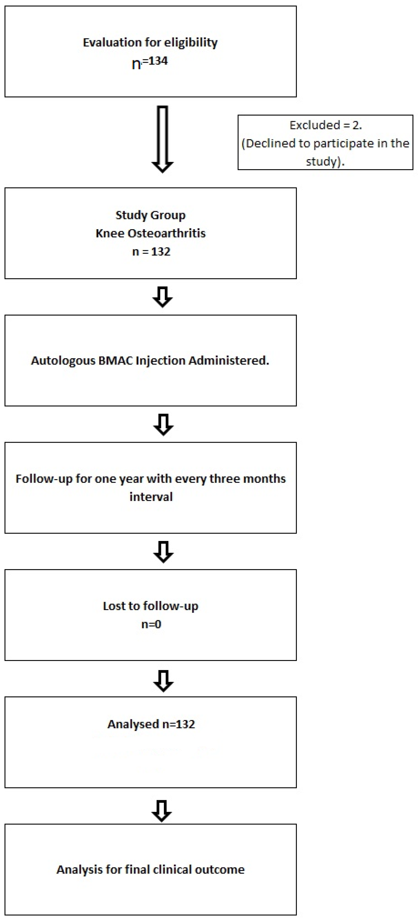
2. Materials and Methods

2.1. Patient Recruitment

One hundred and thirty-two (132) patients with KOA-KL grade II and III (Figure 1) were recruited, and the study was conducted at Kamineni and Yashoda Hospitals in Hyderabad, India.
The study duration was between 4 March 2013 and 11 October 2016. All patients were counseled by a Lead Surgeon and the Certified Genetic Counselor about the single autologous BMAC injection as a treatment option for the patients with KOA. Institutional Ethics Committee approved this study [ECR/58/Inst/AP/2013/RR-19]. The recruitment of all the patients was completed after obtaining written consent (Figure 2).

2.1.1. Inclusion Criteria

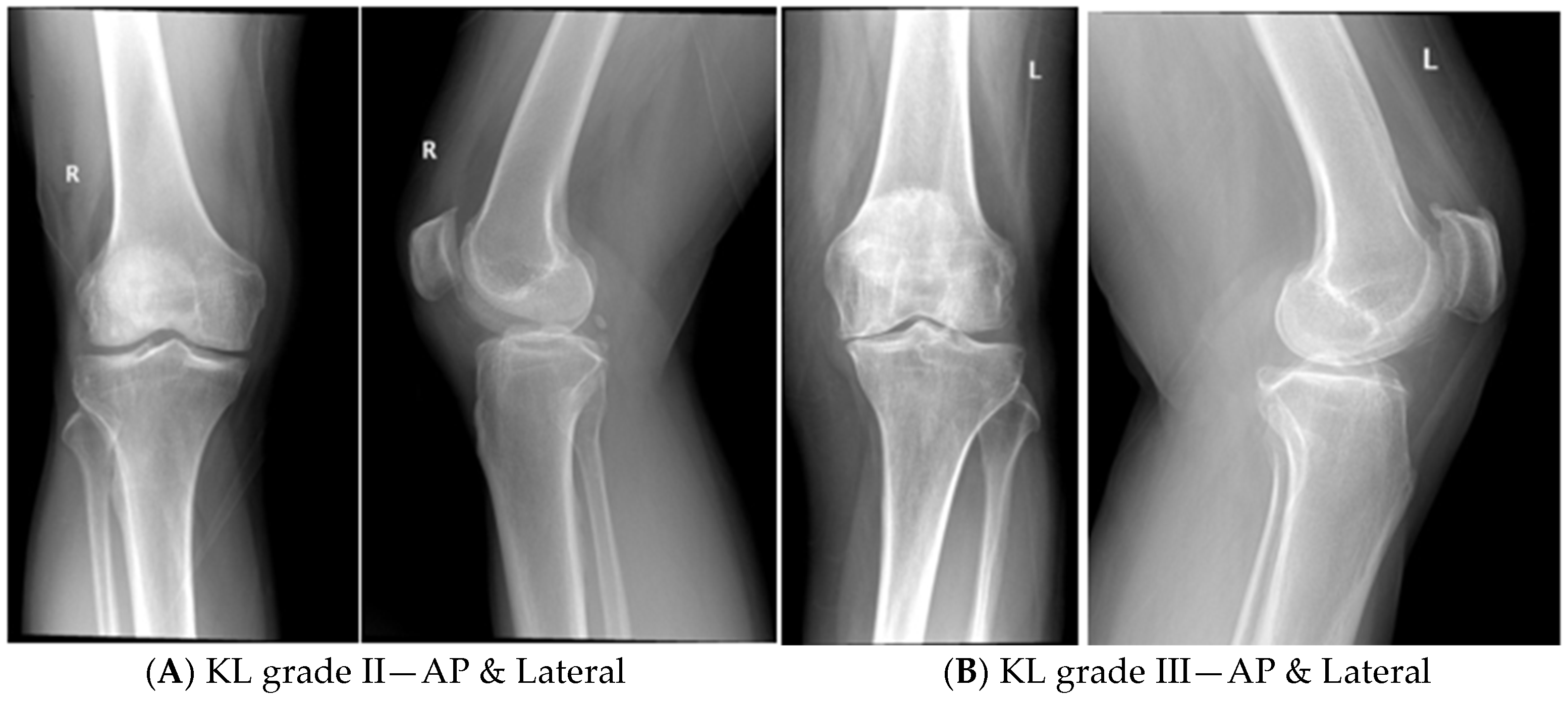
  • Patients of KOA with radiological grading of KL grade II and III.
  • Age group of the patients between 25 and 60 years of either gender.
  • Only patients with primary KOA were included in the study.

2.1.2. Exclusion Criteria

  • KOA patients with KL grade IV were excluded, as it has global cartilage loss with subchondral erosions and deformities.
  • KOA patients with deformities like varus/valgus/genu recurvatum ligament imbalance and any gross anatomical malalignment of the joints were also excluded.
  • Patients with secondary KOA and rheumatoid arthritis were also excluded.
  • Patients with metabolic disorders like hyperuricemia were excluded.

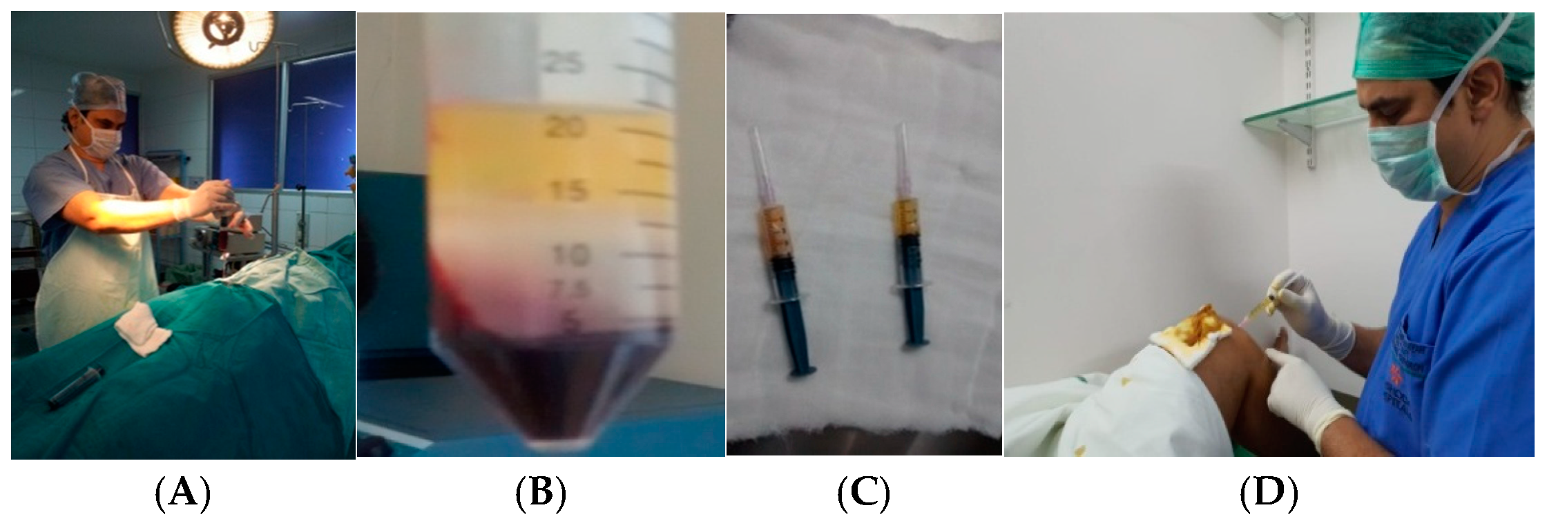
2.2. Procedure for Bone Marrow Aspiration

In all the patients, autologous bone marrow was aspirated under sterile aseptic conditions and harvested from the posterior iliac crest. In the operating room, the patient was positioned in a lateral position, the iliac wing was covered under the ‘O’ drape, and under local anesthesia, 60 mL of the bone marrow was aspirated using a 14-gauge bone marrow aspiration needle and collected into the falcon tubes, which were flushed with anticoagulant citrate dextrose (ACD). Aspiration was carried out by a single surgeon for all the cases (Figure 3A). In cases of patients with bilateral KOA, bone marrow was aspirated from both the iliac crests separately. The collected bone marrow was processed by the density gradient centrifugation method (Figure 3B).

Density Gradient Centrifugation Method for the Separation of Mononuclear Cells Enriched with Stem Cells

After the aspiration, bone marrow was transferred to sterile falcon tubes flushed with ACD and processed using the density gradient centrifugation method [22]. Ficoll and the sample were taken in 1:1 ratio and centrifuged. Total red blood cells (RBCs) were settled at the bottom and the plasma was separated on the top. The buffy coat (creamy middle layer) was aspirated. The cell suspension was washed twice with DPBS and the cell yield was counted. Approximately 7 mL of BMAC (final mononuclear cells enriched with stem cells) was obtained (Figure 3C). The obtained BMAC was injected into the knee joint through the antero-lateral portal using 20-gauge needle while the knee was in flexion under strict aseptic precautions. A compression bandage was applied in all the patients following the injection and post-injection knee Range of Movement (ROM) exercises were initiated for the uniform spread of BMAC. For all the patients, BMAC was injected by a single surgeon (Figure 3D). In all the patients, post-autologous BMAC injection selective COX2 inhibitors were advised for the relief of pain as and when required. All patients were advised of weight relieving exercises like stationary bike/cycling.

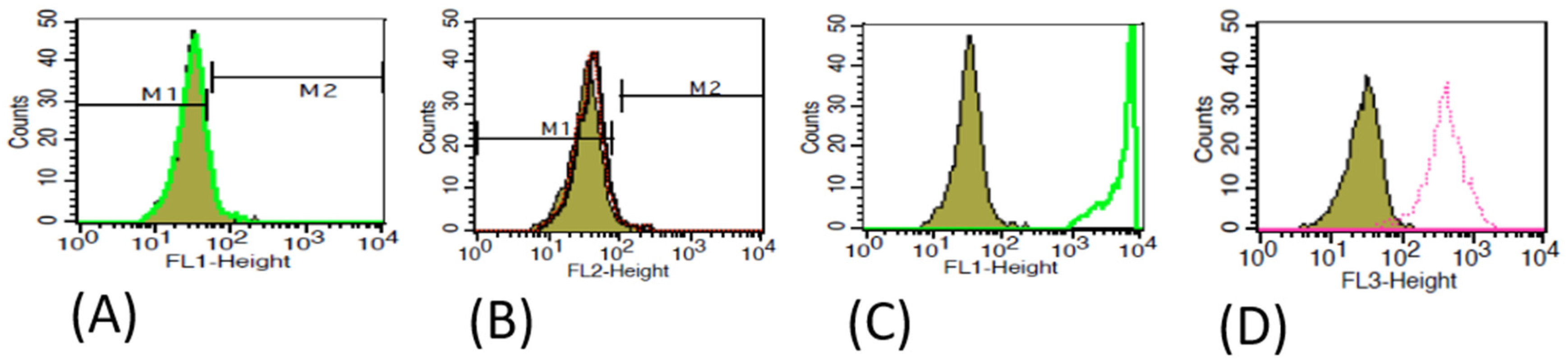
2.3. Characterization of MSCs

During the initial part of the study, we performed flowcytometry to characterize MSCs using specific Cluster of Differentiation (CD) markers (both positive and negative) in the final BMAC before injection into the patient [23]. Positive markers CD105, CD90 and negative markers CD34, CD45 were used. We found an average of 85–90% of positive CD markers in all the samples that were analyzed (Figure 4).

2.4. Primary Clinical Outcome Measurements

The primary clinical outcomes of the patients were evaluated by Visual Analogue Scale (VAS) and Western Ontario and McMaster Universities’ Osteoarthritis Index (WOMAC) index scores. A total of 132 patients were included in this study and evaluated based on the eligibility criteria. Post-injection, all patients were followed for one year, periodically at intervals of 3, 6, and 12 months. The patient outcome measurements were assessed by the following criteria.

2.4.1. Subjective Assessment: It Is Assessed by VAS and WOMAC Index Score

VAS: VAS indicates the degree of pain intensity. It is indicated from 1 to 10, where 1 indicates no pain, 2–4 is mild pain, and 5–7 is moderate pain, and above 7 is considered as severe pain.
WOMAC: WOMAC is widely used in the evaluation of Knee and Hip Osteoarthritis. WOMAC is a self-administered questionnaire to assess the sub-scale of pain, stiffness, and physical/functional activities, like sitting down, bending to the floor, walking on a flat surface, etc. The WOMAC Index for the patients was scored for 96 points, which is subdivided and summed up as follows: pain 0–20, stiffness 0–8, and physical function 0–68 (overall WOMAC Index scored for 98).

2.4.2. Objective Assessment

All patients underwent radiographic examination of both knees (weight bearing) from anterio-posterior and lateral views before BMAC injection and at the end of 12 months following the injection. Radiographs were captured and scored by the radiologist and Orthopedic Surgeon, blinded to the patient’s clinical information using the KL grade system. MRI was performed in 30% of patients to assess the cartilage regeneration. Due to financial constraints, MRI was not carried out for all the patients.
All the patients were clinically evaluated by the Orthopedic Surgeon pre- and post-injection periodically for treatment safety and adverse effects. None of the patients in the study group had any adverse effects.
All the patients were analyzed both subjectively and objectively at the end of 3, 6, and 12 months.

2.5. Statistical Analysis

Statistical analysis was performed using SPSS Inc Chicago, IL. All the continuous data were determined by mean, average, and standard deviation (SD), and standard error of mean (SEM) was also calculated using numerical variables. The main analysis was performed using VAS and WOMAC index scores to evaluate pain and functional outcomes and these were compared with the categorical variables such as percentages. An unpaired t-test was performed to understand the level of significance. The distribution of data was assessed using Chi square analysis; a p-value less than 0.05 (p ≤ 0.05) is considered as statistically significant. Patients with KL grade II and III were separately evaluated to understand the clinical outcomes.

3. Results

3.1. Baseline Characteristics of the Patients

The total number of patients included was 132 and the percentages of females and males were 70% (92) and 30% (40), respectively, in this study. The mean age of patients was 45 ± 8.8 years and the mean age of onset for KOA was 42 ± 08 years. The percentage of patients with unilateral and bilateral KOA was 80% (106) and 20% (26), respectively. The severity of KOA was scored by KL grading in all the patients; 50% (66) of them were in KL grade II, and the remaining 50% (66) were KL grade III (Table 1).

3.2. Primary Clinical Outcome

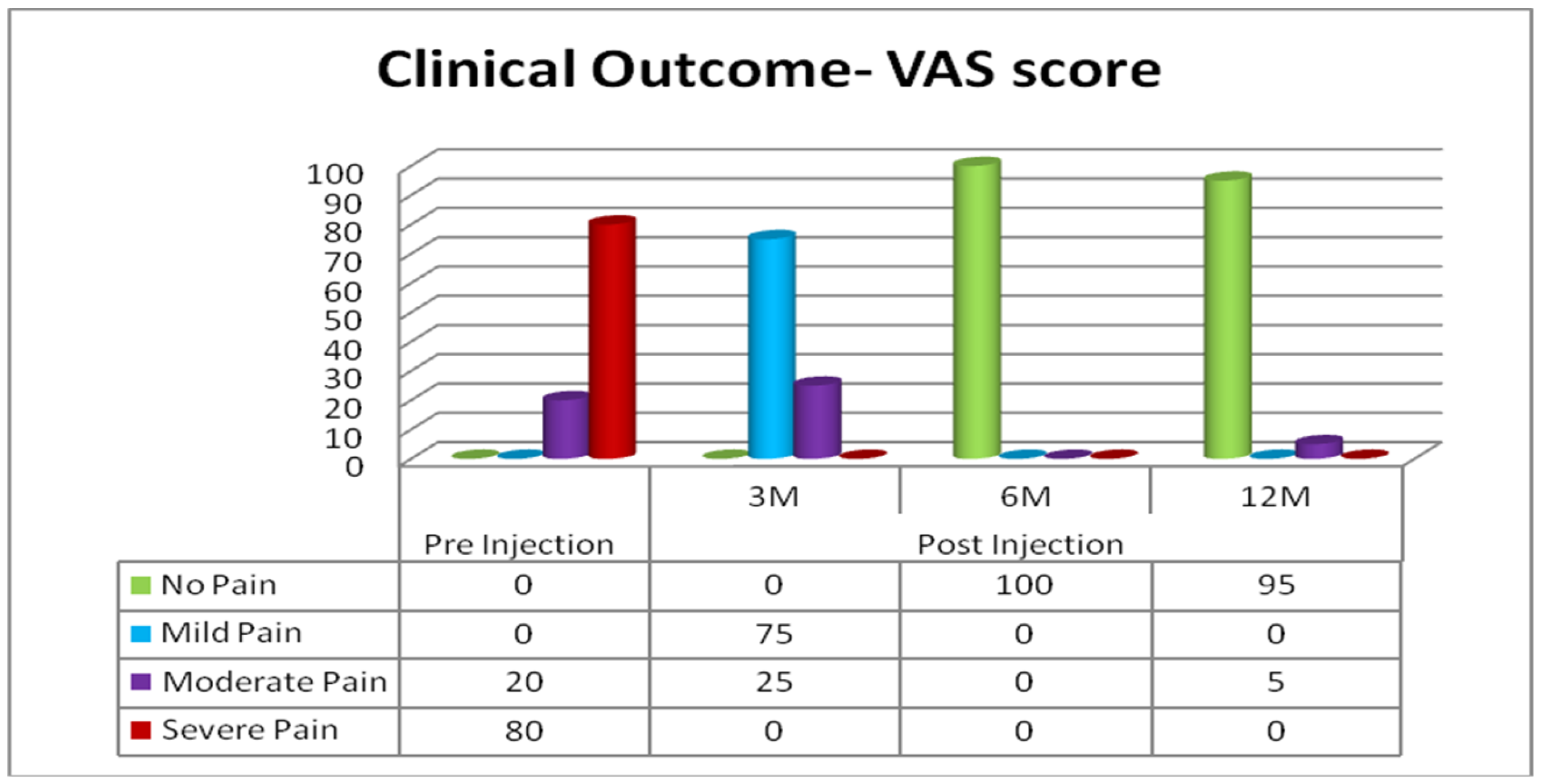
3.2.1. Subjective Assessment: Clinical Outcomes Based on VAS

All the patients with KL grade III had a pre-injection VAS score of 8 and 9 and were unable to attend to most of their daily activities, whereas 90% of the KL grade II had a pre-injection VAS score of 6 and 7, with difficulty in climbing stairs and performing high-demand activities for the knee, like squatting. After proper counseling and consent, a single autologous BMAC injection was administered to the patients. Post-procedure, all the patients were advised to engage in weight relieving exercises like stationary bike and swimming and to take pain medication as and when necessary (selective COX2 inhibitors). Following the BMAC injection, all patients showed improvement of pain as early as from the end of the 3rd month, and steady improvement in their VAS scores at the end of the 6th month. Their VAS scores reached as low as 0–1, which consistently lasted until the last follow-up, i.e., the 12th month (Figure 5).
In all 132 patients, improvement of pain was observed as early as at the end of the 3rd month following BMAC injection to an extent of 75%, compared to pre-injection by VAS, and steadily improved to almost complete relief of pain by the end of the 6th month. This pain relief lasted until the end of the 12th month (final follow-up) in 95% (125) of them, and the remaining 5% (7) of patients started experiencing pain (VAS 5 to 6) with high-demand activities like climbing stairs and squatting during the final follow-up. At the end of one year, 95% of the patients found complete pain relief and no individual was left with severe pain, indicating a significant outcome with this therapy/treatment (Figure 6).

3.2.2. Evaluation of Statistical Significance Using the VAS score

The mean VAS score in the pre-injection stage was 8.3 with an SD of 1.4 for KOA patients. The SEM was 0.12. The average and SD post-injection of KOA patients at the 12th month were 1.25 and 1.10, respectively, with an SEM of 0.09. An unpaired t-test was carried out and the results indicated that the two-tailed p-value is less than 0.0001, and the difference is considered to be extremely significant [95% confidence interval of this difference from 4.8712 to 9.2288] (Table 2).
The distribution of data is evaluated using Chi square with Yates correction pre-injection and at the end of the 12th month. The results indicated that the two tailed p-value is less than 0.001, which is considered to be extremely statistically significant (Chi square equals 225.987 with degree of freedom).

3.3. Subjective Assessment: Clinical Outcomes with WOMAC

WOMAC score was assessed pre-injection and post-injection at the 3rd, 6th, and 12th month for both KL grade II and III patients.
KOA patients with KL grade II: Initially, all patients had moderate WOMAC index score, and gradually, WOMAC improved post-injection. By the end of the 12th month, all patients had mild WOMAC scores.
KOA patients with KL grade III: In this group of patients, 25% of them had moderate WOMAC index and 75% had severe WOMAC score. In the periodic evaluation at different time points, WOMAC score improvement was significant; at the end of study follow-up (12th month), 90% of patients had a mild WOMAC index, and only 10% of cases remained at a moderate WOMAC index (Table 3).

3.3.1. Evaluation of Statistical Significance Using WOMAC

KL Grade II KOA Patients

The mean, SD, and SEM at pre-injection in KOA- KL grade II patients with WOMAC scores were 55.59, 10.44, and 1.28 respectively, whereas, post-injection at the 12th month, the mean, SD, and SEM of KOA-KL grade II patients were 21.60, 8.55, and 1.05.
An unpaired t-test was performed and the results indicated that the two-tailed p-value is less than 0.0001, and the difference is considered to be extremely significant [95% confidence interval of this difference from 30.1747 to 37.2653] (Table 4).

KL Grade III KOA Patients

The mean, SD, and SEM pre-injection in the KOA-KL grade III patients with WOMAC scores were 76.84, 14.33, and 1.76, respectively, whereas, post-injection at the 12th month, the mean, SD, and SEM of KOA-KL grade III patients were 25.31, 13.15, and 1.61.
An unpaired t-test was performed and the results indicated that the two-tailed p-value is less than 0.0001, and the difference is considered to be extremely significant [95% confidence interval of this difference from 26.2108 to 34.3492] (Table 5). The statistical analysis indicates a considerable improvement in the clinical outcomes from pre-injection to post-injection in KOA patients.

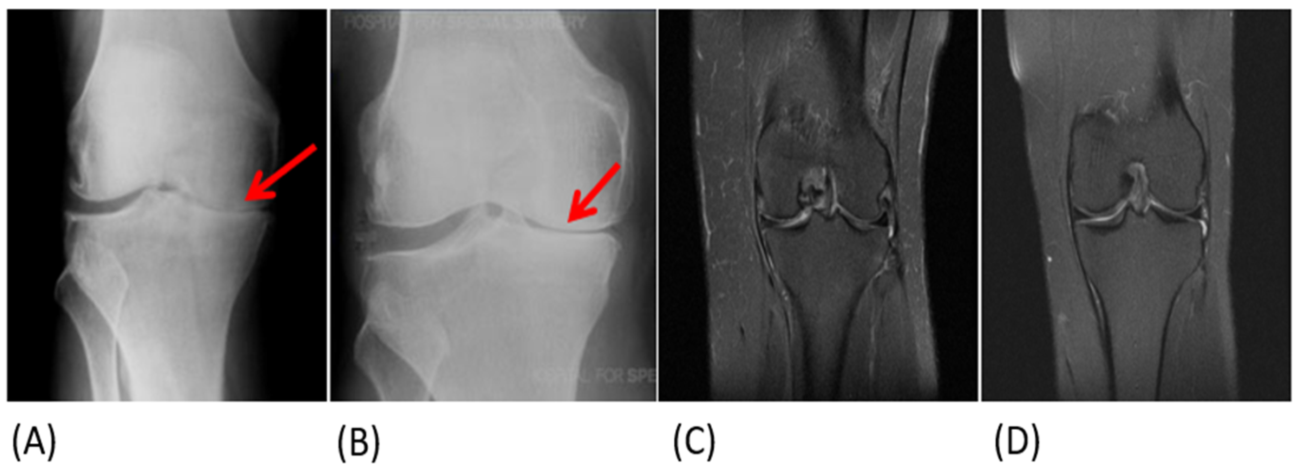
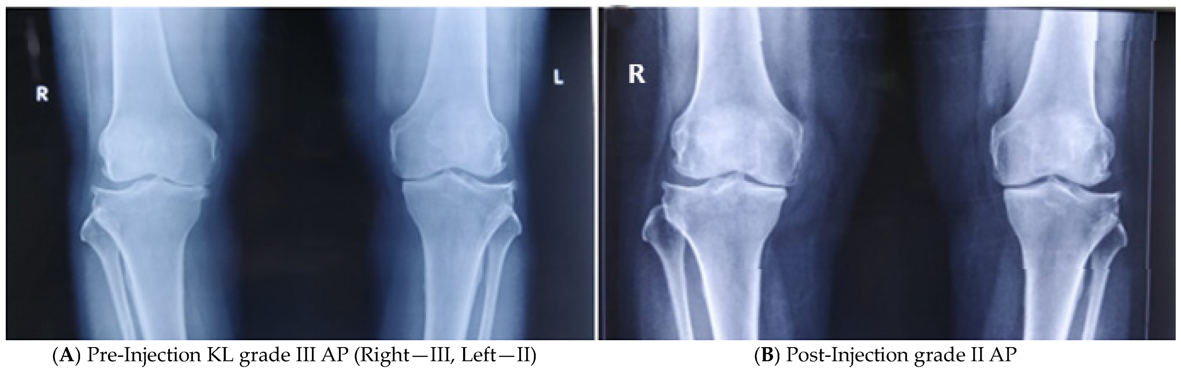
3.4. Objective Assessment

In all patients, weight-bearing knee radiographs (both AP and lateral) were taken pre-injection and at the end of the 12th month following the BMAC injection. At the end of the 12th month, after the procedure, all patients with KL grade II and 85% (56) of patients with KL grade III remained in the same KL grade, whereas 15% (10) of patients in KL grade III improved to KL grade II with the opening of the medial joint space, as shown in the final radiographs (Figure 7).
We have performed an MRI of the knee joint in 30% (40) of the patients before injection and at the end of the 12th month following BMAC injection. All patients in whom the MRI of the knee was performed belonged to KL grade III. Among these, 10 patients showed improvement of the cartilage regeneration, about 20 to 30%, at the end of the 12th month, as compared to pre-injection, based on the MRI. Due to financial constraints, MRI was not performed in all patients.

4. Discussion

BMAC has opened a new horizon in the treatment of KOA and may in future shift the paradigm in clinical practice. BMAC is an attention-drawing therapy which arose from the need to explore the new therapeutic possibilities for orthopedic-related problems [24,25]. Its advantage is the composition of multiple cell components, which are later notorious for their multi-lineage differentiation. BMAC can be applied to problems related to bone, cartilage, and tendon regeneration [26,27]. In the orthopedics field, diseases for which current treatment modalities do not offer satisfactory, efficient, or durable results may be targets of this therapy [6,28,29]. BMAC may provide an innovative, cost-effective, and reliable therapy for knee osteoarthritis [30,31].
Concentrated bone marrow aspirate can interpret cartilage matrix degradation driven by pro-inflammatory cytokines [15]. It is considered as a very minimal cell manipulation and allows one to obtain a product that can be used in clinical practice to treat cartilage lesion in one-step treatment [32,33]. BMAC serves as a powerful source of growth factors like transforming growth factor, platelet-derived growth factor, and bone morphogenic protein, which is important due to their anti-inflammatory and paracrine effect [34]. Also, because of its regenerative potential, we have selected BMAC for the KOA treatment.
During recent years, there has been an increasing interest in using intra-articular BMAC injection in the treatment of KOA. BMAC is essentially a combination of cellular components, soluble growth factors, and cytokines [35,36]. The cellular component of BMAC contains various MSCs, hemopoietic precursors, monocytes, and endothelial cells. MSCs exhibit immune-modulatory, anti-inflammatory, proliferative, and chemo-attractive functions [29]. Therefore, BMACs can induce the differentiation process of cellular regeneration in osteoarthritis of the knee joint [37]. Various growth factors present in BMAC, like TGF beta, VEGF, BMP2, IL 1ra, and platelet-derived growth factors work on the various cellular pathways essential in the regulation of cell catabolic pathways [15,38,39].
Therefore, the combination of cellular components and various biologically active proteins makes BMAC a unique Ortho biological treatment modality by not only reducing the pain in KOA patients, but also potentially altering disease progression [11,12,14].
The advantages of BMAC are ease of obtainability (aspiration from iliac crest), no need for culture and cell expansion, no risk or reduced risk of infection (point of source processing and injection), and no risk of allogeneic disease [40,41]. With all these advantages, the usage of BMAC as a treatment modality in KOA is rapidly growing and gaining popularity [14,15,42].
Earlier studies showed good results, where the bone marrow stem cells were used in KOA (tibiofemoral and patellofemoral joints) and isolated osteochondral lesions, with a follow-up of 12–24 months [16,43,44,45,46,47]. The latest studies in the literature have uniformly emphasized the safety and benefit of intra-articular injection of autologous BMAC in KOA patients [29,48].
In the current study, a single injection of intra-articular autologous BMAC was administered into the KOA joints in 132 patients (KL grade II and III), and we evaluated the functional outcomes both objectively and subjectively for a period of 12 months. We found that there is a uniform improvement of pain score and activity of daily living (quality of life) following the BMAC injection and sustained relief of pain even at the end of 12 months in 125 patients. In seven patients with KL grade III, persistence of knee pain with high-demand activities like stair climbing and squatting was observed, which required the occasional use of COX2 inhibitors for pain relief.
Ten (13%) patients out of one hundred thirty-two showed increased knee joint space and improved KL grade from III to II in the post-BMAC injection weight-bearing knee radiographs at the end of the 12th month, as compared to the pre-injection radiographs. The rest of the patients remained in the same KL grades. No adverse events were observed in any of the patients following BMAC injection. Therefore, the current study showed both subjective and objective improvement in all the patients following single intra-articular autologous BMAC injection in KL grade II and III KOA patients. A study from the literature, Themistocleous et al. 2018 [22], also evaluated the efficacy of BMAC, but in KL III and IV, and concluded that BMAC is a safe and reliable procedure for the clinical improvement of KOA; however, 5% of the study group proceeded further to total knee orthoplasty. The current study is superior in terms of clinical evaluation, as it utilized both VAS and WOMAC indexes periodically, which is a limitation of the above study. Sample size is also comparatively high. Radiological assessment is one of the advantages of this study in terms of evaluating cartilage regeneration. None of the patients further proceeded to total knee orthoplasty in the current study.
Other studies from the literature [12,15,49,50] also conducted BMAC injection for KOA patients; however, our study’s follow-up duration was long, i.e., 12 months, whereas other studies (mostly) followed up for 6 months (Table 6). A recent study by Anz et al. (2020) [51] was almost similar, but in the current study, both subjective assessment by VAS and WOMAC scores was carried out for a period of 12 months, and objective assessment by X-rays and MRI was a novel feature of this study. No patient had any adverse effects following the injection. Standing radiographs of the knee joint were recommended for all patients; however, due to the cost constraints, MRI was only performed in 30% of patients. The patient selection for MRI was carried out randomly and also ruling out any mechanical problems, like meniscal tear.
  • The unique features/highlights of the current study
In the current study, we recruited KOA patients with KL grade II and III, but in most of the studies in the literature, only KL grades I and II were included. The number of patients was equal in both KL grade II and III. We have given a single injection of autologous BMAC harvested from the iliac crest. Post-injection, all patients were followed both subjectively and objectively with weight-bearing knee radiographs, and in 30% of patients, knee MRI was performed to determine the cartilage regeneration, whereas in most of the studies, only a pain evaluation was carried out post-injection.
  • Limitations
Despite encouraging results, there are some limitations in the current study, like the smaller sample size, absence of a control group, lack of instrumental monitoring with MRI studies for all the patients, and follow-up of only one year. As always, a large sample size would provide better insight into the outcome of the study. A control group for any study strengthens the results. If MRI were performed for all patients, cartilage regeneration could be evaluated in a better way. The amount of pain relief and functional improvement of the knee joint could be evaluated in a better way if the follow-up period were longer.

5. Conclusions

BMAC therapy has potential implications in the treatment of KL grade II and III KOA in terms of pain reduction, avoiding the need of analgesics and slowing the degenerative process and regeneration in the diseased cartilage with satisfactory, efficient, and durable results. It can be a safe alternative therapy with early intervention option. BMAC therapy can leverage autologous components to address symptoms and potentially modify the disease trajectory. This preliminary study showed promising results; however, the study could be performed again in a larger cohort and with different ethnic groups for better understanding. To the best of our knowledge, this is the first study of its kind in India. Our future research will focus on longitudinal studies for evaluating long-term outcomes and on planning a more evidence-based approach to the regenerative management of KOA.

Author Contributions

Conceptualization: K.S. and S.P.; methodology: K.S., S.P. and S.K.; formal analysis, K.S. and S.P.; investigation, K.S.; data curation, K.S. and S.P.; writing—original draft preparation: S.P.; writing—review and editing, K.S., S.P. and Q.H.; visualization, K.S., S.K. and Q.H.; supervision, K.S.; project administration, K.S.; All authors have read and agreed to the published version of the manuscript.

Funding

This research received no external funding.

Institutional Review Board Statement

Ethical approval (ECR/58/Inst/AP/2013/RR-19; 19 April 2012) was obtained from the Institutional Ethics committee, Kamineni Hospitals, Hyderabad, India.

Informed Consent Statement

Informed consent was obtained from all patients involved in the study.

Data Availability Statement

The data can be obtained from the corresponding author upon request.

Acknowledgments

The authors are thankful to all the study participants and their family members, as well as hospital and laboratory staff. Sincere thanks to the management of Kamineni and Yashoda hospitals, Hyderabad for their continuous support.

Conflicts of Interest

The authors declare no conflicts of interest.

References

  1. Sharma, L. Osteoarthritis of the Knee. N. Engl. J. Med. 2021, 384, 51–59. [Google Scholar] [CrossRef]
  2. Azad, C.S.; Singh, A.; Pandey, P.; Gambhir, I.S. Osteoarthritis in India: An epidemiologic aspect. Int. J. Recent Sci. Res. 2011, 8, 20918–20922. [Google Scholar]
  3. Mora, J.C.; Przkora, R.; Cruz-Almeida, Y. Knee osteoarthritis: Pathophysiology and current treatment modalities. J. Pain Res. 2018, 11, 2189. [Google Scholar] [CrossRef]
  4. Lim, W.B.; Al-Dadah, O. Conservative treatment of knee osteoarthritis: A review of the literature. World J. Orthop. 2022, 13, 212. [Google Scholar] [CrossRef]
  5. Primorac, D.; Molnar, V.; Matišić, V.; Hudetz, D.; Jeleč, Ž.; Rod, E.; Čukelj, F.; Vidović, D.; Vrdoljak, T.; Dobričić, B.; et al. Comprehensive review of knee osteoarthritis pharmacological treatment and the latest professional societies’ guidelines. Pharmaceuticals 2021, 14, 205. [Google Scholar] [CrossRef]
  6. Belk, J.W.; Lim, J.J.; Keeter, C.; McCulloch, P.C.; Houck, D.A.; McCarty, E.C.; Frank, R.M.; Kraeutler, M.J. Patients with knee osteoarthritis who receive platelet-rich plasma or bone-marrow aspirate concentrate injections have better outcomes than patients who receive hyaluronic acid: Systematic review and meta-analysis. Arthrosc. J. Arthrosc. Relat. Surg. 2023, 39, 1714–1734. [Google Scholar] [CrossRef]
  7. Dulic, O.; Rasovic, P.; Lalic, I.; Kecojevic, V.; Gavrilovic, G.; Abazovic, D.; Maric, D.; Miskulin, M.; Bumbasirevic, M. Bone marrow aspirate concentrate versus platelet rich plasma or hyaluronic acid for the treatment of knee osteoarthritis. Medicina 2021, 57, 1193. [Google Scholar] [CrossRef]
  8. Keeling, L.E.; Belk, J.W.; Kraeutler, M.J.; Kallner, A.C.; Lindsay, A.; McCarty, E.C.; Postma, W.F. Bone marrow aspirate concentrate for the treatment of knee osteoarthritis: A systematic review. Am. J. Sports Med. 2022, 50, 2315–2323. [Google Scholar] [CrossRef]
  9. Bolia, I.K.; Bougioukli, S.; Hill, W.J.; Trasolini, N.A.; Petrigliano, F.A.; Lieberman, J.R.; Weber, A.E. Clinical efficacy of bone marrow aspirate concentrate versus stromal vascular fraction injection in patients with knee osteoarthritis: A systematic review and meta-analysis. Am. J. Sports Med. 2022, 50, 1451–1461. [Google Scholar] [CrossRef]
  10. Al-Najar, M.; Khalil, H.; Al-Ajlouni, J.; Al-Antary, E.; Hamdan, M.; Rahmeh, R.; Alhattab, D.; Samara, O.; Yasin, M.; Abdullah, A.A.; et al. Intra-articular injection of expanded autologous bone marrow mesenchymal cells in moderate and severe knee osteoarthritis is safe: A phase I/II study. J. Orthop. Surg. Res. 2017, 12, 190. [Google Scholar] [CrossRef]
  11. Kon, E.; Boffa, A.; Andriolo, L.; Di Martino, A.; Di Matteo, B.; Magarelli, N.; Marcacci, M.; Onorato, F.; Trenti, N.; Zaffagnini, S.; et al. Subchondral and intra-articular injections of bone marrow concentrate are a safe and effective treatment for knee osteoarthritis: A prospective, multi-center pilot study. Knee Surg. Sports Traumatol. Arthrosc. 2021, 29, 4232–4240. [Google Scholar] [CrossRef]
  12. Mautner, K.; Bowers, R.; Easley, K.; Fausel, Z.; Robinson, R. Functional outcomes following microfragmented adipose tissue versus bone marrow aspirate concentrate injections for symptomatic knee osteoarthritis. Stem Cells Transl. Med. 2019, 8, 1149–1156. [Google Scholar] [CrossRef]
  13. Wells, K.; Klein, M.; Hurwitz, N.; Santiago, K.; Cheng, J.; Abutalib, Z.; Beatty, N.; Lutz, G. Cellular and clinical analyses of autologous bone marrow aspirate injectate for knee osteoarthritis: A pilot study. PM&R 2021, 13, 387–396. [Google Scholar]
  14. Betzler, B.K.; Bin Muhammad Ridzwan Chew, A.H.; Bin Abd Razak, H.R. Intra-articular injection of orthobiologics in patients undergoing high tibial osteotomy for knee osteoarthritis is safe and effective—A systematic review. J. Exp. Orthop. 2021, 8, 83. [Google Scholar] [CrossRef]
  15. Cavallo, C.; Boffa, A.; Andriolo, L.; Silva, S.; Grigolo, B.; Zaffagnini, S.; Filardo, G. Bone marrow concentrate injections for the treatment of osteoarthritis: Evidence from preclinical findings to the clinical application. Int. Orthop. 2021, 45, 525–538. [Google Scholar] [CrossRef]
  16. Ding, W.; Xu, Y.Q.; Zhang, Y.; Li, A.X.; Qiu, X.; Wen, H.J.; Tan, H.B. Efficacy and safety of intra-articular cell-based therapy for osteoarthritis: Systematic review and network meta-analysis. Cartilage 2021, 13, 104S–115S. [Google Scholar] [CrossRef]
  17. Silva, S.; Andriolo, L.; Boffa, A.; Di Martino, A.; Reale, D.; Vara, G.; Miceli, M.; Cavallo, C.; Grigolo, B.; Zaffagnini, S.; et al. Prospective double-blind randomised controlled trial protocol comparing bone marrow aspirate concentrate intra-articular injection combined with subchondral injection versus intra-articular injection alone for the treatment of symptomatic knee osteoarthritis. BMJ Open 2022, 12, e062632. [Google Scholar]
  18. Rodríguez-Merchán, E.C. Intraarticular injections of mesenchymal stem cells in knee osteoarthritis: A review of their current molecular mechanisms of action and their efficacy. Int. J. Mol. Sci. 2022, 23, 14953. [Google Scholar] [CrossRef]
  19. Im, G.I. Clinical use of stem cells in orthopaedics. Eur. Cell Mater. 2017, 33, 183–196. [Google Scholar] [CrossRef]
  20. Gursoy, S.; Akkaya, M.; Simsek, M.E.; Bozkurt, M. Functional outcomes of bone marrow aspirate concentrate application in osteoarthritis of the knee. Int. J. Res. Med. Sci. 2019, 7, 587–591. [Google Scholar] [CrossRef]
  21. Kim, G.B.; Seo, M.S.; Park, W.T.; Lee, G.W. Bone marrow aspirate concentrate: Its uses in osteoarthritis. Int. J. Mol. Sci. 2020, 21, 3224. [Google Scholar] [CrossRef]
  22. Themistocleous, G.S.; Chloros, G.D.; Kyrantzoulis, I.M.; Georgokostas, I.A.; Themistocleous, M.S.; Papagelopoulos, P.J.; Savvidou, O.D. Effectiveness of a single intra-articular bone marrow aspirate concentrate (BMAC) injection in patients with grade 3 and 4 knee osteoarthritis. Heliyon 2018, 4, e00871. [Google Scholar] [CrossRef]
  23. Sundberg, M.; Jansson, L.; Ketolainen, J.; Pihlajamäki, H.; Suuronen, R.; Skottman, H.; Inzunza, J.; Hovatta, O.; Narkilahti, S. CD marker expression profiles of human embryonic stem cells and their neural derivatives, determined using flow-cytometric analysis, reveal a novel CD marker for exclusion of pluripotent stem cells. Stem Cell Res. 2009, 2, 113–124. [Google Scholar] [CrossRef]
  24. Granero-Molto, F.; Weis, J.A.; Longobardi, L.; Spagnoli, A. Role of mesenchymal stem cells in regenerative medicine: Application to bone and cartilage repair. Expert Opin. Biol. Ther. 2008, 8, 255–268. [Google Scholar] [CrossRef]
  25. Davatchi, F.; Abdollahi, B.S.; Mohyeddin, M.; Shahram, F.; Nikbin, B. Mesenchymal stem cell therapy for knee osteoarthritis. Preliminary report of four patients. Int. J. Rheum. Dis. 2011, 14, 211–215. [Google Scholar] [CrossRef]
  26. Boffa, A.; Di Martino, A.; Andriolo, L.; De Filippis, R.; Poggi, A.; Kon, E.; Zaffagnini, S.; Filardo, G. Bone marrow aspirate concentrate injections provide similar results versus viscosupplementation up to 24 months of follow-up in patients with symptomatic knee osteoarthritis. A randomized controlled trial. Knee Surg. Sports Traumatol. Arthrosc. 2022, 30, 3958–3967. [Google Scholar] [CrossRef]
  27. McCarrel, T.; Fortier, L. Temporal growth factor release from platelet-rich plasma, trehalose lyophilized platelets, and bone marrow aspirate and their effect on tendon and ligament gene expression. J. Orthop. Res. 2009, 27, 1033–1042. [Google Scholar] [CrossRef]
  28. Sampson, S.; Botto-van Bemden, A.; Aufiero, D. Stem cell therapies for treatment of cartilage and bone disorders: Osteoarthritis, avascular necrosis, and non-union fractures. PM&R 2015, 7, S26–S32. [Google Scholar]
  29. Pintore, A.; Notarfrancesco, D.; Zara, A.; Oliviero, A.; Migliorini, F.; Oliva, F.; Maffulli, N. Intra-articular injection of bone marrow aspirate concentrate (BMAC) or adipose-derived stem cells (ADSCs) for knee osteoarthritis: A prospective comparative clinical trial. J. Orthop. Surg. Res. 2023, 18, 350. [Google Scholar] [CrossRef]
  30. Im, G.I. Regeneration of articular cartilage using adipose stem cells. J. Biomed. Mater. Res. Part A 2016, 104, 1830–1844. [Google Scholar] [CrossRef]
  31. Vinatier, C.; Bouffi, C.; Merceron, C.; Gordeladze, J.; Brondello, J.M.; Jorgensen, C.; Weiss, P.; Guicheux, J.; Noël, D. Cartilage tissue engineering: Towards a biomaterial-assisted mesenchymal stem cell therapy. Curr. Stem Cell Res. Ther. 2009, 4, 318–329. [Google Scholar] [CrossRef]
  32. Woodell-May, J.; Steckbeck, K.; King, W. Potential Mechanism of Action of Current Point-of-Care Autologous Therapy Treatments for Osteoarthritis of the Knee—A Narrative Review. Int. J. Mol. Sci. 2021, 22, 2726. [Google Scholar] [CrossRef]
  33. Hernigou, J.; Vertongen, P.; Rasschaert, J.; Hernigou, P. Role of scaffolds, subchondral, intra-articular injections of fresh autologous bone marrow concentrate regenerative cells in treating human knee cartilage lesions: Different approaches and different results. Int. J. Mol. Sci. 2021, 22, 3844. [Google Scholar] [CrossRef]
  34. Chahla, J.; Mannava, S.; Cinque, M.E.; Geeslin, A.G.; Codina, D.; LaPrade, R.F. Bone marrow aspirate concentrate harvesting and processing technique. Arthrosc. Tech. 2017, 6, e441-5. [Google Scholar] [CrossRef]
  35. Jeyaraman, M.; Bingi, S.K.; Muthu, S.; Jeyaraman, N.; Packkyarathinam, R.P.; Ranjan, R.; Sharma, S.; Jha, S.K.; Khanna, M.; Rajendran, S.N.; et al. Impact of the process variables on the yield of mesenchymal stromal cells from bone marrow aspirate concentrate. Bioengineering 2022, 9, 57. [Google Scholar] [CrossRef]
  36. El-Kadiry, A.E.; Lumbao, C.; Salame, N.; Rafei, M.; Shammaa, R. Bone marrow aspirate concentrate versus platelet-rich plasma for treating knee osteoarthritis: A one-year non-randomized retrospective comparative study. BMC Musculoskelet. Disord. 2022, 23, 23. [Google Scholar] [CrossRef]
  37. Anzillotti, G.; Conte, P.; Di Matteo, B.; Bertolino, E.M.; Marcacci, M.; Kon, E. Injection of biologic agents for treating severe knee osteoarthritis: Is there a chance for a good outcome? A systematic review of clinical evidence. Eur. Rev. Med. Pharmacol. Sci. 2022, 26, 5447–5459. [Google Scholar]
  38. Baria, M.; Pedroza, A.; Kaeding, C.; Durgam, S.; Duerr, R.; Flanigan, D.; Borchers, J.; Magnussen, R. Platelet-rich plasma versus microfragmented adipose tissue for knee osteoarthritis: A randomized controlled trial. Orthop. J. Sports Med. 2022, 10, 23259671221120678. [Google Scholar] [CrossRef]
  39. Weber, A.E.; Bolia, I.K.; Trasolini, N.A. Biological strategies for osteoarthritis: From early diagnosis to treatment. Int. Orthop. 2021, 45, 335–344. [Google Scholar] [CrossRef]
  40. Chahla, J.; Dean, C.S.; Moatshe, G.; Pascual-Garrido, C.; Serra Cruz, R.; LaPrade, R.F. Concentrated Bone Marrow Aspirate for the Treatment of Chondral Injuries and Osteoarthritis of the Knee: A Systematic Review of Outcomes. Orthop. J. Sports Med. 2016, 4, 2325967115625481. [Google Scholar] [CrossRef]
  41. Lopa, S.; Colombini, A.; Moretti, M.; de Girolamo, L. Injective mesenchymal stem cell-based treatments for knee osteoarthritis: From mechanisms of action to current clinical evidences. Knee Surg. Sports Traumatol. Arthrosc. 2019, 27, 2003–2020. [Google Scholar] [CrossRef]
  42. Wakitani, S.; Nawata, M.; Tensho, K.; Okabe, T.; Machida, H.; Ohgushi, H. Repair of articular cartilage defects in the patello-femoral joint with autologous bone marrow mesenchymal cell transplantation: Three case reports involving nine defects in five knees. J. Tissue Eng. Regen. Med. 2007, 1, 74–79. [Google Scholar] [CrossRef]
  43. Haleem, A.M.; Singergy, A.A.E.; Sabry, D.; Atta, H.M.; Rashed, L.A.; Chu, C.R.; El Shewy, M.T.; Azzam, A.; Aziz, M.T.A. The clinical use of human culture–expanded autologous bone marrow mesenchymal stem cells transplanted on platelet-rich fibrin glue in the treatment of articular cartilage defects: A pilot study and preliminary results. Cartilage 2010, 1, 253–261. [Google Scholar] [CrossRef]
  44. Kasemkijwattana, C.; Hongeng, S.; Kesprayura, S.; Rungsinaporn, V.; Chaipinyo, K.; Chansiri, K. Autologous bone marrow mesenchymal stem cells implantation for cartilage defects: Two cases report. J. Med. Assoc. Thail. 2011, 94, 395. [Google Scholar]
  45. Giannini, S.; Buda, R.; Vannini, F.; Cavallo, M.; Grigolo, B. One-step bone marrow-derived cell transplantation in talar osteochondral lesions. Clin. Orthop. Relat. Res. 2009, 467, 3307–3320. [Google Scholar] [CrossRef]
  46. Giannini, S.; Buda, R.; Cavallo, M.; Ruffilli, A.; Cenacchi, A.; Cavallo, C.; Vannini, F. Cartilage repair evolution in post-traumatic osteochondral lesions of the talus: From open field autologous chondrocyte to bone-marrow-derived cells transplantation. Injury 2010, 41, 1196–1203. [Google Scholar] [CrossRef]
  47. Gobbi, A.; Karnatzikos, G.; Scotti, C.; Mahajan, V.; Mazzucco, L.; Grigolo, B. One-step cartilage repair with bone marrow aspirate concentrated cells and collagen matrix in full-thickness knee cartilage lesions: Results at 2-year follow-up. Cartilage 2011, 2, 286–299. [Google Scholar] [CrossRef]
  48. Pabinger, C.; Lothaller, H.; Kobinia, G.S. Intra-articular injection of bone marrow aspirate concentrate (mesenchymal stem cells) in KL grade III and IV knee osteoarthritis: 4 year results of 37 knees. Sci. Rep. 2024, 14, 2665. [Google Scholar] [CrossRef]
  49. Shapiro, S.A.; Kazmerchak, S.E.; Heckman, M.G.; Zubair, A.C.; O’Connor, M.I. A prospective, single-blind, placebo-controlled trial of bone marrow aspirate concentrate for knee osteoarthritis. Am. J. Sports Med. 2017, 45, 82–90. [Google Scholar] [CrossRef]
  50. Garay-Mendoza, D.; Villarreal-Martínez, L.; Garza-Bedolla, A.; Pérez-Garza, D.M.; Acosta-Olivo, C.; Vilchez-Cavazos, F.; Diaz-Hutchinson, C.; Gómez-Almaguer, D.; Jaime-Pérez, J.C.; Mancías-Guerra, C. The effect of intra-articular injection of autologous bone marrow stem cells on pain and knee function in patients with osteoarthritis. Int. J. Rheum. Dis. 2018, 21, 140–147. [Google Scholar] [CrossRef]
  51. Anz, A.W.; Hubbard, R.; Rendos, N.K.; Everts, P.A.; Andrews, J.R.; Hackel, J.G. Bone marrow aspirate concentrate is equivalent to platelet-rich plasma for the treatment of knee osteoarthritis at 1 year: A prospective, randomized trial. Orthop. J. Sports Med. 2020, 8, 2325967119900958. [Google Scholar] [CrossRef]
Figure 1. Representative X-ray images of KL grade II (AP & Lateral) and KL grade III (AP & Lateral) of a KOA patient.
Biologics 04 00015 g001
Figure 2. Work flow chart of the current study.
Biologics 04 00015 g002
Figure 3. (A) Harvesting of bone marrow from patient; (B) separation of bone marrow aspirate; (C) separated BMAC; (D) injection into the knee joint.
Biologics 04 00015 g003
Figure 4. Flowcytometric analysis of BMAC for both negative (A—CD34; B—CD45) and positive markers (C—CD105; D—CD90).
Biologics 04 00015 g004
Figure 5. (A) X-ray image of a patient pre-injection with VAS score 8 (gross reduction of medial joint space); (B) X-ray image of a patient post-injection with VAS score 2 to 3 (improved medial joint space indicating cartilage regeneration); (C) MR image of knee pre-injection showing KL grade III; (D) MR image of knee post-injection showing KL grade II [all the images are of same patient]. The red arrow is showing improvement to the clinical outcome.
Biologics 04 00015 g005
Figure 6. The percentage of clinical outcome of KOA patients based on VAS.
Biologics 04 00015 g006
Figure 7. X-ray of both weight-bearing knees in a 52-year-old female, with right knee KL grade III pre-injection and post-injection KL grade II after 12-month follow-up, indicating improvement in KL grading in right knee from grade III to II, while the left KL grade II was maintained (same patient).
Biologics 04 00015 g007
Table 1. Demographic details of patients included in the study.
CharactersCases (n = 132)
Age (Years)45 ± 8.8
Sex: (M:F)92:40
Height (cm)155.7 ± 3.1
Weight (kg)74.7 ± 8.7
BMI (kg/m2)30.8 ± 3.4
Age of Onset42 ± 8
Patients with Unilateral KOA80%
Patients with Bilateral KOA20%
Patients with KL grade II50%
Patients with KL grade III50%
Table 2. Mean, SD, and SEM of KOA patients with VAS scores at periodic intervals.
Pre-Injection 3 Months 6 Months 12 Months
Mean 8.33.731.001.25
SD 1.491.4901.10
SEM 0.120.1200.09
Table 3. Percentage of clinical outcome of KOA patients assessed with WOMAC Score.
KL Grade II (n = 66)KL Grade III (n = 66)
WOMAC Index Score
(0–96)
Pre-Injection Post-Injection Pre-Injection Post-Injection
3 Months 6 Months 12 Months 3 Months 6 Months 12 Months
Mild (<35)010010010006010090
Moderate (36–75)1000002540010
Severe (76–96) 000075000
Table 4. Mean, SD, and SEM of KOA KL grade II patients with WOMAC scores at periodic intervals.
Pre-Injection 3 Months 6 Months 12 Months
Mean 55.5922.3420.8321.60
SD 10.448.249.398.55
SEM 1.281.011.151.05
Table 5. Mean, SD, and SEM of KOA KL grade III patients with WOMAC scores at periodic intervals.
Pre-Injection 3 Months 6 Months 12 Months
Mean 76.8433.3621.8325.31
SD 14.3317.158.2613.15
SEM 1.762.111.011.61
Table 6. Clinical studies from literature and their outcomes with mean WOMAC and mean VAS scores.
AuthorsCell TypeDosage Study Duration (Months)No. of JointsPre-Injection (WOMAC)3 Months (WOMAC)6 Months (WOMAC)12 Months (WOMAC)Pre-Injection (VAS)3 Months (VAS)6 Months (VAS)12 Months (VAS)
Shapiro 2017 [49] BMAC6mL 625NA NA NA NA31.015.011.0NA
Garay Mendoza 2018 [50]BMAC10 mL62635.97.928.07.952.79.242.8NA
Mautner 2019 [12] BMAC8 mL 658NANANANA392514NA
Anz 2020 [51]BMAC7 mL 124535.319.415.919.4NANA NANA
Current StudyBMAC7 ml1215853.221.517.2186.823.82.63.2
NA: Not available.
Disclaimer/Publisher’s Note: The statements, opinions and data contained in all publications are solely those of the individual author(s) and contributor(s) and not of MDPI and/or the editor(s). MDPI and/or the editor(s) disclaim responsibility for any injury to people or property resulting from any ideas, methods, instructions or products referred to in the content.

