Review

Sherpabodies—A Highly Versatile and Modular Scaffold for Biomedical Targeting

1
Department of Virology, University of Helsinki, FIN-00014 Helsinki, Finland
2
HUS Diagnostic Centre, HUSLAB, Clinical Microbiology, Helsinki University Hospital, 00290 Helsinki, Finland
*
Author to whom correspondence should be addressed.
Biologics 2025, 5(2), 13; https://doi.org/10.3390/biologics5020013
Submission received: 3 March 2025 / Revised: 17 April 2025 / Accepted: 20 April 2025 / Published: 23 April 2025
(This article belongs to the Section Protein Therapeutics)

Abstract

:
Sherpabodies are a novel class of antibody-mimetic proteins and represent the third generation of SH3 domain-based targeting scaffolds. Sherpabodies have several advantageous biophysical properties, and molecular libraries based on this scaffold provide a rich and facile source of high-quality binders against diverse target proteins of interest. Recent studies have successfully exploited sherpabodies for developing potent antivirals to block SARS-CoV-2 infection and for the advanced guiding of cancer cell killing by chimeric antigen receptor (CAR)-T cells, but many other applications for sherpabody-mediated targeting in biomedicine and biotechnology can be anticipated.

1. Non-Ig Scaffolds

Non-immunoglobulin-derived scaffold proteins have emerged as a versatile and powerful alternative to antibodies, offering significant advantages in numerous therapeutic, diagnostic, and biotechnological applications.
Protein scaffolds are modified to create antibody-mimetic binders by introducing novel binding surfaces into them through combinatorial engineering [1,2]. This involves generating large DNA libraries encoding scaffold proteins containing randomized residues introduced in their exposed loop regions or secondary structure elements. This is followed by the discovery of target-specific binders from such semisynthetic libraries using phage display or related screening methods, enabling high-throughput phenotype/genotype-coupled affinity selection [3,4].
Designed for high stability, modularity, and ease of production, these engineered proteins provide solutions to many of the limitations faced by traditional antibodies. Their small size provides several advantages, including enhanced tissue and tumor penetration. Unlike antibodies, which require costly mammalian cell expression systems, scaffold proteins can be efficiently produced in bacteria, simplifying manufacturing and reducing costs. Their compact, single-chain structure not involving disulfide bonds or glycosylation facilitates their biochemical handling and ensures consistent production. One of the valuable features of scaffold proteins is their modularity, allowing them to be easily fused to other proteins at various positions without disrupting the function of either partner. Additionally, scaffold proteins can be engineered as multimers to provide increased avidity or multi-specific targeting without encountering prohibitive problems with protein expression or solubility.
Early work paving the way to scaffold protein development involved the use of then-emergent combinatorial molecular biology techniques to improve natural binding properties of proteins, such as protease inhibitors [5,6,7,8], and grafting peptide aptamers into heterologous protein frameworks [9,10,11]. This led to the “patch engineering” of ectopic binding sites into suitable host proteins [12], and, subsequently, increasing attention was given to the overall functionality and relevant biophysical and pharmacokinetic properties of such retargeted binder proteins to optimize their utility as novel biologics. Today, over 20 types of scaffold proteins have been engineered as antibody-mimetic targeting tools, with their key features summarized in Table 1. Provided in this table is the earliest scientific reference describing the construction and use of each scaffold, as well as—when available—a more recent reference for a paper describing the current state and biomedical applications of this technology.

2. What Are SH3 Domains?

The Src Homology 3 (SH3) domains are a prominent class of modular interactions domains and serve to mediate inter- and intramolecular protein interactions [65,66]. The human proteome encodes approximately 300 SH3 domains, which occur in more than 200 different proteins (1 protein can contain up to 6 SH3 domains; Figure 1) that are typically involved in the regulation of cellular signal transduction, cytoskeletal organization, or membrane trafficking.
The SH3 domain is a small β-barrel class protein fold composed of a β-sandwich built of five strands connected by three loops and a short 310 helix. SH3 domains show favorable folding kinetics and, despite their small size and lack of disulfide bonds, have high thermal stability [67,68].
Human SH3 domains are composed of 55 or more amino acid residues depending on the length of the non-conserved loops between their β-strands. These loops, in particular the so-called RT-loop (between the β1 and β2 strand) and N-Src-loop (between the β2 and β3 strand) (Figure 2) are involved in providing binding specificity for SH3 domains. The core binding sites of natural SH3 ligands typically involve proline-rich peptides containing a PxxP consensus sequence [69].

3. History of SH3 Scaffold Engineering

The role of the SH3 loop regions in determining target selectivity was discovered through studies on how the HIV-1 pathogenicity factor Nef hijacks host cell regulation via binding to the SH3 domain of the tyrosine kinase Hck [70,71]. These findings inspired the construction of phage libraries of loop-randomized Hck SH3 domains initially used to generate high-affinity Nef inhibitors [23] and later to target other proteins [24]. Engineered Hck SH3 domains thus represent the first generation of SH3 scaffolds and also belong to one of the earliest classes of ligand-targeted non-Ig scaffold proteins reported before the turn of the millennium.
The use of the SH3 fold for biomedical targeting gained wider attention with the advent of Fynomers [40], which are derived from the SH3 domain of human Fyn, a tyrosine kinase closely related to Hck. While based on an SH3 backbone very similar to Hck, more extensive attention was paid to the Fynomers’ binding surface randomization strategy, which therefore can be considered as the second generation of SH3-based targeting scaffolds. The Swiss biotech Covagen (later acquired by Cilag GmbH International, Schaffhausen, Switzerland, a J&J/Janssen subsidiary) has targeted several proteins of therapeutic interest with Fynomers [72,73] and developed bispecific IgG-Fynomer fusion molecules (FynomAbs), including the anti-inflammatory TNF/IL-17A-targeting COVA322. This FynomAb was based on the marketed anti-TNF antibody adalimumab, which was modified by the addition of an IL-17A-targeted Fynomer at the C-termini of its light chains [74]. COVA322 reached a Phase Ib/IIa study for the treatment of psoriasis [74], but its development has been discontinued and no new Fynomer-based drugs have been announced in J&J’s pipeline.

4. Discovery of Sherpabodies

Sherpabodies (SH3-derived recombinant protein affinity) represent the third generation of SH3 domain-based targeting scaffold proteins. They are based on the SH3 domain of the human ciliary protein NPHP1 (nephrocystin), which is ubiquitously expressed in human tissues, including professional antigen-presenting cells of the immune system [75,76]. The NPHP1 SH3 domain was selected as the foundation for sherpabodies after a thorough bioinformatic and experimental analysis of the human SH3 domain repertoire to identify the optimal non-Ig targeting scaffold [77].
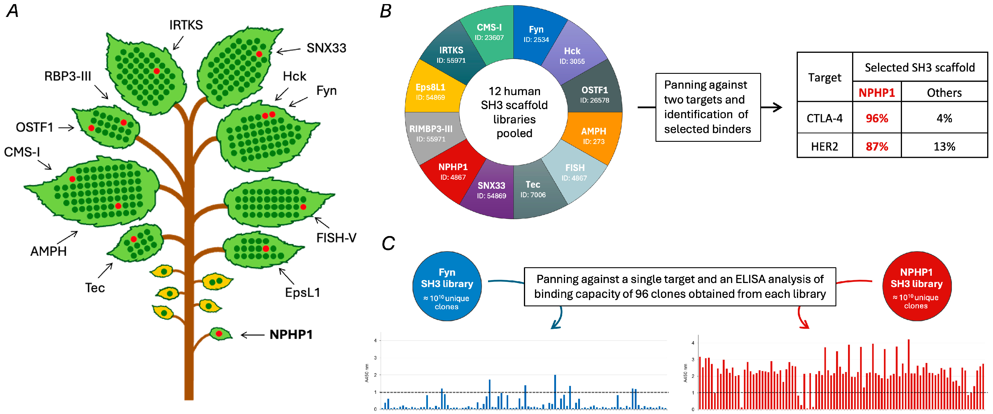
All human SH3 domains were first grouped into 14 subfamilies based on their sequence similarity (Figure 3A). From each subfamily, one or two SH3 domains were selected based on their prior identification as hits in a large proteomic study that screened a comprehensive phage library of natural human SH3 domains to characterize the SH3-mediated human interactome [78]. This ensured that all selected SH3 domains had the capacity to be efficiently displayed in a functional form on the surface of M13 phages and led to the exclusion of a few small SH3 subfamilies, together comprising only six members. Accordingly, 12 different human SH3 domains, including Hck and Fyn—the targeting scaffold of Fynomers—were chosen as templates for constructing large semisynthetic phage display libraries. The same strategy of loop randomization was applied to each of these 12 SH3 domains, where the corresponding RT- and N-src loop regions (identified by the conserved residues flanking these divergent regions) were both replaced with six random residues encoded by NNK nucleotides (where N is any nucleotide and K is G or T, thus encoding any of the 20 natural amino acids), and libraries containing more than 1010 unique phage clones were generated for all of them.
Strikingly, when a combinatorial phage library obtained by pooling these 12 SH3 scaffold libraries together was used for screening campaigns with two therapeutically relevant targets proteins, namely, HER2 and CTLA-4, the enriched clones after successive rounds of affinity panning were almost exclusively derived from the NPHP1 SH3 library (Figure 3B).
The superiority of the NPHP1 SH3 scaffold was also supported by studies where the NPHP1-derived library was used in parallel with a library based on the Fyn SH3 domain (the scaffold of Fynomers [40]) for screening against another target, namely, the NS1 protein of dengue virus type 2. When 96 randomly selected clones obtained from each library were analyzed, 94% of the NPHP1-library derived clones showed relevant binding against NS1 and the majority of these were strong binders, whereas only 7% of the clones from the Fyn SH3 library showed relevant binding and none were strong (Figure 3C).
Robust phage–ELISA signals shown for the NPHP1-derived binders in Figure 3 have been found to correlate with binding affinities in the 10−8 to 10−6 M range [63,64]. Thus, binders directly obtained from the NPHP1 library via only a few panning rounds are useful for diverse targeting applications, but further affinity maturation or biparatopic designs may be needed when a high affinity of binding is essential.
These data indicated that the NPHP1 SH3 scaffold was particularly well suited among the human SH3 domains for use as targeting scaffold. Accordingly, it was chosen as the template for generating additional large semisynthetic third-generation SH3 scaffold libraries, and the novel binders obtained from these libraries were named sherpabodies.
What distinguishes NPHP1 as a superior targeting scaffold compared to other SH3 domains remains an intriguing but unresolved question. As evident from Figure 3A, NPHP1 is not closely related to any other human SH3 domain. Its closest relative is the N-terminal SH3 domain of the Crk adaptor protein, showing only ≈40% amino acid identity with NPHP1 SH3. Nevertheless, the 3D structure of NPHP1 SH3 is very similar to other SH3 domains and, apart from the tips of the RT- and N-src loops, can be closely superimposed with them [79]. However, a distinctive feature of the NPHP1 SH3 domain structure is the unusual S-shaped conformation of its N-Src loop [79], which may contribute to an especially favorable presentation of the randomized residues introduced into this region. The NPHP1 SH3 domain shows remarkable thermal stability (Tm ≈ 80 °C; [80]), which is a highly desirable feature for a small targeting scaffold protein but not exceptional among the family of human SH3 domains [67,68]. Nevertheless, it is likely that a combination of a well-folding and stable SH3 core and a particularly suitable presentation of its RT- and N-src loops are key factors underlying the superior performance of the NPHP1 SH3 domain as a targeting scaffold. Collectively, these properties help to maximize the genuine functional diversity of the NPHP1 SH3-derived sherpabody library and ensure that the vast majority of its > 1010 clones represent viable binding candidates.

5. Biomedical Applications of Sherpabodies

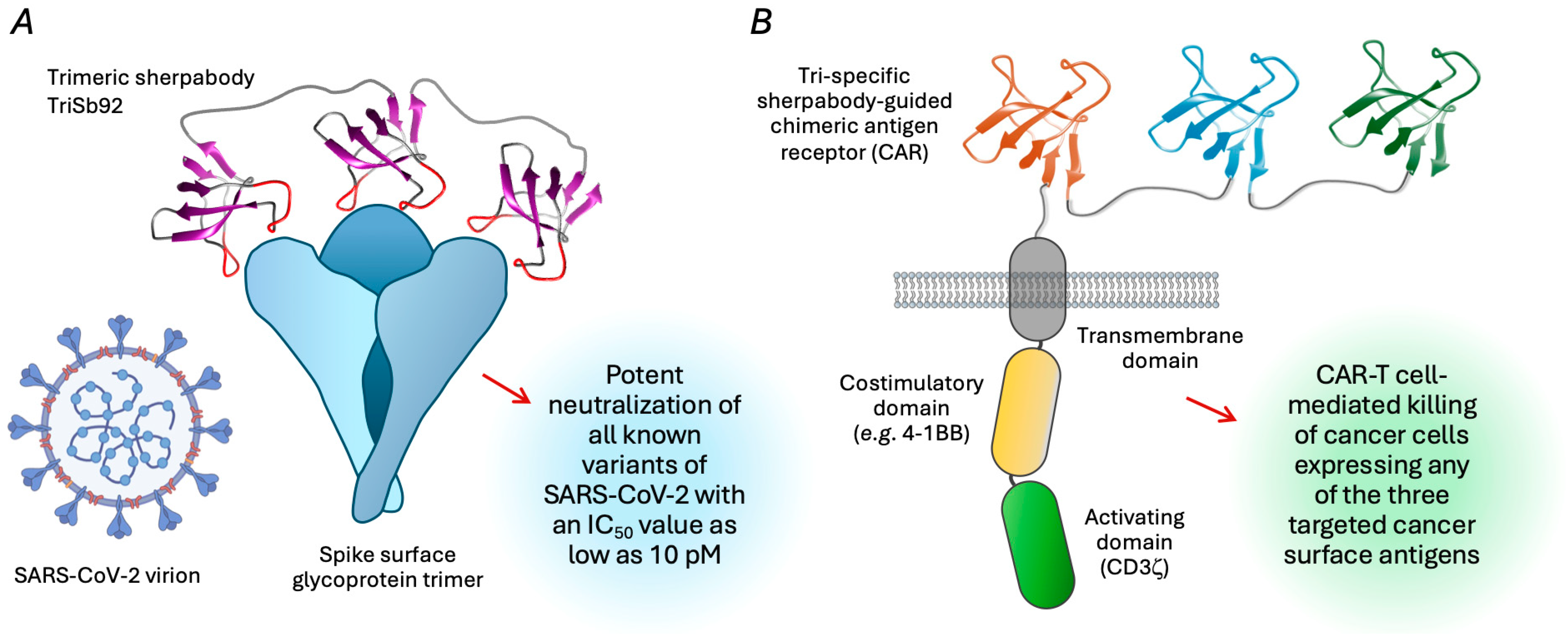
The modularity and facile targeting and manipulation properties of sherpabodies have been successfully exploited in very different types of biomedical targeting applications (Figure 4).

5.1. Multimeric Sherpabodies as Entry-Blocking Antiviral Proteins

The targeting of sherpabodies against a highly conserved site in the receptor-binding domain (RBD) of the SARS-CoV-2 spike glycoprotein gave rise to an antiviral inhibitor TriSb92 that could also potently neutralize the increasingly immunoevasive Omicron variants that recently emerging one after another [63]. The high potency of TriSb92 stems from its homotrimeric architecture, which, upon engaging with an RBD of each of the three protomers of the trimeric spike protein on SARS-CoV-2 virions, can neutralize the virus with half-maximal inhibitory concentration (IC50) values as low as 10 pM. This suggests a strong cooperativity in the binding and neutralizing action of the trimerized sherpabodies, as the binding affinity of an individual Sb92 unit of TriSb92 is estimated to be 30 nM. Remarkably, the intranasal application of TriSb92 was shown to provide several hours of pre- and post-exposure protection for mice challenged with different strains of SARS-CoV-2, including a fatal mouse-adapted strain [63].
This multimeric sherpabody protein demonstrated exceptional stability over time, as research-grade TriSb92 obtained via a single affinity purification step retained its full neutralization potential after storage for over 18 months at room temperature in a physiological salt buffer without protease inhibitors, stabilizers, or other additives [63]. This supports the potential of TriSb92 to be developed into an affordable, long-lasting nasal spray that can block SARS-CoV-2 transmission—a desirable scenario for everyone, but particularly for immunocompromised individuals who remain vulnerable to severe COVID-19 complications despite vaccination.
Thanks to its conserved binding site, TriSb92 was also found to be effective against the related SARS-CoV virus, which emerged as a global health threat during the 2002–2004 outbreak [63]. Therefore, TriSb92 could serve as a prophylactic emergency measure against future coronavirus pandemics, potentially arising from one of the many related sarbecoviruses currently circulating in bat populations.
On the other hand, several other pathogenic human viruses enter their target cells using multimeric surface glycoproteins that are analogous to the coronavirus spike [81] and would be attractive targets for developing similar antivirals based on multimeric sherpabodies. Apart from respiratory viruses, such as H5 avian influenza (see, e.g., Beukenhorst et al. [82]), rabies could be a worthwhile target, and blocking the function of its surface G protein with a sherpabody could replace expensive monoclonal antibodies used in combination with vaccination as a part of prophylaxis for exposed at-risk individuals (for current practices and challenges, see Sparrow et al. [83] and Fan et al. [84]). In addition to being much cheaper than antibodies and allowing storage at ambient temperatures, an anti-rabies sherpabody could better penetrate into tissues around the bite site due to its smaller size.

5.2. Precision Targeting of Cancer for Killing by CAR-T Cells

Chimeric Antigen Receptor (CAR)-T cell therapy involves genetically programming patients’ immune cells to attack cancer and has shown remarkable results in the treatment of blood cell malignancies [85]. However, one of the key obstacles in extending the success of CAR-T cells to therapy for solid cancers has been the lack of unique surface markers that can distinguish cancerous cells from normal cells expressing the same cancer-associated antigens (CAAs) at lower levels, which can lead to severe toxicity in healthy tissues [86]. Additionally, solid tumors are highly heterogeneous, meaning that malignant cells may not uniformly express the same CAAs [87]. To address these challenges, CAR-T cells with enhanced targeting capabilities involving the combinatorial detection of multiple CAAs would be needed.
In a recent study, a large panel of sherpabodies targeted against different CAAs commonly overexpressed in human cancer were generated and found to be well suited for such advanced CAR-T cell targeting [64]. The activation of CAR-T cells and killing of target cells correlated strictly with the engineered specificities of the sherpabodies used as targeting modules. In dual mouse xenograft cancer models, sherpabody-guided CAR-T cells could discriminate between tumors expressing high CAA levels and tumors expressing normal-tissue-like low levels of the same antigen that were implanted in the opposite flanks of the same animal, resulting in the selective destruction of the high-CAA tumors.
A problem that has been limiting the efficacy of FDA-approved CAR-T cell therapies, which are based on cancer targeting by single-chain (scFv) antibody fragments, has been the functional exhaustion of the engineered CAR-T cells [88]. This exhaustion has been linked to both an excessive target-binding affinity and the tonic, target-independent activation caused by the inherent tendency of scFvs to self-associate [89]. In this regard, it was encouraging that sherpabody-guided CAR-T cells exhibited a lower surface expression of exhaustion markers, likely due to the favorable biochemical properties associated with their compact and modular nature [64].
Sherpabody modularity was further exploited by creating CAR-T cells with a trispecific target recognition module consisting of three sherpabodies with different CAA-binding specificities connected to each other by flexible linkers. The activation of multi-targeted CAR-T cells could be triggered by any one of the three target CAAs, whereas undesired antigen-independent basal signaling was equally as low as that observed for the simpler monospecific CAR constructs guided by a single sherpabody [64].
Together, these findings highlight sherpabodies as powerful tools for directing next-generation CAR-T cells involving advanced architectures designed to overcome the remaining challenges in CAR-T cell therapy.
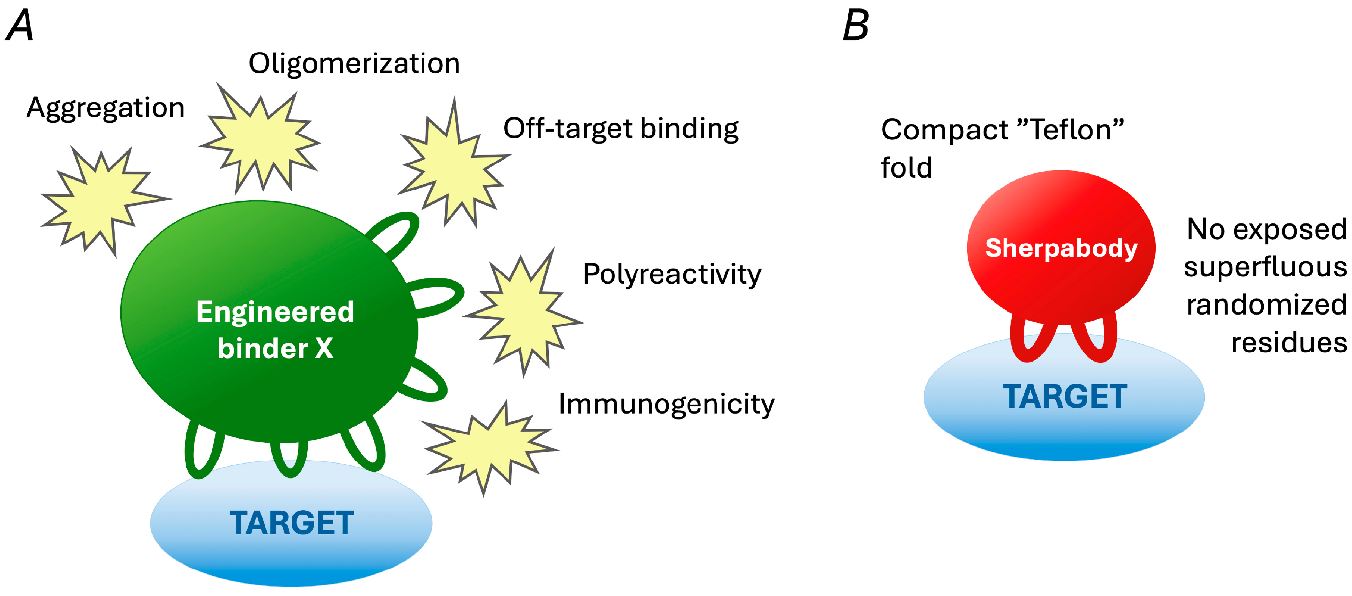
A notable feature of the sherpabodies confirmed in these studies was that besides their apparent specificity in binding to intended targets and not to non-target proteins, they also showed a remarkable absence of polyreactivity [64]. This was assessed by measuring the tendency of the CAA-targeted sherpabodies to interact with a panel of “sticky” macromolecules known for promiscuous binding and used by therapeutic antibody developers to predict problematic antibody behavior [90,91]. The lack of undesirable polyspecific reactivity highlights the favorable biochemical properties of sherpabodies and their utility in advanced biomedical targeting applications (see Figure 5).

6. Conclusions and Future Perspectives

Sherpabody technology has proven to be highly useful in blocking the function of multimeric viral entry glycoproteins, and sherpabodies have served as targeting modules of chimeric antigen receptor constructs for guiding cancer cell killing by CAR-T cells. These successes have been driven by the modularity and favorable biophysical properties of sherpabodies, which, in the above applications, are more relevant than the maximal strength of binding.
The binding affinity of sherpabodies can be enhanced through further engineering, as demonstrated for the structurally related Fynomers having picomolar potency [41]. However, given the rapid and straightforward identification of sherpabodies exhibiting excellent biochemical properties and binding to diverse target proteins with affinities in the low-to-mid-nanomolar range, this technology is particularly well suited for applications where this affinity range is sufficient.
The trimeric architecture of the SARS-CoV-2 inhibitor TriSb92 endowed it with excellent neutralization potency, achieving IC50 values as low as 10 pM, apparently resulting both from an enhanced avidity for the trimeric viral spike protein and from a cooperative inhibition mechanism.
On the other hand, a high target binding affinity can be detrimental for CAR-T cell function, leading to excessive activation and rapid exhaustion. Therefore, antibody fragments used for guiding CAR-T cell therapies have been engineered to have more moderate affinities [92,93]. In contrast, sherpabodies obtained directly from phage libraries are already equipped with binding strengths that appear ideal for developing long-lived and efficacious CAR-T cells.
In the context of the mucosal application of antiviral sherpabody multimers or sherpabody-guided CAR-T cell therapy, the serum half-life of soluble sherpabodies is irrelevant, but it becomes a factor to be considered in other types of therapeutic applications. The retention time in circulation has not been measured for sherpabodies but is likely to be relatively short and similar to the serum half-life of 4.4 h established for the Fynomer backbone (wild-type Fyn SH3 domain) in mice [94]. However, several approaches are available for extending the systemic persistence of small proteins like sherpabodies [95], including the fusion of the therapeutically targeted sherpabody to another sherpabody binding to serum albumin.
Both areas of research and development discussed above can be predicted to greatly benefit from the further exploitation of sherpabody technology to generate novel antiviral strategies against current and emerging viral threats and to improve the state of the art of CAR-T cell therapy in cancer. Beyond these applications, however, the beneficial properties of sherpabodies—such as their compact size, modularity, and favorable biophysical characteristics—suggest a broad range of potential uses in biomedicine and biotechnology as components of innovative diagnostic tools, biosensors, therapeutic agents, and research reagents.

Author Contributions

Conceptualization, A.R.M. and K.S.; writing—original draft preparation, A.R.M. and K.S.; writing—review and editing, K.S.; funding acquisition, K.S. All authors have read and agreed to the published version of the manuscript.

Funding

The authors acknowledge funding to K.S. from Research Council of Finland [grants 331787 and 365346], Cancer Foundation Finland [grant 230119], and the Sakari Alhopuro Foundation [grant 24022021], which supported research discussed in this review.

Data Availability Statement

No new data were created or analyzed in this study. Data sharing is not applicable to this article.

Acknowledgments

We thank all members of the Saksela lab who have contributed to the research discussed in this review.

Conflicts of Interest

K.S. is a founder of Next Biomed Therapies Oy that holds patents on SH3 scaffold targeting technologies. A.M. is a founder of Pandemblock Oy that develops sherpabody-based antivirals against respiratory pathogens.

References

  1. Sidhu, S.S.; Koide, S. Phage display for engineering and analyzing protein interaction interfaces. Curr. Opin. Struct. Biol. 2007, 17, 481–487. [Google Scholar] [CrossRef]
  2. Binz, H.K.; Amstutz, P.; Plückthun, A. Engineering novel binding proteins from nonimmunoglobulin domains. Nat. Biotechnol. 2005, 23, 1257–1268. [Google Scholar] [CrossRef]
  3. Packer, M.S.; Liu, D.R. Methods for the directed evolution of proteins. Nat. Rev. Genet. 2015, 16, 379–394. [Google Scholar] [CrossRef]
  4. Levin, A.M.; Weiss, G.A. Optimizing the affinity and specificity of proteins with molecular display. Mol. Biosyst. 2006, 2, 49–57. [Google Scholar] [CrossRef] [PubMed]
  5. Clackson, T.; Wells, J.A. In vitro selection from protein and peptide libraries. Trends Biotech. 1994, 12, 173–184. [Google Scholar] [CrossRef] [PubMed]
  6. Markland, W.; Ley, A.C.; Lee, S.W.; Ladner, R.C. Iterative optimization of high-affinity proteases inhibitors using phage display. 1. Plasmin. Biochemistry 1996, 35, 8045–8057. [Google Scholar] [CrossRef] [PubMed]
  7. Roberts, B.L.; Markland, W.; Siranosian, K.; Saxena, M.J.; Guterman, S.K.; Ladner, R.C. Protease inhibitor display M13 phage: Selection of high-affinity neutrophil elastase inhibitors. Gene 1992, 121, 9–15. [Google Scholar] [CrossRef]
  8. Röttgen, P.; Collins, J. A human pancreatic secretory trypsin inhibitor presenting a hypervariable highly constrained epitope via monovalent phagemid display. Gene 1995, 164, 243–250. [Google Scholar] [CrossRef]
  9. Abedi, M.R.; Caponigro, G.; Kamb, A. Green fluorescent protein as a scaffold for intracellular presentation of peptides. Nucleic Acids Res. 1998, 26, 623–630. [Google Scholar] [CrossRef]
  10. Norman, T.C.; Smith, D.L.; Sorger, P.K.; Drees, B.L.; O’Rourke, S.M.; Hughes, T.R.; Roberts, C.J.; Friend, S.H.; Fields, S.; Murray, A.W. Genetic selection of peptide inhibitors of biological pathways. Science 1999, 285, 591–595. [Google Scholar] [CrossRef]
  11. Lu, Z.; Murray, K.S.; Van Cleave, V.; LaVallie, E.R.; Stahl, M.L.; McCoy, J.M. Expression of thioredoxin random peptide libraries on the Escherichia coli cell surface as functional fusions to flagellin: A system designed for exploring protein-protein interactions. Biotechnology 1995, 13, 366–372. [Google Scholar] [CrossRef] [PubMed]
  12. Smith, G. Patch engineering: A general approach for creating proteins that have new binding activities. Trends Biochem. Sci. 1998, 23, 457–460. [Google Scholar] [CrossRef] [PubMed]
  13. Colas, P.; Cohen, B.; Jessen, T.; Grishina, I.; McCoy, J.; Brent, R. Genetic selection of peptide aptamers that recognize and inhibit cyclin-dependent kinase 2. Nature 1996, 380, 548–550. [Google Scholar] [CrossRef]
  14. Dennis, M.S.; Herzka, A.; Lazarus, R.A. Potent and selective Kunitz domain inhibitors of plasma kallikrein designed by phage display. J. Biol. Chem. 1995, 270, 25411–25417. [Google Scholar] [CrossRef]
  15. Szakács, D.; Kocsis, A.; Szász, R.; Gál, P.; Pál, G. Novel MASP-2 inhibitors developed via directed evolution of human TFPI1 are potent lectin pathway inhibitors. J. Biol. Chem. 2019, 294, 8227–8237. [Google Scholar] [CrossRef]
  16. Ku, J.; Schultz, P.G. Alternate protein frameworks for molecular recognition. Proc. Natl. Acad. Sci. USA 1995, 92, 6552–6556. [Google Scholar] [CrossRef]
  17. McConnell, S.J.; Hoess, R.H. Tendamistat as a scaffold for conformationally constrained phage peptide libraries. J. Mol. Biol. 1995, 250, 460–470. [Google Scholar] [CrossRef]
  18. Li, R.; Hoess, R.H.; Bennett, J.S.; DeGrado, W.F. Use of phage display to probe the evolution of binding specificity and affinity in integrins. Protein Eng. 2003, 16, 65–72. [Google Scholar] [CrossRef] [PubMed]
  19. Nord, K.; Gunneriusson, E.; Ringdahl, J.; Ståhl, S.; Uhlén, M.; Nygren, P.A. Binding proteins selected from combinatorial libraries of an alpha-helical bacterial receptor domain. Nat. Biotechnol. 1997, 15, 772–777. [Google Scholar] [CrossRef]
  20. Ståhl, S.; Gräslund, T.; Eriksson Karlström, A.; Frejd, F.Y.; Nygren, P.; Löfblom, J. Affibody molecules in biotechnological and medical applications. Trends Biotech. 2017, 35, 691–712. [Google Scholar] [CrossRef]
  21. Koide, A.; Bailey, C.W.; Huang, X.; Koide, S. The fibronectin type III domain as a scaffold for novel binding proteins. J. Mol. Biol. 1998, 284, 1141–1151. [Google Scholar] [CrossRef] [PubMed]
  22. Koide, A.; Koide, S. Monobodies: Antibody mimics based on the scaffold of the fibronectin type III domain. Methods Mol. Biol. 2007, 352, 95–109. [Google Scholar] [CrossRef] [PubMed]
  23. Hiipakka, M.; Poikonen, K.; Saksela, K. SH3 domains with high affinity and engineered ligand specificities targeted to HIV-1 Nef. J. Mol. Biol. 1999, 293, 1097–1106. [Google Scholar] [CrossRef]
  24. Hiipakka, M.; Saksela, K. Versatile retargeting of SH3 domain binding by modification of non-conserved loop residues. FEBS Lett. 2007, 581, 1735–1741. [Google Scholar] [CrossRef]
  25. Beste, G.; Schmidt, F.S.; Stibora, T.; Skerra, A. Small antibody-like proteins with prescribed ligand specificities derived from the lipocalin fold. Proc. Natl. Acad. Sci. USA 1999, 96, 1898–1903. [Google Scholar] [CrossRef]
  26. Skerra, A. Alternative binding proteins: Anticalins-harnessing the structural plasticity of the lipocalin ligand pocket to engineer novel binding activities. FEBS J. 2008, 275, 2677–2683. [Google Scholar] [CrossRef]
  27. Christmann, A.; Walter, K.; Wentzel, A.; Krätzner, R.; Kolmar, H. The cystine knot of a squash-type protease inhibitor as a structural scaffold for Escherichia coli cell surface display of conformationally constrained peptides. Protein Eng. 1999, 12, 797–806. [Google Scholar] [CrossRef]
  28. Moore, S.J.; Cochran, J.R. Engineering knottins as novel binding agents. Methods Enzymol. 2012, 503, 223–251. [Google Scholar] [CrossRef]
  29. Binz, H.K.; Stumpp, M.T.; Forrer, P.; Amstutz, P.; Plückthun, A. Designing repeat proteins: Well-expressed, soluble and stable proteins from combinatorial libraries of consensus ankyrin repeat proteins. J. Mol. Biol. 2003, 332, 489–503. [Google Scholar] [CrossRef]
  30. Stumpp, M.T.; Binz, H.K.; Amstutz, P. DARPins: A new generation of protein therapeutics. Drug Discov. Today 2008, 13, 695–701. [Google Scholar] [CrossRef]
  31. Silverman, J.; Liu, Q.; Bakker, A.; To, W.; Duguay, A.; Alba, B.M.; Smith, R.; Rivas, A.; Li, P.; Le, H.; et al. Multivalent avimer proteins evolved by exon shuffling of a family of human receptor domains. Nat. Biotechnol. 2005, 23, 1556–1561. [Google Scholar] [CrossRef]
  32. Hulme, J.T.; D’Souza, W.N.; McBride, H.J.; Yoon, B.P.; Willee, A.M.; Duguay, A.; Thomas, M.; Fan, B.; Dayao, M.R.; Rottman, J.B.; et al. Novel protein therapeutic joint retention strategy based on collagen-binding Avimers. J. Orthop. Res. 2018, 36, 1238–1247. [Google Scholar] [CrossRef]
  33. Woodman, R.; Yeh, J.T.; Laurenson, S.; Ko Ferrigno, P. Design and validation of a neutral protein scaffold for the presentation of peptide aptamers. J. Mol. Biol. 2005, 352, 1118–1133. [Google Scholar] [CrossRef] [PubMed]
  34. Stadler, L.K.; Hoffmann, T.; Tomlinson, D.C.; Song, Q.; Lee, T.; Busby, M.; Nyathi, Y.; Gendra, E.; Tiede, C.; Flanagan, K.; et al. Structure-function studies of an engineered scaffold protein derived from Stefin A. II: Development and applications of the SQT variant. Protein Eng. Des. Sel. 2011, 24, 751–763. [Google Scholar] [CrossRef] [PubMed]
  35. Tiede, C.; Bedford, R.; Heseltine, S.J.; Smith, G.; Wijetunga, I.; Ross, R.; AlQallaf, D.; Roberts, A.P.; Balls, A.; Curd, A.; et al. Affimer proteins are versatile and renewable affinity reagents. Elife 2017, 6, e24903. [Google Scholar] [CrossRef]
  36. Ebersbach, H.; Fiedler, E.; Scheuermann, T.; Fiedler, M.; Stubbs, M.T.; Reimann, C.; Proetzel, G.; Rudolph, R.; Fiedler, U. Affilin-novel binding molecules based on human gamma-B-crystallin, an all beta-sheet protein. J. Mol. Biol. 2007, 372, 172–185. [Google Scholar] [CrossRef]
  37. Mirecka, E.A.; Hey, T.; Fiedler, U.; Rudolph, R.; Hatzfeld, M. Affilin molecules selected against the human papillomavirus E7 protein inhibit the proliferation of target cells. J. Mol. Biol. 2009, 390, 710–721. [Google Scholar] [CrossRef]
  38. Mouratou, B.; Schaeffer, F.; Guilvout, I.; Tello-Manigne, D.; Pugsley, A.P.; Alzari, P.M.; Pecorari, F. Remodeling a DNA-binding protein as a specific in vivo inhibitor of bacterial secretin PulD. Proc. Natl. Acad. Sci. USA 2007, 104, 17983–17988. [Google Scholar] [CrossRef]
  39. Mouratou, B.; Béhar, G.; Paillard-Laurance, L.; Colinet, S.; Pecorari, F. Ribosome display for the selection of Sac7d scaffolds. Methods Mol. Biol. 2012, 805, 315–331. [Google Scholar] [CrossRef]
  40. Grabulovski, D.; Kaspar, M.; Neri, D. A novel, non-immunogenic Fyn SH3-derived binding protein with tumor vascular targeting properties. J. Biol. Chem. 2007, 282, 3196–3204. [Google Scholar] [CrossRef]
  41. Silacci, M.; Baenziger-Tobler, N.; Lembke, W.; Zha, W.; Batey, S.; Bertschinger, J.; Grabulovski, D. Linker length matters, fynomer-Fc fusion with an optimized linker displaying picomolar IL-17A inhibition potency. J. Biol. Chem. 2014, 289, 14392–14398. [Google Scholar] [CrossRef]
  42. Byla, P.; Andersen, M.H.; Holtet, T.L.; Jacobsen, H.; Munch, M.; Gad, H.H.; Thøgersen, H.C.; Hartmann, R. Selection of a novel and highly specific tumor necrosis factor alpha (TNFalpha) antagonist: Insight from the crystal structure of the antagonist-TNFalpha complex. J. Biol. Chem. 2010, 285, 12096–12100. [Google Scholar] [CrossRef] [PubMed]
  43. Allen, J.E.; Ferrini, R.; Dicker, D.T.; Batzer, G.; Chen, E.; Oltean, D.I.; Lin, B.; Renshaw, M.W.; Kretz-Rommel, A.; El-Deiry, W.S. Targeting TRAIL death receptor 4 with trivalent DR4 Atrimer complexes. Mol. Cancer Ther. 2012, 11, 2087–2095. [Google Scholar] [CrossRef] [PubMed]
  44. Nilvebrant, J.; Alm, T.; Hober, S.; Löfblom, J. Engineering bispecificity into a single albumin-binding domain. PLoS ONE 2011, 6, e25791. [Google Scholar] [CrossRef]
  45. Garousi, J.; Lindbo, S.; Nilvebrant, J.; Åstrand, M.; Buijs, J.; Sandström, M.; Honarvar, H.; Orlova, A.; Tolmachev, V.; Hober, S. ADAPT, a novel scaffold protein-based probe for radionuclide imaging of molecular targets that are expressed in disseminated cancers. Cancer Res. 2015, 75, 4364–4371. [Google Scholar] [CrossRef]
  46. Varadamsetty, G.; Tremmel, D.; Hansen, S.; Parmeggiani, F.; Plückthun, A. Designed Armadillo repeat proteins: Library generation, characterization and selection of peptide binders with high specificity. J. Mol. Biol. 2012, 424, 68–87. [Google Scholar] [CrossRef]
  47. Reichen, C.; Hansen, S.; Plückthun, A. Modular peptide binding: From a comparison of natural binders to designed armadillo repeat proteins. J. Struct. Biol. 2014, 185, 147–162. [Google Scholar] [CrossRef]
  48. Lee, S.C.; Park, K.; Han, J.; Lee, J.J.; Kim, H.J.; Hong, S.; Heu, W.; Kim, Y.J.; Ha, J.S.; Lee, S.G.; et al. Design of a binding scaffold based on variable lymphocyte receptors of jawless vertebrates by module engineering. Proc. Natl. Acad. Sci. USA 2012, 109, 3299–3304. [Google Scholar] [CrossRef]
  49. Jung, Y.H.; Choi, Y.; Seo, H.D.; Seo, M.H.; Kim, H.S. A conformation-selective protein binder for a KRAS mutant inhibits the interaction between RAS and RAF. Biochem. Biophys. Res. Commun. 2023, 645, 110–117. [Google Scholar] [CrossRef]
  50. Jacobs, S.A.; Diem, M.D.; Luo, J.; Teplyakov, A.; Obmolova, G.; Malia, T.; Gilliland, G.L.; O’Neil, K.T. Design of novel FN3 domains with high stability by a consensus sequence approach. Protein Eng. Des. Sel. 2012, 25, 107–117. [Google Scholar] [CrossRef]
  51. Diem, M.D.; Hyun, L.; Yi, F.; Hippensteel, R.; Kuhar, E.; Lowenstein, C.; Swift, E.J.; O’Neil, K.T.; Jacobs, S.A. Selection of high-affinity Centyrin FN3 domains from a simple library diversified at a combination of strand and loop positions. Protein Eng. Des. Sel. 2014, 27, 419–429. [Google Scholar] [CrossRef] [PubMed]
  52. Lorey, S.; Fiedler, E.; Kunert, A.; Nerkamp, J.; Lange, C.; Fiedler, M.; Bosse-Doenecke, E.; Meysing, M.; Gloser, M.; Rundfeldt, C.; et al. Novel ubiquitin-derived high affinity binding proteins with tumor targeting properties. J. Biol. Chem. 2014, 289, 8493–8507. [Google Scholar] [CrossRef] [PubMed]
  53. Job, F.; Settele, F.; Lorey, S.; Rundfeldt, C.; Baumann, L.; Beck-Sickinger, A.G.; Haupts, U.; Lilie, H.; Bosse-Doenecke, E. Ubiquitin is a versatile scaffold protein for the generation of molecules with de novo binding and advantageous drug-like properties. FEBS Open Bio 2015, 5, 579–593. [Google Scholar] [CrossRef] [PubMed]
  54. Tiede, C.; Tang, A.A.; Deacon, S.E.; Mandal, U.; Nettleship, J.E.; Owen, R.L.; George, S.E.; Harrison, D.J.; Owens, R.J.; Tomlinson, D.C.; et al. Adhiron: A stable and versatile peptide display scaffold for molecular recognition applications. Protein Eng. Des. Sel. 2014, 27, 145–155. [Google Scholar] [CrossRef]
  55. Desmet, J.; Verstraete, K.; Bloch, Y.; Lorent, E.; Wen, Y.; Devreese, B.; Vandenbroucke, K.; Loverix, S.; Hettmann, T.; Deroo, S.; et al. Structural basis of IL-23 antagonism by an Alphabody protein scaffold. Nat. Commun. 2014, 5, 5237. [Google Scholar] [CrossRef]
  56. Pannecoucke, E.; Van Trimpont, M.; Desmet, J.; Pieters, T.; Reunes, L.; Demoen, L.; Vuylsteke, M.; Loverix, S.; Vandenbroucke, K.; Alard, P.; et al. Cell-penetrating Alphabody protein scaffolds for intracellular drug targeting. Sci. Adv. 2021, 7, eabe1682. [Google Scholar] [CrossRef]
  57. Steemson, J.D.; Baake, M.; Rakonjac, J.; Arcus, V.L.; Liddament, M.T. Tracking molecular recognition at the atomic level with a new protein scaffold based on the OB-fold. PLoS ONE 2014, 9, e86050. [Google Scholar] [CrossRef]
  58. Suderman, R.J.; Rice, D.A.; Gibson, S.D.; Strick, E.J.; Chao, D.M. Development of polyol-responsive antibody mimetics for single-step protein purification. Protein Expr. Purif. 2017, 134, 114–124. [Google Scholar] [CrossRef]
  59. Suderman, R.J.; Gibson, S.D.; Strecker, M.; Bonner, A.M.; Chao, D.M. Protein engineering of a nanoCLAMP antibody mimetic scaffold as a platform for producing bioprocess-compatible affinity capture ligands. J. Biol. Chem. 2023, 299, 104910. [Google Scholar] [CrossRef]
  60. Wicke, N.; Bedford, M.R.; Howarth, M. Gastrobodies are engineered antibody mimetics resilient to pepsin and hydrochloric acid. Commun. Biol. 2021, 4, 960. [Google Scholar] [CrossRef]
  61. Hokanson, C.A.; Zacco, E.; Cappuccilli, G.; Odineca, T.; Crea, R. AXL-receptor targeted 14FN3 based single domain proteins (PronectinsTM) from 3 synthetic human libraries as components for exploring novel bispecific constructs against solid tumors. Biomedicines 2022, 10, 3184. [Google Scholar] [CrossRef]
  62. Riillo, C.; Polerà, N.; Di Martino, M.T.; Juli, G.; Hokanson, C.A.; Odineca, T.; Signorelli, S.; Grillone, K.; Ascrizzi, S.; Mancuso, A.; et al. A PronectinTM AXL-targeted first-in-class bispecific T cell engager (pAXLxCD3ε) for ovarian cancer. J. Transl. Med. 2023, 21, 301. [Google Scholar] [CrossRef] [PubMed]
  63. Mäkelä, A.R.; Ugurlu, H.; Hannula, L.; Kant, R.; Salminen, P.; Fagerlund, R.; Mäki, S.; Haveri, A.; Strandin, T.; Kareinen, L.; et al. Intranasal trimeric sherpabody inhibits SARS-CoV-2 including recent immunoevasive Omicron subvariants. Nat. Commun. 2023, 14, 1637. [Google Scholar] [CrossRef]
  64. Hernández-López, R.A.; Kesti, T.; Mäkelä, A.R.; Zhao, Z.; Yu, W.; Tonai, Y.; Monzo, H.J.; Kalander, K.; Leppä, S.; Ojala, P.M.; et al. Engineered SH3-derived sherpabodies function as a modular platform for targeted T cell immunotherapy. Cancer Res. 2025, in press. [CrossRef] [PubMed]
  65. Kaneko, T.; Li, L.; Li, S.S. The SH3 domain—A family of versatile peptide- and protein-recognition module. Front. Biosci. 2008, 13, 4938–4952. [Google Scholar] [CrossRef] [PubMed]
  66. Mayer, B.J. SH3 domains: Complexity in moderation. J. Cell Sci. 2001, 114, 1253–1263. [Google Scholar] [CrossRef]
  67. Plaxco, K.W.; Guijarro, J.I.; Morton, C.J.; Pitkeathly, M.; Campbell, I.D.; Dobson, C.M. The folding kinetics and thermodynamics of the Fyn-SH3 domain. Biochemistry 1998, 37, 2529–2537. [Google Scholar] [CrossRef]
  68. Lim, W.A.; Fox, R.O.; Richards, F.M. Stability and peptide binding affinity of an SH3 domain from the Caenorhabditis elegans signaling protein Sem-5. Protein Sci. 1994, 3, 1261–1266. [Google Scholar] [CrossRef]
  69. Saksela, K.; Permi, P. SH3 domain ligand binding: What’s the consensus and where’s the specificity? FEBS Lett. 2012, 586, 2609–2614. [Google Scholar] [CrossRef]
  70. Lee, C.H.; Leung, B.; Lemmon, M.A.; Zheng, J.; Cowburn, D.; Kuriyan, J.; Saksela, K. A single amino acid in the SH3 domain of Hck determines its high affinity and specificity in binding to HIV-1 Nef protein. EMBO J. 1995, 14, 5006–5015. [Google Scholar] [CrossRef]
  71. Lee, C.H.; Saksela, K.; Mirza, U.A.; Chait, B.T.; Kuriyan, J. Crystal structure of the conserved core of HIV-1 Nef complexed with a Src family SH3 domain. Cell 1996, 85, 931–942. [Google Scholar] [CrossRef]
  72. Schlatter, D.; Brack, S.; Banner, D.W.; Batey, S.; Benz, J.; Bertschinger, J.; Huber, W.; Joseph, C.; Rufer, A.; van der Klooster, A.; et al. Generation, characterization and structural data of chymase binding proteins based on the human Fyn kinase SH3 domain. mAbs 2012, 4, 497–508. [Google Scholar] [CrossRef] [PubMed]
  73. Brack, S.; Attinger-Toller, I.; Schade, B.; Mourlane, F.; Klupsch, K.; Woods, R.; Hachemi, H.; von der Bey, U.; Koenig-Friedrich, S.; Bertschinger, J.; et al. A bispecific HER2-targeting FynomAb with superior antitumor activity and novel mode of action. Mol. Cancer Ther. 2014, 13, 2030–2039. [Google Scholar] [CrossRef]
  74. Silacci, M.; Lembke, W.; Woods, R.; Attinger-Toller, I.; Baenziger-Tobler, N.; Batey, S.; Santimaria, R.; von der Bey, U.; Koenig-Friedrich, S.; Zha, W.; et al. Discovery and characterization of COVA322, a clinical-stage bispecific TNF/IL-17A inhibitor for the treatment of inflammatory diseases. mAbs 2016, 8, 141–149. [Google Scholar] [CrossRef] [PubMed]
  75. Protein Atlas. Available online: https://www.proteinatlas.org/ENSG00000144061-NPHP1 (accessed on 3 March 2025).
  76. Otto, E.; Kispert, A.; Schätzle, S.; Lescher, B.; Rensing, C.; Hildebrandt, F. Nephrocystin: Gene expression and sequence conservation between human, mouse, and Caenorhabditis elegans. J. Am. Soc. Nephrol. 2000, 11, 270–282. [Google Scholar] [CrossRef] [PubMed]
  77. Saksela, K. SH3 Domain Derivatives. U.S. Patent US110,060,084 B2, 13 July 2021. [Google Scholar]
  78. Kazlauskas, A.; Schmotz, C.; Kesti, T.; Hepojoki, J.; Kleino, I.; Kaneko, T.; Li, S.S.; Saksela, K. Large-scale screening of preferred interactions of human Src homology-3 (SH3) domains using native target proteins as affinity ligands. Mol. Cell Proteom. 2016, 15, 3270–3281. [Google Scholar] [CrossRef]
  79. le Maire, A.; Weber, T.; Saunier, S.; Broutin, I.; Antignac, C.; Ducruix, A.; Dardel, F. Solution NMR structure of the SH3 domain of human nephrocystin and analysis of a mutation-causing juvenile nephronophthisis. Proteins 2005, 59, 347–355. [Google Scholar] [CrossRef]
  80. Wodarczyk, C.; Distefano, G.; Rowe, I.; Gaetani, M.; Bricoli, B.; Muorah, M.; Spitaleri, A.; Mannella, V.; Ricchiuto, P.; Pema, M.; et al. Nephrocystin-1 forms a complex with polycystin-1 via a polyproline motif/SH3 domain interaction and regulates the apoptotic response in mammals. PLoS ONE 2010, 5, e12719. [Google Scholar] [CrossRef]
  81. White, J.M.; Ward, A.E.; Odongo, L.; Tamm, L.K. Viral membrane fusion: A dance between proteins and lipids. Annu. Rev. Virol. 2023, 10, 139–161. [Google Scholar] [CrossRef]
  82. Beukenhorst, A.L.; Rice, K.L.; Frallicciardi, J.; Koldijk, M.H.; Boudreau, C.M.; Crawford, J.; Cornelissen, L.; da Costa, K.A.S.; de Jong, B.A.; Fischinger, S.; et al. Intranasal administration of a panreactive influenza antibody reveals Fc-independent mode of protection. Sci. Rep. 2025, 15, 10309. [Google Scholar] [CrossRef]
  83. Sparrow, E.; Torvaldsen, S.; Newall, A.T.; Wood, J.G.; Sheikh, M.; Kieny, M.P.; Abela-Ridder, B. Recent advances in the development of monoclonal antibodies for rabies post exposure prophylaxis: A review of the current status of the clinical development pipeline. Vaccine 2019, 37 (Suppl. 1), 132–139. [Google Scholar] [CrossRef]
  84. Fan, L.; Zhang, L.; Li, J.; Zhu, F. Advances in the progress of monoclonal antibodies for rabies. Hum. Vaccin. Immunother. 2022, 18, 2026713. [Google Scholar] [CrossRef] [PubMed]
  85. Lim, W.A.; June, C.H. The principles of engineering immune cells to treat cancer. Cell 2017, 168, 724–740. [Google Scholar] [CrossRef]
  86. Guzman, G.; Reed, M.R.; Bielamowicz, K.; Koss, B.; Rodriguez, A. CAR-T therapies in solid tumors: Opportunities and challenges. Curr. Oncol. Rep. 2023, 25, 479–489. [Google Scholar] [CrossRef] [PubMed]
  87. Kailayangiri, S.; Altvater, B.; Wiebel, M.; Jamitzky, S.; Rossig, C. Overcoming heterogeneity of antigen expression for effective CAR T cell targeting of cancers. Cancers 2020, 12, 1075. [Google Scholar] [CrossRef] [PubMed]
  88. Zhu, X.; Li, Q.; Zhu, X. Mechanisms of CAR T cell exhaustion and current counteraction strategies. Front. Cell Dev. Biol. 2022, 10, 1034257. [Google Scholar] [CrossRef]
  89. Zajc, C.U.; Salzer, B.; Taft, J.M.; Reddy, S.T.; Lehner, M.; Traxlmayr, M.W. Driving CARs with alternative navigation tools—The potential of engineered binding scaffolds. FEBS J. 2021, 288, 2103–2118. [Google Scholar] [CrossRef]
  90. Mouquet, H.; Scheid, J.F.; Zoller, M.J.; Krogsgaard, M.; Ott, R.G.; Shukair, S.; Artyomov, M.N.; Pietzsch, J.; Connors, M.; Pereyra, F.; et al. Polyreactivity increases the apparent affinity of anti-HIV antibodies by heteroligation. Nature 2010, 467, 591–595. [Google Scholar] [CrossRef]
  91. Jain, T.; Sun, T.; Durand, S.; Hall, A.; Houston, N.R.; Nett, J.H.; Sharkey, B.; Bobrowicz, B.; Caffry, I.; Yu, Y.; et al. Biophysical properties of the clinical-stage antibody landscape. Proc. Natl. Acad. Sci. USA 2017, 114, 944–949. [Google Scholar] [CrossRef]
  92. Vander Mause, E.R.; Atanackovic, D.; Lim, C.S.; Luetkens, T. Roadmap to affinity-tuned antibodies for enhanced chimeric antigen receptor T cell function and selectivity. Trends Biotech. 2022, 40, 875–890. [Google Scholar] [CrossRef]
  93. Mao, R.; Kong, W.; He, Y. The affinity of antigen-binding domain on the antitumor efficacy of CAR T cells: Moderate is better. Front. Immunol. 2022, 13, 1032403. [Google Scholar] [CrossRef] [PubMed]
  94. Buller, F.; Wüllner, U.; Zbinden, I.; Attinger-Toller, I.; von der Bey, U.; König-Friedrich, S.; Bertschinger, J.; Grabulovski, D.; Henne, P. Human Serum Albumin Binding Compounds and Fusion Proteins Thereof. U.S. Patent US10,364,419 B2, 30 July 2019. [Google Scholar]
  95. Zaman, R.; Islam, R.A.; Ibnat, N.; Othman, I.; Zaini, A.; Lee, C.Y.; Chowdhury, E.H. Current strategies in extending half-lives of therapeutic proteins. J. Control Release 2019, 301, 176–189. [Google Scholar] [CrossRef] [PubMed]
Figure 1. Architectures of human signaling/adapter proteins containing multiple SH3 domains. GEF stands for a guanine nucleotide exchange factor domain, RING for a zinc-binding RING finger domain, and BAR for a Bin/amphiphysin/Rvs (BAR) domain. Domain sizes are not drawn to scale.
Biologics 05 00013 g001
Figure 2. SH3 domain structure and alignment of representative human SH3 domain sequences. The five β-strands are colored purple, and the 310 helix green. The regions in the RT- and N-Src-loop showing diversity in sequence and length that were replaced by random hexamer peptides in the randomized libraries (see later) are boxed in red. Roman numerals after protein names refer to the order of the SH3 domain when the same protein contains several.
Biologics 05 00013 g002
Figure 3. (A) Phylogenic tree of 296 human SH3 domains drawn based on amino acid sequence homology. Each dot represents one SH3 domain, and those shown in red and indicated by a protein name (and SH3 order when relevant) were chosen as templates for scaffold library construction [77]. One or more of the SH3 domains in the subfamilies depicted as green leaves (unlike the yellow leaves) were identified as hits in the screening of a comprehensive human SH3 domain phage display library to characterize the human SH3 interactome [78]. (B,C) Data adapted from [77] showing functional comparison of 12 large (each comprising ≈ 1010 unique clones) M13 phage-display libraries based on SH3 domain scaffolds chosen from different subfamilies of human SH3 domains, shown in Panel (A). The RT- and N-Src-loop regions of all 12 SH3 domains were randomized in an analogous manner by replacing their highly divergent regions (indicated by red boxes in Figure 2) with random hexamer sequences, a strategy identical to the original design of Fynomers [40]. Panel (B) shows the proportion of clones based on the NPHP1 (nephrocystin) SH3 vs. other SH3 scaffolds among clones selected by the iterative affinity selection of pooled 12 SH3 libraries against two different target proteins (CTLA-4 and HER2). Panel (C) shows the target binding capacity of 96 individual clones obtained from an unmixed NPHP1 or a Fyn SH3 library that were screened in parallel via five rounds of affinity panning using a third target (NS1) as bait.
Biologics 05 00013 g003
Figure 4. Two recently described applications of sherpabody targeting technology are illustrated, namely, (A) design of host cell entry-blocking antiviral proteins [63] and (B) multi-specific targeting modules for guiding chimeric antigen receptor (CAR)-T cell-based anti-cancer therapy [64]. See text for more details.
Biologics 05 00013 g004
Figure 5. Projected benefits of the compact sherpabody-targeting surface and the lack of exposed extraneous (non-targeting) randomly diversified regions. Target engagement of a binder with multiple potential binding loops and surfaces (A) is compared to that of the streamlined sherpabody architecture (B).
Biologics 05 00013 g005
Table 1. Features of non-immunoglobulin scaffold proteins used as templates for construction of libraries for discovery of antibody mimetics used in biomedical targeting. Missing from the list are early studies involving grafting of peptide aptamers into heterologous proteins such as thioredoxin [13] or GFP [9].
ScaffoldProtein of OriginRandomized SurfaceSpecies of OriginMolar MassFirst PublicationReferences
Kunitz domainsBPTI or other protease inhibitorsExposed loopsCow and others6 kDa1992[7,14,15]
Cytochrome b562Bacterial c-type cytochromeExposed loopsEscherichia coli (bacterium)12 kDa1995[16]
TendamistatAlpha-amylase inhibitor HOE 467Exposed loopsStreptomyces tendae (bacterium)8 kDa1995[17,18]
AffibodyProtein A (Z domain)α-helicesStaphylococcus aureus (bacterium)6 kDa1997[19,20]
Monobodies/AdnectinsFibronectin (10th type III domain)Exposed loopsHuman10 kDa1998[21,22]
Hck-SH3Hck kinase (SH3 domain)Exposed loopsHuman6 kDa1999[23,24]
AnticalinLipocalinsExposed loopsPieris brassicae (butterfly); later also human lipocalins20 kDa1999[25,26]
KnottinsKnottin folds of various proteinsExposed loopsSquirting cucumber (plant) and other species 3 kDa1999[27,28]
DARPinHuman ankyrin repeat proteinsβ-turn & α-helixNon-natural protein10–19 kDa2003[29,30]
AvimerHuman LDL receptor-like proteinsExposed loopsDesigned consensus of 197 A-domains9–18 kDa2005[31,32]
AffimerCystatin A (Stefin A)Exposed loopsHuman12–14 kDa2005[33,34,35]
Affilin (γB)Gamma-B crystallinβ-strandsHuman20 kDa2007[36,37]
Affitin/NanofitinSac7d DNA binding proteinβ-strandsSulfolobus acidocaldarius (archaeon)7 kDa2007[38,39]
FynomerFyn kinase (SH3 domain)Exposed loopsHuman6 kDa2007[40,41]
AtrimerTetranectin (C-type lectin domain)Exposed loopsHuman60–70 kDa2010[42,43]
ADAPTProtein G (3rd albumin binding domain)α-helicesStreptococcus strain G148 (bacterium)5 kDa2011[44,45]
ArmRPArmadillo repeat proteinsα-helicesNon-natural protein30kDa2012[46,47]
RepebodyVLR proteins of jawless vertebratesβ-strands and loopsDesigned based on consensus leucine-rich repeats30 kDa2012[48,49]
CentyrinTenascin and fibronectinExposed loopsDesigned consensus of different III domains10 kDa2012[50,51]
AffilinTwo head-to-tail linked ubiquitinsβ-strandsHuman17 kDa2014[52,53]
AdhironCystatinsExposed loopsDesigned consensus of plant cystatins12–14 kDa2014[35,54]
AlphabodyEngineered triple helix coiled coilα-helicesNon-natural protein10 kDa2014[55,56]
ObodyAspartyl tRNA synthetase (OB-fold)β-strands and loopsPyrobaculum aerophilum (archaeon)11 kDa2014[57]
nanoCLAMPNagH (carbohydrate binding module 32-2)Exposed loopsClostridium perfringens (bacterium)16 kDa2017[58,59]
GastrobodySoybean trypsin inhibitor Exposed loopsSoybean (plant)21 kDa2021[60]
PronectinFibronectin (14th type III domain)Exposed loopsHuman10 kDa2022[61,62]
SherpabodyNephrocystin (SH3 domain)Exposed loopsHuman6 kDa2023[63,64]
Disclaimer/Publisher’s Note: The statements, opinions and data contained in all publications are solely those of the individual author(s) and contributor(s) and not of MDPI and/or the editor(s). MDPI and/or the editor(s) disclaim responsibility for any injury to people or property resulting from any ideas, methods, instructions or products referred to in the content.