Brief Report

An Interdisciplinary Approach to Biobanking in Cardiac Surgery: Protocol of a Prospective, Single-Center Research Project Involving Longitudinal Biobanking

1
Department of Cardiothoracic Surgery, University Hospital Augsburg, Stenglinstraße 2, 86156 Augsburg, Germany
2
Augsburg Central BioBank, Medical Faculty, University of Augsburg, Stenglinstraße 2, 86156 Augsburg, Germany
3
Department of General Pathology and Molecular Diagnostics, University Hospital Augsburg, Stenglinstraße 2, 86156 Augsburg, Germany
*
Authors to whom correspondence should be addressed.
Int. J. Transl. Med. 2024, 4(2), 238-246; https://doi.org/10.3390/ijtm4020014
Submission received: 29 January 2024 / Revised: 19 March 2024 / Accepted: 3 April 2024 / Published: 4 April 2024

Abstract

:
Cross-sectional and longitudinal profiling of full sets of nucleic acids, peptides, or proteins as well as metabolites expressed in biospecimens acquired via a cardiovascular disease-oriented biobank may aid in the elucidation of the disease pathways and mechanisms underlying individual cardiovascular diseases, such as degenerative valvular heart disease. This may promote the development of novel and effective, personalized diagnostic and therapeutic strategies to efficiently reduce cardiovascular mortality and morbidity as well as its health and economic burden. This brief report aims to describe the unique, standardized, interdisciplinary, and interprofessional approach to cross-sectional and longitudinal cardiovascular biobanking and databasing at the University Hospital Augsburg. Moreover, we present the study protocol of a specific, well-defined, prospective, single-center research project involving cross-sectional and longitudinal cardiovascular biobanking. The aim of this project is to gain a better insight into the molecular mechanisms underlying aortic valve disease-induced cardiomyopathy and the long-term effect of surgical correction of the aortic valve pathology on the left ventricular myocardial molecule profile.

1. Introduction

Cardiovascular diseases remain the leading cause of death and disability globally, placing an immense burden on the health systems and economies [1,2]. Along with ischemic heart disease, degenerative valvular heart disease is a major contributor to cardiovascular mortality and morbidity with a steadily increasing global prevalence over the past 30 years [3,4]. For the development of novel and effective diagnostic and therapeutic strategies in degenerative valvular heart disease, a better elucidation of the disease pathways and mechanisms through basic and translational research is paramount.
Biobanking refers to the systematic, organized collection and long-term storage of well-characterized biospecimens (e.g., tissue samples (cryopreserved or formalin-fixed and paraffin-embedded), blood samples, various types of cells or nucleic acids) annotated with medical and/or epidemiological data for research purposes. Biobanking can occur either on a small scale for specific single-center research projects or on a national level for comprehensive epidemiological studies [5,6]. The collection of biospecimens can be conducted cross-sectionally or longitudinally, an advantage of the latter possibly being a better characterization of disease evolution and progression [5]. Biobanks can either be population-based, comprising annotated samples of biospecimens from volunteers without specific inclusion or exclusion criteria, or are disease-oriented, containing annotated samples of biospecimens from a specific patient population, such as patients suffering from cardiovascular disease [7]. The evolution of omics-based analyses (i.e., the profiling of full sets of nucleic acids, peptides/proteins or metabolites expressed in a specific specimen) has significantly contributed to progress in the field of disease-oriented biobanking and biomedical research by enabling researchers to gain better insights into disease-related changes on the molecular level [6,7]. Yet, the quality of biospecimens might have a major impact on omics data. Thus, documentation of the pre-analytical factors of the biospecimens (i.e., processing time, centrifugation condition, temperature) in the biobank is crucial to enable successful and reproducible research studies [8,9,10]. Many biobanks, therefore, use the Standard PREanalytical Code (SPREC), which was established by the ISBER Biospecimen Science Working group, in order to properly document critical preanalytical variables of fluid and solid biospecimens within a seven-element code [11].
The aim of this brief report is to describe the general, standardized, interdisciplinary and interprofessional approach to cardiovascular biobanking and databasing at the University Hospital Augsburg, referred to as A-CaRe (for Augsburg Cardiovascular Research), as well as a specific, well-defined, prospective, single-center research project using, and thereby testing, the organizational structure, workflow and institutional infrastructure provided by the A-CaRe protocol. It incorporates longitudinal biobanking to better understand the molecular mechanisms underlying aortic valve disease-induced cardiomyopathy and the long-term effect of surgical correction of aortic valve pathology on the left ventricular myocardial molecule profile.

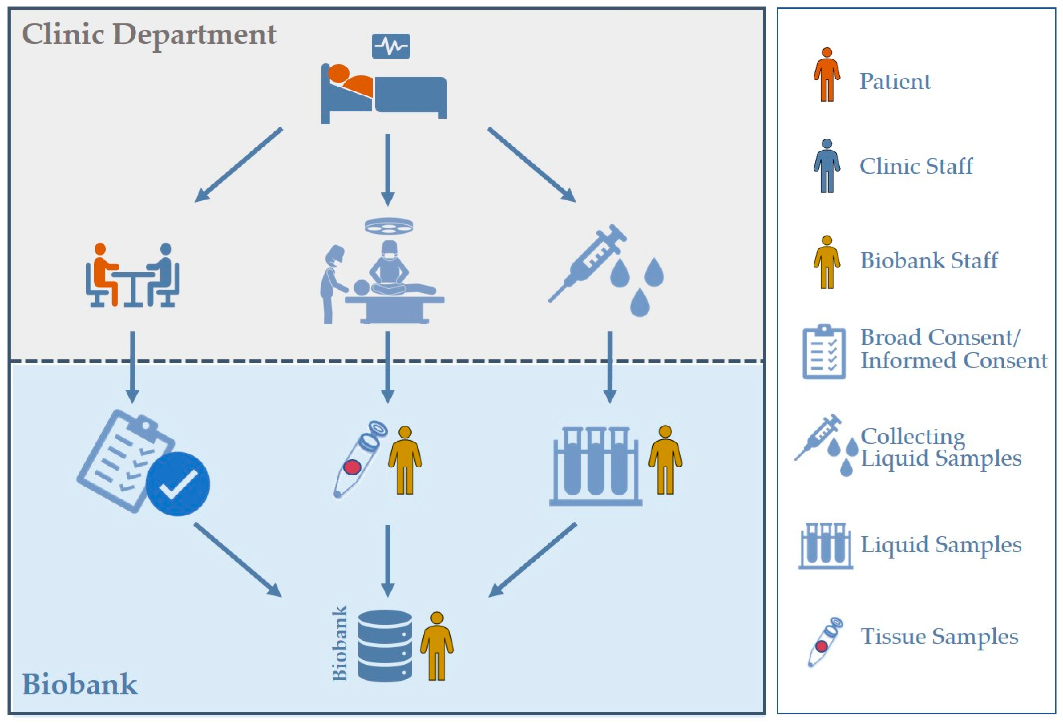
2. Cardiovascular Biobanking at the University Hospital Augsburg

The general process of cardiovascular biobanking at the University Hospital Augsburg (i.e., A-CaRe) is displayed in Figure 1. It is a unique, collaborative, interdisciplinary and interprofessional project involving clinicians, scientists, research assistants and medical (laboratory) assistants from the Department of Cardiothoracic Surgery as well as the Augsburg Central BioBank. It is aimed at the systematic and organized collection of a broad range of biospecimens obtained intraoperatively during elective, urgent and emergency open heart procedures as well as during postoperative follow-up for the purpose of dedicated research on the etiology of cardiovascular diseases and their underlying molecular pathways and mechanisms, thereby intending to improve and personalize diagnostics as well as therapy.

2.1. Broad Consent

Within the scope of the preoperative informed consent procedure, senior surgeons provide information on the planned cardiac operation to the patients. Irrespective of the consent to the surgery, the patients are informed about the possibility to participate in biobanking. Thereby, upon the patient’s consent, biospecimens can be stored in the Augsburg Central BioBank and used for future research projects. Specific emphasis is placed on the fact that only those biospecimens that are routinely excised as part of the procedure (e.g., aortic valve leaflets during aortic valve replacement, aortic tissue during aortic surgery, remains of arterial or venous grafts during coronary artery bypass grafting, etc.) are collected. Hence, patients who agree to the general storage of their biospecimens in the biobank receive the same surgical treatment as patients who deny consent. The only difference between those who consent and those who do not is the collection of additional blood samples with a total volume of approximately 20 to 25 mL via central venous or arterial line. Before the biospecimens and their associated data are released for a specific research project, a positive vote by the independent Ethics Committee and the subsequent approval of the in-house Use and Access Committee must be obtained.

2.2. The A-CaRe Protocol for Collection, Processing and Long-Term Storage of Liquid and Tissue Samples

Key elements of the A-CaRe protocol are dedicated medical (laboratory) assistants in charge of the collection and transfer of biospecimens to the biobank and of processing and handling of the samples before storage along with the appropriate pre-existing equipment and infrastructure.
Blood samples are acquired via central venous or arterial line during induction of anesthesia and comprise 4 tubes with different additives: ethylenediaminetetraacetic acid (EDTA) plasma (9 mL), citrate plasma (5 mL), serum/whole blood (7.5 mL) and PAXgene® for isolation of ribonucleic acids (RNA) (2.5 mL). Tubes are inverted to mix the additives with the whole blood and then immediately transferred to the biobank in an upright position for further processing and storage. Upon arrival in the biobank, tubes are centrifuged at 2000× g for 15 min at 20 °C. Next, 500 μL aliquots of EDTA plasma, citrate plasma and serum as well as 300 μL aliquots of EDTA buffy coat are transferred to cryo-storage tubes. Finally, the cryo-storage tubes are frozen at −80 °C for long-term storage. Meanwhile, RNA-PAXgene® blood is incubated for at least 2 h at ambient temperature, then placed at −20 °C for 24 h before finally being transferred to long-term storage at −80 °C in a freezer. All relevant information about the quality of the samples such as time stamps (i.e., collection, arrival, centrifugation, aliquoting, freezing) as well as any abnormalities and protocol deviations/violations are recorded in the Augsburg Central BioBank’s biobank information management system (BIMS). For all data entries concerning biospecimens of the Augsburg Central BioBank, the Data Information System (DIS, Bitcare GmbH) is used.
Tissue collection comprises the broad collection of any type of tissue that is excised during open heart procedures. Shortly after removal from the patient, the tissue samples are placed into universal collection containers (e.g., sputum collection container), labelled and sent to the biobank via a pneumatic dispatch system. Upon arrival in the biobank, up to 5 aliquots are obtained. Aliquots are then snap-frozen, temporarily stored at −80 °C in a freezer and eventually transferred to long-term storage at lower than −135 °C in the vapor phase of liquid nitrogen. Temperatures are closely monitored at any step via a temperature monitoring system. Again, all relevant times (i.e., excision, arrival, aliquoting, freezing) as well as any protocol deviations/violations are recorded and entered in the biobank information management system (BIMS).
A slightly different protocol exists for patients undergoing emergency cardiac surgery during “off-time” (i.e., incision time after 4 pm or before 7 am or during the weekend). As preoperative consent, a prerequisite for the active collection of biospecimens, can generally not be obtained in such patients, no blood samples are collected. In these cases, only residual material can be used for biobanking and of course, only if the patient agrees to the broad consent postoperatively. If the patient’s consent is not given, the samples and related data are discarded immediately.
Small samples of the tissue that is excised for medical reasons during the emergency procedure are compiled. Yet, in contrast to the above-mentioned approach, the operating room staff are responsible for the processing of samples, including assembly of the aliquots into cryo-tubes, correct labeling and snap-freezing of aliquots in liquid nitrogen at −196 °C. The samples are stored in a liquid nitrogen tank close to the operating room. The aliquots will then be transferred to the biobank for long-term storage at lower than −135 °C in the vapor phase of liquid nitrogen. Excision and fixation times are recorded on a specific worksheet intraoperatively and subsequently entered into the BIMS.
Relevant clinical information, including demographics, comorbidities, perioperative risk scores, pre- and postoperative laboratory tests, pre- and postoperative echocardiography, details of the surgical procedure and major postoperative complications, are extracted from the patients’ hospital charts after discharge. The information is entered into the A-CaRe register of the BIMS, hence linking it to the biospecimens and eventually molecular characteristics.

2.3. Longitudinal Biobanking

The key idea of longitudinal cardiovascular biobanking (i.e., the repeated collection of biospecimens) is to monitor the course of cardiovascular disease from its initial diagnosis through all further stages of disease development, considering specific treatment effects. While the general A-CaRe protocol currently does not include any follow-up visits, longitudinal sampling during the rehabilitation and follow-up period after surgery will be an integral part of specific research projects based on the A-CaRe protocol, such as the below-mentioned AortOmics trial. Longitudinal sampling is an essential tool in biomarker research for the prediction of specific cardiovascular diseases and for the monitoring of their progression during the natural course as well as upon treatment (i.e., medication, intervention, or surgery). Potential areas of application of longitudinal cardiovascular biobanking may include personalized decision-making on the optimal timing of (prophylactic) cardiac intervention/surgery, e.g., in degenerative valvular heart disease or aortic disease, where the benefit of invasive correction of the underlying pathology to prevent future irreversible damage outweighs the risk of intervention- or surgery-associated morbidity and mortality. To allow for coupling with longitudinal clinical data, clinical follow-up information, including symptoms, laboratory tests, echocardiography, and adverse events, may be added to the BIMS for specific follow-up visits.

2.4. Number of Aliquots Obtained and Stored

Table 1 and Table 2 present the numbers of aliquots of samples obtained from November 2021 to September 2023 from 596 individual patients undergoing elective, urgent and emergency open heart surgery via sternotomy or thoracotomy approach.

3. The AortOmics Trial

3.1. Background

Isolated aortic valve disease, including aortic stenosis and regurgitation, is the most prevalent form of degenerative valvular heart disease in Europe and the United States [1,12]. Left ventricular myocardial remodeling, a sequel of long-lasting pressure or volume overload induced by chronic aortic valve dysfunction, may eventually result in valvular cardiomyopathy and progressive chronic heart failure, if valvular pathology is left untreated [13]. Currently, aortic valve surgery is recommended as soon as any symptoms or any echocardiographic evidence of left ventricular dysfunction is detected. It is supposed to set the course for left ventricular reverse remodeling, eventually resulting in recovery of left ventricular function [14,15]. Yet, despite successful aortic valve surgery, cardiomyopathy persists or even worsens postoperatively in a substantial subset of patients [16,17,18]. This phenomenon appears to be more common in patients with aortic regurgitation than in patients with aortic stenosis [18,19,20,21]. Disease pathways and mechanisms that might help to explain the different reactions of the left ventricular myocardium to the reduction in pressure and volume overload by surgical correction of valvular pathology remain widely unexplored. Hence, no biomarkers (neither molecular, nor circulating, nor imaging-based) exist that might serve as predictors of inadequate recovery of left ventricular function following aortic valve surgery [21,22]. Consequently, optimal timing of surgery remains a challenge.

3.2. Objectives

The primary aim of the AortOmics Trial is the exploratory multi-omics-based analysis of left ventricular myocardial molecule signatures that might serve as indicators of the preoperative extent of irreversible myocardial dysfunction (i.e., cardiomyopathy), thereby predicting chances of full recovery of left ventricular function (i.e., left ventricular reverse remodeling) after aortic valve surgery in patients with aortic regurgitation vs. stenosis. Further elucidation of the molecular mechanism of left ventricular remodeling and reverse remodeling may allow for individualized decision-making and better timing of surgery in the future. A secondary aim of the study is the characterization of biomarkers that might serve as predictors of satisfactory long-term outcomes after aortic valve surgery in either phenotype. Moreover, we aim to evaluate potential correlations of myocardial and circulating biomarkers which may simplify diagnosis and allow for reliable detection of the different stages of disease in either phenotype.

3.3. Study Design

The trial is designed as a single-center prospective observational study. A total of 50 adult patients with severe aortic valve disease (i.e., aortic regurgitation or stenosis) in whom elective aortic valve surgery is indicated will be included after informed consent. Minors as well as patients with acute endocarditis, concomitant mitral or tricuspid valve disease or relevant coronary artery disease (i.e., coronary stenosis > 50%) will be excluded.
The study protocol encompasses the collection of blood and left ventricular outflow tract myocardial samples for each individual patient. Blood samples are acquired via the central venous or arterial line during induction of anesthesia and comprise two tubes with different additives: serum/whole blood (7.5 mL) and RNA-PAXgene® (2.5 mL). Tissue samples are obtained intraoperatively by excision from the subaortic interventricular septum at approximately 1 cm below the aortic valve in the area underneath the left/right aortic commissure. Blood samples are processed and stored according to the A-CaRe protocol (see above). Myocardial samples are cut in half if large enough; one half is processed as a fresh-frozen sample following the A-CaRe protocol, while the other half is formalin-fixed and paraffin-embedded (FFPE) in the Department of Pathology. The FFPE tissue samples are archived in the biobank at ambient temperature. Curation of clinical data also follows the A-CaRe protocol using the BIMS. Patients are invited to serial follow-up visits at our institution on a yearly basis for 5 years. Routine follow-up comprises clinical assessment, transthoracic echocardiography, if necessary further imaging-based/invasive diagnostics as well as laboratory tests including the collection of further blood samples (again serum/whole blood (7.5 mL) and RNA-PAXgene® (2.5 mL)) for longitudinal biobanking.

3.4. Timeline

The first patient was included in July 2023. As of December 2023, 33 patients have been enrolled, including 19 patients with isolated or predominant aortic regurgitation and 14 patients with isolated or predominant aortic stenosis. Enrolment of patients is expected to take approximately 12 months. Each patient will be followed up for 5 years after surgery and will return to our institution once per year.

3.5. Formal Analysis

Systematic analysis of all pre- and intraoperatively collected biospecimens will be performed after inclusion of the last patient and will include the extraction of total nucleic acids, peptides/proteins, and metabolites for global profiling of the genome, transcriptome, proteome, and metabolome using high-performance liquid chromatography coupled with tandem mass spectrometry. Identification of nucleic acids, peptides/proteins and metabolites and the extraction of their intensities will be achieved using a dedicated software package, e.g., Spectronaut® (Biognosys, Zürich, Switzerland). For evaluation of molecule association with aortic regurgitation or stenosis phenotype, a logistic regression model will be calculated and adapted for covariates, significantly increasing data variance. Moreover, enrichment analyses of sets of molecules positively associated with either phenotype will be performed. Normality of data distribution will be assessed and correlations between myocardial and circulating biomarkers and clinical parameters will be analyzed using the Pearson correlation (or Spearman correlation in the case of non-normal data distribution). If necessary, further statistical testing, such as association analyses of clinical data with molecular intensities, will be added. Lastly, formalin-fixed, paraffin-embedded tissue samples will be cut, and staining of potential target molecules for validation of results will be carried out. Follow-up blood samples will be analyzed in batches (most likely on a yearly basis) in the same fashion as preoperative blood samples. Comparison of repeated measures will be performed using a one-way repeated measures ANOVA (or the Friedman test in the case of non-normal data distribution). The false discovery rate will be controlled by the Benjamini–Hochberg method [23], and p-values will be considered statistically significant if <0.05.

4. Outlook

In a subsequent study, cross-sectional and longitudinal cardiovascular biobanking could serve as a valuable source of biospecimens for the evaluation of a correlation of myocardial and/or circulating indicators of the extent of valvular cardiomyopathy as a sequel of aortic valve dysfunction with imaging-based biomarkers acquired, for example, through modern photon-counting computed tomography (PCCT) or 68Ga-fibroblast-activation-protein inhibitor positron emission tomography/computed tomography (68Ga-FAPI PET/CT).

5. Conclusions

Cross-sectional and longitudinal biobanking during and after cardiac surgery is a valuable tool in cardiovascular research, requiring interdisciplinary and interprofessional collaboration. The establishment of a systematic intra- and postoperative biobanking protocol, such as the A-CaRe protocol, allows for successful conduction of specific and dedicated trials involving modern omics-based analysis techniques and aimed at better elucidating disease pathways and the mechanisms underlying individual cardiovascular diseases, e.g., degenerative valvular heart disease. The evaluation of the correlation of tissue- and blood-based biomarkers with clinical phenotypes and long-term clinical outcome may potentially provide a solid foundation for personalized medicine and patient-centered decision-making in cardiovascular diseases.

Author Contributions

Conceptualization, T.H., A.L. and T.M.S.G., methodology, T.H., A.L. and T.M.S.G.; software, A.L. and B.M.; formal analysis, A.L. and V.M.; investigation, T.H. and E.G.; resources, B.M. and E.G.; data curation, T.H., N.A., V.M. and L.M.; writing—original draft preparation, T.H.; writing—review and editing, A.L., T.M.S.G., N.A., V.M., L.M., S.S., B.M. and E.G.; visualization, T.H. and A.L.; supervision, S.S., B.M. and E.G.; project administration, B.M. and E.G. All authors have read and agreed to the published version of the manuscript.

Funding

This research currently receives no external funding.

Institutional Review Board Statement

The A-CaRe and AortOmics protocols are set up in accordance with the Declaration of Helsinki and have been approved by the Ethics Committee of the Ludwig Maximilian University of Munich, Germany (EK 17-277: approved on 06/08/2019 and 22-0964: approved on 10/07/2023).

Informed Consent Statement

Informed consent is obtained from all patients prior to inclusion or long-term storage.

Data Availability Statement

The data underlying this article will be shared on reasonable request to the corresponding author.

Acknowledgments

The authors very much appreciate the support of the medical laboratory assistants of the Augsburg Central BioBank in the processing and storage of the cardiovascular biospecimens obtained.

Conflicts of Interest

The authors declare no conflicts of interest.

References

  1. Tsao, C.W.; Aday, A.W.; Almarzooq, Z.I.; Anderson, C.A.M.; Arora, P.; Avery, C.L.; Baker-Smith, C.M.; Beaton, A.Z.; Boehme, A.K.; Buxton, A.E.; et al. Heart Disease and Stroke Statistics-2023 Update: A Report From the American Heart Association. Circulation 2023, 147, e93–e621. [Google Scholar] [PubMed]
  2. Townsend, N.; Kazakiewicz, D.; Lucy Wright, F.; Timmis, A.; Huculeci, R.; Torbica, A.; Gale, C.P.; Achenbach, S.; Weidinger, F.; Vardas, P. Epidemiology of cardiovascular disease in Europe. Nat. Rev. Cardiol. 2022, 19, 133–143. [Google Scholar] [CrossRef] [PubMed]
  3. Santangelo, G.; Bursi, F.; Faggiano, A.; Moscardelli, S.; Simeoli, P.S.; Guazzi, M.; Lorusso, R.; Carugo, S.; Faggiano, P. The Global Burden of Valvular Heart Disease: From Clinical Epidemiology to Management. J. Clin. Med. 2023, 12, 2178. [Google Scholar] [CrossRef] [PubMed]
  4. Roth, G.A.; Mensah, G.A.; Johnson, C.O.; Addolorato, G.; Ammirati, E.; Baddour, L.M.; Barengo, N.C.; Beaton, A.Z.; Benjamin, E.J.; Benziger, C.P.; et al. Global Burden of Cardiovascular Diseases and Risk Factors, 1990–2019: Update From the GBD 2019 Study. J. Am. Coll. Cardiol. 2020, 76, 2982–3021. [Google Scholar] [CrossRef] [PubMed]
  5. Coppola, L.; Cianflone, A.; Grimaldi, A.M.; Incoronato, M.; Bevilacqua, P.; Messina, F.; Baselice, S.; Soricelli, A.; Mirabelli, P.; Salvatore, M. Biobanking in health care: Evolution and future directions. J. Transl. Med. 2019, 17, 172. [Google Scholar] [CrossRef] [PubMed]
  6. Malsagova, K.; Kopylov, A.; Stepanov, A.; Butkova, T.; Sinitsyna, A.; Izotov, A.; Kaysheva, A. Biobanks-A Platform for Scientific and Biomedical Research. Diagnostics 2020, 10, 485. [Google Scholar] [CrossRef]
  7. Zhu, Y.; Jackson, D.; Hunter, B.; Beattie, L.; Turner, L.; Hambly, B.D.; Jeremy, R.W.; Malecki, C.; Robertson, E.N.; Li, A.; et al. Models of cardiovascular surgery biobanking to facilitate translational research and precision medicine. ESC Heart Fail. 2022, 9, 21–30. [Google Scholar] [CrossRef]
  8. Lee, J.E.; Kim, Y.Y. Impact of Preanalytical Variations in Blood-Derived Biospecimens on Omics Studies: Toward Precision Biobanking? Omics 2017, 21, 499–508. [Google Scholar] [CrossRef] [PubMed]
  9. Dollé, L.; Bekaert, S. High-Quality Biobanks: Pivotal Assets for Reproducibility of OMICS-Data in Biomedical Translational Research. Proteomics 2019, 19, e1800485. [Google Scholar] [CrossRef]
  10. Sens, A.; Rischke, S.; Hahnefeld, L.; Dorochow, E.; Schäfer, S.M.G.; Thomas, D.; Köhm, M.; Geisslinger, G.; Behrens, F.; Gurke, R. Pre-analytical sample handling standardization for reliable measurement of metabolites and lipids in LC-MS-based clinical research. J. Mass. Spectrom. Adv. Clin. Lab. 2023, 28, 35–46. [Google Scholar] [CrossRef]
  11. Betsou, F.; Bilbao, R.; Case, J.; Chuaqui, R.; Clements, J.A.; De Souza, Y.; De Wilde, A.; Geiger, J.; Grizzle, W.; Guadagni, F.; et al. Standard PREanalytical Code Version 3.0. Biopreserv. Biobank. 2018, 16, 9–12. [Google Scholar] [CrossRef] [PubMed]
  12. Iung, B.; Delgado, V.; Rosenhek, R.; Price, S.; Prendergast, B.; Wendler, O.; De Bonis, M.; Tribouilloy, C.; Evangelista, A.; Bogachev-Prokophiev, A.; et al. Contemporary Presentation and Management of Valvular Heart Disease: The EURObservational Research Programme Valvular Heart Disease II Survey. Circulation 2019, 140, 1156–1169. [Google Scholar] [CrossRef] [PubMed]
  13. Katz, A.M.; Rolett, E.L. Heart failure: When form fails to follow function. Eur. Heart J. 2016, 37, 449–454. [Google Scholar] [CrossRef] [PubMed]
  14. Vahanian, A.; Beyersdorf, F.; Praz, F.; Milojevic, M.; Baldus, S.; Bauersachs, J.; Capodanno, D.; Conradi, L.; De Bonis, M.; De Paulis, R.; et al. 2021 ESC/EACTS Guidelines for the management of valvular heart disease. Eur. Heart J. 2022, 43, 561–632. [Google Scholar] [CrossRef] [PubMed]
  15. Otto, C.M.; Nishimura, R.A.; Bonow, R.O.; Carabello, B.A.; Erwin, J.P., 3rd; Gentile, F.; Jneid, H.; Krieger, E.V.; Mack, M.; McLeod, C.; et al. 2020 ACC/AHA guideline for the management of patients with valvular heart disease: A report of the American College of Cardiology/American Heart Association Joint Committee on Clinical Practice Guidelines. J. Thorac. Cardiovasc. Surg. 2021, 77, 450–500. [Google Scholar]
  16. Seldrum, S.; de Meester, C.; Pierard, S.; Pasquet, A.; Lazam, S.; Boulif, J.; Vanoverschelde, J.L.; Gerber, B.L. Assessment of Left Ventricular Reverse Remodeling by Cardiac MRI in Patients Undergoing Repair Surgery for Severe Aortic or Mitral Regurgitation. J. Cardiothorac. Vasc. Anesth. 2019, 33, 1901–1911. [Google Scholar] [CrossRef] [PubMed]
  17. Krayenbuehl, H.P.; Hess, O.M.; Monrad, E.S.; Schneider, J.; Mall, G.; Turina, M. Left ventricular myocardial structure in aortic valve disease before, intermediate, and late after aortic valve replacement. Circulation 1989, 79, 744–755. [Google Scholar] [CrossRef] [PubMed]
  18. Villari, B.; Sossalla, S.; Ciampi, Q.; Petruzziello, B.; Turina, J.; Schneider, J.; Turina, M.; Hess, O.M. Persistent diastolic dysfunction late after valve replacement in severe aortic regurgitation. Circulation 2009, 120, 2386–2392. [Google Scholar] [CrossRef] [PubMed]
  19. Sutton, M.; Plappert, T.; Spiegel, A.; Raichlen, J.; Douglas, P.; Reichek, N.; Edmunds, L. Early postoperative changes in left ventricular chamber size, architecture, and function in aortic stenosis and aortic regurgitation and their relation to intraoperative changes in afterload: A prospective two-dimensional echocardiographic study. Circulation 1987, 76, 77–89. [Google Scholar] [CrossRef]
  20. Vollema, E.M.; Singh, G.K.; Prihadi, E.A.; Regeer, M.V.; Ewe, S.H.; Ng, A.C.; Mertens, B.J.; Klautz, R.J.; Ajmone Marsan, N.; Bax, J.J.; et al. Time course of left ventricular remodelling and mechanics after aortic valve surgery: Aortic stenosis vs. aortic regurgitation. Eur. Heart J. Cardiovasc. Imaging 2019, 20, 1105–1111. [Google Scholar] [CrossRef]
  21. Rank, N.; Stoiber, L.; Nasser, M.; Tanacli, R.; Stehning, C.; Knierim, J.; Schoenrath, F.; Pieske, B.; Falk, V.; Kuehne, T.; et al. Assessment of 10-Year Left-Ventricular-Remodeling by CMR in Patients Following Aortic Valve Replacement. Front. Cardiovasc. Med. 2021, 8, 645693. [Google Scholar] [CrossRef] [PubMed]
  22. Petersen, J.; Kloth, B.; Iqbal, S.; Reichenspurner, H.; Geelhoed, B.; Schnabel, R.; Eschenhagen, T.; Christ, T.; Girdauskas, E. Blunted beta-adrenoceptor-mediated inotropy in valvular cardiomyopathy: Another piece of the puzzle in human aortic valve disease. Eur. J. Cardiothorac. Surg. 2021, 60, 56–63. [Google Scholar] [CrossRef] [PubMed]
  23. Hochberg, Y.; Benjamini, Y. Controlling the false discovery rate: A practical and powerful approach to multiple testing. J. R. Stat. Soc. 1995, 57, 289–300. [Google Scholar]
Figure 1. Schematic representation of biobanking at the University Hospital Augsburg. For cardiovascular biobanking, the Department of Cardiothoracic Surgery informs the patient about the Broad Consent. Based on the patient’s consent, intraoperative biospecimens (tissue and blood samples) are collected. The biospecimens are transferred to the biobank, where they are subsequently processed and stored. For ongoing specific projects, e.g., AortOmics (see description below), a study-specific informed consent is additionally used.
Ijtm 04 00014 g001
Table 1. The number of aliquots of fluid samples obtained stratified by type.
Type of Liquid SampleNumber of Aliquots
EDTA plasma3915
Citrate plasma2728
Serum3223
EDTA buffy coat1005
RNA-PAXgene®429
Data presented as absolute frequencies. EDTA: ethylenediaminetetraacetic acid; RNA: ribonucleic acid.
Table 2. The number of aliquots of tissue samples obtained stratified by type.
Type of TissueNumber of Aliquots
Aortic arch87
Ascending aorta, greater curvature256
Ascending aorta, lesser curvature139
Aortic root141
Aortic valve leaflet852
Anterior mitral valve leaflet133
Posterior mitral valve leaflet61
Chordae tendineae121
Papillary muscle29
Left atrium20
Left atrial appendage6
Left ventricle25
Left ventricular outflow tract11
Pulmonary artery13
Pulmonary valve6
Tricuspid valve10
Right atrium17
Right ventricle6
Right ventricular outflow tract6
Pericardium190
Epicardial adipose tissue1046
Left internal mammary artery142
Right internal mammary artery51
Radial artery15
Saphenous vein226
Thymus gland273
Tumor28
Other non-specified tissue198
Data presented as absolute frequencies. Note that the range of biospecimens that is obtained is dependent upon the surgical procedure. Internal mammary artery and saphenous vein are, for instance, specific to coronary artery bypass grafting, while leaflets and valvular tissue are specific to heart valve procedures.
Disclaimer/Publisher’s Note: The statements, opinions and data contained in all publications are solely those of the individual author(s) and contributor(s) and not of MDPI and/or the editor(s). MDPI and/or the editor(s) disclaim responsibility for any injury to people or property resulting from any ideas, methods, instructions or products referred to in the content.

