Review

Non-Invasive In Vivo Bioimaging in Pigs

1
Center for Development of Advanced Medical Technology, Jichi Medical University, Tochigi 329-0498, Japan
2
Division of Regenerative Medicine, Center for Molecular Medicine, Jichi Medical University, Tochigi 329-0498, Japan
3
Department of Kidney Regenerative Medicine, The Jikei University School of Medicine, Tokyo 105-8461, Japan
*
Author to whom correspondence should be addressed.
Int. J. Transl. Med. 2024, 4(3), 570-583; https://doi.org/10.3390/ijtm4030039
Submission received: 14 August 2024 / Revised: 11 September 2024 / Accepted: 16 September 2024 / Published: 18 September 2024

Abstract

:
Imaging technologies are used to observe the morphology and function of various organs in the body and have become indispensable in a multitude of fields, ranging from basic research to clinical medicine. The luminescence technology based on the luciferin–luciferase reaction has been used in many research fields as an imaging technique, enabling quantitative analysis and detection at high sensitivity. Specifically in gene therapy and cell therapy, it has been developed as an in vivo bioimaging technique mainly for small animal models because of its non-invasive and time-sequential analysis. Currently, translational research using this luminescence imaging technology in pigs for clinical applications is ongoing. In this review, we discuss the progress of these technologies and issues for their clinical application, focusing on pigs, by comparing conventional imaging techniques, including fluorescent probes, with luminescence imaging techniques.

1. Introduction

Methods based on imaging techniques are widely used to observe the structure and function of various organs in the body. There are various types of imaging techniques for scientific research and diagnosis, including ultrasound, X-ray computed tomography (CT) scans, magnetic resonance imaging (MRI), and positron emission tomography (PET). Types of imaging modalities employed differ based on the part of the body that the scientists need to view the image and also on the types of imaging modalities that are readily available to the patient [1,2,3]. For many years, medical imaging has been an indispensable tool for the early detection, diagnosis, and treatment of cancer and other diseases. Medical imaging provides a first step in preventing the progression of cancer through early detection and, in many cases, enables the cure or elimination of the cancer [4,5]. Imaging modalities such as CT, MRI, PET, and ultrasound have become pivotal tools in the treatment of a wide variety of diseases.

1.1. Common Imaging Techniques

CT scanning is a type of radiography that uses X-rays to provide detailed 3-D images [6,7]. The CT device consists of a doughnut-shaped scanner, a table, an X-ray tube, a detector, and a computer [7]. The patient lies on the table, and the X-ray tube and detector move 360° around the patient for scanning, producing multiple cross-sectional images of organs in the body. The main advantage of CT scanning is the ability to rapidly obtain clear diagnostic images of wide areas of the body. The disadvantage of CT, on the other hand, is the exposure to radiation [8]. In addition, organs such as the brain, which are surrounded by bone, are more difficult to reach X-rays, making diagnosis more difficult. Some diseases require the use of contrast agents to clarify lesions because there is little contrast between normal tissue and lesions [9,10].
MRI is a diagnostic imaging technique that employs a powerful magnetic field and radio waves to make 3D images of internal organs within the body [11]. MRI does not involve radiation exposure like CT or PET scans. The components of an MRI device are similar to those of a CT or PET scan. During an MRI scan, the patient lies on a table and slides into a tube containing a large magnet. The device’s magnet generates a strong magnetic field. The stable magnetic field results in hydrogen ions aligning with the patient’s body parts. Then, high-frequency waves are applied, allowing the aligned hydrogen ions to move and the ions to return to their equilibrium state. The hydrogen ion signals generated during this process are converted into an image by an attached computer system. MRI offers an excellent contrast of soft tissues. For instance, the white and gray matter structures of the brain can be readily distinguished with this approach [12,13]. While MRI has several advantages, being a painless and non-invasive technique with high spatial resolution and no radiation exposure, the scan takes longer and is louder [14].
PET imaging is a 3D functional imaging technique that uses radioactive tracers to show the physiological activity of tissues and organs in the body [15,16]. The injected radioactive tracer is enriched in the tissue and visualized as a 3D image on a computer. The components of this system are similar to those of CT and MRI, and recent PET scan images can be combined with CT and MRI scans to create unique images [17,18]. PET scan images provide a quantitative view of the most active areas according to the amount of radioactive tracer absorbed in the organ or tissue. PET scanning is primarily used to study and diagnose tumors and diseases related to the brain and heart, including Alzheimer’s disease and Huntington’s disease [19,20]. Advances in PET technology have provided accurate whole-body imaging for the study of early primary and metastatic disease [21]. The technique is painless and non-invasive, but it does involve exposure to radiation released from radioactive tracers.
Ultrasound is a popular imaging technique widely used in laboratories and clinics today [2,22]. Compared to other imaging techniques such as MRI and CT, ultrasound involves no radiation exposure, is relatively inexpensive, and is highly portable [22,23]. This painless, non-invasive technique employs an ultrasound system that emits silent, high-frequency sound waves to make real-time images of the internal body structures, also known as sonograms. This allows examiners to visualize lesions or normal organs and tissues in the body without surgical incision in the area to be observed. The ultrasound system consists of a transducer probe and an imaging computer. In ultrasound imaging, the probe is placed on the surface of the body, which is covered with a gel. The probe, which contains an acoustic transducer, sends and receives millions of ultrasound pulses into the body per second. The ultrasound waves penetrate the body, and when they hit tissue boundaries (e.g., soft tissue and bone), they are reflected back to the probe and then sent to a computer. The computer calculates the time of reflection of the sound waves and the depth of the tissue boundaries that cause the waves and constructs a two-dimensional image. Because ultrasound does not generate radiation in the manner of other medical imaging techniques, this is the recommended medical procedure for imaging the fetus and is greatly used for monitoring normal pregnancies, placenta previa, multiple pregnancies, and various abnormalities during pregnancy and resting state [24,25,26]. However, bone, air, and thick fat make it difficult for ultrasound to penetrate, making it unsuitable for scanning areas surrounded by bone or organs where air is present, such as the lungs and gastrointestinal tract [27].

1.2. Scope and Objectives

These imaging techniques allow non-invasive and temporal observations and are extremely helpful not only in the medical field but also in translational research using animals, a field in which animal welfare is of great importance. Imaging tools for large animals, such as pigs, include CT, MRI, PET, and ultrasound methods based on different principles [28,29,30,31]. Each of these techniques exhibits different characteristics and can be used for various purposes. The remarkable advances in imaging technology that have occurred in recent years have not only enhanced the utility of conventional modalities but have also given rise to the development of novel approaches, including photoacoustic imaging [32] and functional near-infrared spectroscopy [33,34], that can obtain qualitative and quantitative information from a living organism.

1.3. Reason for Selecting Luminescence Imaging

Common imaging techniques mentioned above (i.e., PET, CT, MRI, ultrasound, etc.) can image a part or the whole of a living body at high resolution and in a short time; however, they have not yet been adapted to molecular biological studies such as tracking transplanted cells for cell therapy or vector fate for gene therapy. Luminescence based on the luciferin–luciferase reaction, in contrast, is widely used for in vivo bioimaging of small animals, especially in the field of cell and gene therapy, due to its non-invasive and quantitative properties [35,36]. In this review, we discuss the progress of these technologies and issues for their clinical application, focusing on pigs, by comparing conventional imaging techniques, including fluorescent probes, with luminescence imaging techniques.

2. Conventional Non-Invasive In Vivo Imaging in Pigs

In vivo imaging studies in pigs typically involve PET, CT, ultrasound, and MRI. These methods and devices are the same as those used for humans (Figure 1), which is advantageous for using pigs as a preclinical model. Among imaging modalities, ultrasound, which has been used for a long time and is therefore one of the best-understood techniques, is the easiest modality to employ in medical devices and drug development (Figure 1, column 4). Typical devices used in pig research, such as the Aplio (Canon) and other ultrasound devices, are the same as those used for patients. In recent years, it has contributed to the development of cardiac revascularization procedures and coronary balloon catheters [37]. Echocardiographic measurements have allowed long-term studies of cardiac function and response in pigs as well as in adult patients [37]. Meanwhile, CT and MRI are mainly used to assess morphology, morphological changes (e.g., changes in tissue thickness and shape), volume, and weight of organs (Figure 1, columns 2 and 3). CT has a high spatial resolution, whereas MRI can capture changes in physical properties, such as differences in water and fat content, allowing observation of organs that are poorly delineated based on morphology alone. CT and MRI can also observe organs in areas that are difficult to visualize with echo. CT is immensely useful in a wide range of preclinical studies, including trauma surgery [38], orthopedics [39,40], and renal disease [41]. A typical device of the CT, for example, Somatom (Siemens), is used in research using pigs. CT imaging has been employed for the following applications: (1) visualization to evaluate the reduction in calcification in the heart and kidneys using a new hemodialysis column [41]; (2) angiography to evaluate the efficacy of a new balloon catheter procedure for hemorrhagic shock [38]; (3) evaluation of material strength and fragility after ligament reconstruction [39]; and (4) assessment of bone remodeling conditions of stem cell therapies for osteonecrosis [40]. The development of innovative MRI methods has led to the generation of brain atlases for young and adolescent pigs [42] and is expected to further advance our understanding of the influence of neurodegenerative diseases on the neural network. A functional MRI (fMRI) protocol has been developed to map the brain and reveal changes in blood flow (known as the quiescent state network) that occur when no active tasks are being performed explicitly. Rest-state networks in the pig brain have been shown to resemble human networks [43]. MRI images of pig brains at various time points after traumatic brain injury also provide a template for neuroimaging analysis [44]. Neuropathic pain models have been created in pigs with induced common peroneal nerve injury to monitor pain behavior [45] and investigate pain mechanisms, among others. Research using pigs, such as Magnetom (Siemens), is used as an example of a typical MRI system. PET imaging has been increasingly used in pigs to assess the metabolic function of various organs, particularly the brain (Figure 1, column 1) [46,47]. The PET image of the pig in Figure 1 was taken from Ref. [48]. This image was taken using a Biograph True-Point (Siemens).
Although these non-invasive imaging techniques can acquire images of live bodies either in part or entirety at high resolution and in a short time, they have not yet been adapted for studying cellular dynamics. The anatomical or physiologic functional study is possible with the above popular imaging techniques (i.e., PET, CT, MRI, ultrasound), but these methods are incapable of tracking the transplanted cells. Molecular labeling with fluorescent genes is a common approach for studying cellular dynamics. Although fluorescence can only detect targets near the surface, luciferin–luciferase luminescence can detect targets deeper beneath the surface, making this technology theoretically applicable to cellular dynamic studies in large animals.

3. Luciferase Bioimaging in Pigs

To date, the majority of luciferase bioimaging studies in large animals, including pigs, have employed ex vivo excision of tissues such as the brain and heart [50,51,52]. The in vivo studies using this imaging technique, however, were recently performed in live pigs [53].
Watano et al. [53] evaluated tissue-specific delivery of adeno-associated virus (AAV)-based gene therapy vectors targeted to the liver using live micromini pigs. AAV8, among the serotypes of AAV vectors, showed high directivity to hepatocytes. They systematically injected the AAV8 vector carrying the luciferase gene into pigs and examined whether the luciferase gene was locally expressed in the liver by analyzing luminescence images of the chest to lower abdomen of live pigs [53]. As shown in Figure 2, non-invasive imaging of a live pig 7 days after vector administration using a luminescence detection system (IVIS-Spectrum CT system purchased from PerkinElmer Corp., Caliper Life Sciences, Hopkinton, MA, USA) showed that luminescence was detected only in the liver, indicating the liver specificity of the AAV8 vector. Figure 2 in this review is reproduced from Ref. [53].
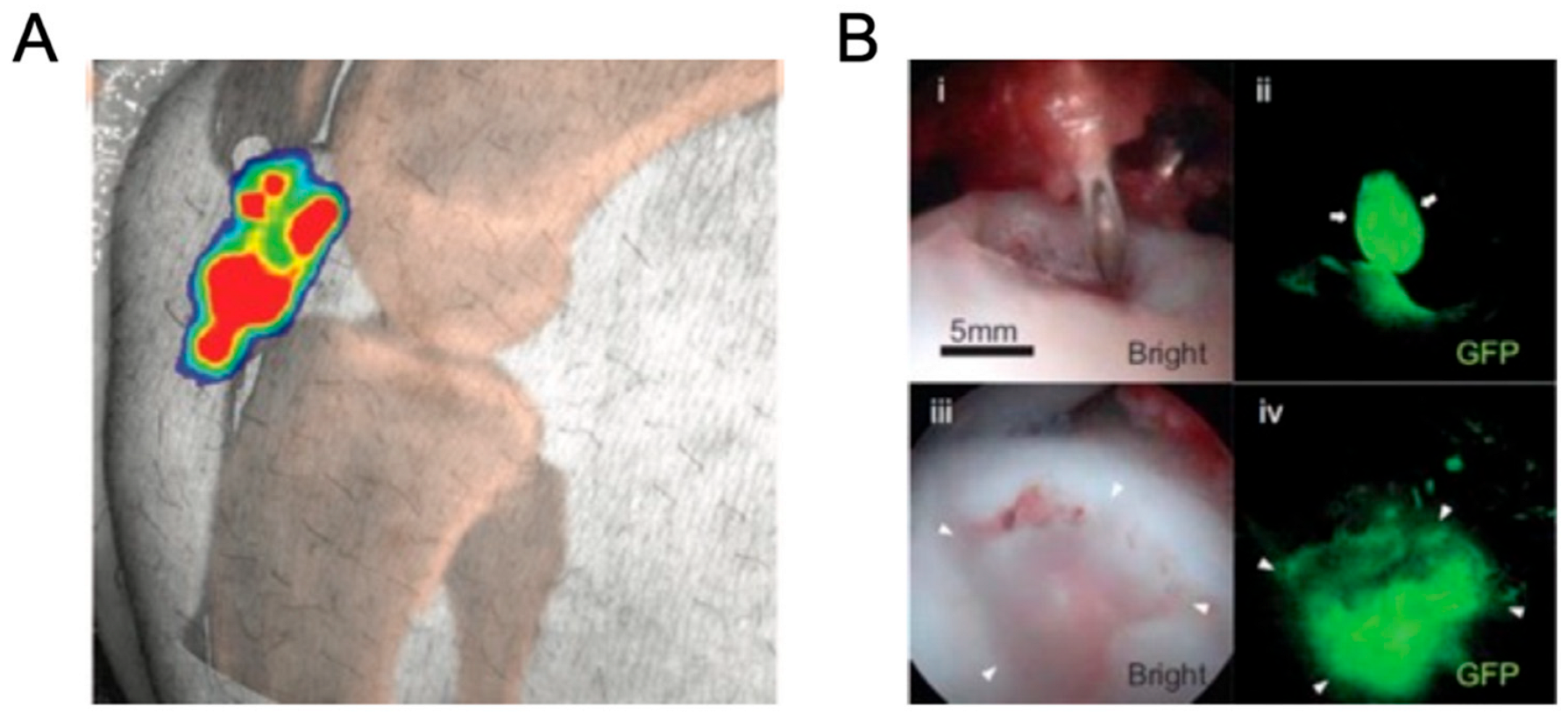
At the same time as Watano et al. [53], Kremen et al. [54] reported that the efficacy of cell transplantation therapy for ligament injury in pigs was tested using this non-invasive luciferase bioimaging. The researchers transplanted human mesenchymal cells that had been transduced with a lentiviral vector carrying a luciferase gene into the knee joint of a pig with a transected anterior cruciate ligament and examined the effect of mesenchymal cells on engraftment and ligament regeneration. In this approach, a knee joint of a live recipient pig was observed by a luminescence detection system (IVIS-Spectrum system from PerkinElmer Corp.) at 2 weeks after transplantation of human mesenchymal cells. As shown in Figure 3A, an accumulation of luciferase-labeled transplanted cells was detected at the site of ligament injury, indicating that the transplanted human mesenchymal cells engrafted at the site of injury [54]. Figure 3A in this review is reproduced from Ref. [54].
Transplantation of pig hearts and kidneys into humans has received considerable attention in recent years [56,57]. As a model animal for pig-to-human xenotransplantation, we transplanted bone marrow cells from luciferase-transgenic rats into pigs (rat-to-pig) and visualized the xenogeneic rat cells in the pig by luminescence imaging [58]. The rats used in this study are the world’s first rat strain developed by our research team and ubiquitously express the luciferase gene under the Rosa26 promoter. Already, these rats have contributed substantially to the advancement of the research field in transplantation and regenerative medicine [59,60]. Luminescence imaging of transplanted pigs over time for 15 days revealed that the rat bone marrow cells remained in the pig bone marrow for some days, and luminescence was continuously detected in the bone marrow. In our experiments, we used the IVIS-Spectrum CT system (PerkinElmer) for the luminescence detection [58]. The transplanted pig under general anesthesia was intravenously administered luciferin and immediately placed in this device, which was tightly shielded from light, and imaging was performed. It is extremely difficult to track fluorescent genes in transplanted cells over time non-invasively, even in deep tissue locations, such as in the lungs and bone marrow [59].
We determined the duration for which this luminescence imaging technique can be continued on the same individual animal. For this, we introduced the following: In our previous study with pigs, the IVIS device could only be observed for up to 15 days due to facility limitations in maintaining it [53,58]. As shown in Figure 4 (reproduced from Ref. [60]), we investigated how long luciferase-transgenic cells can be followed in the same animal by transplanting luciferase-transgenic rat islets and hepatocytes into wild-type rats [60]. Pancreatic islets from luciferase-transgenic rats were transplanted subcutaneously into wild-type diabetic rats in Figure 4A. Also, in Figure 4B, parenchymal hepatocytes from luciferase-transgenic rats were injected into the spleen of wild-type rats. Bioluminescence images were then acquired using the Lago X system (Spectral Instruments Imaging) over a 20–22 week period by intravenous luciferin injection through the tail vein of the recipient rats under general anesthesia. Typical bioluminescence images during the observation period in the recipient are shown in the upper part of each figure. The bottom row of each figure shows the change in bioluminescence intensity. The transplanted cell-derived luminescence was observed for 20–22 weeks, and the luminescence intensity did not decrease significantly after this prolonged observation [60]. Furthermore, chondrocyte cell sheets prepared from these luciferase-transfected rats were transplanted into osteochondral-deficient rats [61]. Continuous monitoring of the graft-derived luminescence by the IVIS system allowed tracking of the grafts for as long as 21 months. If the transplanted cells or tissues are successfully engrafted, luminescence from luciferase can be expected to be detectable in pigs for a long time.

4. Discussion

Conventional imaging systems have enabled anatomical and metabolic in vivo analysis in large animals. Recently, in vivo evaluation using fluorescent reporter genes has become the standard method [62,63], but fluorescent reporters have several disadvantages. Luminescent reporters have been used while working around this weakness. Luminescence imaging research has been technically improved mainly in small animals, but in recent years, where translational research has been actively conducted for clinical applications, luminescence imaging techniques have become feasible in pigs, which are large experimental animals. Here, we discuss the characteristic parameters (depth of detection, study period, and animal size) that are important for luciferase bioimaging in pigs and their comparison with fluorescence labeling.

4.1. Important Parameters for Luciferase Bioimaging in Pigs

4.1.1. Depth

Fluorescent genes, such as green fluorescent protein (GFP), have limitations in observing fluorescence in deep tissues. Since excitation light cannot pass through the tissue, only surface fluorescence can be captured [64,65]. Therefore, to capture the expression of fluorescent genes in deep tissue, one can either use biopsy to observe the expression of fluorescent genes with visual observation or isolate cells by dissecting the tissue and performing flow cytometry analysis. The luminescence detection by luciferase solves this problem. In fact, it has already been widely used for molecular imaging and dynamic analysis of transplanted cells in mice and rats, and Watano et al. have sensitively detected luminescence in organs deep in the pig body, i.e., lung and liver [53,58]. Furthermore, we have shown that luminescence can be detected even in the thick pig femur [58]; although CT and MRI allow anatomical observation within the bone marrow, molecular imaging is not possible with these approaches. These results indicate that luminescence from the luciferase–luciferin reaction is advantageous for molecular imaging of deep tissue.

4.1.2. Study Period

Research in cell therapy and organ transplantation involves dynamic analysis of cells and organs over a long period of one year or more [66,67]. For this purpose, the immunogenicity of the marker gene to be introduced is an important factor. In fact, even in syngeneic transplantation systems, the luminescence of the luciferase gene did not weaken even after more than one year [60], whereas the fluorescence of the GFP gene sometimes disappeared. In addition, fluorescent probes require excitation light, which damages cells and tissues [68]. Conversely, luminescence from the luciferin–luciferase reaction requires no excitation light and can be observed for a long period of time without causing tissue damage. Thus, luminescence from the luciferase–luciferin reaction is also effective for long-term molecular imaging.

4.1.3. Animal Size

With a larger animal, the detection target can be at a deeper location, and the life span can be longer, allowing for longer-term observation. Therefore, the depth to be detected and the observation period are related to the size of the animal. Currently, no technique exists that can perform luminescence detection in an animal the same size as an adult human. A detection system could be originally assembled using a super-sensitive camera, but it would require strict light shielding and would be difficult to collect quantitative data. An example of an animal that is anatomically and physiologically similar to a human is the pig [69], and a micromini pig is smaller than a miniature pig and is the same size as a child or young adult. Therefore, we suggest that translational research using the micromini pig is a first step toward clinical application. Watano et al. and we were able to observe temporal changes in luciferase luminescence (and detection of luminescence deep in organs) in micromini pigs [53,58]. Larger luminescence detection systems will eventually be needed for clinical applications. At the same time, we consider it necessary to lower the cost of the device to increase the frequency of testing in preclinical animals such as pigs.

4.1.4. Disadvantages of Luminescence Imaging for Research Using Pigs

We have discussed the advantages of luminescence imaging for research using pigs. In preclinical research, bridge studies using large animals, such as pigs, are indispensable. Although monkeys, dogs, and sheep can be used as animal models, excluding small animals, such as mice and rats, there are no in vivo luminescence imaging studies using these animals at this time. Therefore, pigs are the first choice of animals for luminescence imaging experiments. However, there is a limitation as only a small breed can be used, i.e., a micromini pig weighing up to 20 kg, or the animal cannot fit into the luminescence detection device. This issue will be resolved as the equipment becomes larger, as mentioned above. We have already discussed the advantages of luminescence imaging for research using pigs.

4.2. Comparison of Fluorescent and Luminescent Labeling in Gene and Cell Therapy Research Using Pigs

In small animal-based studies, fluorescence (e.g., GFP) and luminescence labeling are used as optical imaging techniques to trace cells and facilitate fast image acquisition. Moreover, experiments can be repeated using the same animals. The following two features suggest that luciferase is more effective for cell tracing in pigs than for fluorescent labeling.

4.2.1. Phototoxicity

Fluorescence labeling is excellent for molecular as well as cellular analyses; however, it is not suitable for prolonged fluorescence imaging because the excitation light causes cellular and tissue damage. In addition, using inherently fluorescent proteins, such as GFP, does not require the administration of exogenous fluorescent dyes; however, given the wavelengths of excitation and fluorescence emission, this method is limited to surface structures (1–3 mm depth) of experimental animals [70]. Nakamura et al. [55] created a model of cartilage defect in pigs and transplanted GFP-labeled mesenchymal stem cells derived from allogeneic pig synovium into the defect area [55]. As shown in Figure 3B (reproduced from Ref. [55]), GFP-labeled cells on the surface of the transplanted site could be visualized. However, imaging of deeper organs, where excitation light does not penetrate, requires some modifications. Although the applicability of single-molecule imaging remains challenging due to inadequate optical intensity [71], luminescence imaging offers the major advantage of almost nil background, allowing the acquisition of relatively quantitative data [72]. For example, as shown in Figure 3A, Kremen et al. [54] used luminescence imaging to detect the luminescence of human mesenchymal cells deep within the live pig knee joint in a short time with high sensitivity. Unlike GFP-labeling, the luciferase–luciferin reaction does not require excitation light, so prolonged imaging does not damage cells or tissues. In addition, no surgical exposure is required for the area of interest. Thus, luminescence imaging provides non-invasive, deep, and quantitative data in a short time (typically several seconds to tens of seconds) without phototoxicity.

4.2.2. Immunogenicity

Some proteins used as markers may be immunogenic, and cells expressing them may be cleared faster; even in syngeneic transplant systems, transplanted cells expressing fluorescent proteins may be lost during the observation period. Many marker proteins are of xenogeneic origin and are considered immunogenic [59]; Hakamata et al. reported that while luciferase-expressing skin grafts survived for more than 100 days, GFP-expressing skin grafts were no longer visible after 10 days. These data indicate that luciferase is less immunogenic than fluorescent proteins and has a lower risk of rejection.
Taken together, bioimaging of the distribution of viral vectors [53] and transplanted cells [54,58] has now become possible in large animal bodies, such as pigs, due to the advent of luciferase-based approaches.
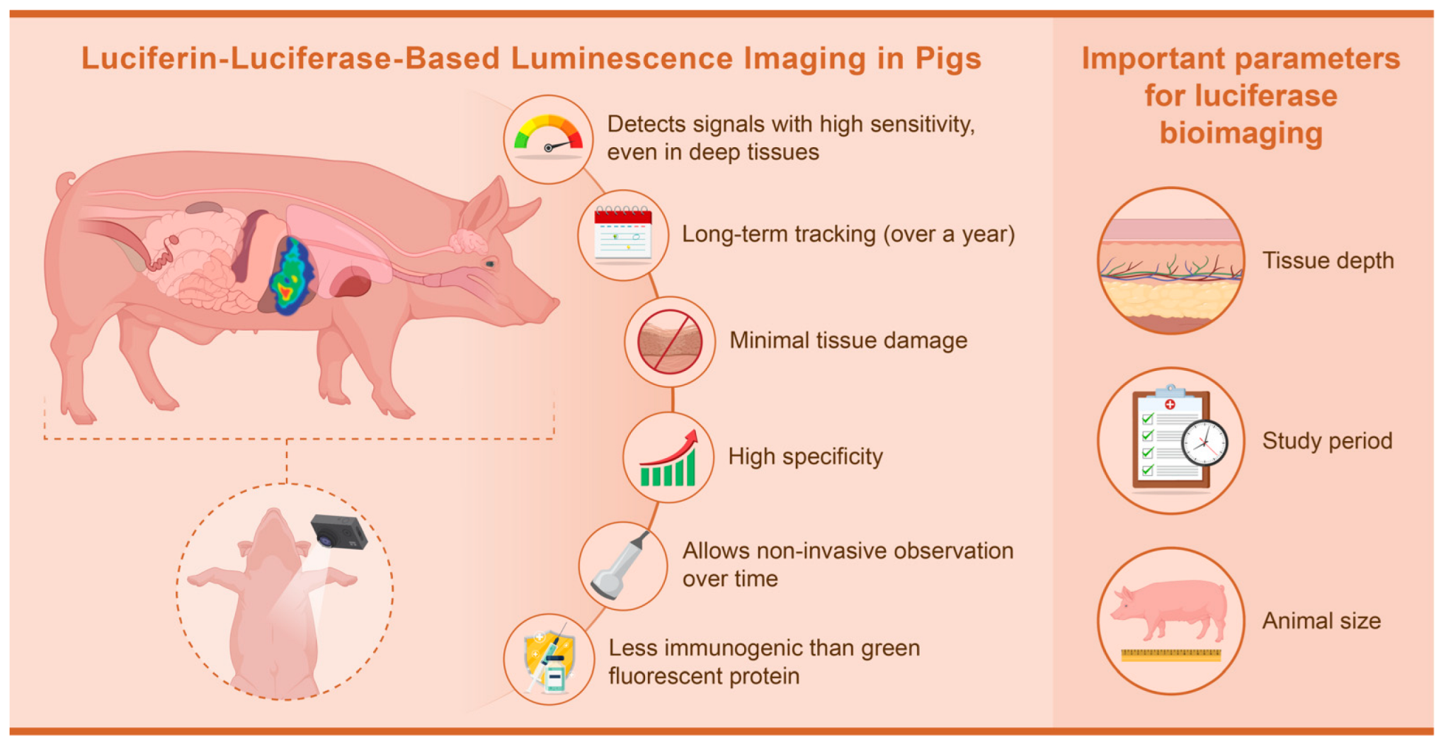
Figure 5 summarizes the characteristics of luminescence imaging using pigs described earlier.

4.3. Specific Points for Luminescence Bioimaging

So far, we described the key aspects of luciferase luminescence imaging in practical use with pigs. In this session, several points that are important to know when adopting luciferase luminescence imaging in one’s own research project are introduced.

4.3.1. Imaging Mechanisms

The luciferase catalyzes the oxidation of luciferin in the presence of ATP and magnesium ions, producing light as the resulting excited state of oxyluciferin returns to its basal state [73]. Only a few organisms, such as fireflies, have luciferase and luciferin naturally [74]. Therefore, prior to performing luminescence imaging, it is necessary to either transplant luciferase-expressing cells into the animals to be observed or to generate luciferase-expressing animals. Luciferin is then administered to target animals immediately before observation to produce luminescence and enable imaging. The luminescence detection system consists of a high-sensitivity CCD camera and lens contained in a light-shielding dark box, which can detect luminescence from live animals inside the dark box. A typical system is PerkinElmer’s IVIS-Spectrum system, for example. For imaging a pig, the entire device must be completely covered with light-shielding curtains because the pig protrudes from the imaging table [58].

4.3.2. Efficacy and Sensitivity

As discussed in Section 4, the detection efficacy of bioluminescence imaging is indeed magnitudes higher than fluorescence imaging, as even targets deep within the pig (approximately 5–10 cm) can be detected. Recently developed near-infrared light-producing substrates allow for clearer imaging even deep in vivo [75]. The imaging system utilizes light in the optical spectrum with a peak at 562 nm produced by firefly-derived luciferase in combination with its natural substrate, luciferin. However, light in the optical spectrum is absorbed by hemoglobin and melanin, which are abundant in the body and thus have low tissue permeability, limiting detection sensitivity. Kuchimaru et al. developed a substrate that produces light in the near-infrared spectrum (650 nm <), which is not easily absorbed by the body and has excellent tissue permeability [75]. This substrate enables the detection of targets deep within the body with 40-fold higher sensitivity.

4.3.3. Challenges and Limitations

Many researchers are interested in applying this imaging technology to humans. However, to date, the materials for luminescence imaging technology have been based on transgenic firefly-derived enzymes or their mutants and not on human-derived genes [71,76,77]. Applying this technology to humans is, therefore, difficult under current conditions. Nakamura and Maki are trying to create artificial materials that react and emit luminescence with substances that are originally present in mammalian organisms [78]. Overcoming the limitation that the materials for the luminescent labeling are derived from non-human genes may allow for clinical applications.

4.3.4. Risk Assessment

The luciferase gene and its substrate, luciferin, have no known oncogenic, genotoxic, or pathogenic properties. Immunogenicity is also thought to be lower than that of fluorescent reporter genes (Section 4.2.2). Unlike fluorescence imaging, luciferase does not require excitation light and does not cause cytotoxic damage due to phototoxicity, as described in Section 4.2.1. No radiation exposure in luminescence imaging, unlike CT or PET (although some models can acquire CT images at the same time). No special protective room is needed, unlike MRI, because it does not generate a strong magnetic field.

4.3.5. Regulatory and Ethical Issues

As mentioned earlier (Section 4.3.1), because luciferase is a foreign gene, the transplantation of luciferase-expressing cells into animals or the generation of luciferase-expressing animals classifies those animals as “genetically modified organisms” in Japan. Therefore, it is subject to the Cartagena Act. The experiments must be conducted in appropriate facilities in accordance with the “Law Concerning the Conservation of Biological Diversity through Regulations on the Use of Living Modified Organisms (Cartagena Act)” and the safety management regulations of the organization to which the researcher belongs. Experiments on animals must be conducted in accordance with the relevant laws and regulations published by the Ministry of the Environment and the Ministry of Education, Culture, Sports, Science, and Technology and must follow the rules of the Ethics Committee for Animal Experiments at the institution.

4.3.6. Diagnostic Applications

The dynamic analysis of transplanted cells and injected viral vectors in the body of a pig, as introduced in this review (Section 3), cannot be demonstrated using instruments such as CT and MRI and requires a new modality of analysis, such as with the luciferase–luciferin reaction. For example, this modality can be used for highly sensitive detection and dynamic analysis of genetically modified immune cells in chimeric antigen receptor-T cell therapy [79] and viral vectors in gene therapy [80]. In principle, in vivo bioimaging is capable of labeling one cell, making it more precise than CT, MRI, ultrasound, and other imaging techniques used in human biomedical diagnostics. If the visualization of one cell, such as a metastatic tumor cell, is possible [76], then treatment can be initiated before the patient’s condition becomes serious.

5. Conclusions

Non-invasive imaging is an essential technique to follow the course of a procedure in the same individual over a long period of time. In preclinical studies with pigs, anatomic and metabolic analyses have been performed using conventional imaging modalities, such as CT and MRI. In the recent field of gene and cell therapy, imaging using fluorescent reporters is a central technique, but it is not suitable for observing deep tissue in large, thick animals, such as pigs. In contrast, biological imaging using a luminescent reporter can capture signals with high sensitivity even in deep tissues and allows non-invasive observation over time. In this review, we introduced examples of actual luminescence imaging using pigs. Through these examples, we also summarized important parameters to consider when using pigs and issues for clinical application. This review lays a foundation for further research in this field.

6. Future Directions

Genes and substrates for luminescence imaging to date have been based on exogenous genes or their mutants derived from fireflies and other organisms [71,76,77]. Therefore, the proteins involved in luminescence may themselves induce an immune response in experimental animals. Unless the recipient animals are immunodeficient, reporter genes of the same animal species should be designed and used for long-term in vivo studies. This challenge hinders the clinical application of luminescence imaging technology and indicates the need to overcome these limitations for clinical application.

Author Contributions

Conceptualization, T.A. and E.K.; writing—review and editing, T.A., K.E., Y.H. and E.K. All authors have read and agreed to the published version of the manuscript.

Funding

This research received no external funding.

Acknowledgments

We extend our heartfelt appreciation to the following individuals whose contributions have been vital to the success of this research: Masayo Kumagai for her long-term care and experimental assistance with luciferase-transgenic rats; Takahiro Ohnuki, Toru Wakui, and Kazushi Miyazawa for their care and experimental assistance with pigs at Jichi Medical University; and Kumiko Uchida for her administrative support. We also acknowledge the dedicated technical staff of the Center for the Development of Advanced Medical Technology, Jichi Medical University, for their involvement in the feeding and management of pigs.

Conflicts of Interest

The authors declare no conflicts of interest.

References

  1. Mettler, F.A. Essentials of Radiology, 2nd ed.; Elsevier Saunders: Philadelphia, PA, USA, 2005; ISBN 978-0-7216-0527-2. [Google Scholar]
  2. Jensen, J.A. Medical Ultrasound Imaging. Prog. Biophys. Mol. Biol. 2007, 93, 153–165. [Google Scholar] [CrossRef] [PubMed]
  3. Hussain, S.; Mubeen, I.; Ullah, N.; Shah, S.S.U.D.; Khan, B.A.; Zahoor, M.; Ullah, R.; Khan, F.A.; Sultan, M.A. Modern Diagnostic Imaging Technique Applications and Risk Factors in the Medical Field: A Review. Biomed. Res. Int. 2022, 2022, 5164970. [Google Scholar] [CrossRef] [PubMed]
  4. Smith-Bindman, R.; Miglioretti, D.L.; Johnson, E.; Lee, C.; Feigelson, H.S.; Flynn, M.; Greenlee, R.T.; Kruger, R.L.; Hornbrook, M.C.; Roblin, D.; et al. Use of Diagnostic Imaging Studies and Associated Radiation Exposure for Patients Enrolled in Large Integrated Health Care Systems, 1996–2010. JAMA 2012, 307, 2400–2409. [Google Scholar] [CrossRef] [PubMed]
  5. Pulumati, A.; Pulumati, A.; Dwarakanath, B.S.; Verma, A.; Papineni, R.V.L. Technological Advancements in Cancer Diagnostics: Improvements and Limitations. Cancer Rep. 2023, 6, e1764. [Google Scholar] [CrossRef] [PubMed]
  6. Gandhi, D.; Ahuja, K.; Grover, H.; Sharma, P.; Solanki, S.; Gupta, N.; Patel, L. Review of X-ray and Computed Tomography Scan Findings with a Promising Role of Point of Care Ultrasound in COVID-19 Pandemic. World J. Radiol. 2020, 12, 195–203. [Google Scholar] [CrossRef]
  7. Seeram, E. Computed Tomography: Physics and Technology: A Self Assessment Guide, 2nd ed.; Wiley-Blackwell: Hoboken, NJ, USA, 2022; ISBN 978-1-119-81932-5. [Google Scholar]
  8. Meulepas, J.M.; Ronckers, C.M.; Smets, A.M.J.B.; Nievelstein, R.A.J.; Jahnen, A.; Lee, C.; Kieft, M.; Laméris, J.S.; van Herk, M.; Greuter, M.J.W.; et al. Leukemia and Brain Tumors among Children after Radiation Exposure from CT Scans: Design and Methodological Opportunities of the Dutch Pediatric CT Study. Eur. J. Epidemiol. 2014, 29, 293–301. [Google Scholar] [CrossRef]
  9. Beckett, K.R.; Moriarity, A.K.; Langer, J.M. Safe Use of Contrast Media: What the Radiologist Needs to Know. Radiographics 2015, 35, 1738–1750. [Google Scholar] [CrossRef]
  10. Yeh, B.M.; FitzGerald, P.F.; Edic, P.M.; Lambert, J.W.; Colborn, R.E.; Marino, M.E.; Evans, P.M.; Roberts, J.C.; Wang, Z.J.; Wong, M.J.; et al. Opportunities for New CT Contrast Agents to Maximize the Diagnostic Potential of Emerging Spectral CT Technologies. Adv. Drug Deliv. Rev. 2017, 113, 201–222. [Google Scholar] [CrossRef]
  11. Burstein, D. MRI for Development of Disease-Modifying Osteoarthritis Drugs. NMR Biomed. 2006, 19, 669–680. [Google Scholar] [CrossRef]
  12. Pando-Naude, V.; Toxto, S.; Fernandez-Lozano, S.; Parsons, C.E.; Alcauter, S.; Garza-Villarreal, E.A. Gray and White Matter Morphology in Substance Use Disorders: A Neuroimaging Systematic Review and Meta-Analysis. Transl. Psychiatry 2021, 11, 29. [Google Scholar] [CrossRef]
  13. Weiskopf, N.; Edwards, L.J.; Helms, G.; Mohammadi, S.; Kirilina, E. Quantitative Magnetic Resonance Imaging of Brain Anatomy and in Vivo Histology. Nat. Rev. Phys. 2021, 3, 570–588. [Google Scholar] [CrossRef]
  14. Zugni, F.; Mariani, L.; Lambregts, D.M.J.; Maggioni, R.; Summers, P.E.; Granata, V.; Pecchi, A.; Di Costanzo, G.; De Muzio, F.; Cardobi, N.; et al. Whole-Body MRI in Oncology: Acquisition Protocols, Current Guidelines, and Beyond. Radiol. Med. 2024, 129, 1352–1368. [Google Scholar] [CrossRef] [PubMed]
  15. Gowrishankar, G.; Namavari, M.; Jouannot, E.B.; Hoehne, A.; Reeves, R.; Hardy, J.; Gambhir, S.S. Investigation of 6-[18F]-Fluoromaltose as a Novel PET Tracer for Imaging Bacterial Infection. PLoS ONE 2014, 9, e107951. [Google Scholar] [CrossRef]
  16. Saboury, B.; Morris, M.A.; Farhadi, F.; Nikpanah, M.; Werner, T.J.; Jones, E.C.; Alavi, A. Reinventing Molecular Imaging with Total-Body PET, Part I: Technical Revolution in Evolution. PET Clin. 2020, 15, 427–438. [Google Scholar] [CrossRef]
  17. Haroon, A.; Zumla, A.; Bomanji, J. Role of Fluorine 18 Fluorodeoxyglucose Positron Emission Tomography-Computed Tomography in Focal and Generalized Infectious and Inflammatory Disorders. Clin. Infect. Dis. 2012, 54, 1333–1341. [Google Scholar] [CrossRef]
  18. Mohan, A.-M.; Beindorff, N.; Brenner, W. Nuclear Medicine Imaging Procedures in Oncology. In Metastasis; Stein, U.S., Ed.; Methods in Molecular Biology; Springer: New York, NY, USA, 2021; Volume 2294, pp. 297–323. ISBN 978-1-07-161349-8. [Google Scholar]
  19. Gonzalez Rojas, N.; Cesarini, M.E.; Peker, G.; Da Prat, G.A.; Etcheverry, J.L.; Gatto, E.M. Review of Huntington’s Disease: From Basics to Advances in Diagnosis and Treatment. J. Neurol. Res. 2022, 12, 93–113. [Google Scholar] [CrossRef]
  20. Malaiya, A.; Singhai, M.; Singh, M.; Prajapati, S.K.; Choudhury, H.; Fatima, M.; Alexander, A.; Dubey, S.K.; Greish, K.; Kesharwani, P. Recent Update on the Alzheimer’s Disease Progression, Diagnosis and Treatment Approaches. Curr. Drug Targets 2022, 23, 978–1001. [Google Scholar] [CrossRef]
  21. Katal, S.; Eibschutz, L.S.; Saboury, B.; Gholamrezanezhad, A.; Alavi, A. Advantages and Applications of Total-Body PET Scanning. Diagnostics 2022, 12, 426. [Google Scholar] [CrossRef]
  22. Chelikam, N.; Vyas, A.; Desai, R.; Khan, N.; Raol, K.; Kavarthapu, A.; Kamani, P.; Ibrahim, G.; Madireddy, S.; Pothuru, S.; et al. Past and Present of Point-of-Care Ultrasound (PoCUS): A Narrative Review. Cureus 2023, 15, e50155. [Google Scholar] [CrossRef]
  23. Chan, V.; Perlas, A. Basics of Ultrasound Imaging. In Atlas of Ultrasound-Guided Procedures in Interventional Pain Management; Narouze, S.N., Ed.; Springer: New York, NY, USA, 2011; pp. 13–19. ISBN 978-1-4419-1679-2. [Google Scholar]
  24. Barik, S.; Spring, J.E.; Jones, M.H.; Luck, C.A. Routine Ultrasound Scanning in Pregnancy. The Benefits Are Clinical. BMJ 1993, 307, 559. [Google Scholar] [CrossRef]
  25. Bourgioti, C.; Konidari, M.; Gourtsoyianni, S.; Moulopoulos, L.A. Imaging during Pregnancy: What the Radiologist Needs to Know. Diagn. Interv. Imaging 2021, 102, 593–603. [Google Scholar] [CrossRef] [PubMed]
  26. Albakri, A.A.; Alzahrani, M.M.; Alghamdi, S.H. Medical Imaging in Pregnancy: Safety, Appropriate Utilization, and Alternative Modalities for Imaging Pregnant Patients. Cureus 2024, 16, e54346. [Google Scholar] [CrossRef] [PubMed]
  27. Quarato, C.M.I.; Lacedonia, D.; Salvemini, M.; Tuccari, G.; Mastrodonato, G.; Villani, R.; Fiore, L.A.; Scioscia, G.; Mirijello, A.; Saponara, A.; et al. A Review on Biological Effects of Ultrasounds: Key Messages for Clinicians. Diagnostics 2023, 13, 855. [Google Scholar] [CrossRef]
  28. Kobayashi, E.; Hanazono, Y.; Kunita, S. Swine used in the Medical University: Overview of 20 years of experience. Exp. Anim. 2018, 67, 7–13. [Google Scholar] [CrossRef] [PubMed]
  29. Greco, A.; Meomartino, L.; Gnudi, G.; Brunetti, A.; Di Giancamillo, M. Imaging Techniques in Veterinary Medicine. Part II: Computed Tomography, Magnetic Resonance Imaging, Nuclear Medicine. Eur. J. Radiol. Open 2023, 10, 100467. [Google Scholar] [CrossRef] [PubMed]
  30. Aziz, A.; Pane, S.; Iacovacci, V.; Koukourakis, N.; Czarske, J.; Menciassi, A.; Medina-Sánchez, M.; Schmidt, O.G. Medical Imaging of Microrobots: Toward In Vivo Applications. ACS Nano 2020, 14, 10865–10893. [Google Scholar] [CrossRef]
  31. Barbagianni, M.S.; Gouletsou, P.G. Modern Imaging Techniques in the Study and Disease Diagnosis of the Mammary Glands of Animals. Vet. Sci. 2023, 10, 83. [Google Scholar] [CrossRef]
  32. Gu, Y.; Sun, Y.; Wang, X.; Li, H.; Qiu, J.; Lu, W. Application of photoacoustic computed tomography in biomedical imaging: A literature review. Bioeng. Transl. Med. 2023, 8, e10419. [Google Scholar] [CrossRef]
  33. Hirai, M.; Sakurada, T.; Ikeda, T.; Monden, Y.; Shimoizumi, H.; Yamagata, T. Developmental changes of the neural mechanisms underlying Level 2 visual perspective-taking: A functional near-infrared spectroscopy study. Dev. Psychobiol. 2022, 64, e22229. [Google Scholar] [CrossRef]
  34. Noel, S.J.; Jørgensen, H.J.H.; Knudsen, K.E.B. The use of near-infrared spectroscopy (NIRS) to determine the energy value of individual feedstuffs and mixed diets for pigs. Anim. Feed. Sci. Technol. 2022, 283, 115156. [Google Scholar] [CrossRef]
  35. James, M.L.; Gambhir, S.S. A molecular imaging primer: Modalities, imaging agents, and applications. Physiol. Rev. 2012, 92, 897–965. [Google Scholar] [CrossRef] [PubMed]
  36. Saito-Moriya, R.; Obata, R.; Maki, S.A. Near-Infrared luciferin Analogs for in vivo optical Imaging. In Bioluminescence—Technology and Biology; Suzuki, H., Ogoh, K., Eds.; IntechOpen: Rijeka, Croatia, 2021; ISBN 978-1-83962-385-1. [Google Scholar]
  37. Camacho, P.; Fan, H.; Liu, Z.; He, J.-Q. Large mammalian animal models of heart disease. J. Cardiovasc. Dev. Dis. 2016, 3, 30. [Google Scholar] [CrossRef] [PubMed]
  38. Matsumura, Y.; Higashi, A.; Izawa, Y.; Hishikawa, S. Organ perfusion during partial REBOA in haemorrhagic shock: Dynamic 4D-CT analyses in swine. Sci. Rep. 2022, 12, 18745. [Google Scholar] [CrossRef] [PubMed]
  39. Sasanuma, H.; Takahashi, T.; Kawai, S.; Saitsu, A.; Kurashina, W.; Iijima, Y.; Saito, T.; Takeshita, K. Morphological and histological evaluation of the tendon-bone junction in porcine shoulders to create a rotator cuff tear and repair model. J. Orthop. Sci. 2023. [Google Scholar] [CrossRef]
  40. Tomaru, Y.; Sugaya, H.; Yoshioka, T.; Arai, N.; Abe, T.; Tsukagoshi, Y.; Kamada, H.; Yamazaki, M.; Mishima, H. Effects of bone marrow-derived mesenchymal stem cell transplantation in piglet legg-calve-perthes disease models: A pilot study. J. Pediatr. Orthop. B 2023, 33, 358–362. [Google Scholar] [CrossRef]
  41. Miura, M.; Miura, Y.; Iwazu, Y.; Mukai, H.; Sugiura, T.; Suzuki, Y.; Kato, M.; Kano, M.; Nagata, D.; Shiizaki, K.; et al. Removal of calciprotein particles from the blood using an adsorption column improves prognosis of hemodialysis miniature pigs. Sci. Rep. 2023, 13, 15026. [Google Scholar] [CrossRef]
  42. Fil, J.E.; Joung, S.; Zimmerman, B.J.; Sutton, B.P.; Dilger, R.N. High-Resolution Magnetic Resonance Imaging-Based Atlases for the Young and Adolescent Domesticated Pig (Sus Scrofa). J. Neurosci. Methods 2021, 354, 109107. [Google Scholar] [CrossRef]
  43. Simchick, G.; Shen, A.; Campbell, B.; Park, H.J.; West, F.D.; Zhao, Q. Pig brains have homologous resting-state networks with human brains. Brain Connect. 2019, 9, 566–579. [Google Scholar] [CrossRef]
  44. Norris, C.; Lisinski, J.; McNeil, E.; VanMeter, J.W.; VandeVord, P.; LaConte, S.M. MRI brain templates of the male Yucatan minipig. Neuroimage 2021, 235, 118015. [Google Scholar] [CrossRef]
  45. Hellman, A.; Maietta, T.; Clum, A.; Byraju, K.; Raviv, N.; Staudt, M.D.; Jeannotte, E.; Nalwalk, J.; Belin, S.; Poitelon, Y.; et al. Development of a common peroneal nerve injury model in domestic swine for the study of translational neuropathic pain Treatments. J. Neurosurg. 2021, 135, 1516–1523. [Google Scholar] [CrossRef]
  46. Alstrup, A.K.O.; Dollerup, M.R.; Simonsen, M.I.T.; Vendelbo, M.H. Preclinical imaging studies: Protocols, preparation, anesthesia, and animal care. Semin. Nucl. Med. 2023, 53, 570–576. [Google Scholar] [CrossRef] [PubMed]
  47. Cumming, P.; Scheidegger, M.; Dornbierer, D.; Palner, M.; Quednow, B.B.; Martin-Soelch, C. Molecular and functional imaging studies of psychedelic drug action in animals and humans. Molecules 2021, 26, 2451. [Google Scholar] [CrossRef] [PubMed]
  48. Pilz, J.; Gloddek, N.; Lindheimer, F.; Lindner, M.J.; Puhr-Westerheide, D.; Ümütlü, M.; Cyran, C.; Seidensticker, M.; Lindner, R.; Kraetzl, M.; et al. Functional maturation and longitudinal imaging of intraportal neonatal porcine islet grafts in genetically diabetic pigs. Am. J. Transplant. 2024, 24, 1395–1405. [Google Scholar] [CrossRef] [PubMed]
  49. Wada, H.; Hyun, H.; Vargas, C.; Gravier, J.; Park, G.; Gioux, S.; Frangioni, J.V.; Henary, M.; Choi, H.S. Pancreas-targeted NIR fluorophores for dual-channel image-guided abdominal surgery. Theranostics 2015, 5, 1–11. [Google Scholar] [CrossRef]
  50. Gyöngyösi, M.; Hemetsberger, R.; Wolbank, S.; Pichler, V.; Kaun, C.; Posa, A.; Petrasi, Z.; Petnehazy, Ö.; Hofer-Warbinek, R.; De Martin, R.; et al. Delayed recovery of myocardial blood flow after intracoronary stem cell administration. Stem Cell Rev. Rep. 2011, 7, 616–623. [Google Scholar] [CrossRef]
  51. Rodenberg, E.J.; Patel, D.S.; Shirley, B.; Young, B.W.; Taylor, A.F.; Steidinger, H.R.; Fisher, S.J.; Patel, A.N. Catheter-based retrograde coronary sinus infusion is a practical delivery technique for introducing biological molecules into the cardiac system. Catheter. Cardiovasc. Interv. 2019, 94, 669–676. [Google Scholar] [CrossRef]
  52. Srivastava, R.K.; Jablonska, A.; Chu, C.; Gregg, L.; Bulte, J.W.M.; Koehler, R.C.; Walczak, P.; Janowski, M. Biodistribution of glial progenitors in a three dimensional-printed model of the piglet cerebral ventricular system. Stem Cells Dev. 2019, 28, 515–527. [Google Scholar] [CrossRef]
  53. Watano, R.; Ohmori, T.; Hishikawa, S.; Sakata, A.; Mizukami, H. Utility of microminipigs for evaluating liver-mediated gene expression in the presence of neutralizing antibody against vector capsid. Gene Ther. 2020, 27, 427–434. [Google Scholar] [CrossRef]
  54. Kremen, T.J.; Stefanovic, T.; Tawackoli, W.; Salehi, K.; Avalos, P.; Reichel, D.; Perez, M.J.; Glaeser, J.D.; Sheyn, D. A translational porcine model for human cell-based therapies in the treatment of posttraumatic osteoarthritis after anterior cruciate ligament injury. Am. J. Sports Med. 2020, 48, 3002–3012. [Google Scholar] [CrossRef]
  55. Nakamura, T.; Sekiya, I.; Muneta, T.; Hatsushika, D.; Horie, M.; Tsuji, K.; Kawarasaki, T.; Watanabe, A.; Hishikawa, S.; Fujimoto, Y.; et al. Arthroscopic, histological and mri analyses of cartilage repair after a minimally invasive method of transplantation of allogeneic synovial mesenchymal stromal cells into cartilage defects in pigs. Cytotherapy 2012, 14, 327–338. [Google Scholar] [CrossRef]
  56. Griffith, B.P.; Goerlich, C.E.; Singh, A.K.; Rothblatt, M.; Lau, C.L.; Shah, A.; Lorber, M.; Grazioli, A.; Saharia, K.K.; Hong, S.N.; et al. Genetically modified porcine-to-human cardiac xenotransplantation. N. Engl. J. Med. 2022, 387, 35–44. [Google Scholar] [CrossRef] [PubMed]
  57. Montgomery, R.A.; Stern, J.M.; Lonze, B.E.; Tatapudi, V.S.; Mangiola, M.; Wu, M.; Weldon, E.; Lawson, N.; Deterville, C.; Dieter, R.A.; et al. Results of two cases of pig-to-human kidney xenotransplantation. N. Engl. J. Med. 2022, 386, 1889–1898. [Google Scholar] [CrossRef] [PubMed]
  58. Abe, T.; Endo, K.; Hanazono, Y.; Kobayashi, E. In vivo luciferin–luciferase reaction in pigs using xenogeneic rat bone marrow transplantation: A case report and consideration of recent advance. Int. J. Mol. Sci. 2024, 25, 8609. [Google Scholar] [CrossRef] [PubMed]
  59. Hakamata, Y.; Murakami, T.; Kobayashi, E. “Firefly rats” as an organ/cellular source for long-term in vivo bioluminescent imaging. Transplantation 2006, 81, 1179–1184. [Google Scholar] [CrossRef]
  60. Kobayashi, E.; Hakamata, Y.; Enosawa, S.; Shang, K.-M.; Komatsu, H. Firefly rats: Illuminating the scientific community in transplantation research. Cell Transplant. 2024, 33, 9636897231224174. [Google Scholar] [CrossRef]
  61. Takaku, Y.; Murai, K.; Ukai, T.; Ito, S.; Kokubo, M.; Satoh, M.; Kobayashi, E.; Yamato, M.; Okano, T.; Takeuchi, M.; et al. In Vivo Cell Tracking by Bioluminescence Imaging after Transplantation of Bioengineered Cell Sheets to the Knee Joint. Biomaterials 2014, 35, 2199–2206. [Google Scholar] [CrossRef]
  62. Ashmore-Harris, C.; Iafrate, M.; Saleem, A.; Fruhwirth, G.O. Non-Invasive Reporter Gene Imaging of Cell Therapies, Including T Cells and Stem Cells. Mol. Ther. 2020, 28, 1392–1416. [Google Scholar] [CrossRef]
  63. Salinas-Jazmín, N.; Rosas-Cruz, A.; Velasco-Velázquez, M. Reporter Gene Systems for the Identification and Characterization of Cancer Stem Cells. World J. Stem Cells 2021, 13, 861–876. [Google Scholar] [CrossRef]
  64. Yoon, S.; Cheon, S.Y.; Park, S.; Lee, D.; Lee, Y.; Han, S.; Kim, M.; Koo, H. Recent Advances in Optical Imaging through Deep Tissue: Imaging Probes and Techniques. Biomater. Res. 2022, 26, 57. [Google Scholar] [CrossRef]
  65. Refaat, A.; Yap, M.L.; Pietersz, G.; Walsh, A.P.G.; Zeller, J.; Del Rosal, B.; Wang, X.; Peter, K. In Vivo Fluorescence Imaging: Success in Preclinical Imaging Paves the Way for Clinical Applications. J. Nanobiotechnol. 2022, 20, 450. [Google Scholar] [CrossRef]
  66. Wu, D.; Lombaert, I.M.A.; DeLeon, M.; Pradhan-Bhatt, S.; Witt, R.L.; Harrington, D.A.; Trombetta, M.G.; Passineau, M.J.; Farach-Carson, M.C. Immunosuppressed Miniswine as a Model for Testing Cell Therapy Success: Experience with Implants of Human Salivary Stem/Progenitor Cell Constructs. Front. Mol. Biosci. 2021, 8, 711602. [Google Scholar] [CrossRef] [PubMed]
  67. Du, X.; Chang, S.; Guo, W.; Zhang, S.; Chen, Z.K. Progress in Liver Transplant Tolerance and Tolerance-Inducing Cellular Therapies. Front. Immunol. 2020, 11, 1326. [Google Scholar] [CrossRef] [PubMed]
  68. Magidson, V.; Khodjakov, A. Circumventing Photodamage in Live-Cell Microscopy. In Methods in Cell Biology; Elsevier: Amsterdam, The Netherlands, 2013; Volume 114, pp. 545–560. ISBN 978-0-12-407761-4. [Google Scholar]
  69. Lunney, J.K.; Van Goor, A.; Walker, K.E.; Hailstock, T.; Franklin, J.; Dai, C. Importance of the Pig as a Human Biomedical Model. Sci. Transl. Med. 2021, 13, eabd5758. [Google Scholar] [CrossRef] [PubMed]
  70. Allport, J.R.; Weissleder, R. In Vivo Imaging of Gene and Cell Therapies. Exp. Hematol. 2001, 29, 1237–1246. [Google Scholar] [CrossRef]
  71. Saito-Moriya, R.; Nakayama, J.; Kamiya, G.; Kitada, N.; Obata, R.; Maki, S.A.; Aoyama, H. How to select firefly luciferin analogues for in vivo imaging. Int. J. Mol. Sci. 2021, 22, 1848. [Google Scholar] [CrossRef]
  72. Srinivas, M.; Aarntzen, E.H.J.G.; Bulte, J.W.M.; Oyen, W.J.; Heerschap, A.; de Vries, I.J.M.; Figdor, C.G. Imaging of cellular therapies. Adv. Drug Deliv. Rev. 2010, 62, 1080–1093. [Google Scholar] [CrossRef]
  73. Sharifian, S.; Homaei, A.; Hemmati, R.; Luwor, R.B.; Khajeh, K. The Emerging Use of Bioluminescence in Medical Research. Biomed. Pharmacother. 2018, 101, 74–86. [Google Scholar] [CrossRef]
  74. Kiyama, M.; Saito, R.; Iwano, S.; Obata, R.; Niwa, H.; Maki, S.A. Multicolor Bioluminescence Obtained Using Firefly Luciferin. Curr. Top. Med. Chem. 2016, 16, 2648–2655. [Google Scholar] [CrossRef]
  75. Kuchimaru, T.; Iwano, S.; Kiyama, M.; Mitsumata, S.; Kadonosono, T.; Niwa, H.; Maki, S.; Kizaka-Kondoh, S. A Luciferin Analogue Generating Near-Infrared Bioluminescence Achieves Highly Sensitive Deep-Tissue Imaging. Nat. Commun. 2016, 7, 11856. [Google Scholar] [CrossRef]
  76. Iwano, S.; Sugiyama, M.; Hama, H.; Watakabe, A.; Hasegawa, N.; Kuchimaru, T.; Tanaka, K.Z.; Takahashi, M.; Ishida, Y.; Hata, J.; et al. Single-cell bioluminescence imaging of deep tissue in freely moving animals. Science 2018, 359, 935–939. [Google Scholar] [CrossRef]
  77. Kim, S.-B.; Paulmurugan, R. Bioluminescent imaging systems for assay developments. Anal. Sci. 2021, 37, 233–247. [Google Scholar] [CrossRef] [PubMed]
  78. Nakamura, A.; Maki, S.A. Precise In Vivo Optical Imaging of the Human Body Based on Artificial Firefly Bioluminescence System:To Challenge Single-Cell Imaging of the Human Body. Kagaku Seibutsu 2022, 60, 72–78. [Google Scholar] [CrossRef]
  79. Uchibori, R.; Teruya, T.; Ido, H.; Ohmine, K.; Sehara, Y.; Urabe, M.; Mizukami, H.; Mineno, J.; Ozawa, K. Functional Analysis of an Inducible Promoter Driven by Activation Signals from a Chimeric Antigen Receptor. Mol. Ther. Oncolytics 2019, 12, 16–25. [Google Scholar] [CrossRef]
  80. Kashiwakura, Y.; Endo, K.; Ugajin, A.; Kikuchi, T.; Hishikawa, S.; Nakamura, H.; Katakai, Y.; Baatartsogt, N.; Hiramoto, T.; Hayakawa, M.; et al. Efficient Gene Transduction in Pigs and Macaques with the Engineered AAV Vector AAV.GT5 for Hemophilia B Gene Therapy. Mol. Ther. Methods Clin. Dev. 2023, 30, 502–514. [Google Scholar] [CrossRef] [PubMed]
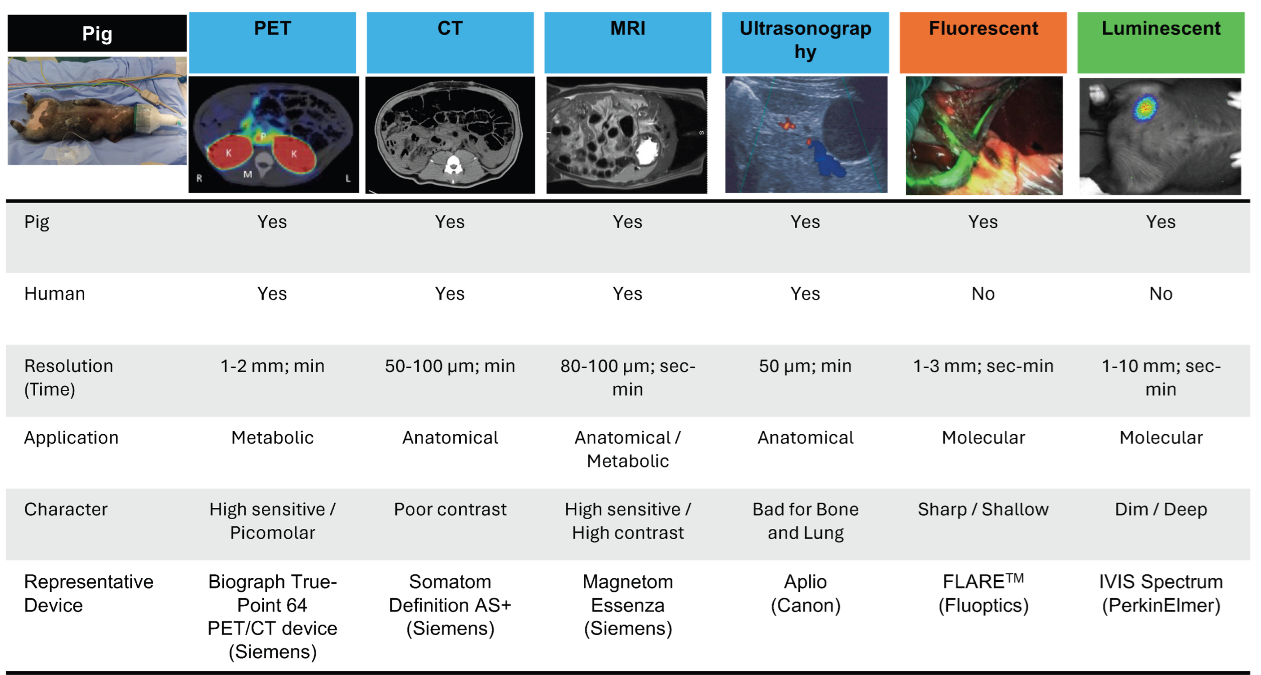
Figure 1. Non-invasive imaging methods used in pigs. Several non-invasive imaging modalities are used in pigs (positron emission tomography [PET], computed tomography [CT], magnetic resonance imaging [MRI], and ultrasonography). Additionally, molecular tracing approaches include fluorescence and luminescence detection. Adapted from Refs. [48,49].
Ijtm 04 00039 g001
Figure 2. Luminescence imaging using pigs in previous studies [53]. Systematic injection of adeno-associated virus (AAV) vectors carrying the luciferase gene showed localized gene expression in the liver. After 7 days of administration of the AAV vector carrying a luciferase gene, imaging of the thorax and abdomen of a live pig was performed, and luminescence was detected localized in the liver [53].
Ijtm 04 00039 g002
Figure 3. Previous luminescence imaging with pigs [54] and comparison with previous fluorescence imaging [55]. (A) A previously published image of the detection of cell-derived luminescence transplanted within the knee joint of a live pig [54]. Deep luminescence was detected without surgically exposing the transplanted site. Reprinted with permission from Ref. [54]. Copyright 2020 SAGE Publications. (B) Previously reported image of a pig knee joint transplanted with GFP-labeled cells [55]. The area where the cells were transplanted is surgically exposed, and fluorescence is detected, but the fluorescence is observed only on the surface of the tissue. Bright field (i) or fluorescence (ii) images of cell droplets during cell injection. Bright field (iii) or fluorescence (iv) images at the cartilage defect site 10 min after cell drop injection [55].
Ijtm 04 00039 g003
Figure 4. (A,B) Long-term tracking of islets and hepatocytes from Luciferase-transgenic rats in the same animal. Luminescence of islets derived from luciferase-transgenic rats transplanted subcutaneously and hepatocytes derived from luciferase-transgenic rats transplanted in the spleen at the site of transplantation (upper panel) and luminescence intensity over time (lower panel) [60].
Ijtm 04 00039 g004
Figure 5. Distinctive features of luminescence imaging using pigs. Based on an actual case, the advantages of luciferase–luciferin luminescence imaging in pigs were highlighted. Specifically, this imaging technique has the following advantages: high sensitivity for detection in deep tissues, long-term tracking, and continuous non-invasive, low-background, non-toxic observations. In addition, important parameters that should be considered when performing this imaging technique are summarized.
Ijtm 04 00039 g005
Disclaimer/Publisher’s Note: The statements, opinions and data contained in all publications are solely those of the individual author(s) and contributor(s) and not of MDPI and/or the editor(s). MDPI and/or the editor(s) disclaim responsibility for any injury to people or property resulting from any ideas, methods, instructions or products referred to in the content.