Article

Efficacy of Ultraviolet Radiations against Coronavirus, Bacteria, Fungi, Fungal Spores and Biofilm

1
School of Optometry and Vision Science, UNSW, Sydney, NSW 2052, Australia
2
Mobile UV Innovations, Melbourne, VIC 3070, Australia
*
Author to whom correspondence should be addressed.
Hygiene 2022, 2(3), 120-131; https://doi.org/10.3390/hygiene2030010
Submission received: 27 June 2022 / Revised: 24 July 2022 / Accepted: 10 August 2022 / Published: 12 August 2022

Abstract

:
Ultra-violet (UV) C (200–280 wavelength) light has long been known for its antimicrobial and disinfecting efficacy. It damages DNA by causing the dimerization of pyrimidines. A newly designed technology (MUVi-UVC; Mobile UV Innovations Pty Ltd., Melbourne, VIC, Australia) that emits UVC at 240 nm is composed of an enclosed booth with three UVC light stands each with four bulbs, and has been developed for disinfecting mobile medical equipment. The aim of this project was to examine the spectrum of antimicrobial activity of this device. The experiments were designed following ASTM E1052-20, EN14561, BSEN14476-2005, BSEN14562-2006 and AOAC-Official-Method-966.04 standards for surface disinfection after drying microbes on surfaces. The disinfection was analyzed using Staphylococcus aureus (ATCC 6538), Pseudomonas aeruginosa (6294), Candida auris (CBS 12373), spores of Aspergillus niger (ATCC 16404), coronavirus (SARS-CoV-2 surrogate ATCC VR-261) as well as a methicillin-resistant Staphylococcus aureus (SA31), a carbapenem- and polymyxin-resistant Pseudomonas aeruginosa (PA219), Escherichia coli K12 (ATCC 10798) and Salmonella typhi (ATCC 700730). The parameters of time, the number of lights and direction of the sample facing the lights were examined. The MUVi-UVC was able to kill 99.999% of all of the tested bacteria, fungi, coronavirus and bacteria in the biofilms if used for 5 min using all three lights in the setup with the glass slides in a vertical position. However, for fungal spores, 30 min were required to achieve 99.999% killing. There was a small but insignificant effect of having the surface horizontally or vertically aligned to the UV lights. Therefore, this UVC device is an effective technology to disinfect medical devices.

1. Introduction

Ultraviolet light (UV) has been used for many years to disinfect contaminated surfaces, as well as for treating infections. Ultraviolet radiation has a shorter wavelength than visible light, but is longer than x-rays [1]. UV radiation is divisible into four spectra, based on its wavelengths, and these are UV (100–200 nm), UVC (200–280 nm), UVB (280–315 nm) and UVA (315–400 nm) [1,2]. UVC is the most effective at killing different types of microorganisms [3].
UVC inactivates microorganisms by damaging their genetic material [4,5]. The UVC range, particularly between 250–270 nm, is absorbed by nucleic acids, with 262 nm being the peak germicidal wavelength [3]. The DNA or RNA of microorganisms is damaged by the dimerization of the nucleic acid bases, particularly pyrimidines, which prevents microbial replication and reduces viability [1,5]. Compared to routine disinfection, UVC has several advantages such as killing a broader range of microorganisms, taking less time to kill vegetative bacteria, being eco-friendly, generally safe to use (provided appropriate protective clothing and equipment are used), having relatively low costs, and the associated technology being generally easy to operate [6,7]. However, as it is light, it does have a disadvantage of shielding or shadowing [8], whereby the places not in the direct line of sight of the UVC source do not obtain adequate disinfection. This can be overcome by adding several sources of UVC, so that the shielding or shadowing is minimized or removed altogether.
Infections can spread via contaminated surfaces [9,10]. Therefore, cleaning and disinfection strategies are important tools to avoid the spread of infection [9]. However, the use of suboptimal disinfectants, reduced exposure time of disinfectants, the improper dilution of disinfectants sometimes as the result of ignoring the manufacturers’ recommendations and non-compliance with hygiene standards, can led to increased cases of infection with microbes, such as methicillin-resistant Staphylococcus aureus (MRSA) in hospitals [11,12]. The efficacy of chemical disinfectants can be affected by different parameters, including the target microorganism, the surface features of the materials on which the microbes are present, the composition of the disinfectants, the concentration of the disinfectants and the cleaning and pre-cleaning protocols [13]. The microbes are able to escape two cycles of cleaning by forming biofilms [14]. Biofilms are aggregations of microbes, often multiple types, encased in a self-made polymer matrix. The consumables used on the patients in hospitals can carry multi-drug resistant (MDR) microorganisms in biofilms [15]. The presence of biofilms in the hospitals may help MDR bacteria to survive and spread in the hospital environment [16]. If the biofilms dry onto surfaces, forming so-called dry biofilms, they can be very difficult to remove, even after 50 wiping actions [17], and the bacteria in these dry biofilms can be transferred to other surfaces [18]. Even extensive room disinfection can leave 50% or more of the surfaces contaminated [12]. Due to these limitations, there is a need to develop improved disinfectants and ways of applying these to machines and surfaces in hospitals to prevent the spread of microbes and infections.
In comparison to liquid disinfectants, UVC has some advantages as it can be used automatically and remotely, and can be applied to liquids and solids, and to decontaminate air in different types of rooms. UVC has been used to aid in manual disinfection in hospitals [19], because of its germicidal efficacy. In addition, UVC has been used to control outbreaks of tuberculosis [20] and the H1N1 influenza virus [21]. UV radiation has had a wide application in sterilizing both critical and non-critical medical devices [22,23] Non-critical patient care items disinfected using UVC showed a reduction in the spores of Clostridium difficile and Adenovirus DNA to below detectable levels [24]. UVC may be a more reliable way to disinfect surfaces, as it can be used remotely and automatically operated with set parameters. The current research focused on the ability of UVC to disinfect surfaces contaminated with various microbes, including a coronavirus surrogate of SARS-CoV-2, using standard protocols.

2. Materials and Methods

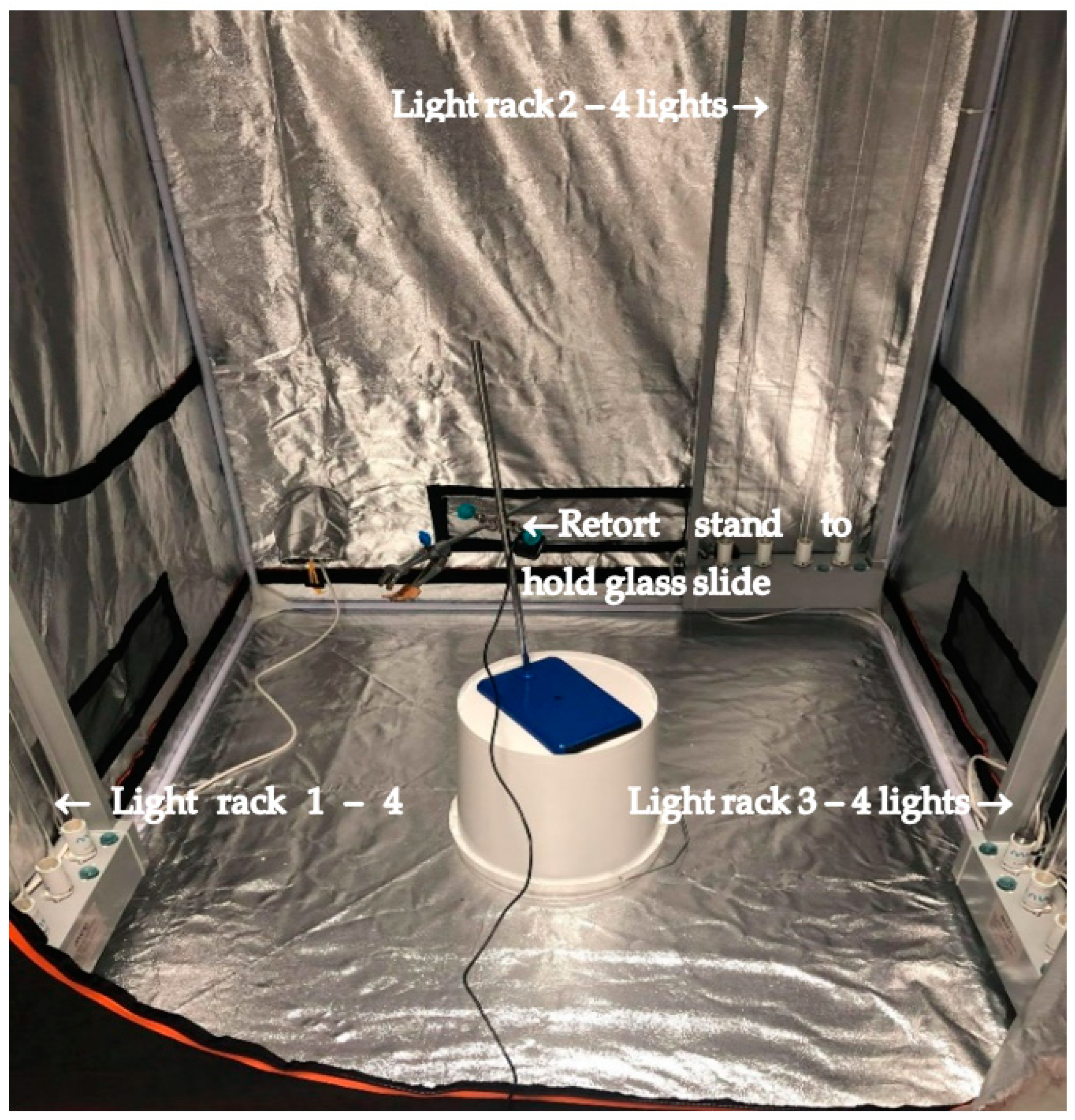
2.1. MUVI-UVC Disinfection System Setup

A newly designed technology, MUVi-UVC (Mobile UV Innovations Pty Ltd., Melbourne, VIC, Australia), which is composed of an enclosed booth with three UVC light stands, each with four bulbs (Figure 1), was developed for disinfecting hospital equipment. MUVi-UVC operates at a wavelength of 254 nm. In the current study, the UVC lights were set up in the corners of the booth and a sample-holding stand was set up in its center. All of the three light racks were operated with separate remote controls, so that different numbers of light racks could be used. The specifications of the setup are given in Table 1.

2.2. Microorganisms Studied

The activity of UVC was analyzed using Staphylococcus aureus ATCC 6538, Pseudomonas aeruginosa 6294, Candia auris CBS 12373, spores of Aspergillus niger ATCC 16404, coronavirus MHV-1 (a SARS-CoV-2 surrogate) ATCC VR-261, Escherichia coli K12 (ATCC 10798) and Salmonella typhi ATCC 700730, as well as clinical isolates of methicillin-resistant Staphylococcus aureus SA31 [25] and carbapenem- and polymyxin-resistant Pseudomonas aeruginosa PA219 [26]. Staphylococcus aureus SA38 (microbial keratitis isolate; known biofilm former) [27] was used for the biofilm assay. The strains labelled ATCC were obtained from the American Type Culture Collection (Manassas, VA, USA) and the others were retrieved from the microbial culture collection of the School of Optometry and Vision Science UNSW, Sydney, Australia.

2.3. Microbial Growth, UV Exposure and Microbial Recovery

The MUVi-UVC was tested for its efficacy against all of the different bacteria, yeast, fungal spores and the virus when dried on glass. Only the S. aureus was tested on vinyl (chlorine, ethylene), Formica (@60% paper and 30–40% cured phenol-formaldehyde resin), stainless steel 304 (chromium 18–20%, nickel 8–10.5%), PVC (ethylene and chloride) and ceramic surfaces. The bacteria, yeast, fungal spores and the coronavirus were processed using different standards: BSEN14476-2005 and ASTM E1053-11 for the viral testing, EN14561 for the bacterial and yeast testing and AOAC-Official-Method-966.04 for the fungal spores. Briefly, the bacteria and vegetative cells of the fungi were grown overnight in tryptic soy broth (TSB; Becton Dickinson, Heidelberg, Germany) at 37 °C. After 24 h growth, 1 × 10⁶ colony forming units (CFU) were spread on the glass or other surfaces. The fungal spores were grown on Sabouraud’s dextrose agar (SDA; Becton Dickinson, Heidelberg, Germany) for 10 days and collected and stored in the sterile water. The stored spores of 1 × 10⁶ CFU were dried on the glass surface. The coronavirus was grown on mouse fibroblasts cells A9 (ATCC/CCL 1.4) and collected in Dulbecco’s modified Eagle medium (DMEM; Remel, KS, USA). The plaque-forming units (PFU) of coronavirus was adjusted to 1 × 10⁶ and dried on the glass surfaces. The surfaces were dried for 30 min and then exposed to UVC.

2.4. Biofilm Formation

Wet biofilms were grown on glass slides as previously described [28]. Briefly, a 100 μL of 1 × 10⁶ CFU of SA38 overnight culture was added onto the slide, spread on the slide, then put in a petri dish containing wet tissue towels to provide a moist environment for the bacteria to develop into biofilms. The bacteria were incubated for 48 h at 37 °C. For the dry biofilms, SA38 was grown as described previously [29]. Briefly, the biofilms were grown on the glass slides by inoculating with 100 μL of 1 × 10⁸ CFU of SA38 from an overnight culture. The slides were kept moist by placing in chambers containing sterilized water on tissues. The slides were incubated for 12 days in alternate wet and dry conditions [29]. The incubation regimen was: 48 h for 35 °C with media on slides; 48 h room temperature after media had been drained off; three cycles of rehydration for 6 h in media; three dehydration phases of 66, 42 and 66 h.

2.5. UVC Exposure

The MUVi-UVC setup consisted of three light racks, each 1.5 m tall and containing four light bulbs, and the racks were placed in up to three corners of the booth (Figure 1). The inoculated slides were held by a retort stand placed in the center, in such a way that the slides were facing one rack at a distance of 43 cm, the second rack at a distance of 53 cm and the third rack at a distance of 73 cm. The parameters that were tested included the number of light racks, the distance of the sample surface from the light racks, time of exposure and the position of the sample surface, either horizontal or vertical. Later, based on the initial trial tests, the parameter of distance was not included. After the biofilm formation, the slides were either placed in the UVC chamber and exposed to UVC light (from two light racks) for 2, 5 or 10 min, or left without exposure for the same time points (un-exposed controls).

2.6. Microbial Recovery

No neutralizing agents were used during the microbial collection, as UVC does not leave any actives present once the light has been switched off. The bacterial and fungal strains and fungal spores were recovered in TSB by placing the UVC-exposed slides in 50 mL centrifuge tubes containing TSB and vortexing for 1–2 min. After the recovery of the supernatants and serial dilution, the bacteria were incubated at 37 °C for 18–24 h on TSA plates. The fungi and fungal spores were incubated on Sabouraud’s dextrose agar (SDA; Becton Dickinson, Heidelberg, Germany) for 30 °C for 48 h. After incubation, the number of colonies forming units were calculated. The controls of the surfaces were also used to determine the number of surviving microbes on surfaces without exposure to UVC.
The amount of biofilms was analyzed in separate experiments by staining with 125 μL 0.1% crystal violet staining for 10–15 min. After washing in tap water, the bound crystal violet was removed, using 30% acetic acid. The aliquots of the released crystal violet were added to 96 well microtiter plates, and the amount analyzed in a spectrophotometer at a wavelength of 550 nm, using 125 μL of 30% acetic acid as a control [30]. After either wet or dry biofilm formation and treatment, the slides were washed three times in phosphate-buffered saline to remove the non-adherent bacteria, then placed with a magnetic stirring bar and vortexed for 2 min at maximum speed to detach the adherent cells [31]. The detached cells were then diluted in phosphate-buffered saline and grown overnight at 37 °C on tryptic soy agar.
The coronavirus, at 1 × 10⁶ plaque-forming units (PFU), was dried on glass and exposed to UVC. The cells were recovered in 50 mL centrifuge tubes containing filtered bovine serum albumin (20% w/v; BSA, Sigma Aldrich, MO, USA) by vortexing and then diluted up to four times. The mouse fibroblasts cells were grown in 12-well plates, using Dulbecco’s modified Eagle medium (DMEM; Remel, KS, USA) containing 10% fetal bovine serum (FBS) and antibiotics (streptomycin sulphate and penicillin G). The UVC-treated or non-treated virus, or sterile filtered BSA were added to 12-well plates covering with agarose. These 12-well plates were inoculated for up to 3–4 days at 37 °C, then fixed with paraformaldehyde for 2–3 h and then stained with 0.1% (w/v) crystal violet staining to count the PFUs of the virus.
A steam disinfection system (manufactured by Duplex Cleaning Machines Australia, Northcote, VIC, Australia) supplied by the MUVi, was used as a comparison in this study. The steam disinfection system operates at the temperature of 161 °C and is used as a commercial steam cleaner for different surfaces. The steam was sprayed onto the virus-inoculated glass slides using the narrow head of the steam system. During the application of steam for 30 s and 1 min, the glass slides were placed in the sterile petri plates to avoid the loss of any of the virus. The whole procedure of steam application and virus recovery from the slides was performed in a biosafety cabinet. The viral recovery from the glass slides was performed as described above.

3. Results

3.1. Efficacy of UVC against Different Bacterial Species

According to the European standard test, EN14561-2:2006, used in the study, a 5 log10 reduction in the number of viable bacteria cells is needed to pass the test. The results indicated a 5 log10 reduction could be achieved on the vertical and horizontal glass surfaces when three light racks were used, and exposure was for five minutes (Table 2). When one light rack was used, it took 10 min to produce a 5 log10 reduction for all of the bacterial strains, whether the glass slides were set up horizontally or vertically.

3.2. Efficacy of UVC against Bacterial Biofilms

As measured using crystal violet staining, SA38 formed the same amount of wet or dry surface biofilm before UVC treatment (Figure 2), that contained the same number of viable cells (Table 3). After UVC treatment, the amount of biofilm reduced to two-thirds of the untreated biofilm. The UVC was able to reduce the amount of the live SA38 cells to the same extent (99.99%) in either the wet or dry biofilms on the glass surface (Table 3) when exposed to two light racks.

3.3. Efficacy of UVC against Coronavirus Fungus and Fungal Spores

UVC reduced the numbers of viable yeast cells and coronavirus by 5 log10 (99.999%) after 5 min exposure when three light racks were used. The coronavirus was also reduced by 5 log10 when one light rack was used for 5 min. However, the spores of the Aspergillus niger needed longer, 30 min, and then reached a 4 log10 reduction (Table 4), whether one or three light racks were used.
As a comparator, a steam disinfection system was used, and it was tested for 30 s and 1 min against the coronavirus surrogate. The steam system produced a 60% and 90% reduction in the numbers of the infectious virus, respectively.

3.4. Efficacy of UVC against S. aureus on Different Surfaces

After analyzing the efficacy of UVC against all of the microorganisms on the glass, S. aureus ATCC 6538 was taken as a representative microbe and tested on different materials. Additionally, the time of exposure was standardized to 5 min and 10 min and the samples only placed horizontally. The number of viable S. aureus was reduced by 5 log10 CFU (99.999%) on most of the materials within 5 min when three light racks were used. The exception was steel, where 10 min was needed to achieve 5 log10 killing (Table 5). When exposed to one light rack for 10 min, the number of S. aureus ATCC 6538 was reduced by 5 log10.

4. Discussion

The current study investigated the efficacy of a new UVC set-up against several microorganisms, including a coronavirus. The results from the present study demonstrated that MUVi-UVC was an effective disinfection system meeting the disinfection criteria of the standards used. These disinfection criteria were the reduction in viable microbes by at least 4 log10 for both of the British standards and 5 log10 for the European standard.
The Therapeutic Goods Administration of Australia identifies the MHV as a suitable surrogate of SARS-CoV-2 for testing the disinfection activity of different solutions (https://www.tga.gov.au/surrogate-viruses-use-disinfectant-efficacy-tests-justify-claims-against-covid-19; accessed on 2 February 2021). MHV is very similar in its mechanism of infection to SARS-Cov-2 and can produce the acute respiratory disease in mice. Together with SARS-Cov-2, SARS-CoV-1 and Middle Eastern Respiratory Syndrome virus (MERS), MHV falls in the group 2 coronaviruses [32]. Previous studies have shown that different chemical-based disinfecting regimes are effective against MHV, including 70% alcohol giving a 4 log10 reduction on stainless steel within a minute [33,34]. In the current study, UVC gave a 5 log10 reduction of MHV on glass after 5 min exposure. Other studies have shown that UVC can be used for disinfection against MHV [35,36,37,38,39,40,41,42]. UVC halts DNA or RNA replication by dimerizing their pyrimidine bases after absorption of UVC, and this leads to the inactivation of viral replication [39,43]. SARS-CoV-2 can survive on glass, stainless steel and paper for up to 28 days at 20 °C [44] and touching contaminated surfaces can initiate infection. Therefore, a technology that can reduce the numbers of culturable coronaviruses is likely to be effective at preventing the spread of the disease.
The finding is important that all of the different bacteria, including the MDR strains, could be reduced by a 5 log10 after exposure to UVC from one light rack for 10 min or three light racks for 5 min. S. aureus ATCC 6538 is a standard strain used to test the efficacy and resistance of disinfectants [45,46]. This S. aureus strain together with P. aeruginosa, Proteus vulgaris and E. coli are required to be tested in order to qualify for the hospital or household disinfection testing, according to TGA guidelines https://www.tga.gov.au/resource/tga-instructions-disinfectant-testing (accessed on 20 March 2021) P. aeruginosa is a common cause of healthcare-acquired infections, where both of the bacteria can form biofilms on the high-touch surfaces in hospitals [47,48]. Antibiotic-resistant isolates are of rising concern, and many are spread through contact with contaminated surfaces. The multidrug-resistant (MDR) bacteria cause nosocomial-, or healthcare-acquired infections. MRSA is disseminated globally and is a leading cause of both community- and hospital-acquired infections [49]. MDR P. aeruginosa has been isolated from multiple hospital outbreaks [50], which increases the global health burden and the cost of treatment due to the prolonged hospitalization [51,52]. Together with antibiotic resistance, MDR isolates may also be resistant to the different disinfectants used in the normal cleaning regimens of hospitals [53,54]. The MDR strains of E. coli, S. aureus and Salmonella typhi can be isolated from community-acquired and healthcare-acquired infections [55,56,57].
Various technologies have been used in hospitals for providing disinfection, including both chemical- and UV-based technologies [58,59,60]. However, different bacteria may escape the effects of disinfectants, such as chlorine and peracetic acid, by developing tolerance or resistance [61,62]. The current study verified that UVC was effective against the standard strains of S. aureus, P. aeruginosa, E. coli and Salmonella typhi along with carbapenem-resistant P. aeruginosa, and it has also been shown to be effective against MRSA [26]. This efficacy of UVC at a wavelength of 254 nm has been shown to be germicidal against influenza A viruses, H1N1 and H3N2, but was less efficient against Mycobacterium tuberculosis, requiring 20 min to produce a 3 log10 reduction [63]. However, the light set-up was different to that in the current study, and it would be of interest to examine the ability of the MUVI-UVC system to kill Mycobacterium tuberculosis.
Other than the development of resistance to disinfectants by mutation or acquisition of genes, biofilm formation can also protect bacteria [64]. For example, sodium hypochlorite was not able to eradicate bacteria from biofilms [29]. In the current study, the SA38 grown in biofilms was more resistant to UVC than when simply dried on glass. However, the UVC treatment for 5 min was able to reduce the numbers of viable cells in the biofilm by 4 log10 CFU (99.99%). The biofilm itself was reduced to two-thirds by UVC treatment, indicating that some form of abrasive treatment is likely to be required after UVC treatment to more thoroughly remove the traces of the biofilm. Dead bacteria remaining in biofilms on surfaces may act as a food source for new bacterial growth and biofilm formation [65]. Even so, a study, examining the effect of dead biofilms on the formation of new biofilms of P. aeruginosa, found that the dead biofilms exerted an effect on the new biofilm formation, with the new biofilms being formed more slowly and were softer (perhaps indicating that they would be easier to remove) [66].
C. auris has been isolated when found in the hospital environment and infections, and responds poorly to antifungals [67]. The Centers for Disease Control and Prevention (CDC) have recommended it be used to test the efficacy of disinfectants (https://www.cdc.gov/fungal/candida-auris/index.html; accessed on 22 August 2021). Aspergillus is an opportunistic human pathogen that can cause nosocomial invasive aspergillosis [68]. Aspergillus can escape many sterilization techniques by producing resistant spores, and it is recommended to be considered in order to test the efficacy of UVC [69]. Previously, studies have found that the fungal spores of this genera can be resistant [70] or susceptible to UVC treatment [71,72]. In addition, the intense use of different chemical disinfectants have been needed to kill fungal spores, and these disinfectants may have significant toxic effects on human and pollute the environment [73]. The finding that the UVC disinfecting system used in the current study was active against the vegetative cells of Candida auris and the spores of Aspergillus niger is therefore of importance, especially as these are often difficult to kill with other chemical or UV disinfection systems [74].
The use of current UVC technology was also tested against S. aureus dried to plastic, vinyl, ceramics, Formica and stainless steel. The application of UVC on the different surfaces was previously shown to be effective in combination with other cleaning methods including ethanol wipes [75] against bacteria, including a 17% reduction in MRSA, VRE, Acinetobacter spp., carbapenem-resistant Enterobacteriaceae and viruses including SARS-CoV-2 [75].
In conclusion, the current study demonstrated that the UVC system could produce a 5 log10 reduction of S. aureus ATCC 6538 when dried onto different surfaces (glass, vinyl, Formica, ceramic, steel, plastic), was able to similarly reduce the numbers of other bacteria including MDR strains on glass, as well as a coronavirus and the yeast C. albicans, when three light racks were used for 5 min. The spores of A. niger were more resistant to the UVC system, requiring 10 min and three light racks to produce a 4 log10 reduction. These reductions in the numbers of microbes on these surfaces, which may be present in hospital wards and as part of medical devices, indicates that the UVC system has the possibility of reducing the transmission of microbes around hospitals. This should be tested in follow-up experiments.

Author Contributions

Conceptualization, M.K. and M.W.; writing—original draft preparation, M.K.; writing—review and editing, M.W.; project administration, K.M.; funding acquisition, M.M. All authors have read and agreed to the published version of the manuscript.

Funding

This research was funded by Mobile UV Innovations and MTP Connect Australia under the program APR.Intern.

Institutional Review Board Statement

Not applicable.

Informed Consent Statement

Not applicable.

Data Availability Statement

Data deposited in https://unsworks.unsw.edu.au, accessed on 27 June 2022.

Acknowledgments

The author of the article would like to acknowledge UNSW, Sydney for the use of the facilities of the Microbiology laboratory at the School of Optometry and Vision Science. The authors would also like to acknowledge the contribution of MTP Connect Australia for their involvement in the study.

Conflicts of Interest

The authors M.M. and K.M. are employees of Mobile UV Innovations that funded this project.

References

  1. Dai, T.; Vrahas, M.S.; Murray, C.K.; Hamblin, M.R. Ultraviolet C irradiation: An alternative antimicrobial approach to localized infections? Expert Rev. Anti-Infect. Ther. 2012, 10, 185–195. [Google Scholar] [CrossRef] [PubMed]
  2. Vázquez, M.; Hanslmeier, A. Ultraviolet Radiation in the Solar System; Springer: Berlin/Heidelberg, Germany, 2005; Volume 331. [Google Scholar]
  3. Gurzadyan, G.G.; Görner, H.; Schulte-Frohlinde, D. Ultraviolet (193, 216 and 254 nm) photoinactivation of Escherichia coli strains with different repair deficiencies. Radiat. Res. 1995, 141, 244–251. [Google Scholar] [CrossRef] [PubMed]
  4. Chang, J.C.; Ossoff, S.F.; Lobe, D.C.; Dorfman, M.H.; Dumais, C.M.; Qualls, R.G.; Johnson, J.D. UV inactivation of pathogenic and indicator microorganisms. Appl. Environ. Microbiol. 1985, 49, 1361–1365. [Google Scholar] [CrossRef] [PubMed]
  5. Cutler, T.D.; Zimmerman, J.J. Ultraviolet irradiation and the mechanisms underlying its inactivation of infectious agents. Anim. Health Res. Rev. 2011, 12, 15–23. [Google Scholar] [CrossRef]
  6. Nerandzic, M.M.; Cadnum, J.L.; Pultz, M.J.; Donskey, C.J. Evaluation of an automated ultraviolet radiation device for decontamination of Clostridium difficile and other healthcare-associated pathogens in hospital rooms. BMC Infect. Dis. 2010, 10, 197. [Google Scholar] [CrossRef]
  7. Rutala, W.A.; Weber, D.J. Disinfectants used for environmental disinfection and new room decontamination technology. Am. J. Infect. Control 2013, 41, S36–S41. [Google Scholar] [CrossRef]
  8. Diffey, B. Physical barriers to protect humans from solar UV radiation exposure. In Sun Protection; IOP Publishing Ltd.: Bristol, UK, 2017; Chapter 6; pp. 6-1–6-24. [Google Scholar] [CrossRef]
  9. Kampf, G. Potential role of inanimate surfaces for the spread of coronaviruses and their inactivation with disinfectant agents. Infect. Prev. Pract. 2020, 2, 100044. [Google Scholar] [CrossRef]
  10. Lei, H.; Li, Y.; Xiao, S.; Yang, X.; Lin, C.; Norris, S.L.; Wei, D.; Hu, Z.; Ji, S. Logistic growth of a surface contamination network and its role in disease spread. Sci. Rep. 2017, 7, 14826. [Google Scholar] [CrossRef]
  11. Carling, P.C.; Briggs, J.; Hylander, D.; Perkins, J. An evaluation of patient area cleaning in 3 hospitals using a novel targeting methodology. Am. J. Infect. Control 2006, 34, 513–519. [Google Scholar] [CrossRef]
  12. Carling, P.C.; Parry, M.M.; Rupp, M.E.; Po, J.L.; Dick, B.; Von Beheren, S.; Group, H.E.H.S. Improving cleaning of the environment surrounding patients in 36 acute care hospitals. Infect. Control Hosp. Epidemiol. 2008, 29, 1035–1041. [Google Scholar] [CrossRef]
  13. Ghedini, E.; Pizzolato, M.; Longo, L.; Menegazzo, F.; Zanardo, D.; Signoretto, M. Which Are the Main Surface Disinfection Approaches at the Time of SARS-CoV-2? Front. Chem. Eng. 2021, 2, 589202. [Google Scholar] [CrossRef]
  14. Vickery, K.; Deva, A.; Jacombs, A.; Allan, J.; Valente, P.; Gosbell, I.B. Presence of biofilm containing viable multiresistant organisms despite terminal cleaning on clinical surfaces in an intensive care unit. J. Hosp. Infect. 2012, 80, 52–55. [Google Scholar] [CrossRef]
  15. Hu, H.; Johani, K.; Gosbell, I.B.; Jacombs, A.; Almatroudi, A.; Whiteley, G.S.; Deva, A.K.; Jensen, S.; Vickery, K. Intensive care unit environmental surfaces are contaminated by multidrug-resistant bacteria in biofilms: Combined results of conventional culture, pyrosequencing, scanning electron microscopy, and confocal laser microscopy. J. Hosp. Infect. 2015, 91, 35–44. [Google Scholar] [CrossRef]
  16. Yezli, S.; Otter, J. Does the discovery of biofilms on dry hospital environmental surfaces change the way we think about hospital disinfection? J. Hosp. Infect. 2012, 81, 293–294. [Google Scholar] [CrossRef]
  17. Parvin, F.; Hu, H.; Whiteley, G.S.; Glasbey, T.; Vickery, K. Difficulty in removing biofilm from dry surfaces. J. Hosp. Infect. 2019, 103, 465–467. [Google Scholar] [CrossRef]
  18. Chowdhury, D.; Tahir, S.; Legge, M.; Hu, H.; Prvan, T.; Johani, K.; Whiteley, G.S.; Glasbey, T.O.; Deva, A.K.; Vickery, K. Transfer of dry surface biofilm in the healthcare environment: The role of healthcare workers’ hands as vehicles. J. Hosp. Infect. 2018, 100, e85–e90. [Google Scholar] [CrossRef]
  19. Ramos, C.C.R.; Roque, J.L.A.; Sarmiento, D.B.; Suarez, L.E.G.; Sunio, J.T.P.; Tabungar, K.I.B.; Tengco, G.S.C.; Rio, P.C.; Hilario, A.L. Use of ultraviolet-C in environmental sterilization in hospitals: A systematic review on efficacy and safety. Int. J. Health Sci. 2020, 14, 52–65. [Google Scholar]
  20. World Health Organization. WHO Guidelines on Tuberculosis Infection Prevention and Control: 2019 Update; World Health Organization: Geneva, Switzerland, 2019. [Google Scholar]
  21. McDevitt, J.J.; Rudnick, S.N.; Radonovich, L.J. Aerosol susceptibility of influenza virus to UV-C light. Appl. Environ. Microbiol. 2012, 78, 1666–1669. [Google Scholar] [CrossRef]
  22. Rogers, W. Steam and dry heat sterilization of biomaterials and medical devices. In Sterilisation of Biomaterials and Medical Devices; Elsevier: Amsterdam, The Netherlands, 2012; pp. 20–55. [Google Scholar]
  23. Iwaguch, S.; Matsumura, K.; Tokuoka, Y.; Wakui, S.; Kawashima, N. Sterilization system using microwave and UV light. Colloids Surf. B Biointerfaces 2002, 25, 299–304. [Google Scholar] [CrossRef]
  24. Moore, G.; Ali, S.; Cloutman-Green, E.A.; Bradley, C.R.; Wilkinson, M.A.C.; Hartley, J.C.; Fraise, A.P.; Wilson, A.P.R. Use of UV-C radiation to disinfect non-critical patient care items: A laboratory assessment of the Nanoclave Cabinet. BMC Infect. Dis. 2012, 12, 174. [Google Scholar] [CrossRef]
  25. Rodríguez-López, P.; Filipello, V.; Di Ciccio, P.A.; Pitozzi, A.; Ghidini, S.; Scali, F.; Ianieri, A.; Zanardi, E.; Losio, M.N.; Simon, A.C.; et al. Assessment of the Antibiotic Resistance Profile, Genetic Heterogeneity and Biofilm Production of Methicillin-Resistant Staphylococcus aureus (MRSA) Isolated from The Italian Swine Production Chain. Foods 2020, 9, 1141. [Google Scholar] [CrossRef] [PubMed]
  26. Khan, M.; Stapleton, F.; Summers, S.; Rice, S.A.; Willcox, M.D. Antibiotic Resistance Characteristics of Pseudomonas aeruginosa Isolated from Keratitis in Australia and India. Antibiotics 2020, 9, 600. [Google Scholar] [CrossRef] [PubMed]
  27. Yasir, M.; Dutta, D.; Willcox, M.D.P. Enhancement of Antibiofilm Activity of Ciprofloxacin against Staphylococcus aureus by Administration of Antimicrobial Peptides. Antibiotics 2021, 10, 1159. [Google Scholar] [CrossRef] [PubMed]
  28. Chen, R.; Cole, N.; Dutta, D.; Kumar, N.; Willcox, M.D.P. Antimicrobial activity of immobilized lactoferrin and lactoferricin. J. Biomed. Mater. Res. Part B Appl. Biomater. 2017, 105, 2612–2617. [Google Scholar] [CrossRef] [PubMed]
  29. Almatroudi, A.; Gosbell, I.B.; Hu, H.; Jensen, S.O.; Espedido, B.A.; Tahir, S.; Glasbey, T.O.; Legge, P.; Whiteley, G.; Deva, A.; et al. Staphylococcus aureus dry-surface biofilms are not killed by sodium hypochlorite: Implications for infection control. J. Hosp. Infect. 2016, 93, 263–270. [Google Scholar] [CrossRef] [PubMed]
  30. O’Toole, G.A. Microtiter dish biofilm formation assay. J. Vis. Exp. 2011, 47, e2437. [Google Scholar] [CrossRef] [PubMed]
  31. Vijay, A.K.; Zhu, H.; Ozkan, J.; Wu, D.; Masoudi, S.; Bandara, R.; Borazjani, R.N.; Willcox, M.D. Bacterial adhesion to unworn and worn silicone hydrogel lenses. Optom. Vis. Sci. Off. Publ. Am. Acad. Optom. 2012, 89, 1095–1106. [Google Scholar] [CrossRef]
  32. Singh, D.; Joshi, K.; Samuel, A.; Patra, J.; Mahindroo, N. Alcohol-based hand sanitisers as first line of defence against SARS-CoV-2: A review of biology, chemistry and formulations. Epidemiol. Infect. 2020, 148, e229. [Google Scholar] [CrossRef]
  33. Hulkower, R.L.; Casanova, L.M.; Rutala, W.A.; Weber, D.J.; Sobsey, M.D. Inactivation of surrogate coronaviruses on hard surfaces by health care germicides. Am. J. Infect. Control 2011, 39, 401–407. [Google Scholar] [CrossRef]
  34. Welch, J.L.; Xiang, J.; Mackin, S.R.; Perlman, S.; Thorne, P.; O’Shaughnessy, P.; Strzelecki, B.; Aubin, P.; Ortiz-Hernandez, M.; Stapleton, J.T. Inactivation of severe acute respiratory coronavirus virus 2 (SARS-CoV-2) and diverse RNA and DNA viruses on three-dimensionally printed surgical mask materials. Infect. Control Hosp. Epidemiol. 2021, 42, 253–260. [Google Scholar] [CrossRef]
  35. Rauth, A.M. The physical state of viral nucleic acid and the sensitivity of viruses to ultraviolet light. Biophys. J. 1965, 5, 257–273. [Google Scholar] [CrossRef]
  36. Budowsky, E.; Bresler, S.; Friedman, E.; Zheleznova, N. Principles of selective inactivation of viral genome. Arch. Virol. 1981, 68, 239–247. [Google Scholar] [CrossRef]
  37. Anderson, D.J.; Gergen, M.F.; Smathers, E.; Sexton, D.J.; Chen, L.F.; Weber, D.J.; Rutala, W.A. Decontamination of targeted pathogens from patient rooms using an automated ultraviolet-C-emitting device. Infect. Control Hosp. Epidemiol. 2013, 34, 466–471. [Google Scholar] [CrossRef]
  38. Andersen, B.; Bånrud, H.; Bøe, E.; Bjordal, O.; Drangsholt, F. Comparison of UV C light and chemicals for disinfection of surfaces in hospital isolation units. Infect. Control Hosp. Epidemiol. 2006, 27, 729–734. [Google Scholar] [CrossRef]
  39. Darnell, M.E.; Subbarao, K.; Feinstone, S.M.; Taylor, D.R. Inactivation of the coronavirus that induces severe acute respiratory syndrome, SARS-CoV. J. Virol. Methods 2004, 121, 85–91. [Google Scholar] [CrossRef]
  40. Kowalski, W. Ultraviolet Germicidal Irradiation Handbook: UVGI for Air and Surface Disinfection; Springer: Berlin/Heidelberg, Germany, 2010. [Google Scholar]
  41. Raeiszadeh, M.; Adeli, B. A critical review on ultraviolet disinfection systems against COVID-19 outbreak: Applicability, validation, and safety considerations. ACS Photonics 2020, 7, 2941–2951. [Google Scholar] [CrossRef]
  42. Biasin, M.; Bianco, A.; Pareschi, G.; Cavalleri, A.; Cavatorta, C.; Fenizia, C.; Galli, P.; Lessio, L.; Lualdi, M.; Tombetti, E.; et al. UV-C irradiation is highly effective in inactivating SARS-CoV-2 replication. Sci. Rep. 2021, 11, 6260. [Google Scholar] [CrossRef]
  43. Pirnie, M.; Linden, K.G.; Malley, J. Ultraviolet disinfection guidance manual for the final long term 2 enhanced surface water treatment rule. US Environ. Prot. Agency 2006, 2, 1–436. [Google Scholar]
  44. Riddell, S.; Goldie, S.; Hill, A.; Eagles, D.; Drew, T.W. The effect of temperature on persistence of SARS-CoV-2 on common surfaces. Virol. J. 2020, 17, 145. [Google Scholar] [CrossRef]
  45. Luppens, S.B.I.; Reij, M.W.; van der Heijden, R.W.L.; Rombouts, F.M.; Abee, T. Development of a standard test to assess the resistance of Staphylococcus aureus biofilm cells to disinfectants. Appl. Environ. Microbiol. 2002, 68, 4194–4200. [Google Scholar] [CrossRef]
  46. Ioannou, C.J.; Hanlon, G.W.; Denyer, S.P. Action of disinfectant quaternary ammonium compounds against Staphylococcus aureus. Antimicrob. Agents Chemother. 2007, 51, 296–306. [Google Scholar] [CrossRef]
  47. Weiner, L.M.; Webb, A.K.; Limbago, B.; Dudeck, M.A.; Patel, J.; Kallen, A.J.; Edwards, J.R.; Sievert, D.M. Antimicrobial-resistant pathogens associated with healthcare-associated infections: Summary of data reported to the National Healthcare Safety Network at the Centers for Disease Control and Prevention, 2011–2014. Infect. Control. Hosp. Epidemiol. 2016, 37, 1288–1301. [Google Scholar] [CrossRef]
  48. Sievert, D.M.; Ricks, P.; Edwards, J.R.; Schneider, A.; Patel, J.; Srinivasan, A.; Kallen, A.; Limbago, B.; Fridkin, S. Antimicrobial-Resistant Pathogens Associated with Healthcare-Associated Infections Summary of Data Reported to the National Healthcare Safety Network at the Centers for Disease Control and Prevention, 2009–2010. Infect. Control. Hosp. Epidemiol. 2013, 34, 1288–1301. [Google Scholar] [CrossRef]
  49. Lee, A.S.; de Lencastre, H.; Garau, J.; Kluytmans, J.; Malhotra-Kumar, S.; Peschel, A.; Harbarth, S. Methicillin-resistant Staphylococcus aureus. Nat. Rev. Dis. Primers 2018, 4, 18033. [Google Scholar] [CrossRef]
  50. Hota, S.; Hirji, Z.; Stockton, K.; Lemieux, C.; Dedier, H.; Wolfaardt, G.; Gardam, M.A. Outbreak of multidrug-resistant Pseudomonas aeruginosa colonization and infection secondary to imperfect intensive care unit room design. Infect. Control Hosp. Epidemiol. 2009, 30, 25–33. [Google Scholar] [CrossRef]
  51. French, G.L. Clinical impact and relevance of antibiotic resistance. Adv. Drug Deliv. Rev. 2005, 57, 1514–1527. [Google Scholar] [CrossRef]
  52. Founou, R.C.; Founou, L.L.; Essack, S.Y. Clinical and economic impact of antibiotic resistance in developing countries: A systematic review and meta-analysis. PLoS ONE 2017, 12, e0189621. [Google Scholar] [CrossRef]
  53. Kadry, A.A.; Serry, F.M.; El-Ganiny, A.M.; El-Baz, A.M. Integron occurrence is linked to reduced biocide susceptibility in multidrug resistant Pseudomonas aeruginosa. Br. J. Biomed. Sci. 2017, 74, 78–84. [Google Scholar] [CrossRef]
  54. Chaoui, L.; Mhand, R.; Mellouki, F.; Rhallabi, N. Contamination of the surfaces of a health care environment by multidrug-resistant (MDR) bacteria. Int. J. Microbiol. 2019, 2019, 3236526. [Google Scholar] [CrossRef]
  55. Ibrahim, M.E.; Bilal, N.E.; Hamid, M.E. Increased multi-drug resistant Escherichia coli from hospitals in Khartoum state, Sudan. Afr. Health Sci. 2012, 12, 368–375. [Google Scholar] [CrossRef]
  56. Mutai, W.C.; Muigai, A.W.T.; Waiyaki, P.; Kariuki, S. Multi-drug resistant Salmonella enterica serovar Typhi isolates with reduced susceptibility to ciprofloxacin in Kenya. BMC Microbiol. 2018, 18, 187. [Google Scholar] [CrossRef] [PubMed]
  57. Hiramatsu, K.; Katayama, Y.; Matsuo, M.; Sasaki, T.; Morimoto, Y.; Sekiguchi, A.; Baba, T. Multi-drug-resistant Staphylococcus aureus and future chemotherapy. J. Infect. Chemother. Off. J. Jpn. Soc. Chemother. 2014, 20, 593–601. [Google Scholar] [CrossRef] [PubMed]
  58. Boyce, J.M. Modern technologies for improving cleaning and disinfection of environmental surfaces in hospitals. Antimicrob. Resist. Infect. Control 2016, 5, 10. [Google Scholar] [CrossRef] [PubMed]
  59. Dancer, S.J. Controlling hospital-acquired infection: Focus on the role of the environment and new technologies for decontamination. Clin. Microbiol. Rev. 2014, 27, 665–690. [Google Scholar] [CrossRef]
  60. Awodele, O.; Emeka, P.; Agbamuche, H.; Akintonwa, A. The antimicrobial activities of some commonly used disinfectants on Bacillus subtilis, Pseudomonas aeruginosa and Candida albicans. Afr. J. Biotechnol. 2007, 6, 987–990. [Google Scholar]
  61. Akinbobola, A.; Sherry, L.; Mckay, W.G.; Ramage, G.; Williams, C. Tolerance of Pseudomonas aeruginosa in in-vitro biofilms to high-level peracetic acid disinfection. J. Hosp. Infect. 2017, 97, 162–168. [Google Scholar] [CrossRef]
  62. Wang, L.; Ye, C.; Guo, L.; Chen, C.; Kong, X.; Chen, Y.; Shu, L.; Wang, P.; Yu, X.; Fang, J. Assessment of the UV/Chlorine Process in the Disinfection of Pseudomonas aeruginosa: Efficiency and Mechanism. Environ. Sci. Technol. 2021, 55, 9221–9230. [Google Scholar] [CrossRef]
  63. Szeto, W.; Yam, W.C.; Huang, H.; Leung, D.Y.C. The efficacy of vacuum-ultraviolet light disinfection of some common environmental pathogens. BMC Infect. Dis. 2020, 20, 127. [Google Scholar] [CrossRef]
  64. Dosler, S.; Mataraci, E. In vitro pharmacokinetics of antimicrobial cationic peptides alone and in combination with antibiotics against methicillin resistant Staphylococcus aureus biofilms. Peptides 2013, 49, 53–58. [Google Scholar] [CrossRef]
  65. Rodriguez Herrero, E.; Boon, N.; Pauwels, M.; Bernaerts, K.; Slomka, V.; Quirynen, M.; Teughels, W. Necrotrophic growth of periodontopathogens is a novel virulence factor in oral biofilms. Sci. Rep. 2017, 7, 1107. [Google Scholar] [CrossRef]
  66. Wang, Z.; Gong, X.; Xie, J.; Xu, Z.; Liu, G.; Zhang, G. Investigation of Formation of Bacterial Biofilm upon Dead Siblings. Langmuir 2019, 35, 7405–7413. [Google Scholar] [CrossRef]
  67. Ku, T.S.N.; Walraven, C.J.; Lee, S.A. Candida auris: Disinfectants and Implications for Infection Control. Front. Microbiol. 2018, 9, 726. [Google Scholar] [CrossRef]
  68. Latgé, J.-P. Aspergillus fumigatus and aspergillosis. Clin. Microbiol. Rev. 1999, 12, 310–350. [Google Scholar] [CrossRef]
  69. und Anlagenbau, V.D.M. Hygienic Filling Machines of VDMA Class IV for Liquid and Viscous Foods Minimum Requirements and Basic Conditions for Operation in Accordance with Specification; Fachverband Nahrungsmittelmaschinen und Verpackungsmaschinen: Frankfurt, Germany, 2005. [Google Scholar]
  70. Racchi, I.; Scaramuzza, N.; Hidalgo, A.; Cigarini, M.; Berni, E. Sterilization of food packaging by UV-C irradiation: Is Aspergillus brasiliensis ATCC 16404 the best target microorganism for industrial bio-validations? Int. J. Food Microbiol. 2021, 357, 109383. [Google Scholar] [CrossRef]
  71. Nourmoradi, H.; Nikaeen, M.; Stensvold, C.R.; Mirhendi, H. Ultraviolet irradiation: An effective inactivation method of Aspergillus spp. in water for the control of waterborne nosocomial aspergillosis. Water Res. 2012, 46, 5935–5940. [Google Scholar] [CrossRef]
  72. Narita, K.; Asano, K.; Naito, K.; Ohashi, H.; Sasaki, M.; Morimoto, Y.; Igarashi, T.; Nakane, A. Ultraviolet C light with wavelength of 222 nm inactivates a wide spectrum of microbial pathogens. J. Hosp. Infect. 2020, 105, 459–467. [Google Scholar] [CrossRef]
  73. Pechter, E.; Rosenman, K.D. Occupational health risks associated with use of environmental surface disinfectants in health care. Am. J. Infect. Control 2016, 44, 1755–1756. [Google Scholar] [CrossRef]
  74. Begum, M.; Hocking, A.D.; Miskelly, D. Inactivation of food spoilage fungi by ultra violet (UVC) irradiation. Int. J. Food Microbiol. 2009, 129, 74–77. [Google Scholar] [CrossRef]
  75. Hosein, I.; Madeloso, R.; Nagaratnam, W.; Villamaria, F.; Stock, E.; Jinadatha, C. Evaluation of a pulsed xenon ultraviolet light device for isolation room disinfection in a United Kingdom hospital. Am. J. Infect. Control 2016, 44, e157–e161. [Google Scholar] [CrossRef]
Figure 1. The MUVi-UVC set-up: within a 120 m wide tent, three 1.5 m tall light racks were placed in three corners. Each light rack contained four bulbs. There is a central retort stand to hold the inoculated surfaces. Each light rack could be operated separately by remote control.
Hygiene 02 00010 g001
Figure 2. Quantification of wet and dry surface biofilms before and after UVC treatment.
Hygiene 02 00010 g002
Table 1. Specifications of MUVi-UVC booth disinfection system.
SpecificationDetails
Dimensions (m)120 × 120 × 2000
Voltage240 V ± 10% 50 Hz
UVC light frequency (Nm)253.7
UV lamp40 W × 4 lights = 160 W per set × 3
UV output (uW/cm2)1620 at 1 m (12 × 135 uW/cm2)
Table 2. Efficacy UVC against different bacterial species dried on glass.
MicroorganismsPosition of SampleNo. of Light RacksTime of Exposure (min)/Killing %
22.5510
S. aureus ATCC 6538Vertical1 light99.999.999.9999.999
3 lights99.999.999.99999.999
Horizontal1 light99.999.999.9999.999
3 lights99.999.999.99999.999
S. aureus SA31 *Vertical1 light99.999.999.999.999
3 lights99.999.999.99999.999
Horizontal1 light99.999.999.9999.999
3 lights99.999.999.99999.999
P. aeruginosa 6294Vertical1 lightND859899.999
3 lightsND9599.99999.999
Horizontal1 lightND9999.9999.999
3 lightsND99.999.99999.999
P. aeruginosa PA219 #Horizontal1 lightNDND99.9999.999
3 lightsNDND99.99999.999
Escherichia coli K12 (ATCC 10798)Vertical1 lightND9999.9999.999
3 lightsND99.999.99999.999
Horizontal1 lightND9099.9999.999
3 lightsND9999.99999.999
Salmonella typhi ATCC 700730Vertical1 lightND9599.9999.999
3 lightsND99.999.99999.999
Horizontal1 lightND9599.9999.999
3 lightsND99.999.99999.999
*, methicillin-resistant; #, carbapenem- and polymyxin-resistant.
Table 3. Efficacy of UVC against SA38 in biofilms.
MicroorganismsMaterialsNo. of Light RacksTime of Exposure (min)/Killing %Viable Bacteria Recovered from the Untreated Biofilm
2510
Wet surface biofilmGlass29299.9999.9946.6 × 106
Dry surface biofilmND99.9999.9943.1 × 106
Table 4. Efficacy of UVC against fungi, fungal spores and coronavirus when dried on glass.
MicroorganismsPosition of SampleNo. of Light RacksTime of Exposure (min)/Killing %
15102030
Candida auris CBS 12373Horizontal1 lightND99.999.99NDND
3 lightsND99.99999.999NDND
Aspergillus niger (spores) ATCC 164041 lightND≤18899.999.99
3 lightsND≤19099.999.99
Coronavirus (SARS-CoV-2 surrogate; MHV-I; ATCC VR-261)1 light99.999.999NDNDND
3 lights99.999.999NDNDND
ND = not determined.
Table 5. Efficacy of UVC against S. aureus on different surfaces.
MicroorganismsTesting SurfacePositionNo. of LightsTime of Exposure (min)/Killing %
510
S. aureus
ATCC 6538
VinylHorizontal18999.999
399.99999.999
Ceramic199.9999.999
399.99999.999
Formica199.9999.999
399.99999.999
Steel18399.999
39699.999
Plastic199.9999.999
399.99999.999
Publisher’s Note: MDPI stays neutral with regard to jurisdictional claims in published maps and institutional affiliations.

