Article

Microbial Eukaryotes in Natural and Artificial Salt Marsh Pools

1
College of Arts and Sciences, Drexel University, Philadelphia, PA 19104, USA
2
Academy of Natural Sciences, Drexel University, Philadelphia, PA 19103, USA
*
Author to whom correspondence should be addressed.
Coasts 2024, 4(2), 287-305; https://doi.org/10.3390/coasts4020015
Submission received: 10 November 2023 / Revised: 29 February 2024 / Accepted: 1 April 2024 / Published: 2 April 2024

Abstract

:
Microscopic eukaryotes are important components of coastal wetland ecosystems. The goal of this study was to investigate the diversity of microeukaryotes in the tidal pools of a New Jersey salt marsh and to compare the assemblages of natural and artificial pools excavated for controlling mosquito populations. We evaluated microeukaryotic assemblages using the amplicon sequencing of 18S and rbcL DNA markers and the microscopic identification of diatoms in water and sediment samples. 18S unique amplicon sequence variants (ASV) representing ciliates, dinoflagellates, diatoms, and cercozoans were the most diverse, while the reads of dinoflagellates, diatoms, ciliates, and nematodes were the most abundant. The dominant ASVs were attributed to organisms that are characteristic of coastal plankton and sediments or those known for their resistance to salinity, desiccation, hypoxia, and UV stress. The sediment assemblages were more diverse compared to those from the water column and contained a larger portion of ASVs that were not assigned to any low-rank taxa, reflecting the current gaps in understanding the diversity of microeukaryotes. Most taxonomic groups were significantly different in their abundance and composition between natural and artificial pools. Dinoflagellates, haptophytes, chrysophytes, pelagophytes, and raphidophytes—the groups that include a large proportion of mixotrophic taxa and species known for forming harmful algal blooms—were more abundant in the artificial than in the natural pools. Fungi, labyrinthulomycetes, and peronosporomycetes were also more abundant in artificial pools, which may be related to organic matter enrichment. Diatoms and foraminifera showed an opposite trend of higher abundance in natural pools.

1. Introduction

Tidal pools are distinct habitats within coastal salt marshes that are critically important for marsh biodiversity, biogeochemistry, and stability [1]. Natural pools are integral components of marsh food webs and serve as shelters, nurseries, and feeding grounds for birds, fish, and aquatic invertebrates [2,3,4]. In addition to plants and macroscopic animals, pools and other salt marsh habitats harbor a much higher, but understudied, diversity of microorganisms that are no less important for ecosystem functioning [5]. Among salt marsh microbes, bacteria and fungi have been studied most extensively considering their key role in decomposition and nutrient cycling [6,7,8,9]. Both the structure and function of their assemblages have been shown to change in response to nutrient enrichment [10,11,12,13], chemical pollution [14], biological invasions [15,16], sea level rise [17], and marsh restoration [18,19].
While recent progress in metabarcoding and metagenomics has led to a surge of studies of prokaryotes in coastal wetlands, including salt marshes [12,16,19,20,21], the knowledge of microbial eukaryotes in these ecosystems is still scarce, although protists (unicellular eukaryotes), fungi, and microscopic animals are essential components of microbial loops and connect them to other components of food webs in both marsh soils and aquatic habitats [22]. Several groups of protists are relatively well studied in salt marshes with traditional microscopy approaches. These are diatoms, foraminifera, and testate amoebae that are often used in paleoecological studies because parts of their cells can fossilize [23,24,25,26,27]. Several other groups of organisms found in salt marshes, such as various soft-bodied algae [28,29,30,31], ciliates [32,33,34], and meiofauna [35,36,37], have been more sporadically investigated using traditional microscopy techniques. High-throughput molecular approaches have several benefits compared to microscopy and are especially promising for the biodiversity assessment of microeukaryotes [38,39]. In the last 15 years, DNA- and RNA-based surveys revolutionized research on marine plankton and benthos [40,41,42,43,44,45,46,47]. DNA metabarcoding was also utilized to characterize microbial eukaryotes in some intertidal habitats [48,49,50,51] and salt marsh soils [52], but not in tidal pools.
Organisms inhabiting salt marsh pools are expected to be mostly recruited from tidal waters and from the surrounding marsh soils. The environment of the pools is harsh, and their biota must be uniquely adapted to these unfavorable conditions. High levels of ultraviolet radiation are characteristic for pools as with any other intertidal habitats [53]. Small water volume and a lack of shading cause high rates of evaporation in tidal pools, leading to fast and pronounced salinity and temperature changes [54,55]. Microbial mats covering sediment surfaces may cause severe diurnal fluctuations in oxygen [1,56]. Desiccation affects tidal pools differentially depending on the frequency of their inundation; therefore, marsh elevation gradient and hydrologic modifications are expected to influence their biota [57].
Many salt marshes of the North American east coast have been severely altered for the purposes of mosquito control, first by the construction of parallel ditches in the first half of the 20th century, and then by the excavation of artificial pools connected by ditches, known as open marsh water management (OMWM) [58]. In the New Jersey coastal marshes, approximately 7000 OMWMs were excavated between 1970 and 2010, and this activity was shown to negatively influence a marsh’s ability to sequester carbon [59]. While these drastic hydrological modifications visibly affect marsh animals and vegetation [57,60,61,62], less is known about their role in restructuring microbial assemblages.
The goal of our study was to assess the diversity of microbial eukaryotes in tidal pools located in a salt marsh in southern New Jersey, USA using a DNA metabarcoding approach combined with the microscopic evaluation of diatom assemblages. We also aimed to compare assemblages of natural pools and OMWMs in terms of their diversity and taxonomic composition.

2. Materials and Methods

2.1. Sampling

Ten pools located in the Great Bay Boulevard Wildlife Management Area, a protected salt marsh near Tuckerton, New Jersey, were sampled on 6 October 2018 (Figure 1, Table S1). This marsh is dominated by cordgrass Spartina alterniflora, with the tall form growing in low-elevation and the short form in higher-elevation areas. Six pools are natural, and four are artificial (OMWMs). The pools have an area of ~50–400 m2 and are 20–60 cm deep. In each pool, three sediment samples and one surface water sample were collected for a total of 40 samples. Sediment samples were collected near shore using turkey basters, at three equidistant locations in each pool, and placed in sterile whirl-pak bags. Water samples were collected by directly immersing 1 L sterile carboys beneath the water surface to a depth of 0.1 m. Samples were transported to the laboratory in a cooler on ice within 4 h of collection. The water samples were fractionated by filtering through 8 μm and 0.2 μm polycarbonate filters. The samples were stored at −80 °C until processing. Water salinity, pH, dissolved oxygen concentration, and temperature were measured in the field with a YSI ProPlus multiparameter meter, YSI Inc., Yellow Springs, OH, USA (Table 1 and Table S1).

2.2. Metabarcoding

Genomic DNA was extracted with a Takara NucleoSpin Soil DNA extraction kit (Takara Bio Inc., Shiga, Japan) following the manufacturer’s instructions. Five sediment samples were extracted twice to estimate variability among extraction replicates, resulting in total of 35 DNA extracts from sediment samples. The 0.2 μm and 8 μm fractions of water samples were extracted separately. DNA yield was quantified with Qubit 3.0 (Invitrogen, Life Technologies, Grand Island, New York, NY, USA), and one extraction (0.2 μm fraction of water sample from the pool “AR”) with very low DNA yield was discarded. The total number of samples used for metabarcoding was 54.
Two markers were used for metabarcoding, the 96–134 base pair (bp) V9 region of the 18S rRNA gene (18S_V9) and a 312 bp fragment of the rbcL plastid gene most often used for diatom metabarcoding [63,64,65,66]. The 18S_V9 region was amplified using the “pan-eukaryotic” primers 1391F and EukB, widely used in protist metabarcoding [46,67]. 18S_V9 libraries were constructed, and sequencing was conducted at the University of Minnesota Genomic Center (UMGC) following protocols by Gohl et al. [68]. The rbcL region was amplified with KAPA HiFi HotStart ReadyMix (Kapa Biosystems, Wilmington, MA, USA) and an equimolar mix of the forward primers Diat_rbcL_708F_1, 708F_2, and 708F_3 and the reverse primers R3_1 and R3_2 [66] modified according to the Illumina protocol by adding universal Illumina tails.
The three replicates of each sample were pooled, purified using the AMPure XP Beads (Agencourt Bioscience Corp., Beverly, MA, USA) according to manufacturer’s instructions, and sent for the last steps of library construction and sequencing to UMGC. The amplicon libraries were sequenced on an Illumina MiSeq platform using the V2 paired-end sequencing kit (2 × 300 bp). Demultiplexed raw Illumina MiSeq reads were archived in the National Center for Biotechnology Information (NCBI) Sequence Read Archive (SRA) under bioproject number PRJNA1035775.

2.3. Bioinformatics Processing and Taxonomic Assignment

Illumina paired-end reads were processed using the dada2 package version 1.18.0 in R version 4.3.1. [69]. Primer sequences were removed with cutadapt version 2.8 [70] using the default parameters (maximum error rate = 10%) and the –g flag, which removes any base upstream of the primers. Read quality was visualized with the plotQualityProfile function. Reads were filtered using the filterAndTrim function, adapting parameters (truncLen, minLen, truncQ, maxEE) according to overall sequence quality. Merging of the forward and reverse reads was carried out with the mergePairs function using the default parameters (minOverlap = 12, maxMismatch = 0). Chimeras were removed using removeBimeraDenovo with default parameters. Parameters in the dada2 script used for processing rbcL reads were those recommended for diatom metabarcoding and available on Github (https://github.com/fkeck/DADA2_diatoms_pipeline, accessed on 1 October 2023).
Two reference databases were used for the taxonomic assignment of 18S_V9 unique sequence amplicons (ASV): SILVA 132 18S [71] and PR2 [72] version 4.14 (https://pr2-database.org, accessed on 1 October 2023). We used the Diat.barcode, version 10 [73] for the taxonomic assignment of diatom rbcL ASVs. Taxonomic assignment was carried out via the assignTaxonomy function in dada2 using 80% as threshold bootstrap values. ASVs matching non-eukaryotic reference sequences and representing predominantly macroscopic groups of organisms were excluded. These included land plants, Rhodophyta (red algae), Pheophyta (brown algae), and most metazoans except Annelida, Gastrotricha, Myxozoa, Nematoda, Plathyhelmintes, and Rotifera.

2.4. Diatom Enumeration

Sediment subsamples were treated with nitric acid, followed by six rinses with distilled water to remove organic matter. Permanent diatom slides were prepared with Naphrax® mounting medium and examined under an AxioImager A1 (Zeiss, Oberkochen, Germany) light microscope equipped with differential contrast optics and oil immersion at 100× objective. At least 400 diatom valves were identified and counted in each sample using several identification resources [74,75,76,77,78].

2.5. Data Analysis

All data manipulations and numerical analyses were conducted in the R environment using packages vegan version 2.6–4 [79], ALDEx2 version 1.32.0 [80,81], indicspecies version 1.7.14 [82], and packages commonly employed for data handling and visualization.
To account for variability in sequencing depth, the raw ASV numbers were rarefied to the smallest number of ASVs per sample in each data subset, namely sediment or water (rrarefy function in vegan). A Welch t-test was used to test for significant difference in mean read abundances of major taxonomic groups in natural and artificial pools. Permutational multivariate analysis of variance (PERMANOVA) with Bray–Curtis dissimilarity (adonis2 function in vegan) was employed to test for differences in assemblage composition [83] using several datasets: all microeukaryotic 18S_V9 metabarcoding data, subsets of 18S data corresponding to major taxonomic groups, rbcL diatom metabarcoding data, and diatom count data. Non-metric multidimensional Scaling (NMDS) with Hellinger-transformed data and a Bray–Curtis distance matrix was used for visualizing differences in assemblage composition (metaMDS function in vegan). The Envfit procedure in vegan was employed to explore correlations between NMDS axes and measured environmental characteristics.
To identify individual ASVs with significantly different abundances in natural and artificial pools, we used the ANOVA-like differential expression tool for high throughput sequencing data implemented in the ALDEx2 Bioconductor R package [80,81], considered as one of the most conservative and reliable tools currently available for differential abundance analysis with high throughput sequencing data [84]. ASVs significantly different in their abundance were identified based on Benjamini–Hochberg corrected p-values of the Welch t-test statistic. To determine which morphologically defined diatom species were characteristic of natural or artificial pools, an indicator species analysis [82] was carried out with the indicspecies package.

3. Results

3.1. Taxonomic Diversity

3.1.1. 18S_V9 Metabarcoding

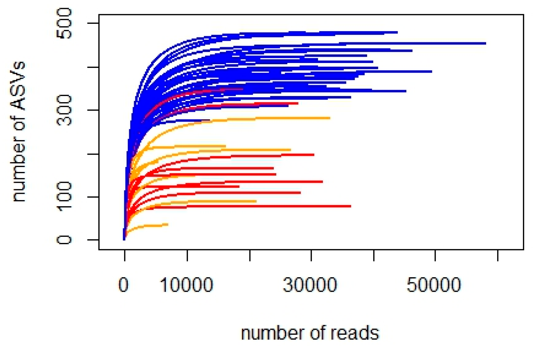
The 18S_V9 sequencing of 54 water and sediment samples yielded a total of 6,123,792 raw reads. A total of 3,604,113 reads remained after quality filtering, merging, denoising, and chimera removal. The total number of ASVs in all 54 samples was 18,455. The removal of ASVs that were not taxonomically assigned to Eukaryota with both reference databases, not assigned to any eukaryotic phylum, or representing groups of predominantly macroscopic organisms, resulted in a dataset of 1,642,367 reads and 4215 ASVs. The rarefaction curves demonstrate a greater number of reads and ASV diversity in sediment compared to water samples, with richness approaching saturation in all samples (Figure 2).
The majority of the ASVs represented ciliates (578), dinoflagellates (427), diatoms (321), and cercozoans (321). A large portion of ASVs could not be assigned to any class within the TSAR (Telonemia + Stramenopila + Alveolata + Rhizaria) phylum (497), the phylum Excavata (85), and below the class Alveolata (145). The proportions of ASVs representing major taxonomic groups were not very different between water and sediment samples, with diatom and dinoflagellate ASVs being slightly more diverse in water, and ciliates in sediment (Figure 3, left panel). Sediment samples had proportionally more reads that belonged to diatoms, nematodes, platyhelmintes, and raphidophytes and fewer green algae (Chlorophyta) reads compared to water samples (Figure 3, right panel).
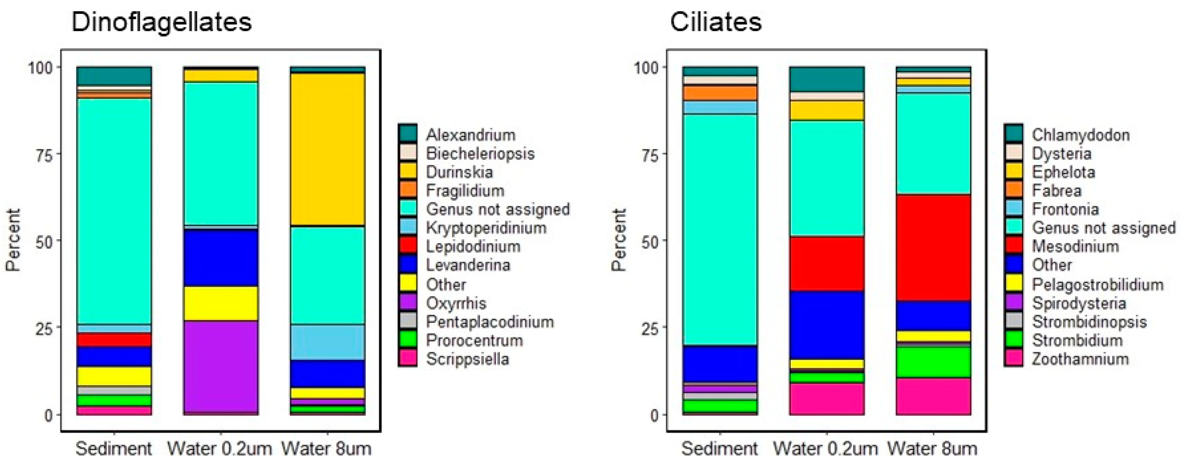
Dinoflagellates were represented by three classes: Dinophyceae (74% of ASVs, 94% of reads), Syndiniales (20% of ASVs, 2.6% of reads), and Oxyrrhea (0.7% of ASVs, 2% of reads). The most abundant genera of dinoflagellates were Durinskia (13.7% of all dinoflagellate reads), Levanderina (6.8%), Kryptoperidinium (4.8%), and Alexandrium (4.0%). The first three genera were each represented by a single species in each, namely Durinskia dybowskii, Kryptoperidinium foliaceum, and Levanderina fissa, and these were the most abundant protistan ASVs identified to the species level in the entire 18S dataset. Alexandrium spp. were proportionally more abundant in sediments, while D. dybowskii and K. foliaceum had the highest contribution in the 8 μm water fraction (Figure 4, left panel). Both D. dybowskii and L. fissa were present in all pools except one located closest to the shore (natural pool “SB”), while K. foliaceum was found only in natural pools. The 0.2 μm water fraction was dominated by reads identified as Oxyrrhis marina.
Among ciliates, most reads were attributed to the genera Fabrea (4.1%), Strombidium (3.9%), Frontonia (3.8%), and Mesodinium (2.5%), with Fabrea, Frontonia, and Strombidium being the most abundant in sediments and Mesodinium and Zoothamnion in water (Figure 4, right panel). Besides these genera, in the 8 μm water fraction, reads assigned to the genus Strombidium were relatively abundant, while in the 0.2 μm fraction, Chlamydodon and Ephelota had higher proportions. The most abundant ASVs identified to the species level belonged to Fabrea salina, Strombidium guangdongense, and Mesodinium rubrum. The proportion of dinoflagellate and ciliate reads not assigned to any genus was higher (approximately 2/3 of all reads) in sediment samples but also substantial (approximately 1/3 of all reads) in water (Figure 4).
Among diatom 18S ASVs assigned a species-level taxonomy, the most abundant were those identified as Chaetoceros tenuissimum, Cyclotella choctawhatcheeana, Melosira dubia, Navicula cryptotenella, and Minidiscus trioculatus. The other abundant reads were only identified to the genus level as Navicula, Haslea, Chaetoceros, Thalassiosira, Skeletonema, and Gyrosigma.
Several organisms notorious for causing harmful algal blooms (HABs) in coastal regions were found. They included dinoflagellates Akashiwo sanguinea, Amphidoma languida, Alexandrium andersonii, A. hiranoi, A. leei, Gymnodinium catenatum, Karlodinium veneficum, Pfiesteria piscicida (at low abundance in two artificial pools), P. shumwayae, Protoceratium reticulatum, Prorocentrum cassubicum, and P. leve (as P. levis in PR2), a diatom Pseudo-nitzschia multiseries, a pelagophyte Aureococcus anafagefferens, and raphidophytes Chatonella subsalsa, C. minima, Chloromorum toxicum, Fibrocapsa japonica, and Heterosigma carterae. The ASVs assigned to Chatonella subsalsa and Chloromorum toxicum were the fifth and sixth most abundant among all protistan reads, respectively.
Within Cercozoa, the relatively highest proportions of reads belonged to the “Protaspa lineage”, Ebria tripartita, and Placopus pusillus, a parasite of Tetraselmis which was found to be most abundant among 18S green algal reads. The most abundant green algal ASVs identified to the species level belonged to Tetraselmis marina, Ostreococcus mediterraneus, O. tauri, and Pyramimonas disomata. The most common euglenid ASVs assigned low-rank taxonomy were Notosolenus urceolatus, N. ostium, Eutreptiella sp., and Rapaza viridis. Among other Euglenozoa classified at least to the genus level, the most abundant were kinetoplastids Neobodo sp., Rhynchomonas nasuta, and Klosteria bodomorphis and diplonemids Rhynchopus serpens, Hemistasia phaeocysticola, and Diplonema aggregatum.
Only 25% of all foraminiferan ASVs were taxonomically assigned at the genus level, and at the class level, 22% of ASVs were assigned to Monothalamids, 16% to Globothalamea, and 6% to Tubothalamea. Within fungi, most reads identified to the class level belonged to Ascomycota (35%), followed by Basidiomycota (8%), while other fungal classes did not reach 1% of relative abundance and 55% of all reads were not assigned to any class.

3.1.2. Diatom rbcL Metabarcoding

RbcL sequencing yielded a total of 2,520,050 raw reads. A total of 1,569,601 reads remained after quality filtering, merging, denoising and chimera removal. Out of 2900 ASVs, 2434 were taxonomically assigned at least to the phylum level with 2404 ASVs classified as diatoms (Bacillariophyta), 29 as other ochrophytes (including 11 Chrysophyceae, five Raphidophyceae and one Xanthophyceae), and one as a red alga (Rhodophyta). A total of 1240 diatom ASVs were unique to benthic samples, 784 were unique to water, and 380 were shared among both habitats.
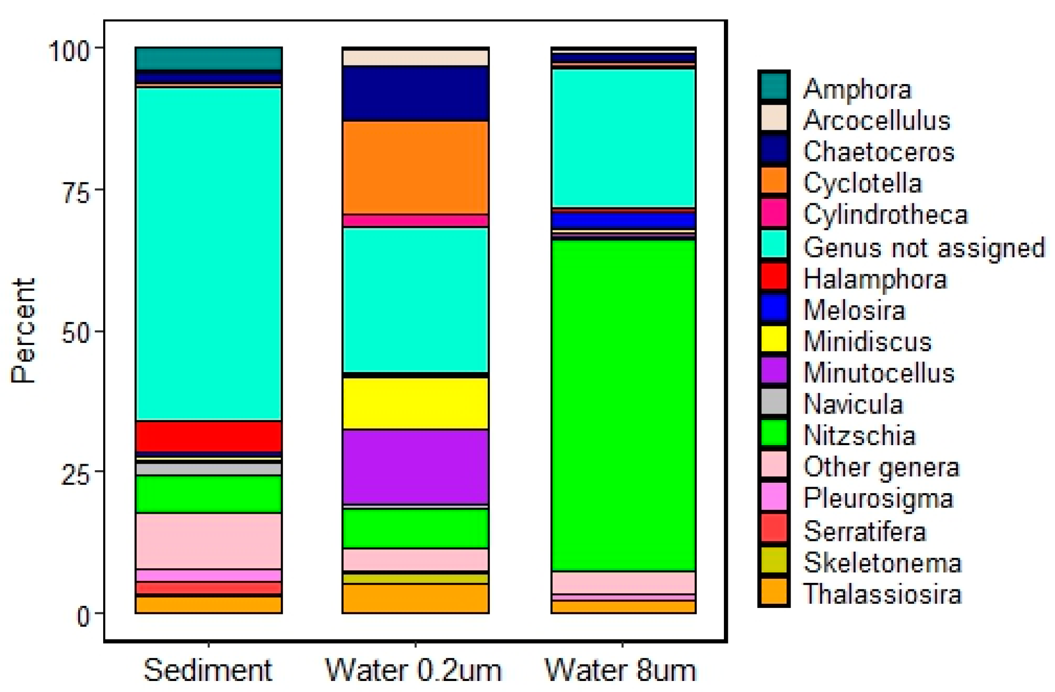
A total of 774 or 32% of diatom ASVs were taxonomically assigned to the genus or species level; these constituted 48% of all diatom reads. The genera most represented in terms of ASV numbers were Nitzschia (21.3% of ASVs identified to genus level), Halamphora (15%), Navicula (7.4%), and Amphora (6.5%). The genera with the highest abundance of reads were Nitzschia (13.8%), Halamphora (4.5%), Amphora (3.2%), and Thalassiosira (3.1%). More than half (51.5%) of all rbcL diatom reads were not classified at the genus level, and most of these unassigned reads were from the sediment samples (Figure 5). As expected, the smallest-size water fraction had a relatively high proportion of amplicons assigned to small-sized genera, such as Arcocellus, Minidiscus, and Minutocellus, or those that have large proportions of small-sized species in coastal waters, such as Cyclotella and Chaetoceros.
Among rbcL ASVs taxonomically assigned to species level, the most abundant (greater than 0.3% of diatom reads) were Nitzschia draveillensis, N. spathulata, N. ardua, N. aurariae, N. laevis, Serratifera andersonii, Minidiscus trioculatus, Amphora fusca, A. ablundens, Tryblionella hungarica, Chaetoceros muelleri, C. tenuissimus, Melosira dubia, Minutocellus polymorphus, Haslea howeana, Halamphora caribaea, H. petusa, Lyrella hennedyi, Thalassiosira minima, Arcocellulus mammifer, and Conticribra weissflogii. Three ASVs were identified as Pseudo-nitzschia: P. americana, P. sp., and toxicogenic P. pungens.

3.1.3. Diatom Counts

A total of 229 diatom morphotaxa were found in 30 sediment samples with 37–69 species per sample (Table S2). The most common and abundant were several species of the genera Navicula (N. salinicola, N. consentanea, N. johnssonii, N. hamiltonii, and N. halinae) and Halamphora (H. adumbratoides, H. tenerrima, and H. subtropica), as well as Serratifera spp., Cocconeis neothumensis var. marina, Nitzschia frustulum, and Opephora spp. A total of 66 morphotaxa did not fully fit descriptions of known species or represented complexes of similar species that were impossible to separate with light microscopy either because of their small size or insufficient information about species delimitation.

3.2. Differences between Natural and Artificial Pools

3.2.1. Assemblage Composition

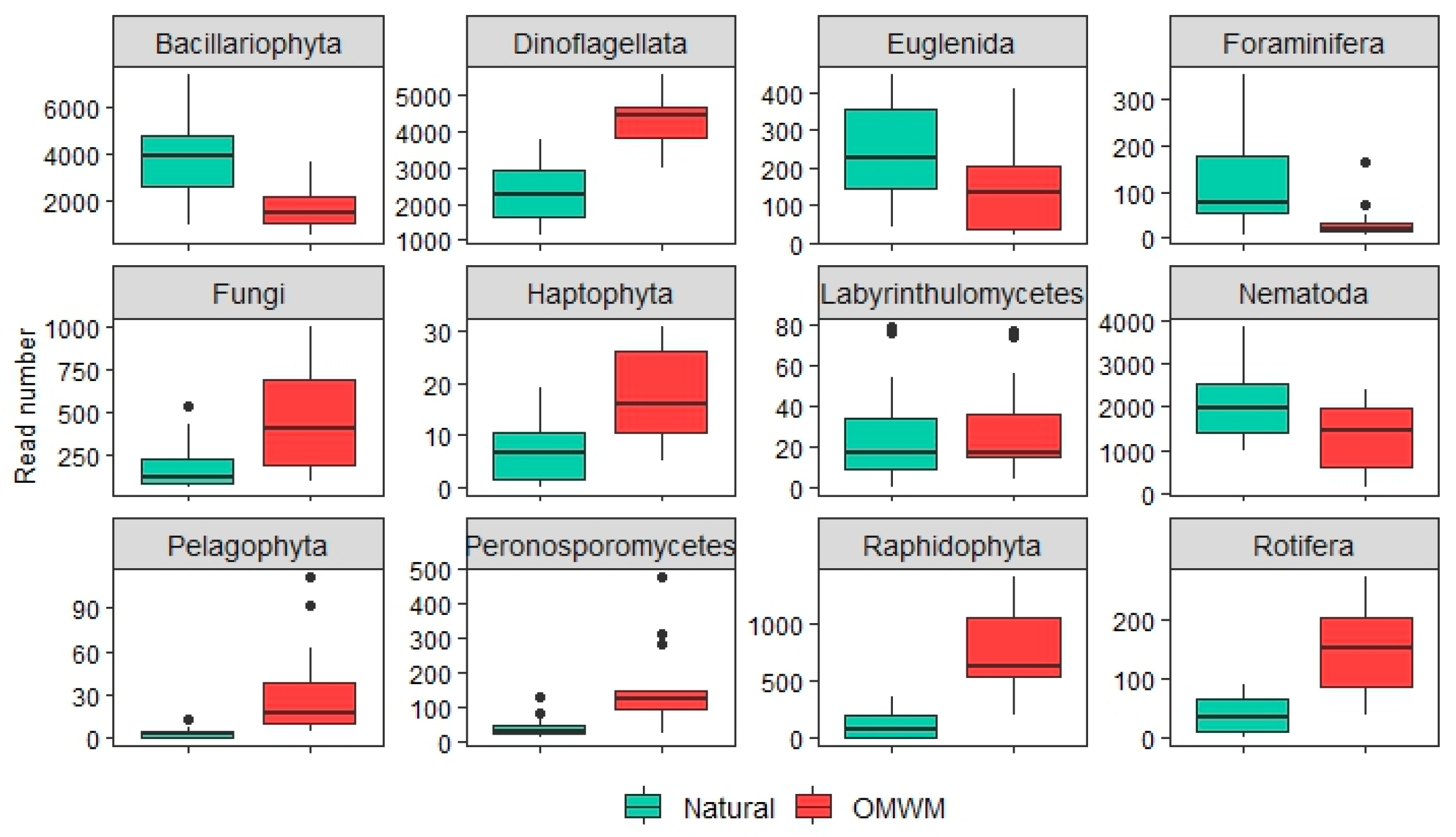
Several taxonomic groups identified by 18S metabarcoding showed significant differences in read abundances between natural and artificial pools (t-test, Table 2). These differences were most pronounced in sediment samples where diatoms, euglenoids, foraminifera, and nematodes were more abundant in natural pools, while several groups of mixotrophic flagellates, such as dinoflagellates, pelagophytes, raphidophytes, and some other groups such as green algae, rotifers, fungi, and fungi-like stramenopiles (peronosporomycetes and labyrinthulomycetes) were proportionally more abundant in OMWMs (Figure 6). Ciliates were more abundant in natural pools, although this trend was marginally significant (Table 2). In water samples, the same main trends were observed, although the differences were rarely statistically significant because of the lower number of samples (Table 2).
Differences in the composition of the whole microeukaryote assemblage between natural and artificial pools were likewise most pronounced in sediment and similar, but often non-significant, in water samples (PERMANOVA, Table 2). The difference in microeukaryotic assemblage composition between natural and artificial pools was mostly driven by dinoflagellates, cercozoans, diatoms, and apicomplexans in sediment and by green algae, diatoms, and cercozoans in water (PERMANOVA, Table 2).
The NMDS plot of sediment samples (Figure 7, left panel) shows a strong separation of samples from natural and artificial pools along the first NMDS axis, which also corresponds to a gradient of water salinity, pH, and turbidity. Samples from the same pools, shown as circles connected by “spider” lines in Figure 7, tend to cluster together, thus showing a greater similarity of assemblages within pools as compared to among-pool similarity. The four samples positioned on the left of the NMDS plot are from a natural pool located closest to the open water of the Barnegat Bay with a sandy bottom and experiencing the most flushing. This indicates that the main gradient of microeukaryotic assemblage variation may also be related to substrate texture and other unmeasured environmental characteristics related to pool position on the marsh. The second NMDS axis appears to be underlain by the water depth gradient and associated characteristics such as temperature (higher in shallow pools) and dissolved oxygen (higher in deeper pools).
Diatom assemblages characterized by rbcL metabarcoding were also found to be significantly different in their composition (Table 2, Figure 7, right panel), while diatom count data revealed only a marginally significant difference between natural and artificial pools (Table 2, Figure S1).

3.2.2. Differential Abundance of ASVs and Morphotaxa

In sediment samples, the ALDeX tool identified 44 18S ASVs as significantly more abundant in natural pools in comparison to OMWMs (Benjamini–Hochberg corrected p-value < 0.05). Those identified to the species level were dinoflagellates Kryptoperidinium foliaceum, Ansanella granifera, Sourniaea diacantha, and Pelagodinium beii (a symbiont of planktonic foraminiferans), diatoms Minidiscus trioculatus, Navicula cryptotenella, Paralia sulcata, and Lithodesmium undulatum, and a cercozoan Ebria tripartita. Among 61 ASVs that were significantly higher in abundance in sediments of OMWMs, the following were identified to the species level: dinoflagellates Alexandrium insuetum, Lepidodinium viride, Biecheleriopsis adriatica, and Prorocentrum cassubicum, raphidophytes Chloromorum toxicum and Chattonella subsalsa, a chrysophyte Paraphysomonas foraminifera, a Chrysomeridophyte Chrysowaernella hieroglyphica, a dictyochopyte Pseudopedinella elastica, an apicomplexan parasite of amphipods Heliospora longissima, and a flat worm Archimacrostomum rubrocinctum.
In water samples, only the dinoflagellate Kryptoperidinium foliaceum was higher in abundance in natural pools as compared to OMWMs, while only one green algal ASV classified as “Chlorellales_X” had a higher abundance in OMWMs.
Thirteen diatom rbcL ASVs had significantly higher abundance in sediments of the natural pools. Among these, four AVSs were assigned taxonomy at the species level: Minidiscus trioculatus, Lyrella hennedyi, Nitzschia draveillensis, and Thalassiosira minima. Twenty-four ASVs were more abundant in OMWMs, and six of them were classified at species level: Chaetoceros muelleri, Haslea howeana, Cylindrotheca closterium, Halamphora foramina, Serratifera andersonii, and Nitzschia laevis. In water samples, no diatom rbcL ASVs were significantly more abundant in one type of pool compared to another if judging by Benjamini–Hochberg corrected p-values, but some of the same ASVs that differentiated pool types in the sediment dataset showed the same trends in water samples based on uncorrected p-values. For example, Minidiscus trioculatus was found to be more abundant in natural samples in comparison to OMWMs, and Chaetoceros muelleri was more abundant in OMWMs.
The indicator species analysis identified one diatom morphospecies, Navicula platyventris, as characteristic of natural pools (p-value < 0.05) and six diatom morphotaxa, Cyclotella katiana, C. meneghiniana, Chaetoceros spp., Navicula cincta, N. microcari, and Nitzschia spathulata, as characteristic of OMWMs.

4. Discussion

4.1. Assemblage Composition

Our metabarcoding results show that the dominant microscopic eukaryotes in studied pools were dinoflagellates, diatoms, ciliates, green algae, cercozoans, and nematodes, the groups commonly reported as dominant in metabarcoding surveys of marine plankton [40,43,44,85,86], deep sediments [41,42], and intertidal mudflats [48]. A large proportion of ASVs recovered in this survey were not attributed to low-rank taxa, which is characteristic of barcoding and metabarcoding surveys regardless of used DNA markers or target taxonomic groups [40,45]. Forster et al. [42] found that about 70% of planktonic and 33% of benthic RNA sequences recovered in the metabarcoding of marine coastal environments could be assigned taxonomy using contemporary reference databases. This ratio reflects the existing knowledge gaps of the taxonomic diversity of benthic organisms, especially protists, and is practically the same as what we found in the water and sediment samples of the tidal pools. The ASV diversity in studied pools was higher in sediment than water, which is also concurrent with findings by Forster et al. [42], who explained this phenomenon by the greater heterogeneity of sediment habitats.
Ciliate ASV richness was the highest among all major taxonomic groups in the studied pools. The most abundant reads were attributed to ciliates known from both coastal plankton and intertidal or inland habitats with high and fluctuating salinity. A characteristic representative of the latter group is Fabrea salina, a euryhaline heterotrich notorious for its ability to withstand high salinity and ultraviolet radiation [87,88,89]. Several other genera abundant in studied pools, such as Strombidium, Chlamydodon, and Frontonia, are common in salt marshes [33,90] and coastal plankton [91], and include species known for their tolerance of low oxygen levels [92]. In particular, Strombidium purpureum, an anaerobic species harboring symbiotic purple bacterium [93], was detected in two artificial pools with relatively low oxygen levels (1.9 and 3.5 mg L−1). The largest proportion of ciliate reads in the water samples of studied pools were attributed to the photosynthetic Mesodinium rubrum, a cosmopolitan and occasionally bloom-forming species common in coastal marine plankton including estuaries of the mid-Atlantic region of USA [94,95]. The presence of amplicons assigned to sessile ciliate genera Zoothamnion and Ephelota in water samples requires further study. Snyder et al. [96] found an unexpected number of non-planktonic ciliates in their survey of microzooplankton in the Gulf of Mexico and explained this by the association of ciliates with floating algae. The high proportion of reads of sessile ciliates in 0.2 μm water fraction in our study is probably due to the capture of extracellular DNA. Zoothamnion DNA, for example, was found in copepod guts [97] and, therefore, may appear in water after excretion.
Dinoflagellate reads were the most abundant in the entire 18S dataset and several dinoflagellate ASVs, all from the class Dinophyceae, were dominant in water, sediments, or both. Their ASV richness was also high, which is often the case in metabarcoding surveys of marine waters, including low-oxygen areas [46]. Dinoflagellates are consistently reported as having a relatively high abundance of reads in metabarcoding surveys, even when their cell numbers are moderate, as their cell DNA content is often extremely high [85]. In studied tidal pools, we also observed dense populations of live and encysted dinoflagellates with light microscopy. The three dinoflagellate species that were most abundant in studied pools, Durinskia dybowskii (syn. D. baltica, Peridinium balticum), Levanderina fissa (syn. Gyrodinium uncatenum, G. instriatum, G. pavillardii), and Kryptoperidinium foliaceum (syn. K. triquetra), have been reported from various coastal systems across the world, including the tidal ponds and coastal lagoons of eastern North America [98,99,100]. Several species of Dinophyceae found in this survey, such as Akashiwo sanguinea, Karlodinium veneficum, and Pfiesteria piscicida, are toxicogenic [101], while others such as Levaderina fissa may cause HABs without producing toxins [102].
The presence of heterotrophic dinoflagellate Oxyrrhis marina in the 0.2 μm fraction of water samples is difficult to explain as this is a benthic species with a body size larger than 8 μm [103]. Dinoflagellate reads including those of Oxyrrhis in this fraction may represent extracellular DNA. While the read numbers of the dinoflagellate class Syndiniales, which includes only parasitoid taxa [104], were not especially high, their high diversity (20% of dinoflagellate ASVs) indicates the complexity of ecological networks in tidal pools.
Among diatoms, the genera Chaetoceros, Thalassiosira, Skeletonema, Minidiscus, and Cyclotella that both rbcL and 18S metabarcoding showed to be abundant in the water samples of the studied pools have been reported as dominant phytoplankters in New Jersey coastal lagoons adjacent to our sampling area [95,105] and in other coastal regions of eastern North America [106]. RbcL ASVs of diatoms Minutocellus polymorphus and Arcocellus mammifer were abundant in the 0.2 μm water fraction in our samples. These diatoms are likely to thrive too in the open water of adjacent coastal lagoons, as M. polymorphus has been reported from Barnegat Bay, while Arcocellus could have been previously reported as Phaeodactylum tricornutum [95,105]. These diatoms are extremely small and, therefore, easy to misidentify or underestimate in routine microscopy analysis. They were also not recoded in our diatom morphology-based counts of sediment samples, indicating that their lightly silicified frustules easily dissolve after cell death. The 8 μm water fraction had a high abundance of ASVs classified as the diatom Nitzschia draveillensis, commonly reported in the plankton of eutrophic inland waters [75]. We also found N. cf. draveillensis, morphologically slightly different from the freshwater N. draveillensis, in our pool sediment samples. Diatoms identified as N. cf. draveillensis have been previously reported from oceanic plankton [107] and identified by the denaturing gradient gel electrophoresis band sequencing from an intertidal mudflat in South Carolina [50]. In sediment samples, our metabarcoding results largely paralleled the morphology-based identification of diatoms at the genus level with Navicula, Halamphora, and Nitzschia as the dominant genera both in terms of morphotaxa and ASV diversity and abundance. These same genera appeared to be dominant in a metabarcoding study of salt marsh diatoms in South Carolina [52]. Neritic planktonic diatoms also appeared abundant in sediment, which can be expected, as many of them (Chaetoceros, Thalassiosira, and Skeletonema) can either produce resting cells and spores [108] or live on the surface of intertidal sediments (Minidiscus) [26]. At the species level, not much concordance between morphological and molecular identifications was found, which is expected given the low coverage of diatom diversity in reference databases [64,65]. Morphology-based identifications revealed that a high proportion of taxa found in studied pools are common for the marsh soils and mudflats of the mid-Atlantic region [26,109].
Small flagellate (Mamiellophyceae, Chlorodendrophyceae, Pyramimonadophyceae, and Chlorophyceae) and coccoid (Trebouxiophyceae) green algae appeared very abundant in the water column of studied pools. They are frequently identified microscopically in coastal plankton and tidal pools [110] including New Jersey coastal lagoons [95,105], have been noted among dominant groups in metabarcoding surveys of marine coastal plankton [111], and were shown to be less abundant in benthic samples than in plankton [42].
Cerocozoans have been noted as one of the most diverse but less studied groups of protists in soils and marine and freshwater sediments [42,112], and we found a high diversity of cerocozoan ASVs in studied pools. Some of the most abundant cercozoan amplicons were attributed to the Protaspa lineage known to parasitize diatoms [113] and identified in metabarcoding surveys of marine plankton [104,113] and intertidal sediments [48]. Another cercozoan abundant in the pools was Placopus pusillus, known for parasitizing flagellate green algae from the order Chlorodendrales [114]. The most abundant free-living cercozoan was Ebria tripartita commonly detected in marine and inland saline waters [110]. Eugenozoa likewise included organisms with a wide range of lifestyles such as free-living (e.g., Eutreptiella) and parasitic (e.g., Rapaza viridis) euglenids [115], mostly free-living and predatory diplonemids, and a variety of kinetoplastids.
There was no correspondence between our metabarcoding identifications of foraminifera apparently dominated by monothalamids and the lists of visually identified species from New Jersey salt marshes [25,27], where multichambered Globothalamea are most common. A similar discord in the taxonomic composition of foraminifera identified morphologically and with metabarcoding was found by Frontalini et al. [116], who also noted a prevalence of soft-walled monothalamids in their 18S metabarcoding dataset from the Adriatic Sea.
The relatively high abundance of nematode ASVs was expected, as they are often the dominant group in the meiofauna of coastal marshes [22,33]. Within fungi, the prevalence of Ascomycota followed by Basidiomycota that we found in studied pools is characteristic of marine waters [86,117] and salt marshes [7,11], in contrast to freshwater habitats where other fungal groups are more common [118].

4.2. Comparison of Assemblages from Natural and Artificial Pools

The natural and artificial pools in this study were found to sustain significantly different microeukaryotic assemblages in terms of the differential abundance of individual ASVs, several major taxonomic groups, and overall assemblage composition. The limited number of sampled pools in our study and their spatial arrangement in two clusters requires this finding to be tested with a larger and spatially and environmentally randomized set of pools. Moreover, our survey was not designed to assess the temporal variation in species composition, while salt marsh microbial assemblages are known to experience considerable seasonal changes [9,48,119]. While further confirmation is needed, these first metabarcoding data provide interesting insights into the impact of hydrological modification on the microeukaryotic diversity of salt marshes. The most striking difference between the two types of pools was the increased abundance of several groups of mixotrophic flagellates in OMWMs. While mixotrophic dinoflagellates, haptophytes, chrysopytes, and raphidophytes are mostly characterized as phagotrophs and less frequently as osmotrophs [120,121], osmotrophy is characteristic of pelagophytes [122] as their growth can be stimulated by dissolved organic compounds [123,124]. The harmful blooms (“brown tides”) of pelagophytes Aureococcus anophagefferens and Aureoumbra lagunenesis in the coastal waters of the eastern USA are associated with elevated concentrations of dissolved organic matter (DOM) [125,126].
DOM concentration was not measured in our study, but it is likely to be higher in OMWMs compared to natural pools because of their relatively greater ratio of shoreline length to pool area (Figure 1D). Narrow ditches with steep banks excavated in marsh peat should inevitably have water that is high in organic matter and, thus, stimulate mixotrophs and decomposers. Besides the direct consumption of DOM, bacterivorous mixotrophs such as dinoflagellates and raphidophytes may benefit from elevated organic matter via an increase in available bacterial biomass [120]. Among bacterivorous mixotrophs, toxigenic raphidophytes Chattonella subsalsa and Chloromorum toxicum showed a remarkable increase in OMWMs in our study. These species are known for their toxicity to fish and mollusks and have been causing HABs with significant economic impacts in the coastal waters of the eastern USA [102,127].
We also found an increase in fungi and fungi-like protists such as peronosporomycetes (water molds) and labyrinthulomycetes in OMWMs, which may also be related to the higher organic matter availability to decomposers. Despite the considerable functional diversity within these groups, together with bacteria, they are ultimately responsible for the degradation of particulate and dissolved organic matter in aquatic ecosystems [128,129,130].
Besides the increased abundance of protistan groups known as predominantly mixotrophs and decomposers, OMWMs were different from natural pools in the composition of several taxonomic groups, for example, diatoms (Figure 7, right panel). It is not obvious from our data what factors may be responsible for this distinction, but the effect of added organic matter and increased turbidity in OMWMs cannot be excluded. For example, some diatom species are known for being partially or even entirely osmotrophic [131], but the extent of osmotrophy in different diatom taxa is largely unknown. As our results show, almost all taxonomic groups of microeukaryotes showed shifts in their assemblage composition if the sample size in terms of ASV and read numbers was sufficiently large. This is not surprising considering that the effects of organic matter increase may propagate through the entire food web, indirectly affecting predatory, parasitic, and symbiotic microeukaryotes through changes in their food sources and hosts [132].
Lewitus et al. [133] found abundant populations of HAB-causing flagellates in artificial ponds excavated in residential areas and golf courses in the coastal areas of South Carolina. Some of the species that formed blooms in these ponds, such as Chattonella subsalsa, were the same as in the tidal pools that we studied in New Jersey. Lewitus et al. [133] further hypothesized that these ponds could be “natural incubators” of harmful bloom-forming protists that proliferate in sheltered and nutrient-rich pond water and subsequently are spread over coastal areas by tides. The ability of most HAB-causing protists to form cysts also makes tidal pools a suitable refuge for surviving unfavorable conditions. Our results support the idea that tidal pools can serve as nurseries for bloom-forming protists, and artificial pools may further promote the growth of mixotrophic flagellates implicated in HABs. As blooms of harmful pelagophytes and raphidophytes began in coastal areas of the eastern USA in the 1980s [102,134], the question arises about the potential link between these blooms and the elevated organic matter concentrations in estuaries and coastal lagoons due to salt marsh modifications and destruction caused by sea level rise.
In conclusion, our survey revealed a diverse assemblage of microbial eukaryotes in salt marsh pools and confirmed the power of the metabarcoding approach for revealing the effects of environmental variation on the biota. It also raised questions about the impacts of salt marsh modifications on coastal ecosystems that could be addressed by further studies.

Supplementary Materials

The following supporting information can be downloaded at https://www.mdpi.com/article/10.3390/coasts4020015/s1. Table S1: Location and characteristics of sampled tidal pools; Table S2: Diatom count data; Figure S1: NMDS plot, sediment diatom count data.

Author Contributions

Conceptualization, M.P.; methodology, M.P. and D.M.; validation, M.P., D.M. and A.K.; formal analysis, A.K. and M.P.; investigation, M.P., D.M. and L.A.; resources, M.P.; data curation, M.P. and L.A.; writing—original draft preparation, M.P.; writing—review and editing, M.P. and L.A.; visualization, A.K. and M.P.; project administration, M.P. All authors have read and agreed to the published version of the manuscript.

Funding

This research received no external funding.

Data Availability Statement

Demultiplexed raw Illumina MiSeq reads are available at the National Center for Biotechnology Information (NCBI) Sequence Read Archive (SRA) under bioproject number PRJNA1035775. Diatom count data are available as Table S2.

Acknowledgments

The following Drexel University students and employees participated in sample collection and laboratory processing: Scott Beadenkopf, Jackie Ng, Hemal Trivedi, Alyssa Norbury, Morgan Sorensen, Shane McCredie, Brooke Herring, Nina Desianti, and Roger Thomas.

Conflicts of Interest

The authors declare no conflicts of interest.

References

  1. Koop-Jakobsen, K.; Gutbrod, M.S. Shallow salt marsh tidal ponds—An environment with extreme oxygen dynamics. Front. Environ. Sci. 2019, 7, 137. [Google Scholar] [CrossRef]
  2. Able, K.W.; Smith, K.J.; Hagan, S.M. Fish composition and abundance in New Jersey salt marsh pools: Sampling technique effects. Northeast. Nat. 2005, 12, 485–502. [Google Scholar] [CrossRef]
  3. Bolduc, F.; Afton, A.D. Relationships between wintering waterbirds and invertebrates, sediments and hydrology of coastal marsh ponds. Waterbirds 2004, 27, 333–341. [Google Scholar] [CrossRef]
  4. Minello, T.J.; Able, K.W.; Weinstein, M.P.; Hays, C.G. Salt marshes as nurseries for nekton: Testing hypotheses on density, growth and survival through meta-analysis. Mar. Ecol. Prog. Ser. 2003, 246, 39–59. [Google Scholar] [CrossRef]
  5. Reed, H.E.; Martiny, J.B.H. Microbial composition affects the functioning of estuarine sediments. ISME J. 2013, 7, 868–879. [Google Scholar] [CrossRef] [PubMed]
  6. Frates, E.S.; Spietz, R.L.; Silverstein, M.R.; Girguis, P.; Hatzenpichler, R.; Marlow, J.J. Natural and anthropogenic carbon input affect microbial activity in salt marsh sediment. Front. Microbiol. 2023, 14, 1235906. [Google Scholar] [CrossRef]
  7. Calabon, M.S.; Jones, E.B.G.; Promputtha, I.; Hyde, K.D. Fungal biodiversity in salt marsh ecosystems. J. Fungi 2021, 7, 648. [Google Scholar] [CrossRef] [PubMed]
  8. Gessner, R.V.; Goos, R.D. Fungi from decomposing Spartina alterniflora. Can. J. Bot. 1973, 51, 51–55. [Google Scholar] [CrossRef]
  9. Rinke, M.; Maraun, M.; Scheu, S. Spatial and temporal variations in salt marsh microorganisms of the Wadden Sea. Ecol. Evol. 2022, 12, e8767. [Google Scholar] [CrossRef]
  10. Kearns, P.; Angell, J.; Howard, E.M.; Deegan, L.A.; Stanley, R.H.R.; Bowen, J.L. Nutrient enrichment induces dormancy and decreases diversity of active bacteria in salt marsh sediments. Nat. Commun. 2016, 7, 12881. [Google Scholar] [CrossRef]
  11. Kearns, P.J.; Bulseco-McKim, A.N.; Hoyt, H.; Angell, J.H.; Bowen, J.L. Nutrient enrichment alters salt marsh fungal communities and promotes putative fungal denitrifiers. Microb. Ecol. 2019, 77, 358–369. [Google Scholar] [CrossRef] [PubMed]
  12. Kim, J.; Chaudhary, D.R.; Kang, H. Nitrogen addition differently alters GHGs production and soil microbial community of tidal salt marsh soil depending on the types of halophyte. Appl. Soil Ecol. 2020, 150, 103440. [Google Scholar] [CrossRef]
  13. Hanley, T.C.; Bowen, J.L.; Kearns, P.J.; Hughes, A.R. Short- and long-term effects of nutrient enrichment on salt marsh plant production and microbial community structure. J. Ecol. 2021, 109, 3779–3793. [Google Scholar] [CrossRef]
  14. Engel, A.S.; Liu, C.; Paterson, A.T.; Anderson, L.C.; Turner, R.E.; Overton, E.B. Salt marsh bacterial communities before and after the Deepwater Horizon oil spill. Appl. Environ. Microbiol. 2017, 83, e00784-17. [Google Scholar] [CrossRef] [PubMed]
  15. An, S.U.; Cho, H.; Jung, U.J.; Kim, B.; Lee, H.; Hyun, J.H. Invasive Spartina anglica greatly alters the rates and pathways of organic carbon oxidation and associated microbial communities in an intertidal wetland of the Han River Estuary, Yellow Sea. Front. Mar. Sci. 2020, 7, 59. [Google Scholar] [CrossRef]
  16. Kim, J.; Chaudhary, D.R.; Lee, J.; Byun, C.; Ding, W.; Kwon, B.O.; Khim, J.S.; Kang, H. Microbial mechanism for enhanced methane emission in deep soil layer of Phragmites-introduced tidal marsh. Environ. Int. 2020, 134, 105251. [Google Scholar] [CrossRef] [PubMed]
  17. Simon, M.R.; Zogg, G.P.; Travis, S.E. Impacts of sea-level rise on sediment microbial community structure and function in two New England salt marshes, USA. J. Soil Sediment 2017, 17, 2847–2855. [Google Scholar] [CrossRef]
  18. Abbott, K.M.; Quirk, T.; Fultz, L.M. Soil microbial community development across a 32-year coastal wetland restoration time series and the relative importance of environmental factors. Sci. Total Environ. 2022, 821, 153359. [Google Scholar] [CrossRef]
  19. Barry, A.; Ooi, S.K.; Helton, A.M.; Steven, B.; Elphick, C.S.; Lawrence, B.A. Vegetation zonation predicts soil carbon mineralization and microbial communities in Southern New England salt marshes. Estuar. Coasts 2022, 45, 168–180. [Google Scholar] [CrossRef]
  20. Armitage, D.; Gallagher, K.; Youngblut, N.; Buckley, D.; Zinder, S. Millimeter-scale patterns of phylogenetic and trait diversity in a salt marsh microbial mat. Front. Microbiol. 2012, 3, 293. [Google Scholar] [CrossRef]
  21. Lee, H.; Heo, Y.M.; Kwon, S.L.; Yoo, Y.; Kim, D.; Lee, J.; Kwon, B.O.; Khim, J.S.; Kim, J.J. Environmental drivers affecting the bacterial community of intertidal sediments in the Yellow Sea. Sci. Total Environ. 2021, 755, 142726. [Google Scholar] [CrossRef]
  22. First, M.R.; Hollibaugh, J.T. Environmental factors shaping microbial community structure in salt marsh sediments. Mar. Ecol. Prog. Ser. 2010, 399, 15–26. [Google Scholar] [CrossRef]
  23. Barnett, R.L.; Newton, T.L.; Charman, D.J.; Gehrels, W.R. Salt-marsh testate amoebae as precise and widespread indicators of sea-level change. Earth-Sci. Rev. 2017, 164, 193–207. [Google Scholar] [CrossRef]
  24. Horton, B.; Sawai, Y. Diatoms as indicators of former sea levels, earthquakes, tsunamis, and hurricanes. In The Diatoms: Applications for the Environmental and Earth Sciences; Smol, J., Stoermer, E., Eds.; Cambridge University Press: Cambridge, UK, 2010; pp. 357–372. [Google Scholar]
  25. Kemp, A.C.; Horton, B.P.; Vann, D.R.; Engelhart, S.E.; Grand Pre, C.A.; Vane, C.H.; Nikitina, D.; Anisfeld, S.C. Quantitative vertical zonation of salt-marsh foraminifera for reconstructing former sea level; an example from New Jersey, USA. Quat. Sci. Rev. 2012, 54, 26–39. [Google Scholar] [CrossRef]
  26. Desianti, N.; Enache, M.D.; Griffiths, M.; Biskup, K.; Degen, A.; DaSilva, M.; Millemann, D.; Lippincott, L.; Watson, E.; Gray, A.; et al. The potential and limitations of diatoms as environmental indicators in mid-Atlantic coastal wetlands. Estuar. Coasts 2019, 42, 1440–1458. [Google Scholar] [CrossRef]
  27. Walker, J.S.; Khan, N.S.; Shaw, T.A.; Barber, D.C.; Switzer, A.D.; Horton, B.P. Spatial and temporal distributions of live salt-marsh foraminifera in southern New Jersey: Implications for sea-level studies. J. Foraminifer. Res. 2023, 53, 3–19. [Google Scholar] [CrossRef]
  28. Quintana, X.D.; Moreno-Amich, R. Phytoplankton composition of Empordà salt marshes, Spain and its response to freshwater flux regulation. J. Coast. Res. 2002, 36, 581–590. [Google Scholar] [CrossRef]
  29. Sullivan, M.J.; Currin, C.A. Community structure and functional dynamics of benthic microalgae in salt marshes. In Concepts and Controversies in Tidal Marsh Ecology; Weinstein, M.P., Kreeger, D.A., Eds.; Springer: Dordrecht, The Netherlands, 2002; pp. 81–106. [Google Scholar]
  30. Underwood, G.J.C. Microalgal colonization in a saltmarsh restoration scheme. Estuar. Coast. Shelf Sci. 1997, 44, 471–481. [Google Scholar] [CrossRef]
  31. Whitcraft, C.R.; Levin, L.A. Regulation of benthic algal and animal communities by salt marsh plants: Impact of shading. Ecology 2007, 88, 904–917. [Google Scholar] [CrossRef]
  32. Barnes, R.S.K.; Sattelle, D.B.; Everton, I.J.; Nicholas, W.; Scott, D.H. Intertidal sands and interstitial fauna associated with different stages of salt-marsh development. Estuar. Coast. Mar. Sci. 1976, 4, 497–511. [Google Scholar] [CrossRef]
  33. Elliott, P.B.; Bamforth, S.S. Interstitial protozoa and algae of Louisiana salt marshes. J. Protozool. 1975, 4, 514–519. [Google Scholar] [CrossRef]
  34. Xu, Y.; Yao, S.; Soetaert, K.; Fan, X. Effects of salt marsh restoration on eukaryotic microbenthic communities in the Yangtze Estuary. Mar. Ecol. Prog. Ser. 2020, 638, 39–50. [Google Scholar] [CrossRef]
  35. Fleeger, J.; Chandler, G.; Fitzhugh, G.; Phillips, F. Effects of tidal currents on meiofauna densities in vegetated salt marsh sediments. Mar. Ecol. Prog. Ser. 1984, 19, 49–53. [Google Scholar] [CrossRef]
  36. Fleeger, W.; Johnson, D.S.; Galván, K.A.; Deegan, L.A. Top-down and bottom-up control of infauna varies across the saltmarsh landscape. J. Exp. Mar. Biol. Ecol. 2008, 357, 20–34. [Google Scholar] [CrossRef]
  37. Teal, J.M.; Wieser, W. The distribution and ecology of nematodes in a Georgia salt marsh. Limnol. Oceanogr. 1966, 11, 217–222. [Google Scholar] [CrossRef]
  38. Alteio, L.; Séneca, J.; Canarini, A.; Angel, R.; Jansa, J.; Guseva, K.; Kaiser, C.; Richter, A.; Schmidt, H. A critical perspective on interpreting amplicon sequencing data in soil ecological research. Soil Biol. Biochem. 2021, 160, 108357. [Google Scholar] [CrossRef]
  39. Pawlowski, J.; Kelly-Quinn, M.; Altermatt, F.; Apothéloz-Perret-Gentil, L.; Beja, P.; Boggero, A.; Borja, A.; Bouchez, A.; Cordier, T.; Domaizon, I.; et al. The future of biotic indices in the ecogenomic era: Integrating (e)DNA metabarcoding in biological assessment of aquatic ecosystems. Sci. Total Environ. 2018, 637–638, 1295–1310. [Google Scholar] [CrossRef]
  40. de Vargas, C.; Audic, S.; Henry, N.; Decelle, J.; Mahe, F.; Logares, R.; Lara, E.; Berney, C.; Le Bescot, N.; Probert, I.; et al. Eukaryotic plankton diversity in the sunlit ocean. Science 2015, 348, 1261605. [Google Scholar] [CrossRef]
  41. Dünn, M.; Arndt, H. Distribution patterns of benthic protist communities depending on depth revealed by environmental sequencing—From the sublittoral to the deep sea. Microorganisms 2023, 11, 1664. [Google Scholar] [CrossRef]
  42. Forster, D.; Dunthorn, M.; Mahé, F.; Dolan, J.R.; Audic, S.; Bass, D.; Bittner, L.; Boutte, C.; Christen, R.; Claverie, J.-M.; et al. Benthic protists: The under-charted majority. FEMS Microbiol. Ecol. 2016, 92, fiw120. [Google Scholar] [CrossRef]
  43. Massana, R.; Gobet, A.; Audic, S.; Bass, D.; Bittner, L.; Boutte, C.; Chambouvet, A.; Christen, R.; Claverie, J.-M.; Decelle, J.; et al. Protist diversity in European coastal areas. Environ. Microbiol. 2015, 17, 4035–4049. [Google Scholar] [CrossRef]
  44. Sommeria-Klein, G.; Watteaux, R.; Ibarbalz, F.M.; Pierella Karlusich, J.J.; Iudicone, D.; Bowler, C.; Morlon, H. Global drivers of eukaryotic plankton biogeography in the sunlit ocean. Science 2021, 374, 594–599. [Google Scholar] [CrossRef]
  45. Stern, R.F.; Horak, A.; Andrew, R.L.; Coffroth, M.-A.; Andersen, R.A.; Küpper, F.C.; Jameson, I.; Hoppenrath, M.; Véron, B.; Kasai, F.; et al. Environmental barcoding reveals massive dinoflagellate diversity in marine environments. PLoS ONE 2010, 5, e13991. [Google Scholar] [CrossRef]
  46. Stoeck, T.; Bass, D.; Nebel, M.; Christen, R.; Jones, M.D.M.; Breiner, H.-W.; Richards, T.A. Multiple marker parallel tag environmental DNA sequencing reveals a highly complex eukaryotic community in marine anoxic water. Mol. Ecol. 2010, 19 (Suppl. S1), 21–31. [Google Scholar] [CrossRef]
  47. Stoeck, T.; Kochems, R.; Forster, D.; Lejzerowicz, F.; Pawlowski, J. Metabarcoding of benthic ciliate communities shows high potential for environmental monitoring in salmon aquaculture. Ecol. Indic. 2018, 85, 153–164. [Google Scholar] [CrossRef]
  48. Kalu, E.I.; Reyes-Prieto, A.; Barbeau, M.A. Community dynamics of microbial eukaryotes in intertidal mudflats in the hypertidal Bay of Fundy. ISME Commun. 2023, 3, 21. [Google Scholar] [CrossRef]
  49. Plante, C.J.; Hill-Spanik, K.; Lowry, J. Controls on diatom biogeography on South Carolina (USA) barrier island beaches. Mar. Ecol. Prog. Ser. 2021, 661, 17–33. [Google Scholar] [CrossRef]
  50. Rivera-Garcia, L.G.; Hill-Spanik, K.M.; Berthrong, S.T.; Plante, C.J. Tidal stage changes in structure and diversity of intertidal benthic diatom assemblages: A case study from two contrasting Charleston harbor flats. Estuar. Coasts 2018, 41, 772–783. [Google Scholar] [CrossRef]
  51. Wang, M.; Yergaliyev, T.; Sun, C.; Martinez, J.G.; Wang, B. Environmental DNA metabarcoding of intertidal meiofauna sheds light on its potential for habitat discovery. Ecol. Indic. 2023, 150, 110223. [Google Scholar] [CrossRef]
  52. Plante, C.J.; Hill-Spanik, K.; Cook, M.; Graham, C. Environmental and spatial influences on biogeography and community structure of saltmarsh benthic diatoms. Estuar. Coasts 2021, 44, 147–161. [Google Scholar] [CrossRef]
  53. Hovel, K.A.; Morgan, S.G. Susceptibility of estuarine crab larvae to ultraviolet radiation. J. Exp. Mar. Biol. Ecol. 1999, 237, 107–125. [Google Scholar] [CrossRef]
  54. Noël, P.E.; Chmura, G.L. Spatial and environmental variability of pools on a natural and a recovering salt marsh in the Bay of Fundy. J. Coast. Res. 2011, 27, 847–856. [Google Scholar] [CrossRef]
  55. Smith, K.J.; Able, K.W. Dissolved oxygen dynamics in salt marsh pools and its potential impacts on fish assemblages. Mar. Ecol. Prog. Ser. 2003, 258, 223–232. [Google Scholar] [CrossRef]
  56. Kearns, P.J.; Holloway, D.; Angell, J.H.; Feinman, S.G.; Bowen, J.L. Effect of short-term, diel changes in environmental conditions on active microbial communities in a salt marsh pond. Aquat. Microb. Ecol. 2017, 80, 29–41. [Google Scholar] [CrossRef]
  57. Barnby, M.A.; Collins, J.N.; Resh, V.H. Aquatic macroinvertebrate communities of natural and ditched potholes in a San Francisco Bay salt marsh. Estuar. Coast. Shelf Sci. 1985, 20, 331–347. [Google Scholar] [CrossRef]
  58. Meredith, W.H.; Saveikis, D.E.; Stachecki, C.J. Guidelines for “Open Marsh Water Management” in Delaware’s salt marshes—Objectives, system designs and installation procedures. Wetlands 1985, 5, 119–133. [Google Scholar] [CrossRef]
  59. Powell, E.B.; Krause, J.R.; Martin, R.M.; Watson, E.B. Pond excavation reduces coastal wetland carbon dioxide assimilation. J. Geophys. Res.-Biogeosciences 2020, 1252, 1–19. [Google Scholar] [CrossRef]
  60. Clarke, J.A.; Harrington, B.A.; Hruby, T.; Waserman, F.E. The effect of ditching for mosquito control on salt marsh use by birds in Rowley, Massachusetts. J. Field Ornithol. 1984, 55, 160–180. [Google Scholar]
  61. James-Pirri, M.J.; Ginsberg, H.S.; Erwin, R.M.; Taylor, J. Effects of open marsh water management on numbers of larval salt marsh mosquitoes. J. Med. Entomol. 2009, 46, 1392–1399. [Google Scholar] [CrossRef]
  62. Rochlin, I.; James-Pirri, M.J.; Adamowicz, S.C.; Dempsey, M.E.; Iwanejko, T.; Ninivaggi, D.V. The effects of integrated marsh management (IMM) on salt marsh vegetation, nekton, and birds. Estuar. Coasts 2012, 35, 727–742. [Google Scholar] [CrossRef]
  63. Kermarrec, L.; Franc, A.; Rimet, F.; Chaumeil, P.; Humbert, J.F.; Bouchez, A. Next-generation sequencing to inventory taxonomic diversity in eukaryotic communities: A test for freshwater diatoms. Mol. Ecol. Resour. 2013, 13, 607–619. [Google Scholar] [CrossRef]
  64. Pérez-Burillo, J.; Valoti, G.; Witkowski, A.P.; Prado, P.; Mann, D.G.; Trobajo, R. Assessment of marine benthic diatom communities: Insights from a combined morphological–metabarcoding approach in Mediterranean shallow coastal waters. Mar. Pollut. Bull. 2022, 174, 113183. [Google Scholar] [CrossRef]
  65. Turk Dermastia., T.; Vascotto, I.; Francé, J.; Stanković, D.; Mozetič, P. Evaluation of the rbcL marker for metabarcoding of marine diatoms and inference of population structure of selected genera. Front. Microbiol. 2023, 14, 1071379. [Google Scholar] [CrossRef]
  66. Vasselon, V.; Rimet, F.; Tapolczai, K.; Bouchez, A. Assessing ecological status with diatoms DNA metabarcoding: Scaling-up on a WFD monitoring network (Mayotte island, France). Ecol. Indic. 2017, 82, 1–12. [Google Scholar] [CrossRef]
  67. Thompson, L.R.; Sanders, J.G.; McDonald, D.; Amir, A.; Jansson, J.K.; Gilbert, J.A.; Knight, R. The Earth Microbiome Project Consortium. A communal catalogue reveals Earth’s multiscale microbial diversity. Nature 2017, 551, 457–463. [Google Scholar] [CrossRef]
  68. Gohl, D.; Vangay, P.; Garbe, J.; MacLean, A.; Hauge, A.; Becker, A.; Gould, T.J.; Clayton, J.B.; Johnson, T.J.; Hunter, R.; et al. Systematic improvement of amplicon marker gene methods for increased accuracy in microbiome studies. Nat. Biotechnol. 2016, 34, 942–949. [Google Scholar] [CrossRef]
  69. R Core Team. R. A Language and Environment for Statistical Computing; R Foundation for Statistical Computing: Vienna, Austria, 2023; Available online: https://www.R-project.org/ (accessed on 1 October 2023).
  70. Martin, M. Cutadapt removes adapter sequences from high-throughput sequencing reads. EMBnet.journal 2011, 17, 10–12. [Google Scholar] [CrossRef]
  71. Morien, E.; Parfrey, L.W. SILVA v128 and v132 dada2 Formatted 18s ‘train sets’ (1.0) [Data Set]. Zenodo. 2018. Available online: https://zenodo.org/records/1447330 (accessed on 1 October 2023).
  72. Guillou, L.; Bachar, D.; Audic, S.; Bass, D.; Berney, C.; Bittner, L.; Boutte, C.; Burgaud, G.; de Vargas, C.; Decelle, J.; et al. The Protist ribosomal reference database (PR2): A catalog of unicellular eukaryote small sub-unit rRNA sequences with curated taxonomy. Nucleic Acids Res. 2013, 41, D597–D604. [Google Scholar] [CrossRef]
  73. Rimet, F.; Gusev, E.; Kahlert, M.; Kelly, M.G.; Kulikovskiy, M.; Maltsev, Y.; Mann, D.G.; Pfannkuchen, M.; Trobajo, R.; Vasselon, V.; et al. Diat.Barcode, an open-access curated barcode library for diatoms. Sci. Rep. 2019, 9, 1. [Google Scholar] [CrossRef]
  74. Desianti, N.; Potapova, M. Diatom Flora of the New Jersey Coastal Wetlands. Report for the New Jersey Department of Environmental Protection. 2019. Available online: https://hdl.handle.net/10929/68423 (accessed on 1 September 2023).
  75. Krammer, K.; Lange-Bertalot, H. Bacillariophyceae. 2. Teil: Bacillariaceae, Epithemiaceae, Surirellaceae. In Susswasserflora von Mitteleuropa; Ettl, H., Gerloff, J., Heynig, H., Mollenhauer, D., Eds.; Gustav Fisher Verlag: Jena, Germany, 1988; Band 2/2; pp. 1–596. [Google Scholar]
  76. Spaulding, S.; Potapova, M.G.; Bishop, I.W.; Lee, S.S.; Gasperak, T.S.; Jovanoska, E.; Furey, P.C.; Edlund, M.B. Diatoms.org: Supporting taxonomists, connecting communities. Diatom Res. 2021, 36, 291–304. [Google Scholar] [CrossRef]
  77. Stepanek, J.G.; Kociolek, J.P. Amphora and Halamphora from coastal and inland waters of the United States and Japan; Bibliotheca Diatomologica; Schweizerbart Science Publishers: Stuttgart, Germany, 2018; Volume 66, pp. 1–260. [Google Scholar]
  78. Witkowski, A.; Lange-Bertalot, H.; Metzeltin, D. Diatom Flora of Marine Coasts I; Iconographia Diatomologica; A.R.G. Gantner Verlag: Ruggell, Liechtenstein, 2000; Volume 7, pp. 1–925. [Google Scholar]
  79. Oksanen, J.; Simpson, G.; Blanchet, F.; Kindt, R.; Legendre, P.; Minchin, P.; O’Hara, R.; Solymos, P.; Stevens, M.; Szoecs, E.; et al. Vegan: Community Ecology Package. R Package Version 2.6-4. 2022. Available online: https://CRAN.R-project.org/package=vegan (accessed on 1 October 2023).
  80. Fernandes, A.D.; Macklaim, J.M.; Linn, T.G.; Reid, G.; Gloor, G.B. ANOVA-Like Differential Expression (Aldex) Analysis for Mixed Population Rna-Seq. PLoS ONE 2013, 8, e67019. [Google Scholar] [CrossRef] [PubMed]
  81. Gloor, G.B.; Macklaim, J.M.; Fernandes, A.D. Displaying variation in large datasets: A visual summary of effect sizes. J. Comput. Graph. Stat. 2016, 25, 971–979. [Google Scholar] [CrossRef]
  82. De Cáceres, M.; Legendre, P. Associations between species and groups of sites: Indices and statistical inference. Ecology 2009, 90, 3566–3574. [Google Scholar] [CrossRef] [PubMed]
  83. Anderson, M.J. Permutational Multivariate Analysis of Variance (PERMANOVA). In Wiley StatsRef: Statistics Reference Online; Balakrishnan, N.T., Colton, T., Everitt, B., Piegorsch, W., Ruggeri, F., Teugels, J.L., Eds.; John Wiley & Sons, Ltd.: Hoboken, NJ, USA, 2017; pp. 1–15. [Google Scholar] [CrossRef]
  84. Nearing, J.T.; Douglas, G.M.; Hayes, M.G.; MacDonald, J.; Desai, D.K.; Allward, N.; Jones, C.M.A.; Wright, R.J.; Dhanani, A.S.; Comeau, A.M.; et al. Microbiome differential abundance methods produce different results across 38 datasets. Nat. Commun. 2022, 13, 342. [Google Scholar] [CrossRef] [PubMed]
  85. Piredda, R.; Tomasino, M.P.; D’Erchia, A.M.; Manzari, C.; Pesole, G.; Montresor, M.; Kooistra, W.H.; Sarno, D.; Zingone, A. Diversity and temporal patterns of planktonic protist assemblages at a Mediterranean Long Term Ecological Research site. FEMS Microbiol. Ecol. 2017, 93, fiw200. [Google Scholar] [CrossRef] [PubMed]
  86. Sildever, S.; Laas, P.; Kolesova, N.; Lips, I.; Lips, U.; Nagai, S. Plankton biodiversity and species co-occurrence based on environmental DNA—A multiple marker study. Metabarcoding Metagenom. 2021, 5, e72371. [Google Scholar] [CrossRef]
  87. Marangoni, R.; Paris, D.; Melck, D.; Fulgentini, L.; Colombetti, G.; Motta, A. In vivo NMR metabolic profiling of Fabrea salina reveals sequential defense mechanisms against ultraviolet radiation. Biophys. J. 2011, 100, 215–224. [Google Scholar] [CrossRef] [PubMed]
  88. Repak, A.J.; Anderson, O.R. The fine structure of the encysting salt marsh heterotrich ciliate Fabrea salina. J. Morphol. 1990, 205, 335–341. [Google Scholar] [CrossRef]
  89. Zhang, B.; Hou, L.; Qi, H.; Hou, L.; Zhang, T.; Zhao, F.; Miao, M. An extremely streamlined macronuclear genome in the free-living protozoan Fabrea salina. Mol. Biol. Evol. 2022, 39, msac062. [Google Scholar] [CrossRef]
  90. Fan, X.; Lin, X.; Liu, W.; Xu, Y.; Al-Farraj, S.A.; Al-Rasheid, K.A.S.; Warren, A. Morphology of three new marine Frontonia species (Ciliophora; Peniculida) with note on the phylogeny of this genus. Eur. J. Protistol. 2013, 49, 312–323. [Google Scholar] [CrossRef]
  91. Liu, W.; Xu, D.; Ma, H.; Al-Farraj, S.A.; Warren, A.; Yi, Z. Taxonomy and molecular systematics of three oligotrich (s.l.) ciliates including descriptions of two new species, Strombidium guangdongense sp. nov. and Strombidinopsis sinicum sp. nov. (Protozoa, Ciliophora). Syst. Biodivers. 2016, 14, 452–465. [Google Scholar] [CrossRef]
  92. Fenchel, T.; Kristensen, L.D.; Rasmussen, L. Water column anoxia: Vertical zonation of planktonic protozoa. Mar. Ecol. Prog. Ser. 1990, 62, 1–10. [Google Scholar] [CrossRef]
  93. Fenchel, T.; Bernard, C. A purple protist. Nature 1993, 362, 300. [Google Scholar] [CrossRef] [PubMed]
  94. Johnson, M.D.; Stoecker, D.K.; Marshall, H.G. Seasonal dynamics of Mesodinium rubrum in Chesapeake Bay. J. Plankton Res. 2013, 35, 877–893. [Google Scholar] [CrossRef]
  95. Ren, L. Baseline Characterization of Phytoplankton and Harmful Algal Blooms in Barnegat Bay-Little Egg Harbor, New Jersey (Year One). Final Report; New Jersey Department of Environmental Protection, Division of Science, Research and Environmental Health: Trenton, NJ, USA, 2013; pp. 1–44. [Google Scholar]
  96. Snyder, R.A.; Moss, J.A.; Santoferrara, L.; Head, M.; Jeffrey, W.H. Ciliate microzooplankton from the Northeastern Gulf of Mexico. ICES J. Mar. Sci. 2021, 78, 3356–3371. [Google Scholar] [CrossRef]
  97. Zamora-Terol, S.; Novotny, A.; Winder, M. Reconstructing marine plankton food web interactions using DNA metabarcoding. Mol Ecol. 2020, 29, 3380–3395. [Google Scholar] [CrossRef] [PubMed]
  98. Carty, S.; Cox, E.R. Kansodinium gen. nov. and Durinskia gen. nov.: Two genera of freshwater dinoflagellates (Pyrrhophyta). Phycologia 1986, 25, 197–204. Phycologia 1986, 25, 197–204. [Google Scholar] [CrossRef]
  99. Moestrup, Ø.; Hakanen, P.; Hansen, G.; Daugbjerg, N.; Ellegaard, M. On Levanderina fissa gen. & comb. nov. (Dinophyceae) (syn. Gymnodinium fissum, Gyrodinium instriatum, Gyr. uncatenum), a dinoflagellate with a very unusual sulcus. Phycologia 2014, 53, 265–292. [Google Scholar] [CrossRef]
  100. Tillmann, U.; Wietkamp, S.; Kretschmann, J.; Chacón, J.; Gottschling, M. Spatial fragmentation in the distribution of diatom endosymbionts from the taxonomically clarified dinophyte Kryptoperidinium triquetrum (=Kryptoperidinium foliaceum, Peridiniales). Sci. Rep. 2023, 13, 8593. [Google Scholar] [CrossRef]
  101. Lundholm, N.; Churro, C.; Escalera, L.; Fraga, S.; Hoppenrath, M.; Iwataki, M.; Larsen, J.; Mertens, K.; Moestrup, Ø.; Tillmann, U.; et al. 2009 Onwards. IOC-UNESCO Taxonomic Reference List of Harmful Micro Algae. Available online: https://www.marinespecies.org/hab (accessed on 1 October 2023).
  102. Anderson, D.M.; Fensin, E.; Gobler, C.J.; Hoeglund, A.E.; Hubbard, K.A.; Kulis, D.M.; Landsberg, J.H.; Lefebvre, K.A.; Provoost, P.; Richlen, M.L.; et al. Marine harmful algal blooms (HABs) in the United States: History, current status and future trends. Harmful Algae 2021, 102, 101975. [Google Scholar] [CrossRef]
  103. Lowe, C.D.; Day, A.; Kemp, S.J.; Montagnes, D.J.S. There are high levels of functional and genetic diversity in Oxyrrhis marina. J. Eukaryot. Microbiol. 2005, 52, 250–257. [Google Scholar] [CrossRef]
  104. Käse, L.; Metfies, K.; Neuhaus, S.; Boersma, M.; Wiltshire, K.H.; Kraberg, A.C. Host-parasitoid associations in marine planktonic time series: Can metabarcoding help reveal them? PLoS ONE 2021, 16, e0244817. [Google Scholar] [CrossRef] [PubMed]
  105. Olsen, P.S.; Mahoney, J.B. Phytoplankton in the Barnegat Bay-Little Egg harbor estuarine system: Species composition and picoplankton bloom development. J. Coast. Res. 2001, SI 32, 115–143. [Google Scholar]
  106. Robicheau, B.M.; Tolman, J.; Bertrand, E.M.; LaRoche, J. Highly-resolved interannual phytoplankton community dynamics of the coastal Northwest Atlantic. ISME Commun. 2022, 2, 38. [Google Scholar] [CrossRef] [PubMed]
  107. Huapaya, K.; Echeveste, P. Physiological responses of Humboldt current system diatoms to Fe and Cu co-limitation. Mar. Environ. Res. 2023, 187, 105937. [Google Scholar] [CrossRef] [PubMed]
  108. McQuoid, M.R.; Hobson, L.A. Diatom resting stages. J. Phycol. 1996, 32, 889–902. [Google Scholar] [CrossRef]
  109. Sullivan, M.J. Diatom communities from a Delaware salt marsh. J. Phycol. 1975, 11, 384–390. [Google Scholar] [CrossRef]
  110. Tomas, C.R. Identifying Marine Phytoplankton; Academic Press: San Diego, CA, USA, 1997; pp. 1–858. [Google Scholar]
  111. Tragin, M.; Vaulot, D. Green microalgae in marine coastal waters: The Ocean Sampling Day (OSD) dataset. Sci. Rep. 2018, 8, 14020. [Google Scholar] [CrossRef] [PubMed]
  112. Bass, D.; Cavalier-Smith, T. Phylum-specific environmental DNA analysis reveals remarkably high global biodiversity of Cercozoa (Protozoa). Int. J. Syst. Evol. Microbiol. 2004, 54, 2393–2404. [Google Scholar] [CrossRef]
  113. Suter, E.A.; Pachiadaki, M.; Taylor, G.T.; Edgcomb, V.P. Eukaryotic parasites are integral to a productive microbial food web in oxygen-depleted waters. Front. Microbiol. 2022, 12, 764605. [Google Scholar] [CrossRef]
  114. More, K.; Simpson, A.G.B.; Hess, S. Two new marine species of Placopus (Vampyrellida, Rhizaria) that perforate the theca of Tetraselmis (Chlordendrales, Viridiplantae). J. Eukaryot. Microbiol. 2019, 66, 560–573. [Google Scholar] [CrossRef]
  115. Yamaguchi, A.; Yubuki, N.; Leander, B.S. Morphostasis in a novel eukaryote illuminates the evolutionary transition from phagotrophy to phototrophy: Description of Rapaza viridis n. gen. et sp. (Euglenozoa, Euglenida). BMC Evol. Biol. 2012, 12, 29. [Google Scholar] [CrossRef]
  116. Frontalini, F.; Cordier, T.; Balassi, E.; Armynot du Chatelet, E.; Cermakova, K.; Apothéloz-Perret-Gentil, L.; Martins, M.V.A.; Bucci, C.; Scantamburlo, E.; Treglia, M.; et al. Benthic foraminiferal metabarcoding and morphology–based assessment around three offshore gas platforms: Congruence and complementarity. Environ. Int. 2020, 144, 106049. [Google Scholar] [CrossRef]
  117. Morales, S.E.; Biswas, A.; Herndl, G.J.; Baltar, F. Global structuring of phylogenetic and functional diversity of pelagic fungi by depth and temperature. Front. Mar. Sci. 2019, 6, 131. [Google Scholar] [CrossRef]
  118. Richards, T.A.; Jones, M.D.; Leonard, G.; Bass, D. Marine fungi: Their ecology and molecular diversity. Ann. Rev. Mar. Sci. 2012, 4, 495–522. [Google Scholar] [CrossRef]
  119. Lee, J.J.; Muller, W.A. Trophic dynamics and niches of salt marsh foraminifera. Am. Zool. 1973, 13, 215–223. [Google Scholar] [CrossRef]
  120. Burkholder, J.M.; Glibert, M.P.; Skelton, H.M. Mixotrophy, a major mode of nutrition for harmful algal species in eutrophic waters. Harmful Algae 2008, 8, 77–93. [Google Scholar] [CrossRef]
  121. Mitra, A.; Caron, D.A.; Faure, E.; Flynn, K.J.; Leles, S.G.; Hansen, P.J.; McManus, G.B.; Not, F.; do Rosario Gomes, H.; Santoferrara, L.F.; et al. The Mixoplankton Database (MDB): Diversity of photo-phago-trophic plankton in form, function, and distribution across the global ocean. J. Eukaryot. Microbiol. 2023, 70, e12972. [Google Scholar] [CrossRef]
  122. Glibert, P.M.; Legrand, C. The diverse nutrient strategies of harmful algae: Focus on osmotrophy. Ecol. Stud. 2006, 189, 163–175. [Google Scholar] [CrossRef]
  123. Lomas, M.W.; Glibert, P.M.; Clougherty, D.A.; Huber, D.E.; Jones, J.; Alexander, J.; Haramoto, E. Elevated organic nutrient ratios associated with brown tide blooms of Aureococcus anophagefferens (Pelagophyceae). J. Plankton Res. 2001, 23, 1339–1344. [Google Scholar] [CrossRef]
  124. Gann, E.R.; Truchon, A.R.; Papoulis, S.E.; Dyhrman, S.T.; Gobler, C.J.; Wilhelm, S.W. Aureococcus anophagefferens (Pelagophyceae) genomes improve evaluation of nutrient acquisition strategies involved in brown tide dynamics. J. Phycol. 2022, 58, 146–160. [Google Scholar] [CrossRef] [PubMed]
  125. Gobler, C.G.; Boneillo, G.E.; Debenham, C.J.; Caron, D.A. Nutrient limitation, organic matter cycling, and plankton dynamics during an Aureococcus anophagefferens bloom. Aquat. Microb. Ecol. 2004, 35, 31–43. [Google Scholar] [CrossRef]
  126. Gobler, C.J.; Koch, F.; Kang, Y.; Berry, D.L.; Tang, Y.Z.; Lasi, M.; Walters, L.; Hall, L.; Miller, J.D. Expansion of harmful brown tides caused by the pelagophyte, Aureoumbra lagunensis DeYoe et Stockwell, to the US east coast. Harmful Algae 2013, 27, 29–41. [Google Scholar] [CrossRef]
  127. Keppler, C.J.; Lewitus, A.J.; Ringwood, A.H.; Hoguet, J.; Staton, T. Sublethal cellular effects of short-term raphidophyte and brevetoxin exposures on the eastern oyster Crassostrea virginica. Mar. Ecol. Prog. Ser. 2006, 312, 141–147. [Google Scholar] [CrossRef]
  128. Dick, M.W. The Peronosporomycetes. In Systematics and Evolution. The Mycota; McLaughlin, D.J., McLaughlin, E.G., Lemke, P.A., Eds.; Springer: Berlin/Heidelberg, Germany, 2001; Volume 7A, pp. 39–72. [Google Scholar]
  129. Fabian, J.; Zlatanovic, S.; Mutz, M.; Premke, K. Fungal–bacterial dynamics and their contribution to terrigenous carbon turnover in relation to organic matter quality. ISME J. 2017, 11, 415–425. [Google Scholar] [CrossRef]
  130. Xie, N.; Wang, Z.; Hunt, D.E.; Johnson, Z.I.; He, Y.; Wang, G. Niche partitioning of labyrinthulomycete protists across sharp coastal gradients and their putative relationships with bacteria and fungi. Front. Microbiol. 2022, 13, 906864. [Google Scholar] [CrossRef]
  131. Hellebust, J.A.; Lewin, J. Heterotrophic nutrition. In The Biology of Diatoms; Werner, D., Ed.; University of California Press: Berkeley, CA, USA, 1977; pp. 169–197. [Google Scholar]
  132. Anderson, O.R.; Gorrell, T.; Bergen, A.; Kruzansky, R.; Levandowsky, M. Naked amoebas and bacteria in an oil-impacted salt marsh community. Microb. Ecol. 2000, 42, 474–481. [Google Scholar] [CrossRef]
  133. Lewitus, A.J.; Schmidt, L.B.; Mason, L.J.; Kempton, J.W.; Wilde, S.B.; Wolny, J.L.; Williams, B.J.; Hayes, K.C.; Hymel, S.N.; Keppler, C.J.; et al. Harmful algal blooms in South Carolina residential and golf course ponds. Pop. Environ. 2003, 24, 387–413. [Google Scholar] [CrossRef]
  134. Sieburth, J.M.; Johnson, P.W.; Hargraves, P.E. Ultrastructure and ecology of Aureococcus anophagefferens gen. et sp. nov. (Chrysophyceae)—The dominant picoplankter during a bloom in Narragansett Bay, Rhode Island, summer 1985. J. Phycol. 1988, 24, 416–425. [Google Scholar] [CrossRef]
Figure 1. Study location. (A): within North America, shown by red dot. (B): within the State of New Jersey. (C): sampled natural pools. (D): sampled artificial pools (OMWMs). (E): one of the sampled natural pools.
Coasts 04 00015 g001
Figure 2. Rarefaction curves showing accumulation of ASVs with read number in individual samples. Blue: sediment samples; red: water samples, 8 μm fraction; yellow: water samples, 0.2 μm fraction.
Coasts 04 00015 g002
Figure 3. Percentage of 18S ASVs (left panel) and reads (right panel) assigned to major taxonomic groups in sediment and water samples.
Coasts 04 00015 g003
Figure 4. Percentages of dinoflagellate (left panel) and ciliate (right panel) 18S reads assigned to the most abundant genera in sediment and water samples.
Coasts 04 00015 g004
Figure 5. Percentages of diatom rbcL reads assigned to the most abundant genera in sediment and water samples.
Coasts 04 00015 g005
Figure 6. Differences in read abundance among natural and artificial pools (OMWM), sediment samples. The horizontal lines represent median values, boxes—interquartile ranges, vertical lines—1.5× interquartile ranges, dots—outliers. Read numbers are rarefied to the smallest number of reads in the sediment dataset.
Coasts 04 00015 g006
Figure 7. NMDS plots of 18S (left panel) and rbcL-diatom (right panel) sediment data showing position of samples (filled circles) from natural and artificial pools (shown by ellipses) separated along the first NMDS axis, which also corresponds to a gradient of water salinity, pH, and turbidity. Samples from individual pools are connected by lines that meet at pool centroids. DO: dissolved oxygen; TEMP: water temperature; TURB: turbidity.
Coasts 04 00015 g007
Table 1. Summary of the environmental characteristics of studied tidal pools (minimum–mean–maximum).
Pool TypeDepth, mSalinity, psupHDissolved Oxygen, mg L−1Temperature, °C
Natural 0.2–0.4–0.631–31–327.2–7.5–7.82.7–4.2–6.220–21–22
OMWM0.3–0.4–0.528–29–307.0–7.2–7.41.9–3.9–5.921–21–22
Table 2. Comparison of assemblages between natural and artificial (OMWM) pools. Differences in assemblage composition assessed by PERMANOVA; differences in read abundance estimated by Welch t-test. “F”: pseudo-F statistic; “T”: T-statistic; “ns”: nonsignificant; “na”: analysis not applicable or not conducted because of low occurrence of ASVs in rarefied datasets. Positive values of T-statistic indicate prevalence of a group in natural pools, and negative numbers indicate prevalence of a group in OMWMs.
GroupSedimentWater
Assemblage Composition
(PERMANOVA)
Read Abundance
(t-Test)
Assemblage Composition
(PERMANOVA)
Read Abundance
(t-Test)
Fp-ValueTp-ValueFp-ValueTp-Value
All 18S ASVs10.40.001nana4.00.001nana
Bacillariophyta_18S13.10.0015.30.0015.00.0014.00.001
Dinoflagellata14.60.001−7.10.0013.70.0010.1ns
Ciliophora6.40.0012.00.0543.40.0013.60.004
Nematoda7.00.0012.60.013nana1.3ns
Rotifera6.80.001−5.30.001nana−1.2ns
Foraminifera4.60.0013.40.002nana−0.5ns
Platyhelminthesnana−2.30.033nana0.7ns
Fungi3.60.003−3.50.0031.40.1132.60.024
Pelagophytanana−3.40.004nana−1.0ns
Raphidophytanana−7.10.001nana−1.0ns
Euglenida6.10.0012.50.0171.80.0051.5ns
Chlorophyta9.50.001−2.50.0248.00.002−2.40.046
Apicomplexa10.70.001−0.5nsnana2.20.046
Cercozoa14.90.001−0.3ns5.40.0010.0ns
Peronosporomycetes4.80.001−3.80.0021.60.0410.0ns
Labyrinthulomycetes4.20.001−3.40.0043.00.0011.9ns
Haptophytanana−4.20.001nana0.3ns
Chrysophytanana−3.30.005nana−0.2ns
Bacillariophyta_rbcL11.60.001nana4.20.001nana
Bacillariophyta_counts1.80.047nananananana
Disclaimer/Publisher’s Note: The statements, opinions and data contained in all publications are solely those of the individual author(s) and contributor(s) and not of MDPI and/or the editor(s). MDPI and/or the editor(s) disclaim responsibility for any injury to people or property resulting from any ideas, methods, instructions or products referred to in the content.