Review

Oxidative Stress and Its Role in Cd-Induced Epigenetic Modifications: Use of Antioxidants as a Possible Preventive Strategy

by
Estefani Yaquelin Hernández-Cruz
1,2,
Yalith Lyzet Arancibia-Hernández
1,
Deyanira Yael Loyola-Mondragón
1 and
José Pedraza-Chaverri
1,*
1
Laboratory F-315, Department of Biology, Faculty of Chemistry, National Autonomous University of Mexico, Mexico City 04510, Mexico
2
Postgraduate in Biological Sciences, National Autonomous University of Mexico, Ciudad Universitaria, Mexico City 04510, Mexico
*
Author to whom correspondence should be addressed.
Oxygen 2022, 2(2), 177-210; https://doi.org/10.3390/oxygen2020015
Submission received: 30 April 2022 / Revised: 21 May 2022 / Accepted: 25 May 2022 / Published: 29 May 2022
(This article belongs to the Special Issue Feature Papers in Oxygen)

Abstract

:
Oxidative stress (OS) represents one of the main mechanisms of toxicity induced by environmental pollutants such as cadmium (Cd). OS is a natural physiological process where the presence of oxidants, such as reactive oxygen-derived species (ROS), outweighs the strategy of antioxidant defenses, culminating in the interruption of signaling and redox control. It has been suggested that Cd increases ROS mainly by inducing damage to the electron transport chain and by increasing the activity of nicotinamide adenine dinucleotide hydrogen phosphate (NADPH) oxidase (NOX) and the concentration of free iron (Fe), as well as causing a decrease in antioxidant defense. On the other hand, OS has been related to changes in the biology of the epigenome, causing adverse health effects. Recent studies show that Cd generates alterations in deoxyribonucleic acid (DNA) methylation, histone modifications, and noncoding RNA (ncRNA) expression. However, the role of OS in Cd-induced epigenetic modifications is still poorly explored. Therefore, this review provides an update on the basic concepts of OS and its relationship with Cd-induced epigenetic changes. Furthermore, the use of antioxidant compounds is proposed to mitigate Cd-induced epigenetic alterations.

1. Introduction

Cadmium (Cd) is a heavy metal with no biological function, which is highly toxic to living beings [1,2]. The primary sources of exposure for humans are water, food, and air pollution [3,4,5,6]. In addition, cigarette smoke also represents an essential source of Cd [7,8]. Exposure to Cd has been a cause for concern as it is associated with numerous health effects such as renal dysfunction, obstructive airway diseases, emphysema, dysregulated blood pressure, bone disorders, immunosuppression, reproductive and pregnancy disorders, and developmental toxicity [9,10]. Cd exposure has also been associated with the development of different types of cancer [11,12]; hence, it was classified as a human carcinogen in group 1 by the International Agency for Research on Cancer [13].
Cd enters the body primarily through inhalation and ingestion and accumulates within human tissues with a biological half-life of 10 to 30 years in the kidney and 4.7 to 9.7 years in the liver [14,15]. Cd excretion is low and occurs through the urine, saliva, and milk during lactation. The absorbed Cd binds with high affinity to metallothionein (MT); however, it is the Cd that does not bind to these proteins that causes cell toxicity [14]. Free Cd can affect different cell organelles and induce oxidative stress (OS), mitochondrial dysfunction, and cell death, such as apoptosis and necrosis [16,17]. Cd-induced OS is primarily associated with the ability of this metal to damage the electron transport chain (ETC), alter the enzyme nicotinamide adenine dinucleotide phosphate (NADPH) oxidase (NOX), increase free iron (Fe), and decrease antioxidant defense [18,19].
Several studies show that OS induced by environmental pollutants has an essential role in programming disease phenotypes through epigenetic processes [20]. Epigenetic changes associated with Cd exposure are methylation of deoxyribonucleic acid (DNA), histone modification, and impaired expression of noncoding ribonucleic acid (ncRNA) [21,22,23]. These Cd-induced changes in the epigenome have been primarily related to kidney and liver damage and cancer [24,25,26]. However, the role of OS in Cd-induced epigenetic modifications is still underexplored. Therefore, this review aims to provide an update on the basics of OS and its relationship to Cd-induced epigenetic changes. In addition, we focus on recent studies demonstrating the use of antioxidant compounds to mitigate Cd-induced epigenetic alterations.

2. Reactive Oxygen Species (ROS)

Living aerobic systems use oxygen (O2) for various metabolic processes, including efficient energy production. For example, to produce energy in the form of adenosine triphosphate (ATP) in mitochondria, O2 is needed as the final electron acceptor in the ETC [27]. However, O2 can be toxic at concentrations >21% to aerobic organisms [28].
O2 in its most abundant form and most stable or fundamental state is a diradical diatomic molecule. It has two unpaired electrons, each located in an antibonding orbital π*. Therefore, both have the same quantum spin number [28,29]. Because of this characteristic, O2 can only react with compounds that have equally unpaired electrons and have a quantum spin number opposite to that of this molecule [29].
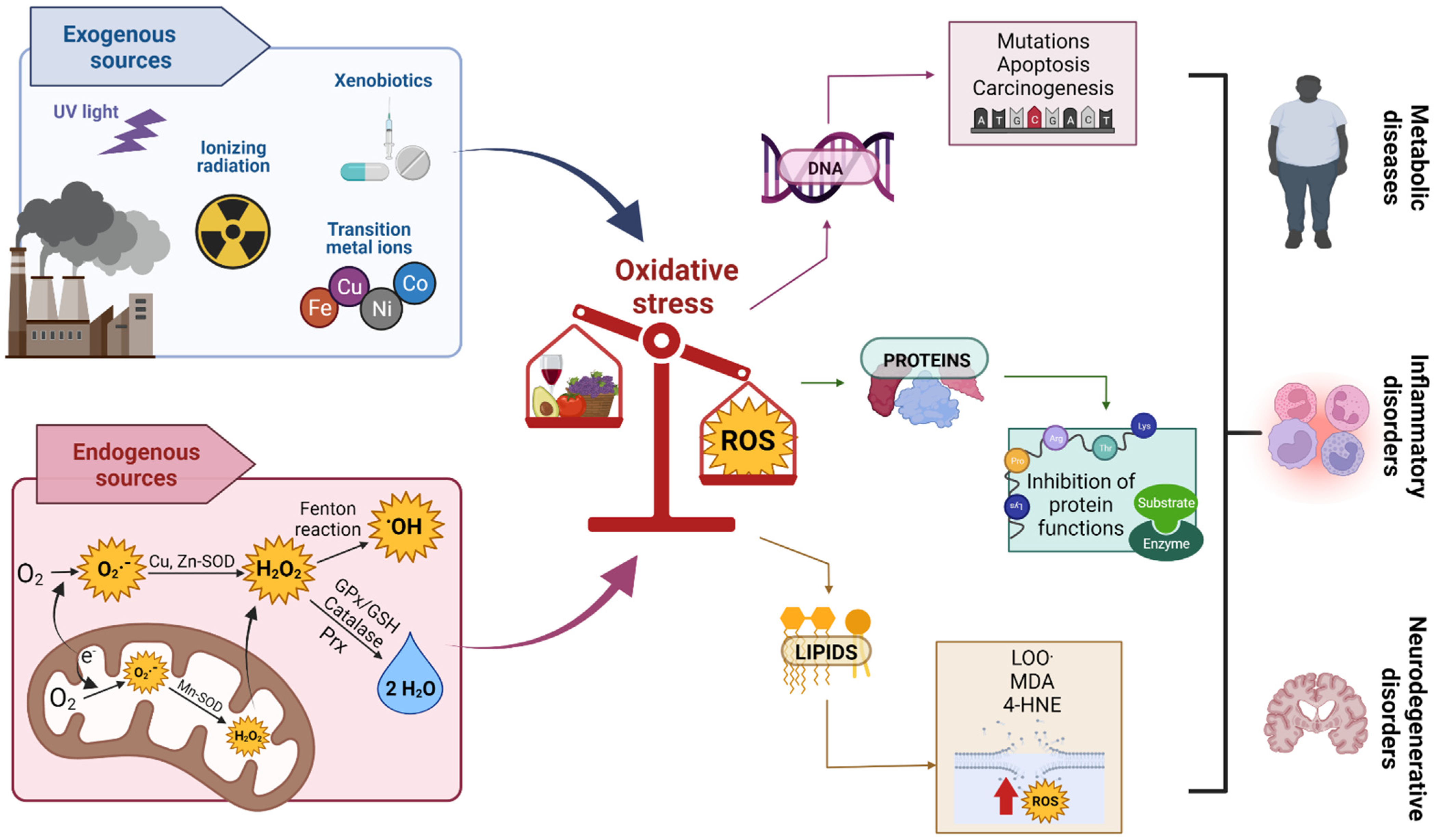
To overcome spin restriction, O2 accepts electrons one at a time to form water (H2O); this process is known as univalent O2 reduction (Figure 1). In univalent reduction, O2 undergoes four successive reductions with one electron. The addition of the first electron produces superoxide radical (O2); adding the second electron plus two protons yields hydrogen peroxide (H2O2); adding a third electron yields the hydroxyl radical (OH) plus the hydroxyl anion (−OH); finally, adding the fourth electron plus two protons produces two H2O molecules. This addition of electrons leads to the formation of O2 reduction intermediates, also called ROS [30,31,32]. On the other hand, the cytochrome c oxidase (COX) complex or complex IV of the mitochondrial respiratory chain acts as a defense by preventing the univalent reduction of O2. COX carries out the tetravalent reduction of O2. In tetravalent O2 reduction, the unique structure of the oxygen-binding site on subunit I of mitochondrial COX simultaneously transfers four electrons to O2 without the release of ROS [33].
The toxic effects of O2 are mainly due to ROS production [34]. ROS is a collective term used to define species derived from O2, which are more reactive than O2. ROS contain O2 that is incompletely reduced (e.g., H2O2, OH) or with a different electronic distribution (e.g., singlet oxygen [1O2]) [28,32,35]. Due to their biological importance in signaling and generating damage to biomolecules (such as DNA, proteins, and lipids), ROS are widely studied.
ROS can be produced from both endogenous and exogenous sources. Endogenous sources of ROS, under aerobic conditions, are generated as a product of metabolic biochemical reactions. These include mitochondria, peroxisomes, microsomes, cellular activation by inflammation, and other enzymatic sources. In humans, mitochondrial ETC has been described as a significant cellular source of ROS production in most tissues [35,36]. Electron leakage in the ETC of complexes I, II, and III reduces O2 to O2 [37]. Under physiological conditions, it is estimated that 1% to 2% of the electrons entering the ETC result in the production of O2 [36,38]. Other ROS such as H2O2, OH, and hypochlorous acid (HOCl) are derived from O2 [37,39]. In contrast, exogenous ROS sources include environmental agents such as heavy metal ions (Cd, lead, arsenic, Fe, and mercury), ultraviolet light, ionizing radiation, or xenobiotics (Figure 2) [34,40,41,42,43].

ROS of Medical and Biological Importance

ROS, which have become important in biology and medicine (due to their harmful effects on biomolecules and signaling functions), can be classified into two large groups: ROS free radicals, which include O2, OH, nitric oxide (NO), nitrogen dioxide (NO2), carbonate (CO3), peroxyl (RO2), and alkoxyl (RO), and non-free radical ROS, including delta-forming singlet oxygen (1O2Δ), ozone (O3), HOCl, nitrous acid (HNO2), peroxynitrite (ONOO), and H2O2 [40]. A free radical ROS is any chemical species (atom, molecule, or ion) derived from O2 that can exist independently and contains one or more unpaired electrons in its outer orbitals [30]. An unpaired electron refers to an electron occupying an atomic or molecular orbital by itself, and their presence in molecules usually makes them highly reactive chemical species [28,34]. On the other hand, a non-radical ROS is a reactive species derived from O2 without unpaired electrons.

3. OS

The first OS concept was formulated by Sies in 1985 [44]. He defined OS as “a disturbance in the prooxidant–antioxidant balance in favor of the former”, and countless authors have accepted it; however, Jones [45] proposed to redefine the term and argued that a more helpful definition is that OS is “an interruption of redox signaling and control”. Finally, an updated definition was reformulated by Sies and Jones in 2007 [46] as “an imbalance between oxidants and antioxidants in favor of the oxidants, leading to a disruption of redox signaling and control and/or molecular damage”; at the moment, this definition the most accepted.

Oxidant Damage Caused by OS

Just as ROS has been observed to have physiological and signaling functions, the dysregulated production of ROS can damage biological molecules. The persistence of OS leads to oxidative damage, which is considered damage to chemical structures (oxidation modifications) of nucleic acids, proteins, lipids, and carbohydrates [47]. ROS that damage nucleic acids affect the nitrogenous bases of purine (adenine, guanine) or pyrimidine (cytosine, thymine) and/or the sugar 2-deoxyribose. Guanine is the DNA base most susceptible to oxidation. DNA damaged by oxidation can generate mutations, can decrease protein synthesis, and may be linked to cancer, the development of aging, or degenerative diseases. The effects depend on the damage’s extent, location, and repair [48].
Oxidative damage to lipids results in the production of malondialdehyde (MDA), 4-hydroxynonenal (4-HNE), oxylipins, isoprostanes, and oxysterols. MDA and 4-HNE are highly reactive and cause DNA damage (Figure 2) [49,50]. Oxylipins are bioactive lipid metabolites derived from polyunsaturated fatty acids (PUFA) associated with inflammation, pain response, cell adhesion, apoptosis, angiogenesis, blood coagulation, and blood vessel permeability [51,52]. These damaging effects are associated with the oxidized metabolites of linoleic acid (LA), including 9- and 13-hydroxyoctadecadienoic acid (HODE) and 9- and 13-oxo-octadecadienoic acid (oxo-ODE) [53,54]. Isopropanes are prostaglandin-like compounds and, like HODEs, are associated with many pathologic conditions (e.g., coronary artery disease and nonalcoholic steatohepatitis) [53,55]. Lastly, oxysterols are oxidized metabolites of cholesterol, and some are cytotoxic (e.g., 7α-hydroxycholesterol (7α-OHC) and 7β-hydroxycholesterol (7β-OHC)), and their dysregulation is associated with various cancer-related degenerative diseases [56,57].
On the other hand, the most common oxidant damage to proteins is the oxidation of thiol groups (–SH) of proteins (amino acids with sulfur, cysteine, and methionine are the most susceptible) and the formation of protein carbonyls (aldehydes and ketones). The amino-acid residues most vulnerable to oxidation are lysine, arginine, proline, and threonine. Protein oxidation leads to inhibition of their function through inhibition of enzyme activity, conformational changes, crosslinking, or aggregation (Figure 2) [32,58]. Therefore, the establishment of cellular antioxidant responses must be fast and efficient to neutralize the possible oxidizing effects of ROS.

4. Antioxidants

Antioxidants are molecules vital in reducing ROS’s oxidizing processes and harmful effects [59]. They include any substance that delays, prevent, or eliminates oxidative damage from a target molecule [28]. Antioxidants can act directly by neutralizing ROS or indirectly by regulating endogenous antioxidant defenses and inhibiting ROS production [60].

4.1. Classification and Description of Antioxidants

Antioxidants can be classified depending on the level of antioxidant action, i.e., the role they play against OS, and they are classified as the first line, second line, third line, and fourth line of defense [61]. They can also be classified by their molecular weight as high or low, by their nature as enzymatic and nonenzymatic, and by their origin as endogenous and exogenous [61,62]. In this article, we focus on the last two classifications.

4.1.1. Endogenous Antioxidants

The endogenous antioxidant system comprises enzymatic antioxidants and nonenzymatic antioxidants [63].

Enzymatic Antioxidants

Enzymatic antioxidants or antioxidant enzymes work by stabilizing or neutralizing free radicals before they attack cellular components. For example, some enzymes convert free radicals, such as OH, into less reactive ROS such as H2O2. Subsequently, other enzymes transform H2O2 into H2O in processes that require cofactors such as copper, zinc, manganese, and iron [64,65]. Examples of antioxidant enzymes include superoxide dismutase (SOD), catalase (CAT), glutathione reductase (GR), glutathione peroxidase (GPx), glutathione-S-transferase (GST), peroxiredoxin (Prx), thioredoxin (Trx), and thioredoxin reductase (TrxR) [63,66,67,68,69,70,71].

Nonenzymatic Antioxidants

Nonenzymatic antioxidants work by interrupting free-radical chain reactions to minimize the damage caused. Examples of nonenzymatic antioxidants are glutathione (GSH), bilirubin (BR), uric acid, and melatonin [72,73,74]. Some of these antioxidants are water-soluble, found mainly in the cytosol or cytoplasmic matrix. Others are lipid-soluble and are present in cell membranes [75].

4.1.2. Exogenous Antioxidants

Exogenous antioxidants or dietary antioxidants (DAs) can be obtained mainly through the diet by consuming fruits, vegetables, byproducts, and/or food supplements. Some examples are vitamins such as A, C, and E, some phenols, and flavonoids [76]. DAs together with endogenous antioxidants can counteract OS and maintain redox homeostasis [62,77]. Previously, in vitro studies suggested that the primary mechanism of DA was through free-radical scavenging, but this is not possible in vivo because DAs, except vitamin E, are not in sufficient concentration and their reactions are not as fast as ROS reactions. It is now known that the main mechanisms of DAs are the maintenance of nucleophilic tone through parahormesis, understood as the generation of signals for the induction of antioxidant enzymes (e.g., induction of nuclear factor E2-related factor 2 (Nrf2)) [78]. In addition, supplementation of essential metals such as zinc (Zn) and magnesium (Mg) has been shown to have beneficial pro-antioxidant effects [79]. Zn has been proposed to act as a pro-antioxidant agent or cofactor through three mechanisms: (1) protection of the free sulfhydryl group in proteins, (2) outperforming redox-active metals, and (3) specific induction of the antioxidant system response [80]. Mg participates in energy metabolism since intracellular adenosine triphosphate (ATP) is in the form of ATP-Mg [81], and its deficiency has been associated with an increase in ROS and a decrease in antioxidant enzymes [81].

5. Role of OS in Cd Toxicity

OS plays an essential role in Cd-induced toxicity, in a wide variety of cell culture systems [82,83,84], in intact animals [85,86,87], or in humans through all routes of exposure [88,89]. However, it has been seen that Cd generates OS via indirect mechanisms since it is not an active redox metal and cannot carry out the Fenton reaction like other metals [90]. The mechanisms via which Cd increases ROS production include (1) damage to ETC, (2) increased NOX activity, (3) increased concentration of free Fe, and (4) decreased antioxidant defense (Figure 3) [18,19].
Excessive production of ROS or reactive nitrogen species (RNS) in the mitochondria is a consequence of Cd-induced ETC damage. Cd inhibits mitochondrial complexes causing electron leakage and the collapse of mitochondrial membrane potential (ΔΨm), which increases ROS and RNS [19]. The primary proposed mechanism via which Cd could be inhibiting the mitochondrial complexes of the ETC is by binding to cysteine residues, Fe–S groups, and the SH groups forming the complexes [91]. In addition, it has been observed that Cd generates unstable semiubiquinones and the accumulation of these promotes the production of O2 [92].
On the other hand, the increase in NOX expression due to Cd exposure has been reported in different studies [93]. NOX is a family of enzymes that use NADPH as a reducing agent to generate O2 [94]. NOX helps regulate metabolism and homeostasis; however, an excess in the production of O2 can be harmful to different cellular components. Cd has been reported to increase NOX1 expression in rats treated with 1 mg/kg Cd chloride (CdCl2) [93]. In addition, Cd also increases ROS production by raising the concentration of active redox metals such as Fe. Cd replaces Fe in several cytoplasmic and membrane proteins, such as ferritin and apoferritin, causing the concentration of the free Fe ion to increase. Being an active redox metal, Fe participates in the Fenton reaction, generating an increase in ROS production [18,95].
Another important mechanism via which Cd generates OS is the decrease in antioxidant defenses, which both direct and indirect interactions achieve. GSH is one of the antioxidants that is decreased, and this decrease occurs due to the affinity of Cd for the thiol group of GSH [96]. GSH deficiency causes alterations in the redox state and, therefore, OS [18,95]. Cd also alters the activity of SOD, CAT, and GPx. The alterations of these enzymes by Cd are given by the fact that this metal can change the enzymatic topography fundamental for the catalytic function of the enzymes [97], replace the metals that are part of the active centers of enzymes [18,98], or deplete the cofactors necessary for functioning; for example, selenium depletion due to the formation of the Cd–Se–Cys complex decreases GPx activity [18,99,100,101]. In addition, Cd indirectly decreases the concentration of antioxidant enzymes by altering the expression of Nrf2 [102,103,104].
In the end, the OS generated by exposure to Cd causes damage to biomolecules such as lipids (formation of MDA and 4 HNE) [85,87,105] and DNA (8-hydroxydeoxyguanosine (8-OHdG) adduct formation) [106,107], causing cell death by apoptosis or necrosis [108,109]. Therefore, OS has been described as one of the causes of several conditions generated by Cd, such as nephrotoxicity, hepatoxicity, and cancer [110]. Furthermore, it is thought to play a role in the epigenetic changes reported by Cd exposure.

6. Epigenetic Effects of Cd Exposure

Growing scientific evidence shows that environmental exposure to heavy metals generates significant epigenetic alterations [111]. In particular, it has been reported that exposure to Cd induces alterations in DNA methylation, post-translational histone modifications, and ncRNA expression.

6.1. DNA Methylation

DNA methylation is the best-studied epigenetic alteration and can be defined as a covalent modification that occurs almost exclusively in the cytosine of the cytosine–guanosine (CpG) dinucleotide sequences [112]. The DNA methylation process is carried out by the DNA methyltransferase (DNMT) family, including DNMT3A, DNMT3B, and DNMT1. DNMT3A and DNMT3B methylate sites that were not previously methylated; that is, they are responsible for de novo methylation [113]. On the other hand, DNMT1 is responsible for maintaining the methylation marks during the mitotic division process; therefore, it is said to be a maintenance methyltransferase [113].
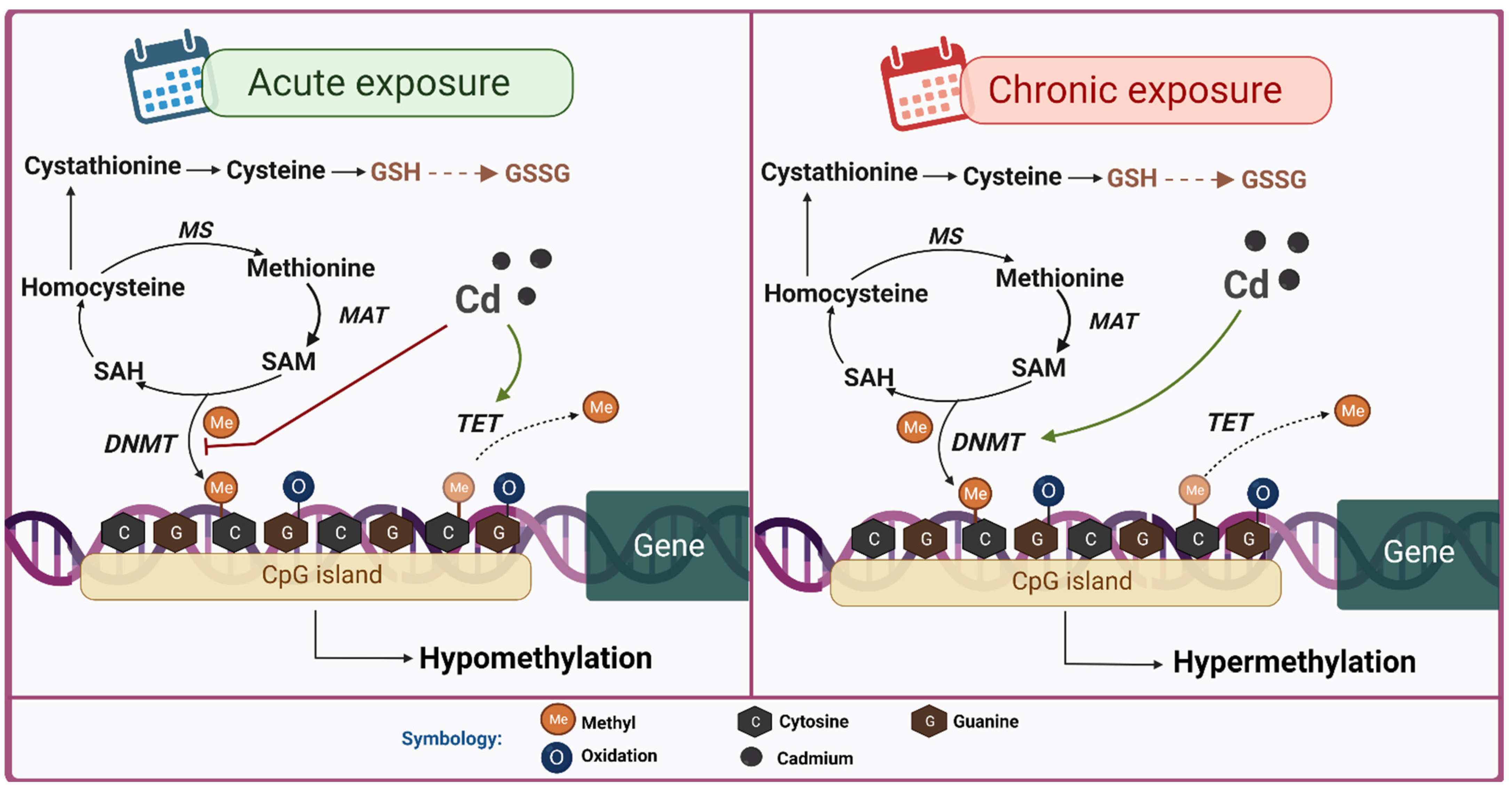
DNA methylation occurs when DNMT transfers a methyl group of the S-adenosylmethionine (SAM) cofactor to cytosine forming 5-methylcytosine (5-mC). In turn, SAM can regenerate again from methionine. When SAM donates the methyl group to cytosine, it is transformed into S-adenosylhomocysteine (SAH), which is hydrolyzed to homocysteine. Homocysteine can generate methionine by the action of methionine synthase (MS). Finally, methionine regenerates SAM via the action of methionine adenosyltransferase (MAT) [114,115,116].
CpG methylation has been mainly associated with gene silencing. Methylation prevents transcription factors from accessing their binding sites directly by methylating transcription factor binding sequences or indirectly by recruiting methyl-CpG binding domain (MBD) proteins. MBD proteins block the binding between transcription factors and DNA or recruit chromatin modifiers that alter chromatin structure in a more transcriptionally repressive environment [117].
DNA can also be demethylated by enzymes of the ten-eleven translocation family (TET) [118]. Demethylation occurs when TETs transform 5-mC to 5-hydroxymethylcytosine (5-hmC) [118]. Subsequently, 5-hmC is converted into 5-formylcytosine (5-fmC) and finally into 5-carboxylcitosin (5-CmC) [118]. Both 5-fmC and 5-CmC are subsequently eliminated by thymine-DNA glycosylase (TDG) [118]. Active genome demethylation has been implicated as a regulatory feature responsible for fine-tuning regulatory methylation marks of CpG.
In the context of environmental pollution, it has been shown that several metals can have a profound impact on DNA methylation with subsequent effects on gene expression [111]. Several research groups have been dedicated to explaining the association between Cd exposure and DNA methylation, and this metal has been found to induce both hypomethylation and hypermethylation [119,120].
DNA hypomethylation by Cd is mainly carried out by decreasing DNMT activity and increasing TETs. A study conducted on nonsmoking women from the Argentinian Andes showed that environmental and dietary exposure to Cd is associated with a decrease in methylation of the long intercalated element-1 (LINE-1) and decreased expression of DNMT3B [121]. LINE-1 is a repetitive DNA retrotransposon that makes up about 17% of the human genome; thus, methylation or demethylation of LINE-1 is considered a marker of global DNA methylation [122].
TET expression also plays an essential role in Cd-induced DNA hypomethylation. It was previously reported that human embryonic kidney cells (HEK293) exposed to CdCl2 in the short term increased messenger RNA (mRNA) and TET protein levels, altering the methylation of complete genomic DNA [123].
On the other hand, DNA hypermethylation by Cd has also been widely reported. In a study conducted in pairs of mothers and children, alterations in the methylation of 61 genes in fetal blood were observed, associated with the Cd of maternal blood at the time of delivery [124]. In addition, Cd exposure was associated with a decrease in gestational age through an increase in DNA methylation at a specific CpG site, cg21010642 [125]. The CpG site was noted in a gene involved in early embryonic development. Therefore, irregular methylation patterns at this site may contribute to preterm birth by mediating irregular biological mechanisms [125].
Change-induced hypermethylation has been widely linked to increased expression of DNMT proteins. In human bronchial epithelial cells (16HBE), Cd elevated the expression of DNMT1 and DNMT3A, causing DNA hypermethylation, inactivating tumor suppressors and genes associated with DNA repair [126]. Likewise, in lymphocytes treated with Cd, hypermethylation of the promoter p16 associated with the increase in DNMT1 and DNMT3B expression levels was observed [127]. Overexpression of DNMT3B is also demonstrated after chronic Cd-induced toxicity in the human prostate [25]. Expression levels of DNMT3B and DNMT3L are crucial in DNA methylation in placentas exposed to Cd [128].
Discrepancies between whether Cd increases or decreases DNA methylation are associated with the duration of Cd exposure. Evidence shows that acute exposures to Cd induce DNA hypomethylation, whereas chronic exposures cause DNA hypermethylation [21,129]. A study conducted on TRL1215 cells derived from rat liver reported that short exposure to Cd (1 week) decreases the activity of DNMT, causing decreased DNA methylation, while prolonged exposure to Cd (10 weeks) produces DNA hypermethylation due to increased DNMT activity [21]. Similar effects were also reported in rat livers [130]. It has also been reported that 24 h to 1 week exposure of Cd in chronic myelogenous leukemia (K562) cells led to DNA hypomethylation through noncompetitive inhibition of DNMT activity by Cd [131]. In contrast, exposure of 8 to 10 weeks to Cd caused global DNA hypermethylation and increased DNMT activity in human embryonic lung fibroblast (HLF) cells [120]. In addition, studies carried out in Lumbricus terrestrial earthworms proved that hypermethylation by Cd has long-lasting effects since it can be extended up to a period of 7 months at a low environmentally relevant concentration of Cd (10 mg/kg) [132].
Alterations in DNA methylation due to Cd exposure are associated with different diseases such as cancer, Alzheimer’s, diabetes, multiple sclerosis, hepatic and renal alterations, and the reproduction and development of organisms [133]. In particular, DNA hypermethylation induced by prolonged exposure to Cd has been associated with malignant transformation in rat liver-derived TRL1215 cells, human prostate epithelial cells, human lung fibroblasts, and human bronchial epithelial cells (BEAS-2B) [21,25,120,134]. Cd has also been shown to silence the expression of enzymes that repair DNA in a dose-dependent manner through hypermethylation [25,120,126].
On the other hand, Cd-induced DNA hypomethylation has been proposed as a cause of alterations in the reproduction and development of organisms. Exposure to Cd for 24 h caused hypomethylation of the HSD11B2 gene in cultured human choriocarcinoma cells, causing increased expression of its protein hydroxysteroid (11-beta) dehydrogenase 2 (HSD11B2), which regulates the steroid hormone cortisol [135]. Likewise, DNA hypomethylation has been observed in babies’ umbilical cord blood with managerial exposure to Cd, which can persist until 9 years of age [136]. Alterations in methylation from exposure to Cd are also associated with alterations in size at birth [137,138], and specific differences have been reported depending on sex [139].
Multiple sclerosis has also been described as a consequence of DNA methylation as an epigenetic change influenced by environmental factors, such as heavy metal contamination. A study conducted with patients with multiple sclerosis reported the correlation of a high concentration of Cd with increased expression of the atypical chemokine receptor 3 (ACKR3) gene by hypomethylation and decreased expression of the apolipoprotein E (APOE) gene by hypermethylation. This indicates that epigenetic changes induced by Cd may be an essential factor in increasing and decreasing the expression of genes involved in the appearance and/or progression of inflammatory processes in multiple sclerosis [140].
Lastly, alterations in DNA methylation have also been associated with liver diseases. It was found that there is a positive relationship between the degree of DNA methylation and the concentration of Cd, and there were many differential sites of methylation promotion in DNA from rat liver tissue [24].
In summary, the alterations in DNA methylation induced by Cd are related to the time of exposure to this metal. The short-term effects induced by Cd are due to a decrease in DNMT activity and an increase in TETs. Meanwhile, the effects of prolonged exposures have been linked to increased levels of DNMT. In addition, Cd-induced DNA hypomethylation and hypermethylation are related to various diseases; thus, understanding how Cd generates these alterations in DNA methylation helps to understand the toxic mechanisms of Cd exposure (Figure 4).

6.2. Histone Modification

Histones are proteins that provide structural support to DNA within units known as nucleosomes [141]. Each nucleosome comprises two identical subunits containing four histones: H2A, H2B, H3, and H4 [142]. In addition, there is another histone, H1, which is not part of the nucleosome itself, but its function is to stabilize the internucleosomal DNA [143]. Post-translational modifications in these histones decide DNA’s interaction with the different components. For example, modifications that disrupt the histone–DNA interaction cause relaxed chromatin, known as euchromatin, where DNA is accessible for transcriptional machinery binding and subsequent gene activation. In contrast, modifications that strengthen histone–DNA interactions create a tightly packed chromatin structure called heterochromatin, where the transcriptional machinery cannot access DNA, causing gene silencing [144].
There are different post-translational modifications in histones, which occur mainly in the tail domains of these proteins. The best-known histone modifications are methylation, phosphorylation, acetylation, and ubiquitylation; however, O-GlcNAcylation, citrullination, nitration, ribosylation, crotonylation, sumoylation, and isomerization are also found [145,146,147,148]. Each modification is added to or removed from histone amino-acid residues by a specific set of enzymes. Dysregulation of histone-modifying enzymes will alter post-histone modification patterns and cause various diseases, including cancer. Consequently, histone-modifying enzymes have become promising biomarkers for disease diagnosis and prognosis [149].
Cd-induced histone modifications have not been very studied compared to other epigenetic modifications. However, few studies have shown that Cd exposure affects histone acetylation, methylation, and phosphorylation primarily by altering the expression of enzymes that modify them.
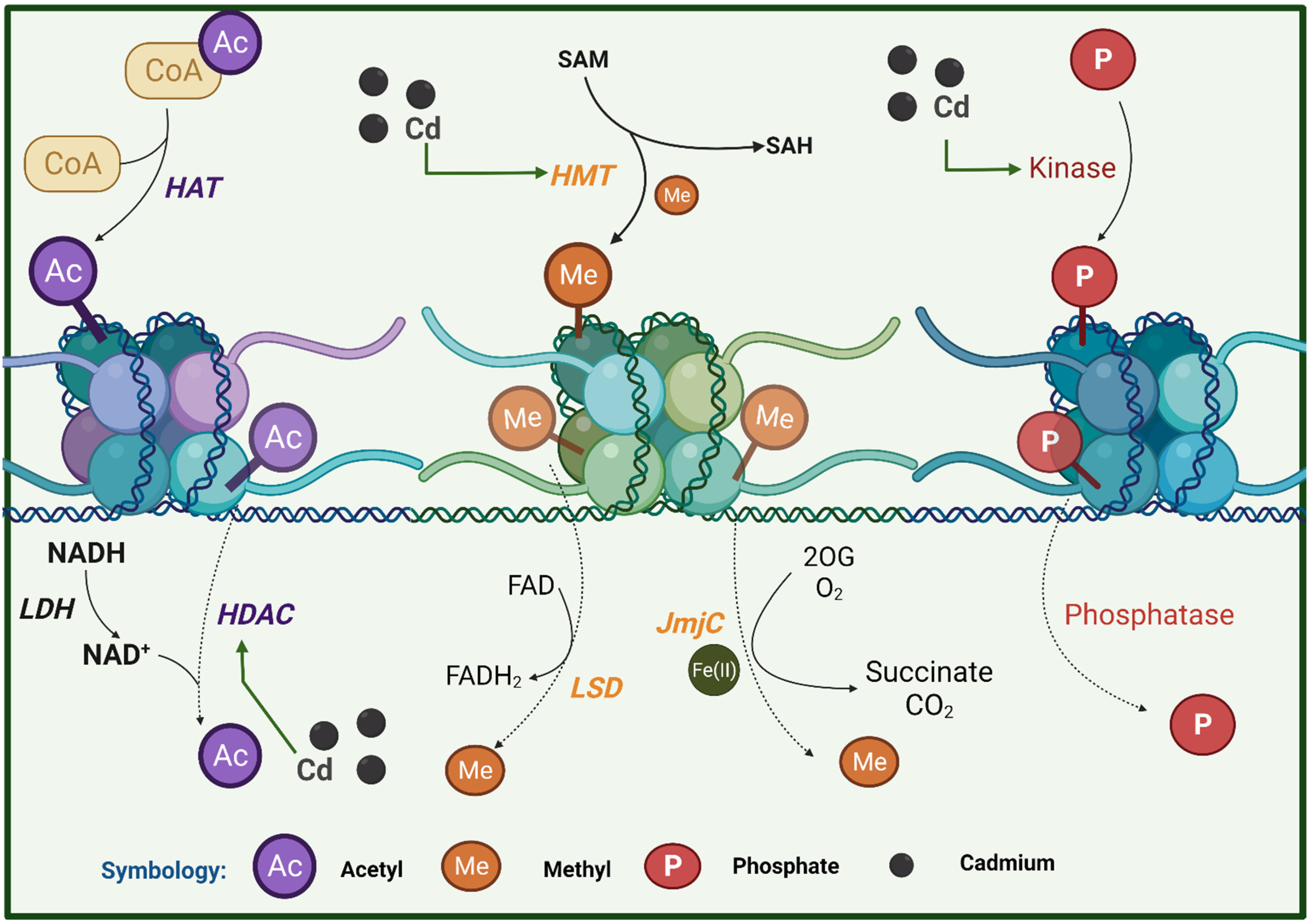
Histone acetylation involves the addition of a negatively charged acetyl group to lysine residues in the tails of histone proteins [150]. When a histone is acetylated, the histone–DNA interaction is weakened; hence, it has been associated with transcriptional activation [151]. There are two enzymes responsible for regulating this process, histone acetyl transferases (HAT), which are responsible for adding acetyl groups, and histone deacetylases (HDAC), which eliminate acetyl groups [152]. A current study reported that Cd exposure increases HDAC2 expression and impairs the learning and memory of rats exposed to 5 ppm of Cd in drinking during weaning and up to three months of age. These effects were most significant when Cd exposure was combined with lead [153]. Likewise, it was observed that maternal exposure to Cd in mice impairs embryonic development and increases the expression of HDAC1 [154].
Histone methylation refers to adding a methyl group to nitrogen atoms in amino-acid side-chains and/or at amino ends [155]. This modification occurs in all the residues of basic amino acids: arginines, lysines, and histidines [156,157,158]. The histone methylation sites are histone H3 lysine 4 (H3K4), H3K9, H3K27, H3K36, H3K79, and H4K20, while arginine methylation sites include H3R2, H3R8, H3R17, H3R26, and H4R3. However, many other basic residues in histone proteins were also identified, including H1, H2A, H2B, H3, and H4 [159]. Histone methylation is carried out by enzymes of the histone family methyltransferases (HMTs); like DNMT, SAM is used as the donor of the methyl group [160]. In contrast, demethylation is carried out by the Jumonji C (JmjC) domain-containing protein family, lysine-specific demethylases (LSD), and peptidylarginine deiminase (PADI) [161]. Histone methylation can repress or activate transcription [162]. Previously, Somji and contributors [22] reported increased levels of H3K4me3, H3K27me3, and H3K9me3 in the MT3 promoter in Cd-transformed urothelial cells, suggesting that chronic exposure to Cd may alter transcriptional responses through histone methylation and, in this case, the silencing of MT3 [22].
Lastly, another modification in histones observed by exposure to Cd includes phosphorylation. Histone phosphorylation confers a negative charge on the histone, resulting in more open chromatin conformation. This modification has been associated with gene expression and is involved in DNA damage repair and chromatin remodeling [163]. Cd has been shown to decrease H3 phosphorylation by inhibiting the human vaccinia-related kinase (VRK1/2) in an in vitro model [164].
In summary, Cd causes histones’ methylation, phosphorylation, and acetylation (Figure 5). Because histones are the most common chromatin proteins, any change in their abundance, structure, or post-translational modifications (PTMs) will severely impact the overall structure of chromatin, influencing gene expression, as well as genome stability and replication.

6.3. ncRNA

ncRNAs are RNA molecules that are not translated into proteins, and it has recently been described that they can participate in epigenetic regulation by adjusting gene expression without altering the DNA sequence [165]. Transcriptional ncRNAs are classified into small ncRNAs and long ncRNAs (LncRNAs). Small ncRNAs can be divided into microRNAs (miRNAs), P-element induced wimpy (PIWI)-RNAs of interference (piRNAs), and small RNAs of interference (siRNAs) [165].
LncRNAs have a length of more than 200 nucleotides and are transcribed by RNA polymerase II (POLII). Depending on their location and their specific interactions with DNA, RNA, and proteins, LncRNAs can modulate chromatin function, obstruct transcriptional machinery, maintain nuclear speckle structure, and regulate nuclear body assembly and function. Without a membrane, they act as molecular sponges for miRNAs, alter the stability and translation of cytoplasmic mRNAs, and interfere with signaling pathways [166,167]. These functions affect gene expression in various biological and pathophysiological contexts, such as neuronal disorders, immune responses, and cancer. Therefore, the expression of LncRNAs can be used as potential biomarkers and justify attacking them clinically [167].
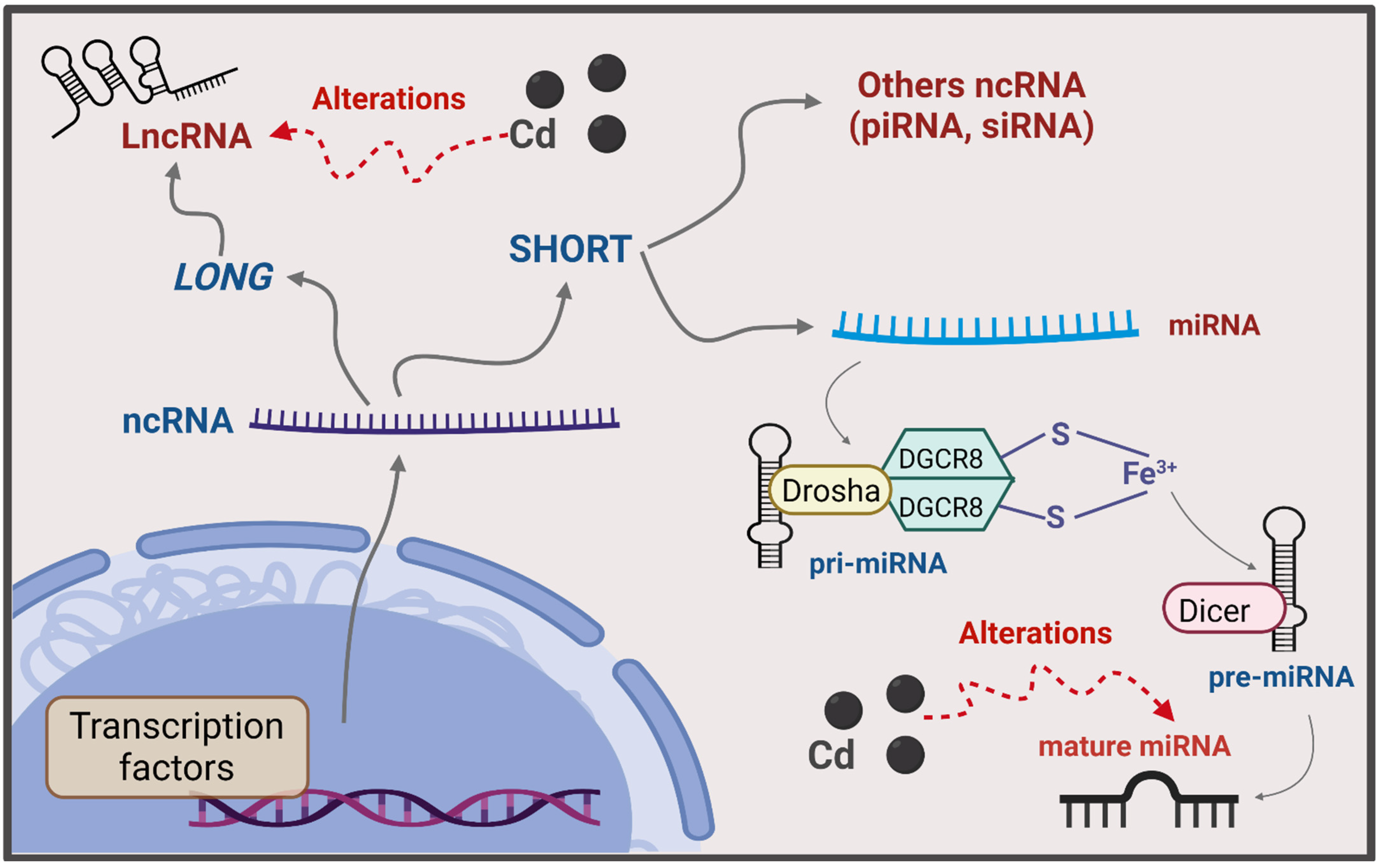
miRNAs are the smallest ncRNAs, ~21 to 23 nucleotides in length, that regulate post-transcriptional gene expression through their complementary binding to untranslated regions 3′ or 5′ (UTR 3′ or 5′) of mRNAs, causing their degradation or translation blocking [168]. miRNAs can also regulate transcription by directly binding to gene promoter sequences and inducing chromatin remodeling [169]. The miRNAs are transcribed by the action of POLII and subsequently processed in the nucleus by RNase III (Drosha), within a nuclear protein complex called Microprocessor, and DiGeorge syndrome critical region 8 (DGCR8) to form structures of hairpin known as pre-miRNAs [170]. The pre-miRNAs are then exported to the cytoplasm, cleaving them by Dicer1 to generate a mature miRNA. More than 17,000 distinct mature miRNAs have been identified in more than 140 species [171]. Alteration in miRNA expression can be associated with various diseases such as cardiovascular and genetic disorders and cancer [172]. Therefore, it has been proposed that they can be used as biomarkers of exposure and disease due to the availability of circulating extracellular miRNAs found in body fluids, such as amniotic fluid, saliva, serum, and plasma [173].
On the other hand, piRNAs have a length of 26 to 32 nucleotides, and their name comes from the fact that they interact with PIWI proteins of the Argonaut family, which have endonuclease properties [174,175]. piRNAs act on somatic cells and are crucial for guiding epigenetic regulation and inducing the silencing of transposons [175,176]. They are synthesized from repetitive intergenic regions, and their processing is carried out independently of Dicer/Drosha. Abnormal piRNA expression is associated with several types of cancer, such as gastric, breast, renal, colorectal, and lung cancer [175].
siRNAs have a length of 20 to 25 nucleotides and, like miRNAs, rely on Dicer enzymes to separate them from their precursors [177]. siRNAs bind to the nucleotide sequence in their target mRNA and interfere with the expression of the respective gene, primarily by degrading mRNA. In addition, they also participate in the organization of the structure of chromatin in a genome and defend the cell from foreign nucleic acids [178]. siRNAs resemble miRNAs; when miRNAs are presented with a substrate with perfect complementarity, they can act as siRNA and guide multiple rounds of mRNA degradation [179].
In the context of environmental pollution, Cd has been shown to affect the expression of ncRNAs. Recent evidence shows that heavy-metal toxicity is related to aberrant alterations of endogenous miRNAs [26]. For example, exposure to Cd telluride (CdTe) induced new miRNAs in mouse embryonic fibroblast cells (NIH/3T3). These results were associated with the ability of CdTe to reprogram gene expression even after the initial signal was removed. In addition, alterations in miRNA biogenesis suggest that these molecules participate in the cytotoxicity of CdTe quantum dots by inducing apoptosis-like cell death [180]. In another study, 4 h exposure to Cd in murine ovarian granulosa cells resulted in the altered expression of five miRNAs involved in alternative splicing of kit ligand (kitl) pre-mRNA [181]. Furthermore, it has been seen that the accumulation of Cd can generate preeclampsia. miRNA-26a and miRNA-155 were affected in preeclamptic placentas and trophoblasts treated with Cd [182].
Alterations in miRNAs may also be associated with Cd-induced kidney damage [26,183]. A study conducted on the renal cortex of Sprague-Dawley rats subcutaneously exposed to CdCl2 (0.6 mg/kg 5 days a week for 12 weeks) showed increased miRNA-34a-5p and miRNA-224-5p and decreased miRNA-455-3p [184]. Increased miRNA-132-3p was also found in primary human proximal tubular epithelial cells treated with CdCl2 [185]. These findings demonstrate that Cd significantly alters the expression profile of miRNA in the kidney and raises the possibility that these alterations play an essential role in the physiology of Cd-induced kidney injury.
Cancer is another pathology associated with alterations in miRNAs induced by Cd. Cd causes malignant transformation of RWPE-1 prostate epithelial cells along with increased levels of Kirsten rat viral sarcoma oncogene homolog (KRAS) and alteration in miRNAs [186]. It was observed that about 12 miRNAs decreased (miRNA-373 was the lowest) in the transformed RWPE-1 cells [186]. In addition, Cd exposure affects the expression of p21 (Cip1/WAF-1) in a hepatoma cell line (HepG2) by increasing miRNA-372 [187]. p21 is a protein downstream of p53, a protein with an essential role in regulating apoptosis and preventing cancer development. Therefore, Cd-induced changes in p21 also alter the functions of p53 [188]. Similarly, exposure to Cd through smoking in bronchial epithelial cells modified multiple miRNAs via both up- and downregulations [189]. Lastly, it was found that exposure to Cd is also related to pancreatic ductal adenocarcinoma (PDAC). An in vitro study reported that exposure to CdCl2 was overexpressed to miRNA-221 and miRNA-155, while miRNA-126 was downregulated [23].
Other ncRNAs that have been affected by Cd exposure are LncRNAs. A study conducted on workers exposed to Cd found a significant positive correlation between the level of Cd in the blood and LncRNA-ENST00000414355 and serine/threonine kinase (ATM) expression and a significant negative correlation between the level of Cd in the blood and the ΔΨm [190]. Another study reported that a maternally expressed tumor suppressor LncRNA 3 (MEG3) is significantly downregulated in BEAS-2B cells transformed with Cd [191]. Cd exposure was also reported to alter LncRNAs expression during T-cell activation [192].
In summary, it can be said that Cd alters the expression of ncRNAs, particularly miRNAs and LncRNAs, which is related to kidney and liver damage and cancer progression (Figure 6).

7. Relationship of Epigenetic Modifications with Cd-Induced OS

Exposure to different environmental factors, such as heavy metals, can change the biology of the epigenome and lead to adverse health effects. As discussed in the last section, Cd exposure causes epigenetic changes, including DNA methylation, histone modifications, and ncRNA expression. OS may play an essential role in the epigenetic changes induced by Cd [26,193,194].

7.1. DNA Methylation

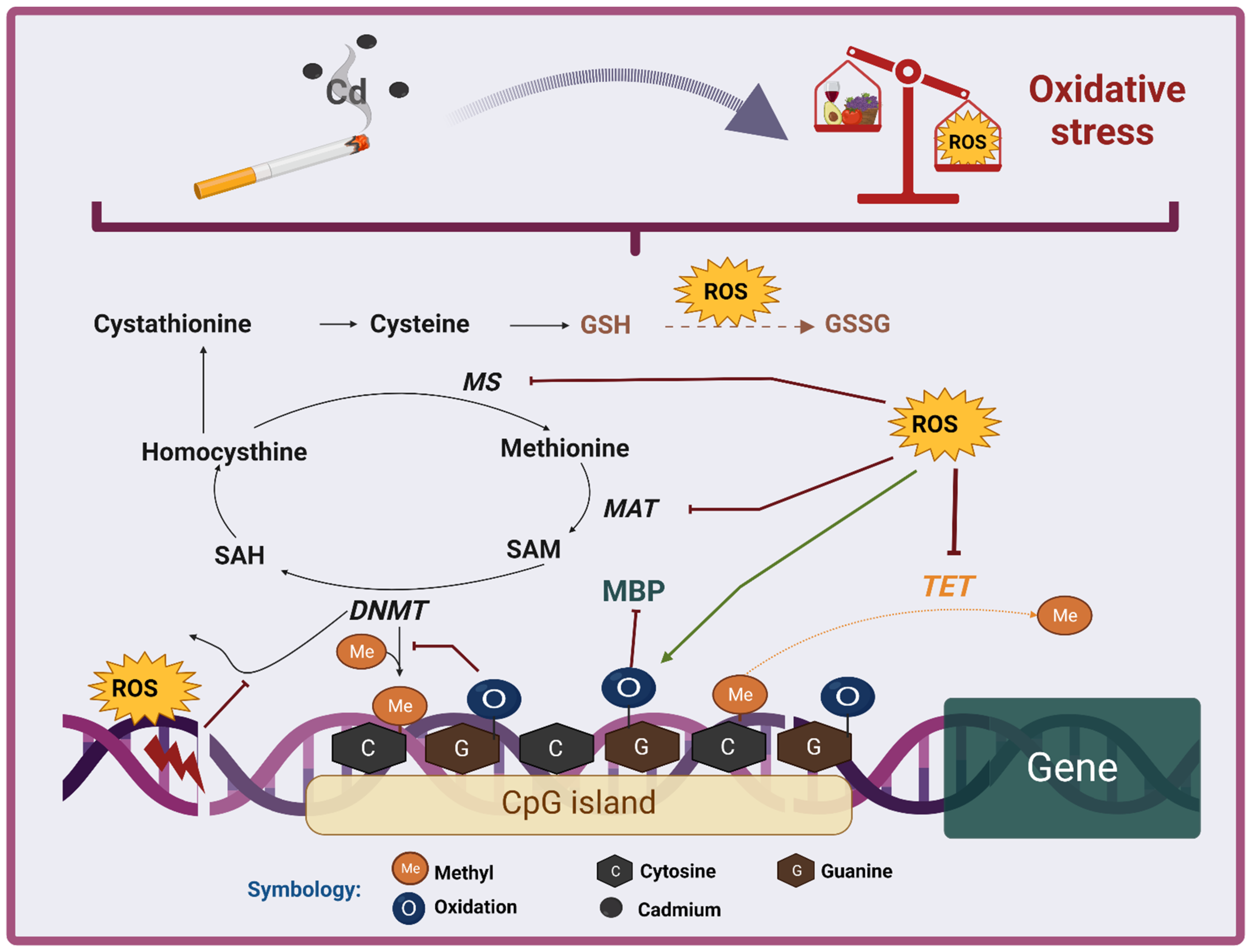
OS is an indirect mechanism via which Cd overexposure influences DNA methylation. Exposure to Cd has been associated with both hypomethylation and hypermethylation of DNA (Figure 7). In hypomethylation, there are different mechanisms via which OS could be participating [195,196]. One of the mechanisms is the depletion of SAM levels. It has been seen that OS can inhibit the activity of MAT and MS by oxidizing them, which causes a decrease in SAM [114,115,116]. Because SAM is the substrate for DNMT, its decrease causes less DNA to be methylated. Moreover, the trans-sulfuration pathway to regenerate GSH under OS conditions causes SAM depletion because this molecule is an intermediate of this pathway [197,198].
Another mechanism of hypomethylation by OS is the generation of DNA lesions due to increased free radicals, mainly the OH radical. Excessive OH production is known to cause DNA damage, including base modifications, deletions, strand breaks, and chromosomal rearrangements [199,200]. Because DNA is damaged in these lesions, it cannot be used as a substrate for DNMT, resulting in global hypomethylation [201].
DNA hypomethylation may also be related to the oxidation of DNA bases in CpGs. Guanine is the preferred nucleotide for ROS to be oxidized, forming mainly (but not exclusively) 8-OHdG [202,203,204]. Guanine oxidation prevents methylation of the neighboring cytokine at CpG due to inhibition of the binding of DNMT enzymes to DNA [205,206]. In addition, the oxidation of guanine prevents the binding of the MBP complex to DNA because the formation of 8-OHdG causes the N7 position of guanine to be converted into a hydrogen bond donor instead of a hydrogen bond acceptor [193,207]. Furthermore, if the CpG cytokine where guanine is oxidized is methylated, then it becomes more susceptible to TET action [208].
A study conducted in Nile tilapias (Oreochromis niloticus) showed that exposure to Cd induces OS, including reduced antioxidant activities and increased MDA and 8-OHdG content, which was related to DNA hypomethylation. A decrease in DNMT and increased expression of TET1 and TET2 were also observed [209]. It should be noted that this study carried out in tilapia was chronic (45 and 90 days); thus, it contradicts the statement that acute Cd exposures are related to hypomethylation, while chronic exposures are related to hypermethylation [21,129]. However, the hypomethylation in the tilapia model could be explained because the OS was evident, and this is more related to DNA hypomethylation than to hypermethylation due to the mechanisms explained previously in this section.
In other words, OS has been shown to appear on acute exposures to Cd, and acute exposures are related to DNA hypomethylation, whereas prolonged exposures to Cd are more related to DNA hypermethylation; however, ROS production in chronic exposures becomes absent [210]. This could be because chronic exposure induces adaptation mechanisms to compensate for the ROS induced by Cd and OS, such as the induction of MT, the increase in cellular GSH, and the activation of the antioxidant transcription factor Nrf2 and other antioxidant components [210]. For example, it has been reported that DNA hypermethylation in prostate cells exposed to Cd is associated with increased DNMT activity and decreased OS and redox-sensitive signal transduction pathways, such as the c-Jun N-terminal kinase (JNK) [25,211].
Although most of the evidence shows that the OS in chronic exposures to Cd is low, there are studies where the excessive production of ROS has been reported. In a study carried out on zebrafish liver, it was found that heavy-metal contamination, mainly Cd, in the Le’an river caused OS and a significant increase in global methylation [212]. Furthermore, overproduction of O2 has been shown to increase DNA methylation in some types of cancer, such as melanoma and possibly endometriosis [213]. In Cd-transformed human bronchial epithelial cells, promoter hypermethylation resulted in decreased expression of DNA repair genes [126]. In addition, it has been seen that the OS could inhibit the activity of the TETs. It is known that these proteins are dependent on Fe(II), which, during the TET catalytic cycle, is oxidized to Fe(III) and Fe(IV) [214], while it is regenerated by the action of ascorbate [215]. Under OS conditions, ascorbate levels are reduced; hence, Fe(II) is not regenerated, and TETs are inhibited, causing hypermethylation [216]. In a study performed on TRL1215 rat liver cells exposed to 2.5 μM Cd for 10 weeks and then cultured in a Cd-free medium for an additional 4 weeks, harmful modulation of TET1 was found. In addition, the expression of tissue inhibitors of metalloproteinases 2 and 3 (TIMP2 and TIMP3), which are positively regulated by TET1, was decreased by Cd. All of the above caused a decrease in the expression of apolipoprotein E (apoE), possibly due to DNA hypermethylation [217].
In summary, it can be said that Cd-induced OS plays a crucial role in DNA methylation and demethylation, particularly in acute exposures. However, the relationship of OS to DNA methylation in chronic Cd exposures needs further investigation.

7.2. Histone Modification

The chemical modification of histones is another epigenetic mechanism that can be altered by OS induced by environmental factors such as Cd [218,219]. It has been shown that OS and nitrosative stress (excessive increase in RNS) modify histones, affecting their folding and stability and their ability to modify themselves post-translationally (Figure 8) [219].
In particular, it has been observed that Cd alters histone acetylation, and ROS production could be a cause [153,220,221]. Previously, maternal exposure to Cd was reported to affect embryo development prior to implantation by inducing DNA damage and increasing HDAC1 levels and ROS production [154]. The increase in HDAC could be attributed to the production of H2O2. It is known that H2O2 generates an increase in lactate dehydrogenase (LDH), an enzyme that catalyzes the reduction of lactate to pyruvate, producing NAD+ [222]. NAD+ is used by HDACs, which stimulates deacetylation [222]. Notably, other studies have reported that ROS can also inhibit HDACs, which is achieved via different mechanisms, including carbonylation, phosphorylation, nitrosylation, or glutathionylation, due to ROS increase [223] and reactive aldehydes [224,225,226]. The inactivation of HDAC2 results in its ubiquitination and proteasome degradation, which increases histone acetylation. Cd-induced stress has also been associated with H4K5 acetylation and increased DNA damage in bean seedlings (V. faba) [227].
Cd also affects histone methylation, and OS could be involved. OS is related to increased histone methylation by inhibiting JmjC by oxidizing Fe(II) to Fe(III), which makes Fe(II) unavailable for use by JmjC [216]. Another way to inhibit this type of protein is when NO binds directly to the catalytic Fe [228,229]. However, the production of ROS also generates a decrease in histone methylation. One of the reasons is that ROS decreases SAM levels. As explained above, under OS conditions, enzymes involved in SAM synthesis are inhibited and, thus, histone methyltransferases (HMTs) are blocked [197,230].
OS could also be involved in histone phosphorylation caused by Cd. ROS production generally generates breaks in double-stranded DNA, causing histone phosphorylation to trigger DNA repair [194,231]. In addition, it is known that H2O2 increases histone phosphorylation via an ATR serine/threonine kinase (ATR)-dependent pathway [232]. However, it has also been found that OS can oxidize the catalytic metal ion within protein phosphatases, causing their inhibition [233].
Lastly, OS is related to other histone modifications; however, these have not yet been much explored with Cd exposure. One of them is the nitration of histones. ONOO is known to nitrate histones, leading to increased β-sheet structures and increased thermostability. Furthermore, it is known that the in vivo nitration–denitration activities of histones could be involved in the control of numerous vital cellular events to maintain apoptosis [234,235]. Cd has been shown to increase ONOO production by generating protein nitration [236].
On the other hand, another modification associated with OS is the modification of histones by the action of oxidized lipids. It has been reported that 4-oxo-2-nonenal (4-ONE) forms adducts with histones causing inhibition of nucleosome assembly [237]. In addition, 4-HNE has been shown to alter the binding of histones to DNA [238].
In summary, OS could have an essential role in Cd-induced histone modifications; however, there is a great need for more studies in this area. In addition, like DNA methylation, the few studies that have seen alterations in histone modifications due to OS are only in studies of acute exposure to Cd (24 and 48 h) [154,212].

7.3. ncRNA

ncRNAs are a newly identified group of epigenetic regulators sensitive to ROS and function according to cellular redox status (Figure 9) [165]. For example, the anormal expression of ncRNAs, mainly miRNAs, is partly attributed to the dysregulation of their transcription factors generated by increased ROS production. Some of these ROS-sensitive transcription factors are c-Myc, p53, and nuclear kappa light chain enhancer of activated B cells (NF-κB) [239,240]. A study conducted on Daphnia pulex found that exposure to Cd plus hypoxia increases ROS production by triggering increased extracellular signal-regulated kinases (ERK), protein kinase B (Akt), and hypoxia-inducible factor 1α (HIF1α), which in turn promotes miRNA-210 expression [241].
In miRNAs, biosynthesis may be affected by ROS exposure. The formation of pre-miRNA is carried out by the Drosha–DGCR8 complex, which depends on Fe(III) for its action. The pre-miRNAs then leave the core and mature by Dicer processing. ROS has been shown to promote the processing power of DGRC8 [239,242]. However, ROS inhibits Dicer activity to delay the production of mature miRNAs, thereby coordinating cellular behavior [239]. Oxidative modifications can also influence pri-miRNA protrusions and loops [243].
miRNA expression may also be affected by increased ROS. ROS has been reported to affect the methylation status of specific promoter regions of miRNA genes [244]. Previously, the expression of miRNA-199a and miRNA-125b was shown to be decreased in the presence of ROS, mainly by upregulation of DNMT1 [245]. In addition, inhibition of miRNA-122 expression in tilapia livers by increased ROS production induced by Cd exposure has been demonstrated. The decrease in miRNA-122 was related to increased metallothionein levels [246].
On the other hand, it has been observed that most miRNAs that respond to ROS influence the Nrf2 system [247]. A study conducted on the human hepatocellular carcinoma (HepG2) cell line showed that Cd induces the expression of transcription factor MTF1, which activates the expression of MT1DP, a pseudogene in the MT family. Subsequently, MT1DP raises the levels of miRNA-365, which causes a decrease in Nrf2 levels, generating OS [248].
In addition, 74 LncRNAs were involved in CdCl2-induced OS in broiler livers [249]. Likewise, 322 LncRNA were found in rats exposed to Cd [250]. Furthermore, it has been seen that LncRNAs modified with N6-methyladenosine (LncRNA-TUG1, LncRNA-PVT1, LncRNA-MALAT1, LncRNA-XIST, and LncRNA-NEAT1) are involved in oxidative damage induced by Cd [251].
In general, it could be said that OS plays an essential role in the maturation of ncRNAs, and, in turn, some of the ncRNAs are also involved in the cellular response to OS. In addition, most of the studies in which alterations in ncRNA expression have been seen as a consequence of OS were carried out with acute exposures to Cd (6 and 24 h) [154,227].

8. Use of Antioxidants to Mitigate Cd-Induced Epigenetic Alterations

There is no specific or 100% effective treatment for chronic Cd intoxication. However, metal chelators have been widely used [1,252]. Chelators are chemical agents that work by binding tightly to metals in the bloodstream, leading to sequestration of the metal for later excretion [253]. Chelators used for Cd poisoning include ethylenediaminetetraacetic acid (EDTA), penicillamine (DPA), dimercaprol (anti-British Lewisite (BAL)), 2,3-dimercaptopropanesulfonic acid (DMPS), and dimercaptosuccinic acid (DMSA) [1]. However, the use of chelators in Cd intoxication faces various problems, among which the increase in the body load of Cd in the kidney and the competition of chelators with albumin, macroglobulin, and MT stand out.
Another strategy used to treat Cd toxicity is to enhance the binding of this metal to MT. The latter can be achieved by Zn supplementation [254]. Zn is an essential metal that has been shown to attenuate Cd-induced OS because Zn functions as a cofactor for the antioxidant enzyme copper/zinc superoxide dismutase (Cu/Zn SOD) [255]. Furthermore, Zn increases the expression of MT [256]. MTs are low-molecular-weight proteins with a high affinity for Cd and other heavy metals due to the –SH on their cysteine residues, which aid detoxification [257]. However, given the critical role that OS plays in Cd toxicity, the use of compounds with antioxidant properties has become one of the most promising strategies [258]. In addition, the use of natural compounds represents advantages compared to chelating compounds, including high safety and few or no side-effects.
Among the compounds or plant extracts with antioxidant properties used to attenuate the toxic effects induced by Cd are green and black tea (Camellia sinensis), blueberries (Aronia melanocarpa), garlic (Allium sativum), onion (Allium cepa), holy basil (Ocimum sanctum), cape gooseberry (Physalis peruviana), ginger (Zingiber officinale), quercetin, lupeol, S-adenosyl-methionine, lipoic acid, glutathione, selenium, N-acetylcysteine (NAC), methionine, cysteine, alpha-tocopherol, ascorbic acid, and resveratrol [258]. Many of these compounds have direct antioxidant effects by trapping ROS, while others exert their effects indirectly by inducing Nrf2 expression [258]. In addition, it should be noted that some of these compounds, such as epigallocatechin-3-gallate, have functional groups within their structure capable of chelating metals such as Cd [259]. Table 1 shows the antioxidants that have already been tested against epigenetic alterations induced by Cd.
Selenium is one of the antioxidants with an antagonistic effect against Cd-induced carcinogenesis [260]. Selenium is an essential trace element in humans involved in numerous processes, including immune function and antioxidant defense. Selenium supplementation is protective against an extensive range of harmful factors, including heavy metals [265]. It has been shown that selenium can alleviate the proliferative effect of Cd on human breast cancer cells (MCF-7). One of selenium’s mechanisms against carcinogenesis was the epigenetic regulation of 10 genes that Cd had modified. Within these genes, selenium downregulated APBA2 and KIAA0895 while upregulating DHX35, CPEB3, SVIL, MYLK, ZFYVE28, ABLIM2, GRB10, and PCDH9 [260]. Analyses of biological functions suggested that these epigenetically regulated genes are involved in multiple cancer-related pathways, such as focal adhesion and the PI3K/Akt pathway [260]. The main mechanisms via which selenium modified these genes were the regulation of DNA methylation and the expression of miRNA and LncRNA.
Quercetin has also been used against Cd toxicity. Quercetin is a polyphenolic flavonoid abundant in cabbages, onions, berries, apples, red grapes, broccoli, cherries, tea, and red wine [266]. Previously, 50 mg/kg quercetin treatment in male rats prevented hepatic steatosis and CdCl2-induced fibrosis by downregulating miRNA-21 transcription via an increase in ROS production [261]. miRNA-21 has a crucial role in various biological functions and diseases, including development, cancer, cardiovascular disease, and inflammation [267]. In addition, it has been shown to stimulate fibrosis in different organs by promoting fibroblast activation and subsequent deposition of extracellular matrix protein (e.g., collagen and fibronectin) [268], whereas quercetin can decrease ROS production and increase levels of Nrf2 and antioxidant enzymes [261]. Therefore, it could be said that quercetin protects against epigenetic alterations in the liver induced by Cd, thanks to its potent antioxidant potential.
On the other hand, N-acetyl-l-cysteine (NAC) is a soluble component of garlic, to which various properties, including antioxidant effects, have been attributed [269]. In addition, NAC has been shown to protect against Cd-induced carcinogenesis. Cd causes malignant transformation of rat liver cells (TRL1215) simultaneously with a negative regulation of ApoE through DNA hypermethylation inhibiting TET activity [217]. However, it has been observed that treatment with NAC restores TET levels, which have been associated with NAC properties to decrease the production of ROS generated by Cd [217].
Resveratrol is another antioxidant that has been used to attenuate epigenetic changes induced by Cd exposure. Resveratrol is a phytoalexin inedible material, including grape skin, peanuts, and red wine [270]. NF-κB, pi3K/Akt pathway, mTOR signaling, MAPK signaling, cyclooxygenases, phosphodiesterases, estrogen receptors, microRNAs, and various protein kinases are targets of this antioxidant [271,272,273]. These targets are involved in several biochemical pathways, including inflammation, cellular metabolism, cell-cycle regulation, cell signaling, and post-translational modification [274]. In vivo and in vitro studies indicate that resveratrol regulates epigenetic mechanisms, including enzymes such as DNMT, HDAC, and LSD1 [275]. Previously, a study conducted in rats exposed to 4.5 mg/kg CdCl2 intraperitoneally and JEG-3 cells with 20 μM CdCl2 showed that resveratrol could decrease Cd-induced hypermethylation. This effect was associated with decreased PI3K/Akt pathway [262].
Another antioxidant used against Cd-induced damage is flavone isoorientin (ISO), an extract of traditional Chinese medicine with proven antioxidant and anti-inflammatory properties. ISO is extracted from plants such as valerian and many fruits, foods, and herbs [276]. It has been proven that ISO can also protect from DNA damage. A study in rat proximal tubular cells (NRK2E and rPT) showed that 80 μM ISO administration decreased ROS production, H2AX histone phosphorylation, and oxidative DNA damage induced by exposure to 2.5 μM Cd. In addition, ISO relieved cell-cycle arrest caused by this metal. [263]. This indicates that ISO protects from Cd-induced OS and DNA damage.
Cyanidin-3-O-glucoside (C3G), an anthocyanin present in fruits, vegetables, and grains, has also been considered a potential antagonist of Cd toxicity [277]. In particular, C3G has been shown to protect against Cd-induced male reproductive dysfunction. A study in CdCl2-fed male pubertal mice showed that treatment with 500 mg/kg C3G effectively protected spermatogenesis by normalizing histone modification, restoring histone to protamine exchanges, and enhancing the antioxidant system alleviating Cd-induced apoptosis [264].
Methionine has also been studied to mitigate the epigenetic effect of Cd [131]. Previously, Cd was shown to increase ROS production and DNA damage, stimulate cell proliferation, and induce global DNA hypomethylation in the K562 chronic myelogenous leukemia cell line. However, treatment with 1.0 mM of methionine significantly suppressed cell proliferation and raised the level of overall DNA methylation [131]. Methionine is one of the sulfur amino acids found in proteins and to which antioxidant properties have been attributed [278]. Methionine is thought to raise methylation levels by increasing the availability of SAM, the methyl donor for methylation, which has been seen to decrease with Cd treatment [279,280].
In summary, the data discussed in this section suggest that compounds with antioxidant properties are a promising treatment to decrease epigenetic alterations and the consequences of Cd exposure. The primary mechanism associated with this protection is the ability of these compounds to reduce the OS. However, it should also be taken into account that, in some instances, antioxidants not only do not help to counteract the toxicity of Cd, but can enhance it. A study in human HaCaT cells found a cumulative toxic effect with the coadministration of NAC and Cd [281]. In addition, it was observed that NAC does not act as a chelator, nor does it reduce the toxic effects induced by Cd in a model of Caenorhabditis elegans [282]. Something similar happened with the joint exposure of CdCl2 and vitamin C in a zebrafish cell line (Z3 cells). In this investigation, the authors found that Cd is responsible for inhibiting the enzyme δ-aminolevulinate dehydratase in the rat lung and vitamin C increases the inhibitory effect [283].

9. Final Comments and Future Perspectives

Cd is one of the most toxic heavy metals associated with epigenetic alterations, including DNA methylation, histone modification, and ncRNA expression. OS is one of the mechanisms involved in these epigenetic alterations induced by exposure to Cd. The OS generated by Cd modifies DNA methylation by causing DNA damage, increasing the oxidation of nitrogenous bases, decreasing the concentration of SAM, and interfering with Fe homeostasis. It has also been related to histone modification since increased ROS production causes acetylation, methylation, phosphorylation, nitration, ribosylation, ubiquitination, sumoylation, or glycosylation of these proteins. Likewise, the OS induced by Cd alters the expression of ncRNAs by modulating their transcription factors and the enzymes necessary for their production and maturation. In addition, studies have shown that acute exposures to Cd are related to the induction of OS and the possible association with epigenetic alterations (DNA hypomethylation, histone modification, and alteration in ncRNA), while very few studies have related chronic exposures to OS. The alterations induced by Cd result in health problems such as alterations in development and reproduction, as well as kidney and liver diseases, multiple sclerosis, and cancer. Therefore, epigenetic marks associated with Cd exposure could be used to predict adverse health outcomes. In addition, taking into account the critical role of OS in the alterations induced by Cd and the evidence shown to date, the use of antioxidants should be considered a promising therapy to prevent these epigenetic alterations.

Author Contributions

E.Y.H.-C. designed the work, coordinated and carried out the bibliographic search, and wrote the manuscript. Y.L.A.-H. conducted the literature search and wrote the manuscript for the oxidative stress section. D.Y.L.-M. conducted the literature search and wrote the manuscript for the antioxidant section. J.P.-C. reviewed and supervised the manuscript. All authors read and agreed to the published version of the manuscript.

Funding

This research was funded by Consejo Nacional de Ciencia y Tecnología (CONACYT) México, Grants Numbers A1-S-7495, by Programa de Apoyo a Proyectos de Investigación e Innovación Tecnológica (PAPIIT), Grant Numbers IN202219 and IN200922 of the Universidad Nacional Autónoma de México (UNAM), and by Programa de Apoyo a la Investigación y el Posgrado (PAIP), Grant Number 5000-9105. E.Y.H.-C is doctoral student from Programa de Doctorado en Ciencias Biológicas from, National Autonomous University of Mexico (UNAM) and she received fellowship from CONACYT (779741).

Conflicts of Interest

The authors declare no conflict of interest.

References

  1. Rafati Rahimzadeh, M.; Rafati Rahimzadeh, M.; Kazemi, S.; Moghadamnia, A.-A. Cadmium toxicity and treatment: An update. Casp. J. Intern. Med. 2017, 8, 135–145. [Google Scholar] [CrossRef]
  2. Gamero Esparza, C. El rastro del Cadmio. Vivat Acad. 2003, 33, 10–28. [Google Scholar] [CrossRef]
  3. Chunhabundit, R. Cadmium Exposure and Potential Health Risk from Foods in Contaminated Area, Thailand. Toxicol. Res. 2016, 32, 65–72. [Google Scholar] [CrossRef]
  4. Kim, K.; Melough, M.; Vance, T.; Noh, H.; Koo, S.; Chun, O. Dietary Cadmium Intake and Sources in the US. Nutrients 2018, 11, 2. [Google Scholar] [CrossRef] [Green Version]
  5. Hou, S.; Zheng, N.; Tang, L.; Ji, X.; Li, Y.; Hua, X. Pollution characteristics, sources, and health risk assessment of human exposure to Cu, Zn, Cd and Pb pollution in urban street dust across China between 2009 and 2018. Environ. Int. 2019, 128, 430–437. [Google Scholar] [CrossRef]
  6. Zhang, T.; Ruan, J.; Zhang, B.; Lu, S.; Gao, C.; Huang, L.; Bai, X.; Xie, L.; Gui, M.; Qiu, R. Heavy metals in human urine, foods and drinking water from an e-waste dismantling area: Identification of exposure sources and metal-induced health risk. Ecotoxicol. Environ. Saf. 2019, 169, 707–713. [Google Scholar] [CrossRef]
  7. Prokopowicz, A.; Sobczak, A.; Szuła-Chraplewska, M.; Ochota, P.; Kośmider, L. Exposure to Cadmium and Lead in Cigarette Smokers Who Switched to Electronic Cigarettes. Nicotine Tob. Res. 2019, 21, 1198–1205. [Google Scholar] [CrossRef]
  8. Dinh, Q.P.; Novirsa, R.; Jeong, H.; Nugraha, W.C.; Addai-Arhin, S.; Viet, P.H.; Tominaga, N.; Ishibashi, Y.; Arizono, K. Mercury, cadmium, and lead in cigarettes from international markets: Concentrations, distributions and absorption ability of filters. J. Toxicol. Sci. 2021, 46, 401–411. [Google Scholar] [CrossRef]
  9. Schoeters, G.; Den Hond, E.; Zuurbier, M.; Naginiene, R.; Van Den Hazel, P.; Stilianakis, N.; Ronchetti, R.; Koppe, J. Cadmium and children: Exposure and health effects. Acta Paediatr. 2006, 95, 50–54. [Google Scholar] [CrossRef]
  10. Fatima, G.; Raza, A.M.; Hadi, N.; Nigam, N.; Mahdi, A.A. Cadmium in Human Diseases: It’s More than Just a Mere Metal. Indian J. Clin. Biochem. 2019, 34, 371–378. [Google Scholar] [CrossRef]
  11. Grioni, S.; Agnoli, C.; Krogh, V.; Pala, V.; Rinaldi, S.; Vinceti, M.; Contiero, P.; Vescovi, L.; Malavolti, M.; Sieri, S. Dietary cadmium and risk of breast cancer subtypes defined by hormone receptor status: A prospective cohort study. Int. J. Cancer 2019, 144, 2153–2160. [Google Scholar] [CrossRef] [PubMed]
  12. Hartwig, A. Cadmium and Cancer. In Cadmium: From Toxicity to Essentiality. Metal Ions in Life Sciences; Springer: Berlin/Heidelberg, Germany, 2013; Volume 11, pp. 491–507. [Google Scholar]
  13. IARC. Cadmium and Cadmium Compounds. In IARC Monographs on the Evaluation of Carcinogenic Risks to Humans; Arsenic, Metals, Fibres, and Dusts: Lyon, France, 2012. [Google Scholar]
  14. Nordberg, M.; Nordberg, G.F. Metallothionein and Cadmium Toxicology—Historical Review and Commentary. Biomolecules 2022, 12, 360. [Google Scholar] [CrossRef] [PubMed]
  15. Ramírez, A. Toxicología del cadmio. Conceptos actuales para evaluar exposición ambiental u ocupacional con indicadores biológicos. An. Fac. Med. 2013, 63, 51. [Google Scholar] [CrossRef] [Green Version]
  16. Gobe, G.; Crane, D. Mitochondria, reactive oxygen species and cadmium toxicity in the kidney. Toxicol. Lett. 2010, 198, 49–55. [Google Scholar] [CrossRef]
  17. Genchi, G.; Sinicropi, M.S.; Lauria, G.; Carocci, A.; Catalano, A. The Effects of Cadmium Toxicity. Int. J. Environ. Res. Public Health 2020, 17, 3782. [Google Scholar] [CrossRef]
  18. Cuypers, A.; Plusquin, M.; Remans, T.; Jozefczak, M.; Keunen, E.; Gielen, H.; Opdenakker, K.; Nair, A.R.; Munters, E.; Artois, T.J.; et al. Cadmium stress: An oxidative challenge. BioMetals 2010, 23, 927–940. [Google Scholar] [CrossRef]
  19. Yan, L.-J.; Allen, D.C. Cadmium-Induced Kidney Injury: Oxidative Damage as a Unifying Mechanism. Biomolecules 2021, 11, 1575. [Google Scholar] [CrossRef]
  20. Baccarelli, A.; Bollati, V. Epigenetics and environmental chemicals. Curr. Opin. Pediatr. 2009, 21, 243–251. [Google Scholar] [CrossRef] [Green Version]
  21. Takiguchi, M.; Achanzar, W.E.; Qu, W.; Li, G.; Waalkes, M.P. Effects of cadmium on DNA-(Cytosine-5) methyltransferase activity and DNA methylation status during cadmium-induced cellular transformation. Exp. Cell Res. 2003, 286, 355–365. [Google Scholar] [CrossRef]
  22. Somji, S.; Garrett, S.H.; Toni, C.; Zhou, X.; Zheng, Y.; Ajjimaporn, A.; Sens, M.; Sens, D.A. Differences in the epigenetic regulation of MT-3 gene expression between parental and Cd+2 or As+3 transformed human urothelial cells. Cancer Cell Int. 2011, 11, 2. [Google Scholar] [CrossRef] [Green Version]
  23. Mortoglou, M.; Buha Djordjevic, A.; Djordjevic, V.; Collins, H.; York, L.; Mani, K.; Valle, E.; Wallace, D.; Uysal-Onganer, P. Role of microRNAs in response to cadmium chloride in pancreatic ductal adenocarcinoma. Arch. Toxicol. 2022, 96, 467–485. [Google Scholar] [CrossRef] [PubMed]
  24. Ren, C.; Ren, L.; Yan, J.; Bai, Z.; Zhang, L.; Zhang, H.; Xie, Y.; Li, X. Cadmium causes hepatopathy by changing the status of DNA methylation in the metabolic pathway. Toxicol. Lett. 2021, 340, 101–113. [Google Scholar] [CrossRef] [PubMed]
  25. Benbrahim-Tallaa, L.; Waterland, R.A.; Dill, A.L.; Webber, M.M.; Waalkes, M.P. Tumor Suppressor Gene Inactivation during Cadmium-Induced Malignant Transformation of Human Prostate Cells Correlates with Overexpression of de Novo DNA Methyltransferase. Environ. Health Perspect. 2007, 115, 1454–1459. [Google Scholar] [CrossRef] [PubMed] [Green Version]
  26. Wallace, D.R.; Taalab, Y.M.; Heinze, S.; Tariba Lovaković, B.; Pizent, A.; Renieri, E.; Tsatsakis, A.; Farooqi, A.A.; Javorac, D.; Andjelkovic, M.; et al. Toxic-Metal-Induced Alteration in miRNA Expression Profile as a Proposed Mechanism for Disease Development. Cells 2020, 9, 901. [Google Scholar] [CrossRef] [Green Version]
  27. Holmström, K.M.; Finkel, T. Cellular mechanisms and physiological consequences of redox-dependent signalling. Nat. Rev. Mol. Cell Biol. 2014, 15, 411–421. [Google Scholar] [CrossRef]
  28. Halliwell, B.; Gutteridge, J.M.C. Free Radicals in Biology and Medicine; Oxford University Press: Oxford, UK, 2015; ISBN 9780198717478. [Google Scholar]
  29. Ho, R.Y.N.; Liebman, J.F.; Valentine, J.S. Overview of the Energetics and Reactivity of Oxygen. In Active Oxygen in Chemistry; Springer: Dordrecht, The Netherlands, 1995; pp. 1–23. [Google Scholar]
  30. Di Meo, S.; Venditti, P. Evolution of the Knowledge of Free Radicals and Other Oxidants. Oxid. Med. Cell. Longev. 2020, 2020, 9829176. [Google Scholar] [CrossRef]
  31. Fridovich, I. Oxygen: How Do We Stand It? Med. Princ. Pract. 2013, 22, 131–137. [Google Scholar] [CrossRef]
  32. Bayr, H. Reactive oxygen species. Crit. Care Med. 2005, 33, S498–S501. [Google Scholar] [CrossRef]
  33. Kadenbach, B. Complex IV—The regulatory center of mitochondrial oxidative phosphorylation. Mitochondrion 2021, 58, 296–302. [Google Scholar] [CrossRef]
  34. Buonocore, G.; Perrone, S.; Tataranno, M.L. Oxygen toxicity: Chemistry and biology of reactive oxygen species. Semin. Fetal Neonatal Med. 2010, 15, 186–190. [Google Scholar] [CrossRef]
  35. Turrens, J.F. Mitochondrial formation of reactive oxygen species. J. Physiol. 2003, 552, 335–344. [Google Scholar] [CrossRef] [PubMed]
  36. Nolfi-Donegan, D.; Braganza, A.; Shiva, S. Mitochondrial electron transport chain: Oxidative phosphorylation, oxidant production, and methods of measurement. Redox Biol. 2020, 37, 101674. [Google Scholar] [CrossRef] [PubMed]
  37. Kowaltowski, A.J.; de Souza-Pinto, N.C.; Castilho, R.F.; Vercesi, A.E. Mitochondria and reactive oxygen species. Free Radic. Biol. Med. 2009, 47, 333–343. [Google Scholar] [CrossRef]
  38. Inoue, M.; Sato, E.F.; Nishikawa, M.; Park, A.-M.; Kira, Y.; Imada, I.; Utsumi, K. Mitochondrial Generation of Reactive Oxygen Species and its Role in Aerobic Life. Curr. Med. Chem. 2003, 10, 2495–2505. [Google Scholar] [CrossRef] [Green Version]
  39. Nordberg, J.; Arnér, E.S.J. Reactive oxygen species, antioxidants, and the mammalian thioredoxin system1 1This review is based on the licentiate thesis “Thioredoxin reductase—Interactions with the redox active compounds 1-chloro-2,4-dinitrobenzene and lipoic acid” by Jonas Nordberg. Free Radic. Biol. Med. 2001, 31, 1287–1312. [Google Scholar] [CrossRef]
  40. Pham-Huy, L.A.; He, H.; Pham-Huy, C. Free radicals, antioxidants in disease and health. Int. J. Biomed. Sci. 2008, 4, 89–96. [Google Scholar]
  41. Daenen, K.; Andries, A.; Mekahli, D.; Van Schepdael, A.; Jouret, F.; Bammens, B. Oxidative stress in chronic kidney disease. Pediatr. Nephrol. 2019, 34, 975–991. [Google Scholar] [CrossRef] [Green Version]
  42. Havránková, R. Biological effects of ionizing radiation. Cas. Lek. Cesk. 2020, 159, 258–260. [Google Scholar]
  43. Milkovic, L.; Cipak Gasparovic, A.; Cindric, M.; Mouthuy, P.-A.; Zarkovic, N. Short Overview of ROS as Cell Function Regulators and Their Implications in Therapy Concepts. Cells 2019, 8, 793. [Google Scholar] [CrossRef] [Green Version]
  44. Sies, H. Oxidative Stress, 1st ed.; Sies, H., Ed.; Academic Press: London, UK, 1985; ISBN 9781483289113. [Google Scholar]
  45. Jones, D.P. Redefining Oxidative Stress. Antioxid. Redox Signal. 2006, 8, 1865–1879. [Google Scholar] [CrossRef]
  46. Sies, H.; Jones, D. Oxidative stress. In Encyclopedia of Stress; Fink, G., Ed.; Elsevier: Amsterdam, The Netherlands, 2007; pp. 45–48. [Google Scholar]
  47. Sies, H.; Berndt, C.; Jones, D.P. Oxidative Stress. Annu. Rev. Biochem. 2017, 86, 715–748. [Google Scholar] [CrossRef] [PubMed]
  48. Chao, M.-R.; Rossner, P.; Haghdoost, S.; Jeng, H.A.; Hu, C.-W. Nucleic Acid Oxidation in Human Health and Disease. Oxid. Med. Cell. Longev. 2013, 2013, 368651. [Google Scholar] [CrossRef] [PubMed]
  49. Ayala, A.; Muñoz, M.F.; Argüelles, S. Lipid Peroxidation: Production, Metabolism, and Signaling Mechanisms of Malondialdehyde and 4-Hydroxy-2-Nonenal. Oxid. Med. Cell. Longev. 2014, 2014, 360438. [Google Scholar] [CrossRef] [PubMed]
  50. Bartsch, H.; Nair, J. Chronic inflammation and oxidative stress in the genesis and perpetuation of cancer: Role of lipid peroxidation, DNA damage, and repair. Langenbecks Arch. Surg. 2006, 391, 499–510. [Google Scholar] [CrossRef] [PubMed]
  51. Dyall, S.C.; Balas, L.; Bazan, N.G.; Brenna, J.T.; Chiang, N.; da Costa Souza, F.; Dalli, J.; Durand, T.; Galano, J.-M.; Lein, P.J.; et al. Polyunsaturated fatty acids and fatty acid-derived lipid mediators: Recent advances in the understanding of their biosynthesis, structures, and functions. Prog. Lipid Res. 2022, 86, 101165. [Google Scholar] [CrossRef]
  52. Vangaveti, V.; Baune, B.T.; Kennedy, R.L. Review: Hydroxyoctadecadienoic acids: Novel regulators of macrophage differentiation and atherogenesis. Ther. Adv. Endocrinol. Metab. 2010, 1, 51–60. [Google Scholar] [CrossRef] [Green Version]
  53. Schuster, S.; Johnson, C.D.; Hennebelle, M.; Holtmann, T.; Taha, A.Y.; Kirpich, I.A.; Eguchi, A.; Ramsden, C.E.; Papouchado, B.G.; McClain, C.J.; et al. Oxidized linoleic acid metabolites induce liver mitochondrial dysfunction, apoptosis, and NLRP3 activation in mice. J. Lipid Res. 2018, 59, 1597–1609. [Google Scholar] [CrossRef] [Green Version]
  54. Ramsden, C.E.; Ringel, A.; Feldstein, A.E.; Taha, A.Y.; MacIntosh, B.A.; Hibbeln, J.R.; Majchrzak-Hong, S.F.; Faurot, K.R.; Rapoport, S.I.; Cheon, Y.; et al. Lowering dietary linoleic acid reduces bioactive oxidized linoleic acid metabolites in humans. Prostaglandins Leukot. Essent. Fat. Acids 2012, 87, 135–141. [Google Scholar] [CrossRef] [Green Version]
  55. Shishehbor, M.H.; Zhang, R.; Medina, H.; Brennan, M.-L.; Brennan, D.M.; Ellis, S.G.; Topol, E.J.; Hazen, S.L. Systemic elevations of free radical oxidation products of arachidonic acid are associated with angiographic evidence of coronary artery disease. Free Radic. Biol. Med. 2006, 41, 1678–1683. [Google Scholar] [CrossRef] [Green Version]
  56. Zhang, X.; Alhasani, R.H.; Zhou, X.; Reilly, J.; Zeng, Z.; Strang, N.; Shu, X. Oxysterols and retinal degeneration. Br. J. Pharmacol. 2021, 178, 3205–3219. [Google Scholar] [CrossRef]
  57. Samadi, A.; Sabuncuoglu, S.; Samadi, M.; Isikhan, S.Y.; Chirumbolo, S.; Peana, M.; Lay, I.; Yalcinkaya, A.; Bjørklund, G. A Comprehensive Review on Oxysterols and Related Diseases. Curr. Med. Chem. 2020, 28, 110–136. [Google Scholar] [CrossRef]
  58. Fuentes-Lemus, E.; Hägglund, P.; López-Alarcón, C.; Davies, M.J. Oxidative Crosslinking of Peptides and Proteins: Mechanisms of Formation, Detection, Characterization and Quantification. Molecules 2021, 27, 15. [Google Scholar] [CrossRef]
  59. Gulcin, İ. Antioxidants and antioxidant methods: An updated overview. Arch. Toxicol. 2020, 94, 651–715. [Google Scholar] [CrossRef] [Green Version]
  60. Christensen, L.P.; Christensen, K.B. The Role of Direct and Indirect Polyphenolic Antioxidants in Protection Against Oxidative Stress. In Polyphenols in Human Health and Disease; Elsevier: Amsterdam, The Netherlands, 2014; pp. 289–309. [Google Scholar]
  61. Pisoschi, A.M.; Pop, A.; Iordache, F.; Stanca, L.; Predoi, G.; Serban, A.I. Oxidative stress mitigation by antioxidants—An overview on their chemistry and influences on health status. Eur. J. Med. Chem. 2021, 209, 112891. [Google Scholar] [CrossRef]
  62. Pisoschi, A.M.; Pop, A. The role of antioxidants in the chemistry of oxidative stress: A review. Eur. J. Med. Chem. 2015, 97, 55–74. [Google Scholar] [CrossRef]
  63. He, L.; He, T.; Farrar, S.; Ji, L.; Liu, T.; Ma, X. Antioxidants Maintain Cellular Redox Homeostasis by Elimination of Reactive Oxygen Species. Cell. Physiol. Biochem. 2017, 44, 532–553. [Google Scholar] [CrossRef]
  64. Krishnamurthy, P.; Wadhwani, A. Antioxidant Enzymes and Human Health. In Antioxidant Enzyme; InTech: Rijeka, Croatia, 2012. [Google Scholar]
  65. Nimse, S.B.; Pal, D. Free radicals, natural antioxidants, and their reaction mechanisms. RSC Adv. 2015, 5, 27986–28006. [Google Scholar] [CrossRef] [Green Version]
  66. Glorieux, C.; Zamocky, M.; Sandoval, J.M.; Verrax, J.; Calderon, P.B. Regulation of catalase expression in healthy and cancerous cells. Free Radic. Biol. Med. 2015, 87, 84–97. [Google Scholar] [CrossRef]
  67. Flohé, L.; Brigelius-Flohé, R. Selenoproteins of the Glutathione Peroxidase Family. In Selenium; Springer: New York, NY, USA, 2011; pp. 167–180. [Google Scholar]
  68. Lu, S.C. Glutathione synthesis. Biochim. Biophys. Acta—Gen. Subj. 2013, 1830, 3143–3153. [Google Scholar] [CrossRef] [Green Version]
  69. Forman, H.J.; Zhang, H.; Rinna, A. Glutathione: Overview of its protective roles, measurement, and biosynthesis. Mol. Aspects Med. 2009, 30, 1–12. [Google Scholar] [CrossRef] [Green Version]
  70. Cárdenas-Rodríguez, N.; Pedraza-Chaverri, J. Especies reactivas de oxígeno y sistemas antioxidantes: Aspectos básicos. Educ. Química 2006, 17, 164–173. [Google Scholar]
  71. Rhee, S.G.; Kil, I.S. Multiple Functions and Regulation of Mammalian Peroxiredoxins. Annu. Rev. Biochem. 2017, 86, 749–775. [Google Scholar] [CrossRef] [PubMed]
  72. Gazzin, S.; Vitek, L.; Watchko, J.; Shapiro, S.M.; Tiribelli, C. A Novel Perspective on the Biology of Bilirubin in Health and Disease. Trends Mol. Med. 2016, 22, 758–768. [Google Scholar] [CrossRef] [PubMed]
  73. Sautin, Y.Y.; Johnson, R.J. Uric Acid: The Oxidant-Antioxidant Paradox. Nucleosides Nucleotides Nucleic Acids 2008, 27, 608–619. [Google Scholar] [CrossRef] [Green Version]
  74. Reiter, R.J.; Mayo, J.C.; Tan, D.-X.; Sainz, R.M.; Alatorre-Jimenez, M.; Qin, L. Melatonin as an antioxidant: Under promises but over delivers. J. Pineal Res. 2016, 61, 253–278. [Google Scholar] [CrossRef]
  75. Moussa, Z.; Judeh, Z.M.A.; Ahmed, S.A. Nonenzymatic Exogenous and Endogenous Antioxidants. In Free Radical Medicine and Biology; IntechOpen: Rijeka, Croatia, 2020. [Google Scholar]
  76. Bouayed, J.; Bohn, T. Exogenous Antioxidants—Double-Edged Swords in Cellular Redox State: Health Beneficial Effects at Physiologic Doses versus Deleterious Effects at High Doses. Oxid. Med. Cell. Longev. 2010, 3, 228–237. [Google Scholar] [CrossRef]
  77. Neha, K.; Haider, M.R.; Pathak, A.; Yar, M.S. Medicinal prospects of antioxidants: A review. Eur. J. Med. Chem. 2019, 178, 687–704. [Google Scholar] [CrossRef]
  78. Forman, H.J.; Davies, K.J.A.; Ursini, F. How do nutritional antioxidants really work: Nucleophilic tone and para-hormesis versus free radical scavenging in vivo. Free Radic. Biol. Med. 2014, 66, 24–35. [Google Scholar] [CrossRef] [Green Version]
  79. Prasad, A.S.; Bao, B. Molecular Mechanisms of Zinc as a Pro-Antioxidant Mediator: Clinical Therapeutic Implications. Antioxidants 2019, 8, 164. [Google Scholar] [CrossRef] [Green Version]
  80. Lee, S.R. Critical Role of Zinc as Either an Antioxidant or a Prooxidant in Cellular Systems. Oxid. Med. Cell. Longev. 2018, 2018, 9156285. [Google Scholar] [CrossRef] [Green Version]
  81. Morais, J.B.S.; Severo, J.S.; dos Santos, L.R.; de Sousa Melo, S.R.; de Oliveira Santos, R.; de Oliveira, A.R.S.; Cruz, K.J.C.; do Nascimento Marreiro, D. Role of Magnesium in Oxidative Stress in Individuals with Obesity. Biol. Trace Elem. Res. 2017, 176, 20–26. [Google Scholar] [CrossRef] [PubMed]
  82. Wätjen, W.; Beyersmann, D. Cadmium-induced apoptosis in C6 glioma cells: Influence of oxidative stress. Biometals 2004, 17, 65–78. [Google Scholar] [CrossRef] [PubMed]
  83. Zhang, T.; Hu, Y.; Tang, M.; Kong, L.; Ying, J.; Wu, T.; Xue, Y.; Pu, Y. Liver Toxicity of Cadmium Telluride Quantum Dots (CdTe QDs) Due to Oxidative Stress In Vitro and In Vivo. Int. J. Mol. Sci. 2015, 16, 23279–23299. [Google Scholar] [CrossRef] [PubMed] [Green Version]
  84. Yano, C.; Marcondes, M. Cadmium chloride-induced oxidative stress in skeletal muscle cells in vitro. Free Radic. Biol. Med. 2005, 39, 1378–1384. [Google Scholar] [CrossRef]
  85. Ashour, T.H.; El-Shemi, A.G. Caffeic acid phenyl ester prevents cadmium intoxication induced disturbances in erythrocyte indices and blood coagulability, hepatorenal dysfunction and oxidative stress in rats. Acta Haematol. Pol. 2014, 45, 272–278. [Google Scholar] [CrossRef]
  86. El-Boshy, M.E.; Risha, E.F.; Abdelhamid, F.M.; Mubarak, M.S.; Hadda, T. Ben Protective effects of selenium against cadmium induced hematological disturbances, immunosuppressive, oxidative stress and hepatorenal damage in rats. J. Trace Elem. Med. Biol. 2015, 29, 104–110. [Google Scholar] [CrossRef]
  87. Pizzino, G.; Irrera, N.; Bitto, A.; Pallio, G.; Mannino, F.; Arcoraci, V.; Aliquò, F.; Minutoli, L.; De Ponte, C.; D’andrea, P.; et al. Cadmium-Induced Oxidative Stress Impairs Glycemic Control in Adolescents. Oxid. Med. Cell. Longev. 2017, 2017, 6341671. [Google Scholar] [CrossRef]
  88. Hormozi, M.; Mirzaei, R.; Nakhaee, A.; Payandeh, A.; Izadi, S.; Haghighi, J.D. Effects of coenzyme Q10 supplementation on oxidative stress and antioxidant enzyme activity in glazers with occupational cadmium exposure: A randomized, double-blind, placebo-controlled crossover clinical trial. Toxicol. Ind. Health 2019, 35, 32–42. [Google Scholar] [CrossRef]
  89. Alkharashi, N.A.O.; Periasamy, V.S.; Athinarayanan, J.; Alshatwi, A.A. Cadmium triggers mitochondrial oxidative stress in human peripheral blood lymphocytes and monocytes: Analysis using in vitro and system toxicology approaches. J. Trace Elem. Med. Biol. 2017, 42, 117–128. [Google Scholar] [CrossRef]
  90. Nuran Ercal, B.S.P.; Hande Gurer-Orhan, B.S.P.; Nukhet Aykin-Burns, B.S.P. Toxic Metals and Oxidative Stress Part I: Mechanisms Involved in Me-tal induced Oxidative Damage. Curr. Top. Med. Chem. 2001, 1, 529–539. [Google Scholar] [CrossRef]
  91. Kurochkin, I.O.; Etzkorn, M.; Buchwalter, D.; Leamy, L.; Sokolova, I.M. Top-down control analysis of the cadmium effects on molluscan mitochondria and the mechanisms of cadmium-induced mitochondrial dysfunction. Am. J. Physiol. Integr. Comp. Physiol. 2011, 300, R21–R31. [Google Scholar] [CrossRef] [PubMed] [Green Version]
  92. Wang, Y.; Fang, J.; Leonard, S.S.; Krishna Rao, K.M. Cadmium inhibits the electron transfer chain and induces Reactive Oxygen Species. Free Radic. Biol. Med. 2004, 36, 1434–1443. [Google Scholar] [CrossRef] [PubMed]
  93. Pinheiro Júnior, J.E.G.; Moraes, P.Z.; Rodriguez, M.D.; Simões, M.R.; Cibin, F.; Pinton, S.; Barbosa Junior, F.; Peçanha, F.M.; Vassallo, D.V.; Miguel, M.; et al. Cadmium exposure activates NADPH oxidase, renin–angiotensin system and cyclooxygenase 2 pathways in arteries, inducing hypertension and vascular damage. Toxicol. Lett. 2020, 333, 80–89. [Google Scholar] [CrossRef] [PubMed]
  94. Sedeek, M.; Nasrallah, R.; Touyz, R.M.; Hébert, R.L. NADPH Oxidases, Reactive Oxygen Species, and the Kidney: Friend and Foe. J. Am. Soc. Nephrol. 2013, 24, 1512–1518. [Google Scholar] [CrossRef] [PubMed]
  95. Dorta, D.J.; Leite, S.; DeMarco, K.C.; Prado, I.M.R.; Rodrigues, T.; Mingatto, F.E.; Uyemura, S.A.; Santos, A.C.; Curti, C. A proposed sequence of events for cadmium-induced mitochondrial impairment. J. Inorg. Biochem. 2003, 97, 251–257. [Google Scholar] [CrossRef]
  96. López, E.; Arce, C.; Oset-Gasque, M.J.; Cañadas, S.; González, M.P. Cadmium induces reactive oxygen species generation and lipid peroxidation in cortical neurons in culture. Free Radic. Biol. Med. 2006, 40, 940–951. [Google Scholar] [CrossRef]
  97. Casalino, E.; Calzaretti, G.; Sblano, C.; Landriscina, C. Molecular inhibitory mechanisms of antioxidant enzymes in rat liver and kidney by cadmium. Toxicology 2002, 179, 37–50. [Google Scholar] [CrossRef]
  98. Wroñska-Nofer, T.; Wisniewska-Knypl, J.; Dziubaltowska, E.; Wyszyñska, K. Prooxidative and genotoxic effect of transition metals (cadmium, nickel, chromium, and vanadium) in mice. Trace Elem. Electrolytes 1999, 16, 87–92. [Google Scholar]
  99. Jihen, E.H.; Imed, M.; Fatima, H.; Abdelhamid, K. Protective effects of selenium (Se) and zinc (Zn) on cadmium (Cd) toxicity in the liver of the rat: Effects on the oxidative stress. Ecotoxicol. Environ. Saf. 2009, 72, 1559–1564. [Google Scholar] [CrossRef]
  100. Newairy, A.A.; El-Sharaky, A.S.; Badreldeen, M.M.; Eweda, S.M.; Sheweita, S.A. The hepatoprotective effects of selenium against cadmium toxicity in rats. Toxicology 2007, 242, 23–30. [Google Scholar] [CrossRef]
  101. Ognjanović, B.; Marković, S.; Pavlović, S.; Žikić, R.; Štajn, A.; Saičić, Z. Effect of chronic cadmium exposure on antioxidant defense system in some tissues of rats: Protective effect of selenium. Physiol. Res. 2008, 57, 403–411. [Google Scholar] [CrossRef] [PubMed]
  102. Albasher, G.; Albrahin, T.; Aljarba, N.; Alharbi, R.I.; Alsultan, N.; Alsairi, J.; Rizwana, H. Involvement of redox status and the nuclear-related factor 2 in protecting against cadmium-induced renal injury with Sana Makki (Cassia senna L.) pre-treatment in male rats. An. Acad. Bras. Cienc. 2020, 92, e20191237. [Google Scholar] [CrossRef] [PubMed]
  103. Almeer, R.S.; AlBasher, G.I.; Alarifi, S.; Alkahtani, S.; Ali, D.; Abdel Moneim, A.E. Royal jelly attenuates cadmium-induced nephrotoxicity in male mice. Sci. Rep. 2019, 9, 5825. [Google Scholar] [CrossRef] [PubMed] [Green Version]
  104. Ge, J.; Zhang, C.; Sun, Y.-C.; Zhang, Q.; Lv, M.-W.; Guo, K.; Li, J.-L. Cadmium exposure triggers mitochondrial dysfunction and oxidative stress in chicken (Gallus gallus) kidney via mitochondrial UPR inhibition and Nrf2-mediated antioxidant defense activation. Sci. Total Environ. 2019, 689, 1160–1171. [Google Scholar] [CrossRef] [PubMed]
  105. Ramamurthy, C.H.; Subastri, A.; Suyavaran, A.; Subbaiah, K.C.V.; Valluru, L.; Thirunavukkarasu, C. Solanum torvum Swartz. fruit attenuates cadmium-induced liver and kidney damage through modulation of oxidative stress and glycosylation. Environ. Sci. Pollut. Res. 2016, 23, 7919–7929. [Google Scholar] [CrossRef]
  106. Xu, P.; Mo, Z.; Wu, L.; Chen, W.; He, S.; Chen, Y.; Xu, D.; Xiang, J.; Chen, Z.; Lou, X.; et al. Elevated cadmium and 8-hydroxy-2’-deoxyguanosine (8-OHdG) levels in residents living near electroplating industries. Environ. Sci. Pollut. Res. 2021, 28, 34427–34435. [Google Scholar] [CrossRef]
  107. Xu, X.; Liao, W.; Lin, Y.; Dai, Y.; Shi, Z.; Huo, X. Blood concentrations of lead, cadmium, mercury and their association with biomarkers of DNA oxidative damage in preschool children living in an e-waste recycling area. Environ. Geochem. Health 2018, 40, 1481–1494. [Google Scholar] [CrossRef]
  108. Wang, Y.; Wu, Y.; Luo, K.; Liu, Y.; Zhou, M.; Yan, S.; Shi, H.; Cai, Y. The protective effects of selenium on cadmium-induced oxidative stress and apoptosis via mitochondria pathway in mice kidney. Food Chem. Toxicol. 2013, 58, 61–67. [Google Scholar] [CrossRef]
  109. Chen, Y.Y.; Zhu, J.Y.; Chan, K.M. Effects of cadmium on cell proliferation, apoptosis, and proto-oncogene expression in zebrafish liver cells. Aquat. Toxicol. 2014, 157, 196–206. [Google Scholar] [CrossRef]
  110. Matović, V.; Buha, A.; Ðukić-Ćosić, D.; Bulat, Z. Insight into the oxidative stress induced by lead and/or cadmium in blood, liver and kidneys. Food Chem. Toxicol. 2015, 78, 130–140. [Google Scholar] [CrossRef]
  111. Martinez-Zamudio, R.; Ha, H.C. Environmental epigenetics in metal exposure. Epigenetics 2011, 6, 820–827. [Google Scholar] [CrossRef] [PubMed] [Green Version]
  112. Dor, Y.; Cedar, H. Principles of DNA methylation and their implications for biology and medicine. Lancet 2018, 392, 777–786. [Google Scholar] [CrossRef]
  113. Jones, P.A. Functions of DNA methylation: Islands, start sites, gene bodies and beyond. Nat. Rev. Genet. 2012, 13, 484–492. [Google Scholar] [CrossRef] [PubMed]
  114. Avila, M.A.; Corrales, F.J.; Ruiz, F.; Sánchez-Góngora, E.; Mingorance, J.; Carretero, M.V.; Mato, J.M. Specific interaction of methionine adenosyltransferase with free radicals. BioFactors 1998, 8, 27–32. [Google Scholar] [CrossRef] [Green Version]
  115. Jarrett, J.T.; Hoover, D.M.; Ludwig, M.L.; Matthews, R.G. The Mechanism of Adenosylmethionine-Dependent Activation of Methionine Synthase: A Rapid Kinetic Analysis of Intermediates in Reductive Methylation of Cob(II)alamin Enzyme. Biochemistry 1998, 37, 12649–12658. [Google Scholar] [CrossRef]
  116. Pajares, M.A.; Durán, C.; Corrales, F.; Pliego, M.M.; Mato, J.M. Modulation of rat liver S-adenosylmethionine synthetase activity by glutathione. J. Biol. Chem. 1992, 267, 17598–17605. [Google Scholar] [CrossRef]
  117. Newell-Price, J.; Clark, A.J.L.; King, P. DNA Methylation and Silencing of Gene Expression. Trends Endocrinol. Metab. 2000, 11, 142–148. [Google Scholar] [CrossRef]
  118. Kohli, R.M.; Zhang, Y. TET enzymes, TDG and the dynamics of DNA demethylation. Nature 2013, 502, 472–479. [Google Scholar] [CrossRef] [Green Version]
  119. Doi, T.; Puri, P.; McCann, A.; Bannigan, J.; Thompson, J. Epigenetic Effect of Cadmium on Global De Novo DNA Hypomethylation in the Cadmium-Induced Ventral Body Wall Defect (VBWD) in the Chick Model. Toxicol. Sci. 2011, 120, 475–480. [Google Scholar] [CrossRef] [Green Version]
  120. Jiang, G.; Xu, L.; Song, S.; Zhu, C.; Wu, Q.; Zhang, L.; Wu, L. Effects of long-term low-dose cadmium exposure on genomic DNA methylation in human embryo lung fibroblast cells. Toxicology 2008, 244, 49–55. [Google Scholar] [CrossRef]
  121. Hossain, M.B.; Vahter, M.; Concha, G.; Broberg, K. Low-Level Environmental Cadmium Exposure Is Associated with DNA Hypomethylation in Argentinean Women. Environ. Health Perspect. 2012, 120, 879–884. [Google Scholar] [CrossRef] [PubMed] [Green Version]
  122. Baba, Y.; Yagi, T.; Sawayama, H.; Hiyoshi, Y.; Ishimoto, T.; Iwatsuki, M.; Miyamoto, Y.; Yoshida, N.; Baba, H. Long Interspersed Element-1 Methylation Level as a Prognostic Biomarker in Gastrointestinal Cancers. Digestion 2018, 97, 26–30. [Google Scholar] [CrossRef] [PubMed]
  123. Li, J.; Li, W.; Yin, H.; Zhang, B.; Zhu, W. Effect of cadmium on TET enzymes and DNA methylation changes in human embryonic kidney cell. Zhonghua Yu Fang Yi Xue Za Zhi 2015, 49, 822–827. [Google Scholar] [PubMed]
  124. Sanders, A.; Smeester, L.; Rojas, D.; DeBussycher, T.; Wu, M.; Wright, F.; Zhou, Y.-H.; Laine, J.; Rager, J.; Swamy, G.; et al. Cadmium exposure and the epigenome: Exposure-associated patterns of DNA methylation in leukocytes from mother-baby pairs. Epigenetics 2014, 9, 212–221. [Google Scholar] [CrossRef] [PubMed]
  125. Koh, E.J.; Yu, S.Y.; Kim, S.H.; Lee, J.S.; Hwang, S.Y. Prenatal Exposure to Heavy Metals Affects Gestational Age by Altering DNA Methylation Patterns. Nanomaterials 2021, 11, 2871. [Google Scholar] [CrossRef] [PubMed]
  126. Zhou, Z.; Lei, Y.; Wang, C. Analysis of Aberrant Methylation in DNA Repair Genes During Malignant Transformation of Human Bronchial Epithelial Cells Induced by Cadmium. Toxicol. Sci. 2012, 125, 412–417. [Google Scholar] [CrossRef] [PubMed] [Green Version]
  127. Yuan, D.; Ye, S.; Pan, Y.; Bao, Y.; Chen, H.; Shao, C. Long-term cadmium exposure leads to the enhancement of lymphocyte proliferation via down-regulating p16 by DNA hypermethylation. Mutat. Res. Toxicol. Environ. Mutagen. 2013, 757, 125–131. [Google Scholar] [CrossRef]
  128. Vilahur, N.; Vahter, M.; Broberg, K. The Epigenetic Effects of Prenatal Cadmium Exposure. Curr. Environ. Health Rep. 2015, 2, 195–203. [Google Scholar] [CrossRef] [Green Version]
  129. Cribiu, P.; Chaumot, A.; Geffard, O.; Ravanat, J.-L.; Bastide, T.; Delorme, N.; Quéau, H.; Caillat, S.; Devaux, A.; Bony, S. Natural variability and modulation by environmental stressors of global genomic cytosine methylation levels in a freshwater crustacean, Gammarus fossarum. Aquat. Toxicol. 2018, 205, 11–18. [Google Scholar] [CrossRef]
  130. Wang, B.; Li, Y.; Shao, C.; Tan, Y.; Cai, L. Cadmium and Its Epigenetic Effects. Curr. Med. Chem. 2012, 19, 2611–2620. [Google Scholar] [CrossRef]
  131. Huang, D.; Zhang, Y.; Qi, Y.; Chen, C.; Ji, W. Global DNA hypomethylation, rather than reactive oxygen species (ROS), a potential facilitator of cadmium-stimulated K562 cell proliferation. Toxicol. Lett. 2008, 179, 43–47. [Google Scholar] [CrossRef] [PubMed]
  132. Šrut, M.; Drechsel, V.; Höckner, M. Low levels of Cd induce persisting epigenetic modifications and acclimation mechanisms in the earthworm Lumbricus terrestris. PLoS ONE 2017, 12, e0176047. [Google Scholar] [CrossRef] [PubMed] [Green Version]
  133. Heyn, H.; Esteller, M. DNA methylation profiling in the clinic: Applications and challenges. Nat. Rev. Genet. 2012, 13, 679–692. [Google Scholar] [CrossRef] [PubMed]
  134. Zeng, L.; Zhou, J.; Zhang, Y.; Wang, X.; Wang, M.; Su, P. Differential Expression Profiles and Potential Intergenerational Functions of tRNA-Derived Small RNAs in Mice After Cadmium Exposure. Front. Cell Dev. Biol. 2022, 9, 791784. [Google Scholar] [CrossRef] [PubMed]
  135. Ronco, A.M.; Llaguno, E.; Epuñan, M.J.; Llanos, M.N. Effect of cadmium on cortisol production and 11β-hydroxysteroid dehydrogenase 2 expression by cultured human choriocarcinoma cells (JEG-3). Toxicol. Vitr. 2010, 24, 1532–1537. [Google Scholar] [CrossRef]
  136. Gliga, A.R.; Malin Igra, A.; Hellberg, A.; Engström, K.; Raqib, R.; Rahman, A.; Vahter, M.; Kippler, M.; Broberg, K. Maternal exposure to cadmium during pregnancy is associated with changes in DNA methylation that are persistent at 9 years of age. Environ. Int. 2022, 163, 107188. [Google Scholar] [CrossRef]
  137. Vidal, A.C.; Semenova, V.; Darrah, T.; Vengosh, A.; Huang, Z.; King, K.; Nye, M.D.; Fry, R.; Skaar, D.; Maguire, R.; et al. Maternal cadmium, iron and zinc levels, DNA methylation and birth weight. BMC Pharmacol. Toxicol. 2015, 16, 20. [Google Scholar] [CrossRef] [Green Version]
  138. Everson, T.M.; Armstrong, D.A.; Jackson, B.P.; Green, B.B.; Karagas, M.R.; Marsit, C.J. Maternal cadmium, placental PCDHAC1, and fetal development. Reprod. Toxicol. 2016, 65, 263–271. [Google Scholar] [CrossRef] [Green Version]
  139. Mohanty, A.F.; Farin, F.M.; Bammler, T.K.; MacDonald, J.W.; Afsharinejad, Z.; Burbacher, T.M.; Siscovick, D.S.; Williams, M.A.; Enquobahrie, D.A. Infant sex-specific placental cadmium and DNA methylation associations. Environ. Res. 2015, 138, 74–81. [Google Scholar] [CrossRef] [Green Version]
  140. Hasani Nourian, Y.; Beh-Pajooh, A.; Aliomrani, M.; Amini, M.; Sahraian, M.A.; Hosseini, R.; Mohammadi, S.; Ghahremani, M.H. Changes in DNA methylation in APOE and ACKR3 genes in multiple sclerosis patients and the relationship with their heavy metal blood levels. Neurotoxicology 2021, 87, 182–187. [Google Scholar] [CrossRef]
  141. Smith, M.M. Histone structure and function. Curr. Opin. Cell Biol. 1991, 3, 429–437. [Google Scholar] [CrossRef]
  142. Biterge, B.; Schneider, R. Histone variants: Key players of chromatin. Cell Tissue Res. 2014, 356, 457–466. [Google Scholar] [CrossRef] [PubMed]
  143. Roque, A.; Ponte, I.; Suau, P. Interplay between histone H1 structure and function. Biochim. Biophys. Acta—Gene Regul. Mech. 2016, 1859, 444–454. [Google Scholar] [CrossRef] [PubMed]
  144. Weake, V.M.; Workman, J.L. Inducible gene expression: Diverse regulatory mechanisms. Nat. Rev. Genet. 2010, 11, 426–437. [Google Scholar] [CrossRef]
  145. Bannister, A.J.; Kouzarides, T. Regulation of chromatin by histone modifications. Cell Res. 2011, 21, 381–395. [Google Scholar] [CrossRef]
  146. Yang, X.; Qian, K. Protein O-GlcNAcylation: Emerging mechanisms and functions. Nat. Rev. Mol. Cell Biol. 2017, 18, 452–465. [Google Scholar] [CrossRef]
  147. Zhang, Y.; Sun, Z.; Jia, J.; Du, T.; Zhang, N.; Tang, Y.; Fang, Y.; Fang, D. Overview of Histone Modification. Adv. Exp. Med. Biol. 2021, 1283, 1–16. [Google Scholar]
  148. Portela, A.; Esteller, M. Epigenetic modifications and human disease. Nat. Biotechnol. 2010, 28, 1057–1068. [Google Scholar] [CrossRef]
  149. Ma, F.; Zhang, C. Histone modifying enzymes: Novel disease biomarkers and assay development. Expert Rev. Mol. Diagn. 2016, 16, 297–306. [Google Scholar] [CrossRef]
  150. Gräff, J.; Tsai, L.-H. Histone acetylation: Molecular mnemonics on the chromatin. Nat. Rev. Neurosci. 2013, 14, 97–111. [Google Scholar] [CrossRef]
  151. Shahbazian, M.D.; Grunstein, M. Functions of Site-Specific Histone Acetylation and Deacetylation. Annu. Rev. Biochem. 2007, 76, 75–100. [Google Scholar] [CrossRef]
  152. Gujral, P.; Mahajan, V.; Lissaman, A.C.; Ponnampalam, A.P. Histone acetylation and the role of histone deacetylases in normal cyclic endometrium. Reprod. Biol. Endocrinol. 2020, 18, 84. [Google Scholar] [CrossRef] [PubMed]
  153. Zhou, R.; Zhao, J.; Li, D.; Chen, Y.; Xiao, Y.; Fan, A.; Chen, X.-T.; Wang, H.-L. Combined exposure of lead and cadmium leads to the aggravated neurotoxicity through regulating the expression of histone deacetylase 2. Chemosphere 2020, 252, 126589. [Google Scholar] [CrossRef] [PubMed]
  154. Zhu, J.; Huang, Z.; Yang, F.; Zhu, M.; Cao, J.; Chen, J.; Lin, Y.; Guo, S.; Li, J.; Liu, Z. Cadmium disturbs epigenetic modification and induces DNA damage in mouse preimplantation embryos. Ecotoxicol. Environ. Saf. 2021, 219, 112306. [Google Scholar] [CrossRef] [PubMed]
  155. Jambhekar, A.; Dhall, A.; Shi, Y. Roles and regulation of histone methylation in animal development. Nat. Rev. Mol. Cell Biol. 2019, 20, 625–641. [Google Scholar] [CrossRef] [PubMed]
  156. Murray, K. The Occurrence of iε-N-Methyl Lysine in Histones. Biochemistry 1964, 3, 10–15. [Google Scholar] [CrossRef]
  157. Byvoet, P.; Shepherd, G.R.; Hardin, J.M.; Noland, B.J. The distribution and turnover of labeled methyl groups in histone fractions of cultured mammalian cells. Arch. Biochem. Biophys. 1972, 148, 558–567. [Google Scholar] [CrossRef]
  158. Fischle, W.; Franz, H.; Jacobs, S.A.; Allis, C.D.; Khorasanizadeh, S. Specificity of the Chromodomain Y Chromosome Family of Chromodomains for Lysine-methylated ARK(S/T) Motifs. J. Biol. Chem. 2008, 283, 19626–19635. [Google Scholar] [CrossRef] [Green Version]
  159. Greer, E.L.; Shi, Y. Histone methylation: A dynamic mark in health, disease and inheritance. Nat. Rev. Genet. 2012, 13, 343–357. [Google Scholar] [CrossRef] [Green Version]
  160. Bannister, A.J.; Schneider, R.; Kouzarides, T. Histone Methylation. Cell 2002, 109, 801–806. [Google Scholar] [CrossRef] [Green Version]
  161. Klose, R.J.; Zhang, Y. Regulation of histone methylation by demethylimination and demethylation. Nat. Rev. Mol. Cell Biol. 2007, 8, 307–318. [Google Scholar] [CrossRef]
  162. Rice, J.C.; Briggs, S.D.; Ueberheide, B.; Barber, C.M.; Shabanowitz, J.; Hunt, D.F.; Shinkai, Y.; Allis, C.D. Histone Methyltransferases Direct Different Degrees of Methylation to Define Distinct Chromatin Domains. Mol. Cell 2003, 12, 1591–1598. [Google Scholar] [CrossRef]
  163. Singh, D.; Nishi, K.; Khambata, K.; Balasinor, N.H. Introduction to epigenetics: Basic concepts and advancements in the field. In Epigenetics and Reproductive Health; Elsevier: Amsterdam, The Netherlands, 2020; pp. xxv–xliv. [Google Scholar]
  164. Barcia-Sanjurjo, I.; Vázquez-Cedeira, M.; Barcia, R.; Lazo, P.A. Sensitivity of the kinase activity of human vaccinia-related kinase proteins to toxic metals. JBIC J. Biol. Inorg. Chem. 2013, 18, 473–482. [Google Scholar] [CrossRef] [PubMed]
  165. García-Guede, Á.; Vera, O.; Ibáñez-de-Caceres, I. When Oxidative Stress Meets Epigenetics: Implications in Cancer Development. Antioxidants 2020, 9, 468. [Google Scholar] [CrossRef] [PubMed]
  166. Marchese, F.P.; Raimondi, I.; Huarte, M. The multidimensional mechanisms of long noncoding RNA function. Genome Biol. 2017, 18, 206. [Google Scholar] [CrossRef] [PubMed] [Green Version]
  167. Statello, L.; Guo, C.-J.; Chen, L.-L.; Huarte, M. Gene regulation by long non-coding RNAs and its biological functions. Nat. Rev. Mol. Cell Biol. 2021, 22, 96–118. [Google Scholar] [CrossRef]
  168. Gebert, L.F.R.; MacRae, I.J. Regulation of microRNA function in animals. Nat. Rev. Mol. Cell Biol. 2019, 20, 21–37. [Google Scholar] [CrossRef]
  169. Zardo, G.; Ciolfi, A.; Vian, L.; Starnes, L.M.; Billi, M.; Racanicchi, S.; Maresca, C.; Fazi, F.; Travaglini, L.; Noguera, N.; et al. Polycombs and microRNA-223 regulate human granulopoiesis by transcriptional control of target gene expression. Blood 2012, 119, 4034–4046. [Google Scholar] [CrossRef] [Green Version]
  170. Miguel, V.; Lamas, S.; Espinosa-Diez, C. Role of non-coding-RNAs in response to environmental stressors and consequences on human health. Redox Biol. 2020, 37, 101580. [Google Scholar] [CrossRef]
  171. Kozomara, A.; Griffiths-Jones, S. miRBase: Integrating microRNA annotation and deep-sequencing data. Nucleic Acids Res. 2011, 39, D152–D157. [Google Scholar] [CrossRef] [Green Version]
  172. Mendell, J.T.; Olson, E.N. MicroRNAs in Stress Signaling and Human Disease. Cell 2012, 148, 1172–1187. [Google Scholar] [CrossRef] [Green Version]
  173. Ray, P.D.; Yosim, A.; Fry, R.C. Incorporating epigenetic data into the risk assessment process for the toxic metals arsenic, cadmium, chromium, lead, and mercury: Strategies and challenges. Front. Genet. 2014, 5, 201. [Google Scholar] [CrossRef] [Green Version]
  174. Weng, W.; Li, H.; Goel, A. Piwi-interacting RNAs (piRNAs) and cancer: Emerging biological concepts and potential clinical implications. Biochim. Biophys. Acta—Rev. Cancer 2019, 1871, 160–169. [Google Scholar] [CrossRef]
  175. Pérez-Alvarado, J.; Moreno-Ortiz, J.M. piRNAs, un nuevo campo de biomarcadores en cáncer. Rev. BIOMÉDICA 2017, 28, 117–122. [Google Scholar] [CrossRef]
  176. Sarkar, A.; Maji, R.K.; Saha, S.; Ghosh, Z. piRNAQuest: Searching the piRNAome for silencers. BMC Genom. 2014, 15, 555. [Google Scholar] [CrossRef] [Green Version]
  177. Ketting, R.F.; Fischer, S.E.J.; Bernstein, E.; Sijen, T.; Hannon, G.J.; Plasterk, R.H.A. Dicer functions in RNA interference and in synthesis of small RNA involved in developmental timing in C. elegans. Genes Dev. 2001, 15, 2654–2659. [Google Scholar] [CrossRef] [Green Version]
  178. Clark, J.; Rager, J.E. Epigenetics: An overview of CpG methylation, chromatin remodeling, and regulatory/noncoding RNAs. In Environmental Epigenetics in Toxicology and Public Health; Elsevier: Amsterdam, The Netherlands, 2020; pp. 3–32. [Google Scholar]
  179. Dorsett, Y.; Tuschl, T. siRNAs: Applications in functional genomics and potential as therapeutics. Nat. Rev. Drug Discov. 2004, 3, 318–329. [Google Scholar] [CrossRef]
  180. Li, S.; Wang, H.; Qi, Y.; Tu, J.; Bai, Y.; Tian, T.; Huang, N.; Wang, Y.; Xiong, F.; Lu, Z.; et al. Assessment of nanomaterial cytotoxicity with SOLiD sequencing-based microRNA expression profiling. Biomaterials 2011, 32, 9021–9030. [Google Scholar] [CrossRef]
  181. Wang, W.; Chen, J.; Luo, L.; Li, Y.; Liu, J.; Zhang, W. Effect of cadmium on kitl pre-mRNA alternative splicing in murine ovarian granulosa cells and its associated regulation by miRNAs. J. Appl. Toxicol. 2018, 38, 227–239. [Google Scholar] [CrossRef]
  182. Brooks, S.A.; Martin, E.; Smeester, L.; Grace, M.R.; Boggess, K.; Fry, R.C. miRNAs as common regulators of the transforming growth factor (TGF)-β pathway in the preeclamptic placenta and cadmium-treated trophoblasts: Links between the environment, the epigenome and preeclampsia. Food Chem. Toxicol. 2016, 98, 50–57. [Google Scholar] [CrossRef] [Green Version]
  183. Satarug, S. Dietary Cadmium Intake and Its Effects on Kidneys. Toxics 2018, 6, 15. [Google Scholar] [CrossRef] [Green Version]
  184. Fay, M.; Alt, L.; Ryba, D.; Salamah, R.; Peach, R.; Papaeliou, A.; Zawadzka, S.; Weiss, A.; Patel, N.; Rahman, A.; et al. Cadmium Nephrotoxicity Is Associated with Altered MicroRNA Expression in the Rat Renal Cortex. Toxics 2018, 6, 16. [Google Scholar] [CrossRef] [Green Version]
  185. Pellegrini, K.L.; Gerlach, C.V.; Craciun, F.L.; Ramachandran, K.; Bijol, V.; Kissick, H.T.; Vaidya, V.S. Application of small RNA sequencing to identify microRNAs in acute kidney injury and fibrosis. Toxicol. Appl. Pharmacol. 2016, 312, 42–52. [Google Scholar] [CrossRef] [Green Version]
  186. Ngalame, N.N.O.; Waalkes, M.P.; Tokar, E.J. Silencing KRAS Overexpression in Cadmium-Transformed Prostate Epithelial Cells Mitigates Malignant Phenotype. Chem. Res. Toxicol. 2016, 29, 1458–1467. [Google Scholar] [CrossRef] [Green Version]
  187. Urani, C.; Melchioretto, P.; Fabbri, M.; Bowe, G.; Maserati, E.; Gribaldo, L. Cadmium Impairs p53 Activity in HepG2 Cells. ISRN Toxicol. 2014, 2014, 976428. [Google Scholar] [CrossRef] [Green Version]
  188. Batinac, T.; Gruber, F.; Lipozencić, J.; Zamolo-Koncar, G.; Stasić, A.; Brajac, I. Protein p53--structure, function, and possible therapeutic implications. Acta Dermatovenerol. Croat. 2003, 11, 225–230. [Google Scholar]
  189. Liu, Q.; Zheng, C.; Shen, H.; Zhou, Z.; Lei, Y. MicroRNAs-mRNAs Expression Profile and Their Potential Role in Malignant Transformation of Human Bronchial Epithelial Cells Induced by Cadmium. Biomed Res. Int. 2015, 2015, 902025. [Google Scholar] [CrossRef]
  190. Moawad, A.M.; Hassan, F.M.; Sabry Abdelfattah, D.; Basyoni, H.A.M. Long non-coding RNA ENST00000414355 as a biomarker of cadmium exposure regulates DNA damage and apoptosis. Toxicol. Ind. Health 2021, 37, 745–751. [Google Scholar] [CrossRef]
  191. Lin, H.-P.; Rea, M.; Wang, Z.; Yang, C. Down-regulation of lncRNA MEG3 promotes chronic low dose cadmium exposure-induced cell transformation and cancer stem cell-like property. Toxicol. Appl. Pharmacol. 2021, 430, 115724. [Google Scholar] [CrossRef]
  192. McCall, J.L.; Varney, M.E.; Rice, E.; Dziadowicz, S.A.; Hall, C.; Blethen, K.E.; Hu, G.; Barnett, J.B.; Martinez, I. Prenatal Cadmium Exposure Alters Proliferation in Mouse CD4+ T Cells via LncRNA Snhg7. Front. Immunol. 2022, 12, 720635. [Google Scholar] [CrossRef]
  193. Valinluck, V. Oxidative damage to methyl-CpG sequences inhibits the binding of the methyl-CpG binding domain (MBD) of methyl-CpG binding protein 2 (MeCP2). Nucleic Acids Res. 2004, 32, 4100–4108. [Google Scholar] [CrossRef] [Green Version]
  194. Ye, B.; Hou, N.; Xiao, L.; Xu, Y.; Xu, H.; Li, F. Dynamic monitoring of oxidative DNA double-strand break and repair in cardiomyocytes. Cardiovasc. Pathol. 2016, 25, 93–100. [Google Scholar] [CrossRef] [PubMed] [Green Version]
  195. Donkena, K.V.; Young, C.Y.F.; Tindall, D.J. Oxidative Stress and DNA Methylation in Prostate Cancer. Obstet. Gynecol. Int. 2010, 2010, 302051. [Google Scholar] [CrossRef] [PubMed] [Green Version]
  196. Kreuz, S.; Fischle, W. Oxidative stress signaling to chromatin in health and disease. Epigenomics 2016, 8, 843–862. [Google Scholar] [CrossRef] [PubMed] [Green Version]
  197. Cyr, A.R.; Domann, F.E. The Redox Basis of Epigenetic Modifications: From Mechanisms to Functional Consequences. Antioxid. Redox Signal. 2011, 15, 551–589. [Google Scholar] [CrossRef] [Green Version]
  198. García-Giménez, J.L.; Romá-Mateo, C.; Pérez-Machado, G.; Peiró-Chova, L.; Pallardó, F.V. Role of glutathione in the regulation of epigenetic mechanisms in disease. Free Radic. Biol. Med. 2017, 112, 36–48. [Google Scholar] [CrossRef]
  199. Valko, M.; Izakovic, M.; Mazur, M.; Rhodes, C.J.; Telser, J. Role of oxygen radicals in DNA damage and cancer incidence. Mol. Cell. Biochem. 2004, 266, 37–56. [Google Scholar] [CrossRef]
  200. Valko, M.; Rhodes, C.J.; Moncol, J.; Izakovic, M.; Mazur, M. Free radicals, metals and antioxidants in oxidative stress-induced cancer. Chem. Biol. Interact. 2006, 160, 1–40. [Google Scholar] [CrossRef]
  201. Wachsman, J.T. DNA methylation and the association between genetic and epigenetic changes: Relation to carcinogenesis. Mutat. Res. Mol. Mech. Mutagen. 1997, 375, 1–8. [Google Scholar] [CrossRef]
  202. Cheng, K.C.; Cahill, D.S.; Kasai, H.; Nishimura, S.; Loeb, L.A. 8-Hydroxyguanine, an abundant form of oxidative DNA damage, causes G→T and A→C substitutions. J. Biol. Chem. 1992, 267, 166–172. [Google Scholar] [CrossRef]
  203. Shibutani, S.; Takeshita, M.; Grollman, A.P. Insertion of specific bases during DNA synthesis past the oxidation-damaged base 8-oxodG. Nature 1991, 349, 431–434. [Google Scholar] [CrossRef]
  204. Dizdaroglu, M. Formation of 8-hydroxyguanine moiety in deoxyribonucleic acid on.gamma.-irradiation in aqueous solution. Biochemistry 1985, 24, 4476–4481. [Google Scholar] [CrossRef] [PubMed]
  205. Turk, P.W.; Laayoun, A.; Smith, S.S.; Weitzman, S.A. DNA adduct 8-hydroxyl-2′-deoxyguanosine (8-hydroxyguanine) affects function of human DNA methyltransferase. Carcinogenesis 1995, 16, 1253–1255. [Google Scholar] [CrossRef] [PubMed]
  206. Udomsinprasert, W.; Kitkumthorn, N.; Mutirangura, A.; Chongsrisawat, V.; Poovorawan, Y.; Honsawek, S. Global methylation, oxidative stress and relative telomere length in biliary atresia patients. Sci. Rep. 2016, 6, 26969. [Google Scholar] [CrossRef] [Green Version]
  207. Weitzman, S.A.; Turk, P.W.; Milkowski, D.H.; Kozlowski, K. Free radical adducts induce alterations in DNA cytosine methylation. Proc. Natl. Acad. Sci. USA 1994, 91, 1261–1264. [Google Scholar] [CrossRef] [Green Version]
  208. Chia, N.; Wang, L.; Lu, X.; Senut, M.-C.; Brenner, C.A.; Ruden, D.M. Hypothesis: Environmental regulation of 5-hydroxymethylcytosine by oxidative stress. Epigenetics 2011, 6, 853–856. [Google Scholar] [CrossRef] [Green Version]
  209. Hu, F.; Yin, L.; Dong, F.; Zheng, M.; Zhao, Y.; Fu, S.; Zhang, W.; Chen, X. Effects of long-term cadmium exposure on growth, antioxidant defense and DNA methylation in juvenile Nile tilapia (Oreochromis niloticus). Aquat. Toxicol. 2021, 241, 106014. [Google Scholar] [CrossRef]
  210. Liu, J.; Qu, W.; Kadiiska, M.B. Role of oxidative stress in cadmium toxicity and carcinogenesis. Toxicol. Appl. Pharmacol. 2009, 238, 209–214. [Google Scholar] [CrossRef] [Green Version]
  211. Qu, W.; Ke, H.; Pi, J.; Broderick, D.; French, J.E.; Webber, M.M.; Waalkes, M.P. Acquisition of Apoptotic Resistance in Cadmium-Transformed Human Prostate Epithelial Cells: Bcl-2 Overexpression Blocks the Activation of JNK Signal Transduction Pathway. Environ. Health Perspect. 2007, 115, 1094–1100. [Google Scholar] [CrossRef] [Green Version]
  212. Hu, J.; Liu, J.; Li, J.; Lv, X.; Yu, L.; Wu, K.; Yang, Y. Metal contamination, bioaccumulation, ROS generation, and epigenotoxicity influences on zebrafish exposed to river water polluted by mining activities. J. Hazard. Mater. 2021, 405, 124150. [Google Scholar] [CrossRef]
  213. Cerda, S.; Weitzman, S. Influence of oxygen radical injury on DNA methylation. Mutat. Res. Mutat. Res. 1997, 386, 141–152. [Google Scholar] [CrossRef]
  214. Ponnaluri, V.K.C.; Maciejewski, J.P.; Mukherji, M. A mechanistic overview of TET-mediated 5-methylcytosine oxidation. Biochem. Biophys. Res. Commun. 2013, 436, 115–120. [Google Scholar] [CrossRef] [PubMed]
  215. Dickson, K.M.; Gustafson, C.B.; Young, J.I.; Züchner, S.; Wang, G. Ascorbate-induced generation of 5-hydroxymethylcytosine is unaffected by varying levels of iron and 2-oxoglutarate. Biochem. Biophys. Res. Commun. 2013, 439, 522–527. [Google Scholar] [CrossRef] [PubMed] [Green Version]
  216. Niu, Y.; DesMarais, T.L.; Tong, Z.; Yao, Y.; Costa, M. Oxidative stress alters global histone modification and DNA methylation. Free Radic. Biol. Med. 2015, 82, 22–28. [Google Scholar] [CrossRef] [PubMed] [Green Version]
  217. Hirao-Suzuki, M.; Takeda, S.; Sakai, G.; Waalkes, M.P.; Sugihara, N.; Takiguchi, M. Cadmium-stimulated invasion of rat liver cells during malignant transformation: Evidence of the involvement of oxidative stress/TET1-sensitive machinery. Toxicology 2021, 447, 152631. [Google Scholar] [CrossRef]
  218. Vrijens, K.; Trippas, A.-J.; Lefebvre, W.; Vanpoucke, C.; Penders, J.; Janssen, B.G.; Nawrot, T.S. Association of Prenatal Exposure to Ambient Air Pollution With Circulating Histone Levels in Maternal Cord Blood. JAMA Netw. Open 2020, 3, e205156. [Google Scholar] [CrossRef]
  219. García-Giménez, J.L.; Romá-Mateo, C.; Pallardó, F.V. Oxidative post-translational modifications in histones. BioFactors 2019, 45, 641–650. [Google Scholar] [CrossRef]
  220. Agudelo, M.; Gandhi, N.; Saiyed, Z.; Pichili, V.; Thangavel, S.; Khatavkar, P.; Yndart-Arias, A.; Nair, M. Effects of Alcohol on Histone Deacetylase 2 (HDAC2) and the Neuroprotective Role of Trichostatin A (TSA). Alcohol. Clin. Exp. Res. 2011, 35, 1550–1556. [Google Scholar] [CrossRef]
  221. Hwang, J.; Yao, H.; Caito, S.; Sundar, I.K.; Rahman, I. Redox regulation of SIRT1 in inflammation and cellular senescence. Free Radic. Biol. Med. 2013, 61, 95–110. [Google Scholar] [CrossRef] [Green Version]
  222. Castonguay, Z.; Auger, C.; Thomas, S.C.; Chahma, M.; Appanna, V.D. Nuclear lactate dehydrogenase modulates histone modification in human hepatocytes. Biochem. Biophys. Res. Commun. 2014, 454, 172–177. [Google Scholar] [CrossRef]
  223. Hu, S.; Liu, H.; Ha, Y.; Luo, X.; Motamedi, M.; Gupta, M.P.; Ma, J.-X.; Tilton, R.G.; Zhang, W. Posttranslational modification of Sirt6 activity by peroxynitrite. Free Radic. Biol. Med. 2015, 79, 176–185. [Google Scholar] [CrossRef] [Green Version]
  224. Doyle, K.; Fitzpatrick, F.A. Redox Signaling, Alkylation (Carbonylation) of Conserved Cysteines Inactivates Class I Histone Deacetylases 1, 2, and 3 and Antagonizes Their Transcriptional Repressor Function. J. Biol. Chem. 2010, 285, 17417–17424. [Google Scholar] [CrossRef] [PubMed] [Green Version]
  225. Moodie, F.M.; Marwick, J.A.; Anderson, C.S.; Szulakowski, P.; Biswas, S.K.; Bauter, M.R.; Kilty, I.; Rahman, I. Oxidative stress and cigarette smoke alter chromatin remodeling but differentially regulate NF-κB activation and proinflammatory cytokine release in alveolar epithelial cells. FASEB J. 2004, 18, 1897–1899. [Google Scholar] [CrossRef] [PubMed]
  226. Fritz, K.S.; Galligan, J.J.; Smathers, R.L.; Roede, J.R.; Shearn, C.T.; Reigan, P.; Petersen, D.R. 4-Hydroxynonenal Inhibits SIRT3 via Thiol-Specific Modification. Chem. Res. Toxicol. 2011, 24, 651–662. [Google Scholar] [CrossRef] [PubMed] [Green Version]
  227. Żabka, A.; Winnicki, K.; Polit, J.T.; Wróblewski, M.; Maszewski, J. Cadmium (II)-Induced Oxidative Stress Results in Replication Stress and Epigenetic Modifications in Root Meristem Cell Nuclei of Vicia faba. Cells 2021, 10, 640. [Google Scholar] [CrossRef] [PubMed]
  228. Chowdhury, R.; Flashman, E.; Mecinović, J.; Kramer, H.B.; Kessler, B.M.; Frapart, Y.M.; Boucher, J.-L.; Clifton, I.J.; McDonough, M.A.; Schofield, C.J. Studies on the Reaction of Nitric Oxide with the Hypoxia-Inducible Factor Prolyl Hydroxylase Domain 2 (EGLN1). J. Mol. Biol. 2011, 410, 268–279. [Google Scholar] [CrossRef]
  229. Hickok, J.R.; Vasudevan, D.; Antholine, W.E.; Thomas, D.D. Nitric Oxide Modifies Global Histone Methylation by Inhibiting Jumonji C Domain-containing Demethylases. J. Biol. Chem. 2013, 288, 16004–16015. [Google Scholar] [CrossRef] [Green Version]
  230. Hitchler, M.J.; Domann, F.E. Redox regulation of the epigenetic landscape in Cancer: A role for metabolic reprogramming in remodeling the epigenome. Free Radic. Biol. Med. 2012, 53, 2178–2187. [Google Scholar] [CrossRef] [Green Version]
  231. Mah, L.-J.; El-Osta, A.; Karagiannis, T.C. γH2AX: A sensitive molecular marker of DNA damage and repair. Leukemia 2010, 24, 679–686. [Google Scholar] [CrossRef] [Green Version]
  232. Katsube, T.; Mori, M.; Tsuji, H.; Shiomi, T.; Wang, B.; Liu, Q.; Nenoi, M.; Onoda, M. Most hydrogen peroxide-induced histone H2AX phosphorylation is mediated by ATR and is not dependent on DNA double-strand breaks. J. Biochem. 2014, 156, 85–95. [Google Scholar] [CrossRef]
  233. Rusnak, F.; Reiter, T. Sensing electrons: Protein phosphatase redox regulation. Trends Biochem. Sci. 2000, 25, 527–529. [Google Scholar] [CrossRef]
  234. KKhan, M.A.; Dixit, K.; Moinuddin; Arif, Z.; Alam, K. Studies on peroxynitrite-modified H1 histone: Implications in systemic lupus erythematosus. Biochimie 2014, 97, 104–113. [Google Scholar] [CrossRef] [PubMed]
  235. Dixit, K.; Khan, M.A.; Moinuddin; Arif, Z.; Alam, K. Physicochemical studies on peroxynitrite-modified H3 histone. Int. J. Biol. Macromol. 2010, 46, 20–26. [Google Scholar] [CrossRef]
  236. Alvarez-Olmedo, D.G.; Biaggio, V.S.; Koumbadinga, G.A.; Gómez, N.N.; Shi, C.; Ciocca, D.R.; Batulan, Z.; Fanelli, M.A.; O’Brien, E.R. Recombinant heat shock protein 27 (HSP27/HSPB1) protects against cadmium-induced oxidative stress and toxicity in human cervical cancer cells. Cell Stress Chaperones 2017, 22, 357–369. [Google Scholar] [CrossRef] [PubMed] [Green Version]
  237. Galligan, J.J.; Rose, K.L.; Beavers, W.N.; Hill, S.; Tallman, K.A.; Tansey, W.P.; Marnett, L.J. Stable Histone Adduction by 4-Oxo-2-nonenal: A Potential Link between Oxidative Stress and Epigenetics. J. Am. Chem. Soc. 2014, 136, 11864–11866. [Google Scholar] [CrossRef] [Green Version]
  238. Drake, J.; Petroze, R.; Castegna, A.; Ding, Q.; Keller, J.N.; Markesbery, W.R.; Lovell, M.A.; Butterfield, D.A. 4-Hydroxynonenal oxidatively modifies histones: Implications for Alzheimer’s disease. Neurosci. Lett. 2004, 356, 155–158. [Google Scholar] [CrossRef]
  239. Lan, J.; Huang, Z.; Han, J.; Shao, J.; Huang, C. Redox regulation of microRNAs in cancer. Cancer Lett. 2018, 418, 250–259. [Google Scholar] [CrossRef]
  240. He, J.; Jiang, B.-H. Interplay Between Reactive Oxygen Species and MicroRNAs in Cancer. Curr. Pharmacol. Rep. 2016, 2, 82–90. [Google Scholar] [CrossRef] [Green Version]
  241. Chen, S.; McKinney, G.J.; Nichols, K.M.; Colbourne, J.K.; Sepúlveda, M.S. Novel Cadmium Responsive MicroRNAs in Daphnia pulex. Environ. Sci. Technol. 2015, 49, 14605–14613. [Google Scholar] [CrossRef]
  242. Barr, I.; Smith, A.T.; Chen, Y.; Senturia, R.; Burstyn, J.N.; Guo, F. Ferric, not ferrous, heme activates RNA-binding protein DGCR8 for primary microRNA processing. Proc. Natl. Acad. Sci. USA 2012, 109, 1919–1924. [Google Scholar] [CrossRef] [Green Version]
  243. Jaksik, R.; Lalik, A.; Skonieczna, M.; Cieslar-Pobuda, A.; Student, S.; Rzeszowska-Wolny, J. MicroRNAs and reactive oxygen species: Are they in the same regulatory circuit? Mutat. Res. Toxicol. Environ. Mutagen. 2014, 764–765, 64–71. [Google Scholar] [CrossRef]
  244. Menezo, Y.J.R.; Silvestris, E.; Dale, B.; Elder, K. Oxidative stress and alterations in DNA methylation: Two sides of the same coin in reproduction. Reprod. Biomed. Online 2016, 33, 668–683. [Google Scholar] [CrossRef] [PubMed] [Green Version]
  245. He, J.; Xu, Q.; Jing, Y.; Agani, F.; Qian, X.; Carpenter, R.; Li, Q.; Wang, X.; Peiper, S.S.; Lu, Z.; et al. Reactive oxygen species regulate ERBB2 and ERBB3 expression via miR-199a/125b and DNA methylation. EMBO Rep. 2012, 13, 1116–1122. [Google Scholar] [CrossRef] [PubMed] [Green Version]
  246. Qiang, J.; Tao, Y.-F.; He, J.; Xu, P.; Bao, J.-W.; Sun, Y.-L. miR-122 promotes hepatic antioxidant defense of genetically improved farmed tilapia (GIFT, Oreochromis niloticus) exposed to cadmium by directly targeting a metallothionein gene. Aquat. Toxicol. 2017, 182, 39–48. [Google Scholar] [CrossRef]
  247. Zhang, Y.; Tao, X.; Yin, L.; Xu, L.; Xu, Y.; Qi, Y.; Han, X.; Song, S.; Zhao, Y.; Lin, Y.; et al. Protective effects of dioscin against cisplatin-induced nephrotoxicity via the microRNA-34a/sirtuin 1 signalling pathway. Br. J. Pharmacol. 2017, 174, 2512–2527. [Google Scholar] [CrossRef] [PubMed] [Green Version]
  248. Gao, M.; Li, C.; Xu, M.; Liu, Y.; Cong, M.; Liu, S. LncRNA MT1DP Aggravates Cadmium-Induced Oxidative Stress by Repressing the Function of Nrf2 and is Dependent on Interaction with miR-365. Adv. Sci. 2018, 5, 1800087. [Google Scholar] [CrossRef]
  249. Yu, C.; Yang, C.; Song, X.; Li, J.; Peng, H.; Qiu, M.; Yang, L.; Du, H.; Jiang, X.; Liu, Y. Long Non-coding RNA Expression Profile in Broiler Liver with Cadmium-Induced Oxidative Damage. Biol. Trace Elem. Res. 2021, 199, 3053–3061. [Google Scholar] [CrossRef]
  250. Huang, S. Microarray profiling of long noncoding RNAs and mRNA expression in the kidney of rats damaged by cadmium. Am. J. Biomed. Sci. Res. 2020, 10, 168–180. [Google Scholar] [CrossRef]
  251. Qu, T.; Mou, Y.; Dai, J.; Zhang, X.; Li, M.; Gu, S.; He, Z. Changes and relationship of N6-methyladenosine modification and long non-coding RNAs in oxidative damage induced by cadmium in pancreatic β-cells. Toxicol. Lett. 2021, 343, 56–66. [Google Scholar] [CrossRef]
  252. Smith, S.W. The Role of Chelation in the Treatment of Other Metal Poisonings. J. Med. Toxicol. 2013, 9, 355–369. [Google Scholar] [CrossRef] [Green Version]
  253. van Lith, R.; Ameer, G.A. Antioxidant Polymers as Biomaterial. In Oxidative Stress and Biomaterials; Elsevier: Amsterdam, The Netherlands, 2016; pp. 251–296. [Google Scholar]
  254. Zhang, D.; Zhang, T.; Liu, J.; Chen, J.; Li, Y.; Ning, G.; Huo, N.; Tian, W.; Ma, H. Zn Supplement-Antagonized Cadmium-Induced Cytotoxicity in Macrophages In Vitro: Involvement of Cadmium Bioaccumulation and Metallothioneins Regulation. J. Agric. Food Chem. 2019, 67, 4611–4622. [Google Scholar] [CrossRef]
  255. Amara, S.; Abdelmelek, H.; Garrel, C.; Guiraud, P.; Douki, T.; Ravanat, J.-L.; Favier, A.; Sakly, M.; Ben Rhouma, K. Preventive Effect of Zinc Against Cadmium-induced Oxidative Stress in the Rat Testis. J. Reprod. Dev. 2008, 54, 129–134. [Google Scholar] [CrossRef] [PubMed] [Green Version]
  256. Formigari, A.; Alberton, P.; Cantale, V.; Nadal, V.D.; Feltrin, M.; Ferronato, S.; Santon, A.; Schiavon, L.; Irato, P. Relationship Between Metal Transcription Factor-1 and Zinc in Resistance to Metals Producing Free Radicals. Curr. Chem. Biol. 2008, 2, 256–266. [Google Scholar] [CrossRef]
  257. Nordberg, M.N.G. Toxicological aspects of metallothionein. Cell. Mol. Biol. 2000, 46, 451–463. [Google Scholar] [PubMed]
  258. Brzóska, M.M.; Borowska, S.; Tomczyk, M. Antioxidants as a Potential Preventive and Therapeutic Strategy for Cadmium. Curr. Drug Targets 2016, 17, 1350–1384. [Google Scholar] [CrossRef] [PubMed]
  259. Abib, R.T.; Peres, K.C.; Barbosa, A.M.; Peres, T.V.; Bernardes, A.; Zimmermann, L.M.; Quincozes-Santos, A.; Fiedler, H.D.; Leal, R.B.; Farina, M.; et al. Epigallocatechin-3-gallate protects rat brain mitochondria against cadmium-induced damage. Food Chem. Toxicol. 2011, 49, 2618–2623. [Google Scholar] [CrossRef] [PubMed]
  260. Liang, Z.-Z.; Zhang, Y.-X.; Zhu, R.-M.; Li, Y.-L.; Jiang, H.-M.; Li, R.-B.; Chen, Q.-X.; Wang, Q.; Tang, L.-Y.; Ren, Z.-F. Identification of epigenetic modifications mediating the antagonistic effect of selenium against cadmium-induced breast carcinogenesis. Environ. Sci. Pollut. Res. 2022, 29, 22056–22068. [Google Scholar] [CrossRef]
  261. Alshammari, G.M.; Al-Qahtani, W.H.; AlFaris, N.A.; Alzahrani, N.S.; Alkhateeb, M.A.; Yahya, M.A. Quercetin prevents cadmium chloride-induced hepatic steatosis and fibrosis by downregulating the transcription of miR-21. BioFactors 2021, 47, 489–505. [Google Scholar] [CrossRef]
  262. Wang, W.; Liu, G.; Jiang, X.; Wu, G. Resveratrol ameliorates toxic effects of cadmium on placental development in mouse placenta and human trophoblast cells. Birth Defects Res. 2021, 113, 1470–1483. [Google Scholar] [CrossRef]
  263. Chen, S.; Luo, T.; Yu, Q.; Dong, W.; Zhang, H.; Zou, H. Isoorientin plays an important role in alleviating Cadmium-induced DNA damage and G0/G1 cell cycle arrest. Ecotoxicol. Environ. Saf. 2020, 187, 109851. [Google Scholar] [CrossRef]
  264. Li, X.; Yao, Z.; Yang, D.; Jiang, X.; Sun, J.; Tian, L.; Hu, J.; Wu, B.; Bai, W. Cyanidin-3-O-glucoside restores spermatogenic dysfunction in cadmium-exposed pubertal mice via histone ubiquitination and mitigating oxidative damage. J. Hazard. Mater. 2020, 387, 121706. [Google Scholar] [CrossRef]
  265. Kiełczykowska, M.; Kocot, J.; Paździor, M.; Musik, I. Selenium—A fascinating antioxidant of protective properties. Adv. Clin. Exp. Med. 2018, 27, 245–255. [Google Scholar] [CrossRef] [PubMed]
  266. Xu, D.; Hu, M.-J.; Wang, Y.-Q.; Cui, Y.-L. Antioxidant Activities of Quercetin and Its Complexes for Medicinal Application. Molecules 2019, 24, 1123. [Google Scholar] [CrossRef] [Green Version]
  267. Kumarswamy, R.; Volkmann, I.; Thum, T. Regulation and function of miRNA-21 in health and disease. RNA Biol. 2011, 8, 706–713. [Google Scholar] [CrossRef] [PubMed] [Green Version]
  268. Cao, W.; Shi, P.; Ge, J.-J. miR-21 enhances cardiac fibrotic remodeling and fibroblast proliferation via CADM1/STAT3 pathway. BMC Cardiovasc. Disord. 2017, 17, 88. [Google Scholar] [CrossRef] [Green Version]
  269. Shih, W.; Chang, C.; Chen, H.; Fan, K. Antioxidant activity and leukemia initiation prevention in vitro and in vivo by N-acetyl-L-cysteine. Oncol. Lett. 2018, 16, 2046–2052. [Google Scholar] [CrossRef] [Green Version]
  270. Bhat, K.P.L.; Kosmeder, J.W.; Pezzuto, J.M. Biological Effects of Resveratrol. Antioxid. Redox Signal. 2001, 3, 1041–1064. [Google Scholar] [CrossRef]
  271. Kulkarni, S.S.; Cantó, C. The molecular targets of resveratrol. Biochim. Biophys. Acta—Mol. Basis Dis. 2015, 1852, 1114–1123. [Google Scholar] [CrossRef] [Green Version]
  272. Lançon, A.; Kaminski, J.; Tili, E.; Michaille, J.-J.; Latruffe, N. Control of MicroRNA Expression as a New Way for Resveratrol To Deliver Its Beneficial Effects. J. Agric. Food Chem. 2012, 60, 8783–8789. [Google Scholar] [CrossRef]
  273. Latruffe, N.; Lançon, A.; Frazzi, R.; Aires, V.; Delmas, D.; Michaille, J.-J.; Djouadi, F.; Bastin, J.; Cherkaoui-Malki, M. Exploring new ways of regulation by resveratrol involving miRNAs, with emphasis on inflammation. Ann. N. Y. Acad. Sci. 2015, 1348, 97–106. [Google Scholar] [CrossRef]
  274. Britton, R.G.; Kovoor, C.; Brown, K. Direct molecular targets of resveratrol: Identifying key interactions to unlock complex mechanisms. Ann. N. Y. Acad. Sci. 2015, 1348, 124–133. [Google Scholar] [CrossRef]
  275. Fernandes, G.; Silva, G.; Pavan, A.; Chiba, D.; Chin, C.; Dos Santos, J. Epigenetic Regulatory Mechanisms Induced by Resveratrol. Nutrients 2017, 9, 1201. [Google Scholar] [CrossRef] [PubMed] [Green Version]
  276. Wu, Q.-Y.; Wong, Z.C.-F.; Wang, C.; Fung, A.H.-Y.; Wong, E.O.-Y.; Chan, G.K.-L.; Dong, T.T.-X.; Chen, Y.; Tsim, K.W.-K. Isoorientin derived from Gentiana veitchiorum Hemsl. flowers inhibits melanogenesis by down-regulating MITF-induced tyrosinase expression. Phytomedicine 2019, 57, 129–136. [Google Scholar] [CrossRef] [PubMed]
  277. Li, X.; Guo, J.; Jiang, X.; Sun, J.; Tian, L.; Jiao, R.; Tang, Y.; Bai, W. Cyanidin-3-O-glucoside protects against cadmium-induced dysfunction of sex hormone secretion via the regulation of hypothalamus-pituitary-gonadal axis in male pubertal mice. Food Chem. Toxicol. 2019, 129, 13–21. [Google Scholar] [CrossRef] [PubMed]
  278. Levine, R.L.; Mosoni, L.; Berlett, B.S.; Stadtman, E.R. Methionine residues as endogenous antioxidants in proteins. Proc. Natl. Acad. Sci. USA 1996, 93, 15036–15040. [Google Scholar] [CrossRef] [Green Version]
  279. Tao, L. Effect of Trichloroethylene and Its Metabolites, Dichloroacetic Acid and Trichloroacetic Acid, on the Methylation and Expression of c-Jun and c-Myc Protooncogenes in Mouse Liver: Prevention by Methionine. Toxicol. Sci. 2000, 54, 399–407. [Google Scholar] [CrossRef] [Green Version]
  280. Zeng, T.; Liang, Y.; Dai, Q.; Tian, J.; Chen, J.; Lei, B.; Yang, Z.; Cai, Z. Application of machine learning algorithms to screen potential biomarkers under cadmium exposure based on human urine metabolic profiles. Chin. Chem. Lett. 2022, in press. [Google Scholar] [CrossRef]
  281. Nzengue, Y.; Steiman, R.; Garrel, C.; Lefèbvre, E.; Guiraud, P. Oxidative stress and DNA damage induced by cadmium in the human keratinocyte HaCaT cell line: Role of glutathione in the resistance to cadmium. Toxicology 2008, 243, 193–206. [Google Scholar] [CrossRef]
  282. Hirota, K.; Matsuoka, M. N-acetylcysteine restores the cadmium toxicity of Caenorhabditis elegans. BioMetals 2021, 34, 1207–1216. [Google Scholar] [CrossRef]
  283. Luchese, C.; Zeni, G.; Rocha, J.B.T.; Nogueira, C.W.; Santos, F.W. Cadmium inhibits δ-aminolevulinate dehydratase from rat lung in vitro: Interaction with chelating and antioxidant agents. Chem. Biol. Interact. 2007, 165, 127–137. [Google Scholar] [CrossRef]
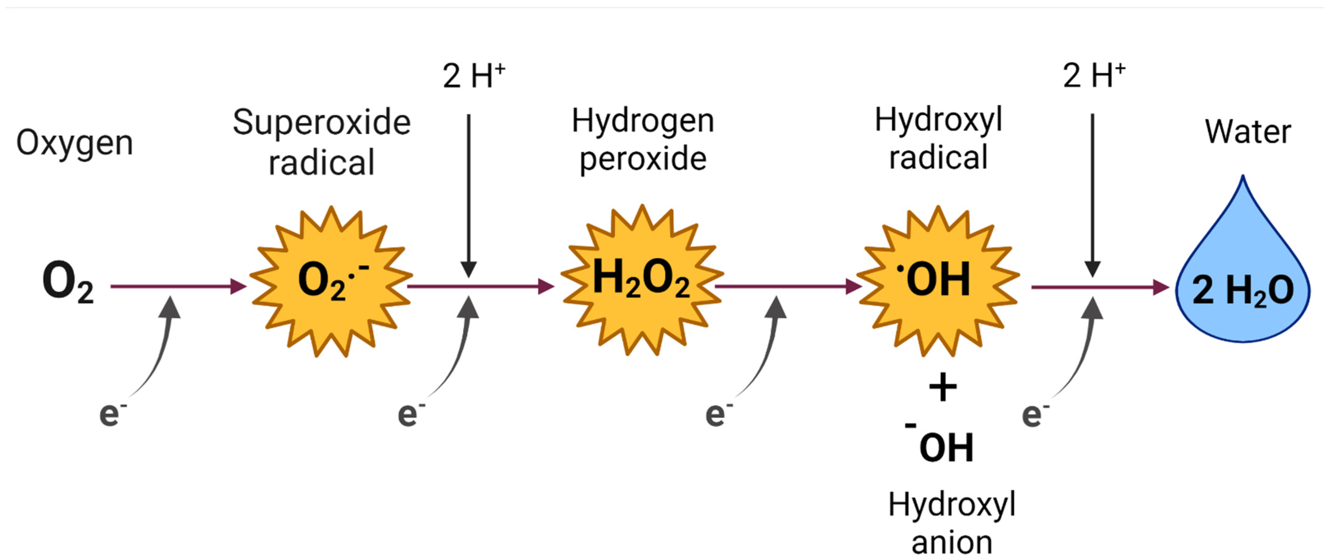
Figure 1. Univalent reduction of oxygen (O2). Through four successive reductions, O2 accepts electrons (e) one by one, generating incompletely reduced O2 intermediates: superoxide radical (O2•−), hydrogen peroxide (H2O2), and hydroxyl radical (OH), until reaching its complete reduction: water (H2O). Four e and four protons (H+) are required to be added to produce two H2O molecules. Created with biorender.com, accessed on 25 April 2022 (published with permission from biorender.com, accessed on 25 April 2022).
Oxygen 02 00015 g001
Figure 2. Reactive oxygen species (ROS) production and consequences of its dysregulated production. ROS can be produced from endogenous or exogenous substances. Under physiological conditions, the mitochondria represent a significant cellular source of ROS production. It is estimated that 1–2% of the electrons that enter the mitochondrial electron transport chain (ETC) produce superoxide radical (O2•−). O2•− at the intracellular level leads to the formation of other ROS due to antioxidant mechanisms and cellular components. Two O2•− molecules dismutate spontaneously or by superoxide dismutase (SOD), producing hydrogen peroxide (H2O2), which can break through cellular compartments. H2O2 can be enzymatically metabolized to water (H2O) by enzyme systems such as glutathione peroxidase (GPx) by reacting with glutathione (GSH), peroxiredoxin (Prx), and catalase, or it can be converted to a hydroxyl radical (OH). On the other hand, exogenous ROS are formed from environmental agents such as transition metal ions (e.g., iron (Fe), copper (Cu), nickel (Ni), cobalt (Co), cadmium (Cd), and lead (Pb)), ultraviolet (UV) light, and ionizing radiation or xenobiotics. When an imbalance in ROS production occurs, this leads to oxidative stress (OS). The persistence of OS leads to oxidative damage to biomolecules such as deoxyribonucleic acid (DNA), proteins, and lipids. DNA damaged by oxidation can generate mutations and may be related to cancer, the development of aging, or degenerative diseases. Protein oxidation leads to inhibition of their function through inhibition of enzyme activity, conformational changes, crosslinking, or aggregation. Oxidative damage to lipids results in the production of malondialdehyde (MDA) and 4-hydroxynonenal (4-HNE), as well as lipid peroxides (LOO). The damage caused leads to the development of pathologies. Created with biorender.com, accessed on 25 April 2022 (published with permission from biorender.com, accessed on 25 April 2022).
Oxygen 02 00015 g002
Figure 3. Cadmium (Cd)-induced oxidative stress (OS). Cd is a non-redox metal that causes OS via different indirect mechanisms. The mechanisms via which Cd increases reactive oxygen species (ROS) production include damage to the electron transport chain (ETC). Cd binds to the thiol groups (–SH or –S–) of the complexes, generating a decrease in the mitochondrial membrane potential (ΔΨm) and mitochondrial uncoupling that leads to lower production of triphosphate (ATP). Moreover, Cd increases the activity of the enzyme nicotinamide adenine dinucleotide phosphate (NADPH) oxidase (NOX), which increases the production of superoxide radical (O2•−). Likewise, Cd generates OS by increasing the concentration of redox metals such as iron (Fe2+). Fe2+ can participate in the Fenton reaction and increase radical hydroxyl (OH) production. Lastly, Cd also causes a decrease in the activity of antioxidant enzymes such as superoxide dismutase (SOD), manganese superoxide dismutase (MnSOD), catalase (CAT), and glutathione peroxidase (GPx). Ultimately, the OS generated by Cd exposure causes damage to biomolecules such as lipids and deoxyribonucleic acid (DNA). MDA: malondialdehyde, 4-hydroxynonenal: 4-HNE, 8-OHdG: 8-hydroxydeoxyguanosine, O2: oxygen, H2O2: hydrogen peroxide, H2O: water, Cyt c: cytochrome c, I, II, II, and IC: Complexes 1, II, III, and 1V of the ETC. Created with biorender.com, accessed on 25 April 2022 (published with permission from biorender.com, accessed on 25 April 2022).
Oxygen 02 00015 g003
Figure 4. Cadmium (Cd)-induced DNA methylation. Alterations in DNA methylation induced by Cd are related to the time of exposure to this metal. The short-term effects induced by Cd are due to a decrease in DNA methyltransferase (DNMT) activity and an increase in the ten-eleven translocation family (TET). Meanwhile, the effects of prolonged exposures have been linked to elevated levels of DNMT. SAH: S-adenosylhomocysteine, SAM: S-adenosylmethionine, MAT: methionine adenosyltransferase, MS: methionine synthase, GSH: glutathione, GSSG: glutathione disulfide, CpG: cytosine-guanosine dinucleotide sequences. Created with biorender.com, accessed on 25 April 2022 (published with permission from biorender.com, accessed on 25 April 2022).
Oxygen 02 00015 g004
Figure 5. Cadmium (Cd)-induced histone modification. Cd induces histone modifications altering the stability and replication of the genome. Cd increases the activity of histone deacetylases (HDAC), methyltransferases (HMT), and kinases, which cause histone acetylation, methylation, and phosphorylation, respectively. LDH: lactate dehydrogenase, NADH: nicotinamide adenine dinucleotide, HAT: histones acetyltransferases, CoA: coenzyme A, SAH: S-adenosylhomocysteine, SAM: S-adenosylmethionine, LSD: lysine-specific demethylases, FADH2: flavin adenine dinucleotide, JmjC: proteins containing the C domain of Jumonji, O2: oxygen, CO2: carbon dioxide, 2OG: 2-oxoglutarate, H2O2: hydrogen peroxide, CoA: coenzyme A, Fe: iron. Created with biorender.com, accessed on 25 April 2022 (published with permission from biorender.com, accessed on 25 April 2022).
Oxygen 02 00015 g005
Figure 6. Alterations in cadmium (Cd)-induced noncoding RNA (ncRNA) expression. Different transcription factors are involved in the expression of noncoding RNA (ncRNA). Transcriptional ncRNAs are classified into small ncRNAs and long ncRNAs (LncRNA). Small ncRNAs can be divided into microRNAs (miRNAs), P-element induced wimpy (PIWI)-RNAs of interference (piRNAs), and small RNAs of interference (siRNAs). Cd can alter LncRNA expression and miRNA biosynthesis. Drosha: subsequently processed in the nucleus by RNase III, DGCR8: DiGeorge syndrome critical region 8. Created with biorender.com, accessed on 25 April 2022 (published with permission from biorender.com, accessed on 25 April 2022).
Oxygen 02 00015 g006
Figure 7. Role of oxidative stress (OS) in cadmium (Cd)-induced DNA methylation. The OS generated by Cd is involved in DNA methylation. Reactive oxygen species (ROS) cause DNA damage, which interferes with the activity of DNA methyltransferases (DNMT); they also oxidize guanine from CpGs, which means that DNMT cannot methylate the cytosine or that binding to the methyl binding protein (MBP) complex does not take place. Guanine oxidation also makes the methylated cytosine more susceptible to oxidation by ten-eleven translocation (TET). In addition, ROS interfere with DNMT activity by depleting S-adenosylmethionine (SAM) by oxidizing methionine adenosyltransferase (MAT) and methionine synthase (MS) or by using homocysteine to regenerate glutathione (GSH). These effects cause DNA hypomethylation; however, ROS can also cause DNA to be hypermethylated by inhibiting TETs. SAH: S-adenosylhomocysteine, GSH: glutathione, GSSG: glutathione disulfide; CpG: cytosine–guanosine dinucleotide sequences. Created with biorender.com, accessed on 25 April 2022 (published with permission from biorender.com, accessed on 25 April 2022).
Oxygen 02 00015 g007
Figure 8. Role of oxidative stress (OS) in cadmium (Cd)-induced histone modifications. Cd-induced OS plays a vital role in histone modification, mainly nitration, acetylation, methylation, and phosphorylation. Peroxynitrite (ONOO−) nitrates histones, while other ROS can increase histone methylation by inhibiting the Jumonji domain C (JmjC) family of protein demethylases or decrease methylation by attenuating the activity of histone methyltransferases (HMT) by the reduction of S-adenosylmethionine (SAM). ROS also increases histone acetylation by inhibiting histone deacetylases (HDACs) and stimulating acetyl transferases (HAT). However, ROS could inhibit acetylation by causing an increase in NAD+, which stimulates HDACs. ROS increase the activity of LDH to increase NAD+. Histones can also be modified by phosphorylation when there is DNA damage. LDH: lactate dehydrogenase, NADH: nicotinamide adenine dinucleotide, CoA: coenzyme A, SAH: S-adenosylhomocysteine, LSD: lysine-specific demethylases, FADH2: flavin adenine dinucleotide, O2: oxygen, CO2: carbon dioxide 2OG: 2-oxoglutarate, H2O2: hydrogen peroxide, CoA: coenzyme A, Fe: iron. Created with biorender.com, accessed on 25 April 2022 (published with permission from biorender.com, accessed on 25 April 2022).
Oxygen 02 00015 g008
Figure 9. Role of oxidative stress (OS) in cadmium (Cd)-induced alterations in noncoding RNA (ncRNA) expression. The OS induced by Cd modifies the expression of ncRNAs, such as long ncRNAs (LncRNA) and microRNAs (miRNAs). Reactive oxygen species (ROS) cause deregulation of transcription factors, promote the generation of pre-miRNA by interaction with iron (Fe3+), and inhibit Dicer activity, which delays the production of mature miRNA. Modifications in the ncRNA can cause the decrease of nuclear factor 2 related factor E2 (Nrf2). Drosha: subsequently processed in the nucleus by RNase III, DGCR8: DiGeorge syndrome critical region 8. Created with biorender.com, accessed on 25 April 2022 (published with permission from biorender.com, accessed on 25 April 2022).
Oxygen 02 00015 g009
Table 1. Antioxidant compounds that mitigate epigenetic changes induced by cadmium (Cd).
AntioxidantModelCd Compound Relevant Effects of the Antioxidant Appointment
Selenium MCF-7 cellsCdCl2Epigenetic regulation of genes affected by Cd: APBA2, KIAA0895, DHX35, CPEB3, SVIL, MYLK, ZFYVE28, ABLIM2, GRB10, and PCDH9↓ Carcinogenesis
↓ PI3K/Akt
[260]
Quercetin Rats CdCl2↓ miRNA-21
↑ Nrf2 ↑ GSH
↑ SOD ↓ MDA
↓ ALT ↓ AST
↓ ROS ↓ IL6↓ TNF-α
↓ Total cholesterol
↓ Triglycerides
[261]
N-acetyl-l-cysteineTRL1215 CellsCdCl2↑ TET1
↑ ApoE
↓ MT2A
[217]
ResveratrolCD-1 mice and JEG-3 cellsCdCl2↓ DNMT activity
↓ DNMT3B expression
↓ Apoptosis
↓ TNF-α ↓ IFN-γ
↓ CCM-1 ↓ MIP-2
↓ KC ↑ SIRT1
↓ PI3K/Akt
[262]
IsoorientineNRK-52E cells and primary cultures (rPT)CdCl2↓ p-H2AX
↓ ROS
↓ 8-OHdG
[263]
Cyanidin-3-O-glucosidePubescent miceCdCl2↑ Spermatogenesis
↓Histone H2A
↓ Histone H2B
↑ Ubiquitination of H2A
↑ SOD ↑ GSH
↑ GSH/GSSG
↓ MDA
↓ p-JNK/JNK
↑ p-ERK/ERK
↓ p-p38/p38
↓ Caspase 3
↓ Bax ↓ Bad
↑ bcl-2
[264]
MethionineK562 cellsCdCl2↑ Global DNA methylation
↓ ROS
↓ 8-OHdG
[131]
8-OHdG: 8-hydroxy-2-deoxyguanosine, ABLIM2: Actin-binding LIM member of the protein family 2, Akt: protein kinase B, ALT: alanine transaminase, APBA2: amyloid beta precursor protein A, ApoE: apolipoprotein E, AST: aspartate aminotransferase, bad: Bcl-2 associated agonist of cell death, bax: Bcl-2 Associated X-protein, bcl-2: B-cell lymphoma 2, CCM-1: cerebral cavernous malformations, CdCl2: cadmium chloride, CPEB3: cytoplasmic polyadenylation element 3-binding protein, DHX35: DEAH helicase box 35, DNMT: DNA methyltransferase, ERK: extracellular signal-regulated kinases, GRB10: growth factor receptor 10-bound protein, GSH: glutathione, GSSG: glutathione disulfide, H2: histone 2, IFN-γ: interferon-gamma, IL6: interleukin 6, JNK: c-Jun N-terminal kinase, KC: chemokine ligand 1, MCP: monocyte chemoattractant protein, MCP-1: monocyte chemoattractant protein 1, MDA: malondialdehyde, MIP-2: macrophage inflammatory protein-2, miRNAs: microRNAs, MT2A: Metallothionein 2A, MYLK: myosin light chain kinase, Nrf2: nuclear factor 2 related factor E2, p38: protein 38, PCDH9: protocadherin 9, PI3K: phosphoinositide-3-kinase, p-H2AX: histone H2AX phosphorylation, ROS: reactive oxygen species, SIRT1: sirtuin 1, SOD: dismutase superoxide, SVIL: supervillain, TET1: Ten-eleven translocation methylcytosine dioxygenase 1, TNF-α: tumor necrosis factor-α, ZFYVE28: FYVE type zinc finger containing 28.
Publisher’s Note: MDPI stays neutral with regard to jurisdictional claims in published maps and institutional affiliations.