Review

Energy Metabolism as a Therapeutic Target in Cancer: The Role of Coenzyme Q10

1
Pharma Nord (UK) Ltd., Morpeth NE61 2DB, UK
2
School of Pharmacy and Biomolecular Sciences, Liverpool John Moores University, Liverpool L3 3AF, UK
3
Department of Physiology, Anatomy and Cell Biology, Andalusian Centre of Developmental Biology, Universidad Pablo de Olavide, 41013 Seville, Spain
*
Author to whom correspondence should be addressed.
Oxygen 2024, 4(2), 122-138; https://doi.org/10.3390/oxygen4020008
Submission received: 24 February 2024 / Revised: 30 March 2024 / Accepted: 9 April 2024 / Published: 11 April 2024

Abstract

:
The generation of energy within cells is a fundamental process enabling cell survival, and as such it represents a potential target in cancer therapy. In this article, we therefore review the relative contributions of glycolysis and oxidative phosphorylation/mitochondrial function to cancer cell energy generation, and we highlight their respective potential value as chemotherapeutic targets. This article is particularly focussed on the potential role of coenzyme Q10 in the prevention and treatment of cancer.

1. Introduction

The generation of energy within cells is a fundamental process enabling cell survival, and as such it represents a potential target in cancer therapy. The two principal mechanisms of cellular energy generation are glycolysis (within the cytoplasm) and oxidative phosphorylation (within the mitochondria), respectively. In higher animals, oxidative phosphorylation is the primary source of ATP generation, since the latter process is more efficient (producing approximately 15-fold more ATP per unit glucose substrate) compared to glycolysis. However, although glycolysis yields less ATP than oxidative phosphorylation, the rate of ATP generation in the former process is greater (by approximately 100-fold) than in the latter, which is more suited to the energy demands of rapidly proliferating cancer cells. Thus, compared to normal cells, in cancer cells, glycolysis was considered to be relatively enhanced and oxidative phosphorylation reduced, a phenomenon first described in the 1920s by Otto Warburg. However, more recently, oxidative phosphorylation (and normal mitochondrial function) has also been found to play a significant role in energy generation in cancer cells. The aim of this article is therefore to review the role of glycolysis and oxidative phosphorylation in cancer therapy, with a particular focus on mitochondria and coenzyme Q10 as potential therapeutic targets.

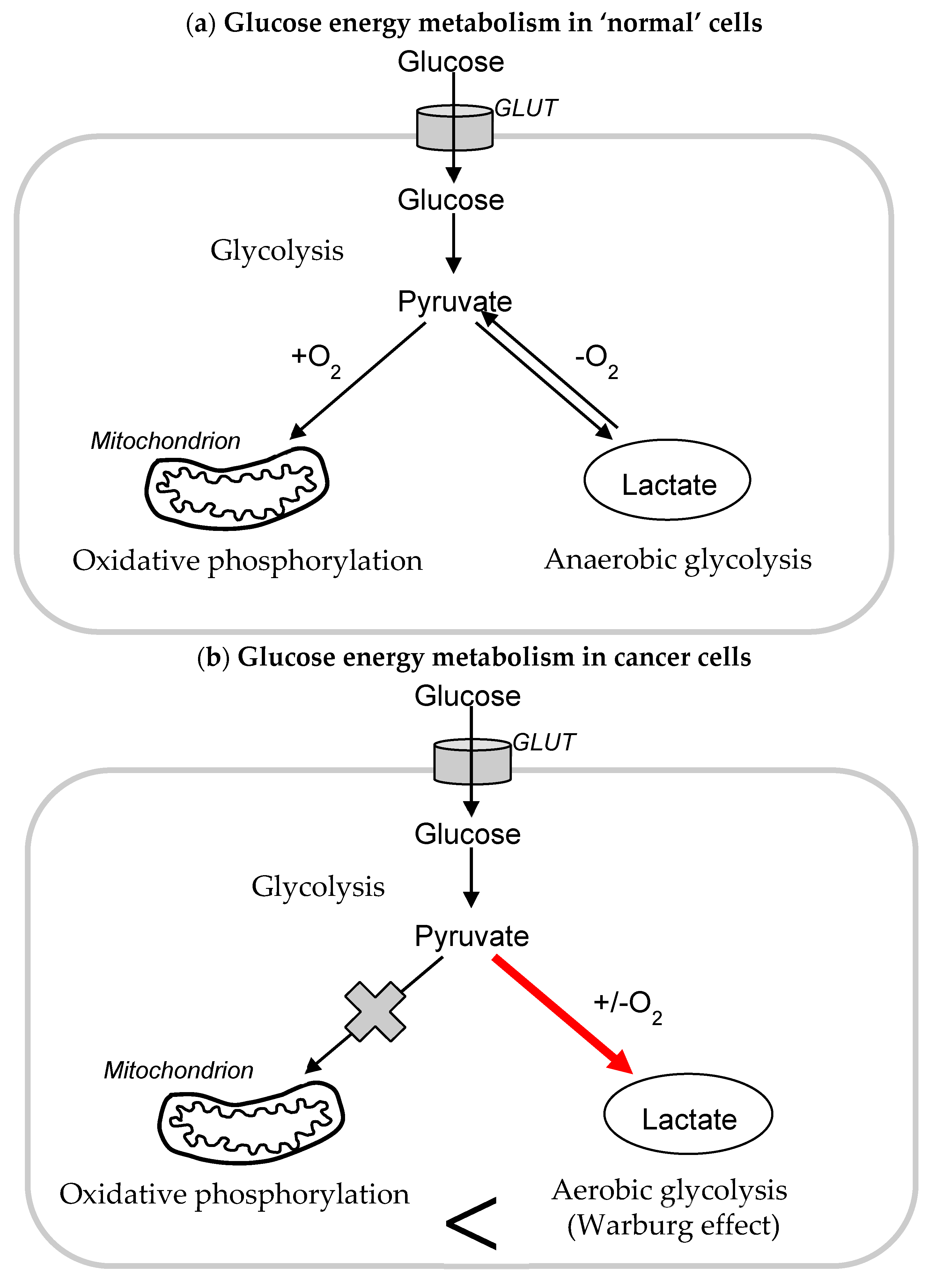
1.1. Glycolysis and Cancer

Glycolysis is an anaerobic metabolic pathway that provides energy to the host by the splitting of glucose into two pyruvate molecules. The process comprises two phases: the investment phase, in which two ATP molecules are used to initiate the process, and the payoff phase, in which additional ATP is produced. In normal cells, oxidative phosphorylation is responsible for 70% of energy generation, with the production of 32 molecules of ATP per unit glucose substrate, compared to only 2 molecules of ATP produced by glycolysis. However, glycolysis provides the primary energy source in cells devoid of mitochondria (such as those found in the eye lens), and, in addition, glycolysis acts as an emergency backup in cells that primarily undergo aerobic respiration.
In contrast to normal cells, cancer cells have been shown to utilise glycolysis as their primary source of energy production regardless of the presence of oxygen (Figure 1); this was first noted in the 1920s by Otto Warburg, subsequently known as the Warburg effect and identified as a hallmark of cancer [1]. Rapidly growing tumours may experience a hostile hypoxic environment as the tumour expands beyond the diffusion limit of local blood supply; cancer cell metabolism generally switches to glycolysis by enhancing the production of glycolytic enzymes, glucose transporter proteins, and mitochondrial metabolic inhibitors, a process known as metabolic reprogramming [2]. As well as the biosynthesis of ATP, glycolysis involves the production of many intermediate metabolites of potential relevance in cancer for macromolecular biosynthesis, conferring a selective advantage to cancer cells under diminished nutrient supply. In addition, many enzymes of the glycolytic pathway also play significant roles in several non-glycolytic processes that enable the cancer cells to meet other cellular demands. Numerous studies have demonstrated that glycolysis promotes tumour growth, metastasis, and chemo-resistance, while also inhibiting the apoptosis of tumour cells [3].
Despite the reliance on glycolysis, cancer cells have been shown to maintain normal mitochondrial function; therefore, the mechanism by which this metabolic shift to glycolysis occurs is of interest to researchers. Three factors have been identified to potentially explain the shift: the rapid production of ATP generated by glycolysis in comparison to oxidative phosphorylation, the high glycolytic flux providing precursors for rapidly proliferating cells to create macromolecules, and resistance to chemotherapeutics enabled by high intracellular levels of glutathione and glutathione transferase associated with high rates of glycolysis [4].
Akt, otherwise known as the Warburg kinase, is a serine kinase that facilitates the metabolic shift in tumour cells under normoxic conditions. Akt activation leads to the increased expression of glucose transporters (such as GLUT1) and glycolytic enzymes (such as HKII) [5]. Interestingly, Akt activation leads to fructose-2,6-bisphosphate production; fructose has been shown to be a driving force in obesity and metabolic syndrome which have both been associated with a higher risk of developing cancer, which in turn could be due to fructose promoting the Warburg effect [6].
Since glycolysis provides cancer cells with their primary source of energy, blocking this metabolic pathway represents a potential therapeutic strategy. This could be achieved, for example, through the inhibition of either glucose transporters or of enzymes involved in the glycolytic pathway. Hexokinase II (HKII) is an isoform of the enzyme that initiates glycolysis, catalysing the conversion of glucose to glucose-6-phosphate; HK II promotes tumour growth and higher levels of HKII have been associated with poor prognosis and recurrence across multiple cancer forms, including lung and breast cancer. The use of an HK inhibitor (such as 2-deoxy-D-glucose) is currently under investigation to help improve treatment outcomes, as it has been shown to increase the sensitivity of cancer cells to chemotherapeutics [7]. For example, a study by Sun et al. [8] demonstrated that 2-deoxy-D-glucose enhanced the action of the anti-tumour agent chloroethylnitrosourea in glioblastoma cells; in addition, the combination of 2-deoxy-D-glucose and chloroethylnitrosourea significantly suppressed tumour growth in tumour-bearing mice. Other enzymes of the glycolytic pathway investigated with regard to possible inhibition include phosphofructokinase, pyruvate kinase, and lactate dehydrogenase [9]. The application of glycolytic inhibitors in the clinical practice of cancer treatment to date has been hindered by the problem of adverse effects [9].

1.2. Mitochondria and Cancer

Mitochondria have a key role in cell metabolism; in addition to their role in cellular energy generation via oxidative phosphorylation, they also have roles in free radical metabolism and redox homeostasis, lipid and nucleic acid metabolism, calcium homeostasis, cell signalling, and apoptosis through the activation of the mitochondrial permeability transition pore (mtPTP) and the release of proapoptotic factors [10]. Changes in these parameters can impinge on biosynthetic pathways, cellular signal transduction pathways, transcription factors, and chromatin structures to shift the cell from a quiescent, differentiated state to an actively proliferating one; thus, mitochondria can influence all processes linked to oncogenesis, starting from malignant transformation to metastatic dissemination [11]. In contrast to the above, it should also be noted that mitochondria can also exert anticancer action via roles in ferroptosis, mitophagy, and antitumour immunity [12,13,14].
The pathophysiology of cancer varies amongst different cancer types, and hence so do alterations in mitochondrial metabolism. As noted in the previous section of this article, glycolysis is upregulated in many forms of cancer, thus raising interest in how cancer cells metabolise the excess pyruvate that is produced, and whether the upregulation of glycolysis is responsible for dysfunctional mitochondria in cancer. Defective protein complexes in the mitochondrial respiratory chain (MRC) have been noted to have genomic mutations that reduce function. This has been exploited in upregulating complex I activity in order to inhibit breast cancer tumour growth via the maintenance of elevated NAD+/NADH levels, although further investigation is needed to determine the extent to which mitochondrial genomic mutations affect cancer progression.
Mitochondria in malignant cells differ structurally and functionally from those in normal cells and participate actively in metabolic reprogramming [15]. Mutations in the mitochondrial DNA (and in nuclear DNA) in cancer cells have been recognized for more than two decades; although mutations in mitochondrial genes are common in cancer cells, they do not inactivate mitochondrial energy metabolism but rather alter mitochondrial metabolism to enhance tumorigenesis and permit cancer cell survival. These altered states communicate with the nucleus (termed retrograde signalling) to modulate signal transduction pathways, transcriptional circuits, and chromatin structures in order to meet the perceived mitochondrial and nuclear requirements of the cancer cell. Through the release of mitochondrial-derived metabolic compounds, cancer cells can then reprogram adjacent stromal cells in order to optimize the cancer cell environment [16,17].
Retrograde signalling may affect the activity of specific enzymes involved in mitochondrial metabolism. Mutations in enzymes and metabolites associated with the TCA cycle may play important roles in tumour invasiveness and metastasis. The intermediates in TCA cycles, such as succinate, citrate, and NAD+, have been shown to possess signalling capacity and influence the immunity associated with tumours and cancer. Mutations in corresponding enzymes include succinate dehydrogenase (SDH), fumarate hydratase (FH), and isocitrate dehydrogenases 1 and 2 (IDH1 and IDH2); for SDH defects, this involves the activation of HIF1 signalling; and for FH defects, this involves alterations in NRF2 signalling. Another important aspect is the role of succinate and acetylcysteine in the epigenetic regulation of various genes [18]. All of these effects can contribute to tumorigenesis [19].
Mitochondria in cancer cells are characterized by the overproduction of reactive oxygen species (ROS) which promote all steps of oncogenesis, from tumour initiation to proliferation and metastasis, by inducing genomic instability, modifying gene expression, and participating in signalling pathways [20]. Increased ROS levels may lead to increases in mutations, particularly in the mitochondrial DNA (mtDNA) owing to the close proximity of mitochondrial ROS and mtDNA, the lack of protective histones, and the limited capacity for DNA repair [21]. The mitochondrial genome mainly contains genes encoding for electron transport chain proteins, and mutations in these genes can therefore affect electron transport chain signalling pathways. This can then create an endless cycle in which mutated electron transport chain proteins cause an increased leakage of electrons and more ROS production, which can lead to more mutations. Thus, mitochondrial and nuclear DNA mutations caused by oxidative damage that impair the oxidative phosphorylation process will result in further mitochondrial ROS production, completing the “vicious cycle” between mitochondria, ROS, genomic instability, and cancer development [22].
The change in cellular redox status may cause the activity of transcription factors, such as HIF1α and FOS–JUN, to change gene expression and stimulate cancer cell proliferation [23]; there is also evidence that ROS can themselves act as signalling molecules that promote oncogenic pathways. Whilst increased ROS production facilitates tumour development and progression, if ROS levels become too high, then intracellular damage may become too great, inducing cell death. To survive, cancerous cells respond to such high ROS levels by increasing the expression of antioxidant enzymes, such as superoxide dismutase (SOD), glutathione peroxidase (GPx), and catalase (CAT), as well as high levels of non-enzymatic antioxidants.
Thus, it has been argued that if ROS levels in cancer cells are close to a threshold and are pushed over that threshold via a therapeutic modality, apoptosis can be triggered; normal cells with intrinsic lower levels of ROS compared to cancer cells would be spared the same fate of undergoing apoptosis, ensuring the potential safety profile of the therapeutic modality [24]. Enhancing ROS production or inhibiting antioxidant action has therefore been identified as a therapeutic target, as noted in the following section. In fact, major chemotherapeutic agents such as cisplatin, doxorubicin, and irinotecan all substantially increase ROS levels in cancer cells as a secondary effect because their main mechanism of action is to produce damage in DNA by crosslinking purine bases forming DNA adducts, intercalating into DNA and blocking topoisomerase-II, or disturbing the elevation of DNA by interfering in the binding of topoisomerase-I and DNA.

1.3. Mitochondria as a Target for Cancer Therapy

The multiple essential roles of mitochondria have been utilized for designing novel mitochondria-targeted anticancer agents. Due to changes in mitochondrial metabolism and changes in membrane potential, cancer cells are more susceptible to mitochondria-targeted therapy. The specificity of this approach is aided by the capacity of non-proliferating non-cancerous cells to withstand oxidative insult induced by oxidative phosphorylation inhibition. The loss of functional mitochondria in cancer cells leads to the arrest of cancer progression and/or cancer cell death, probably due to the disruption of the cancer-associated metabolism that depends on the production of many Krebs-cycle-dependent intermediates, mainly amino acids and pyridine nucleotides [25]. The analysis of over 30 cancer types revealed that mitochondria with mtDNA mutations that are pathogenic are less likely to be maintained in cancer cells, suggesting that there is a positive selection for functional mitochondria to drive tumour growth [26]. In addition, genetic defects leading to defective mitochondrial respiratory function produce a metabolic checkpoint that prevents malignant transformation [27]. These studies indicate that mitochondrial metabolism is an active essential process for tumour growth.
Several classes of compounds have been studied for their mitochondria-targeting ability in cancer cells. These compounds have been described as mitocans, defined as a category of drugs known to precisely target the cancer cells’ mitochondria [28,29]. Based on their mode of action, mitocans have been divided into eight classes; these include drugs targeting the TCA cycle (Class 7 mitocans), drugs targeting the electron transport chain (Class 5 mitocans), drugs targeting free radical production (Class 3 mitocans), and drugs targeting the permeability transition pore (Class 4 mitocans).
With regard to the inhibition of oxidative phosphorylation, decreasing ETC function prevents the oxidative TCA cycle from functioning, thus diminishing macromolecule synthesis to support tumour growth. Several compounds have been identified, including metformin and atovaquone [30]. Metformin exerts its anticancer effects through the inhibition of mitochondrial ETC complex I; metformin also decreases circulating insulin levels, a known mitogen for tumours [31].
With regard to the inhibition of the TCA cycle, drugs which enable the latter would be predicted to be effective due to the central role of the TCA cycle in producing the intermediate metabolites for growth. CPI-613 is a lipoate analogue the first of its kind that can inhibit two major TCA cycle enzyme complexes that require lipoate for their activity, α-ketoglutarate dehydrogenase (α-KGDH) and pyruvate dehydrogenase (PDH). Although the mechanism by which CPI-613 exerts its anti-cancer activity is not fully understood, it has displayed a significant therapeutic index in promising phase I and II results in pancreatic cancer and AML [32].
With regard to the targeting of free radical production, elesclomol sodium is an example of a drug that targets ROS levels in cancer cells. Its role is to inhibit electron transport flux and enhance ROS production by inducing oxidative stress in both transformed and healthy cells. However, as tumour cells already have elevated levels of ROS, this drug will be able to induce cytotoxicity selectively in malignant cells, resulting in the activation of apoptotic cell death [33].
With regard to the targeting of the transition pore, the latter (PTPC) is a highly dynamic supramolecular complex found in the mitochondria membrane and is responsible for mitochondria membrane permeabilization. PTPC is composed of different molecular components: the voltage-dependent anion channel in the outer membrane, the adenine nucleotide translocase (ANT) in the mitochondrial inner membrane, and cyclophilin D in the mitochondrial matrix. The most promising drug targeting ANT that efficiently triggers apoptosis is lonidamine or 1-(2,4-dichlorobenzyl)-1-H-indazole-3-carboxylic acid. Lonidamine is an ANT ligand that acts as a HK and induces a conformational change in the ANT, leading to mitochondrial channel formation. It has been reported that lonidamine is able to target the respiratory activity of complex II, suppressing the formation of fumarate and malate, leading to the accumulation of succinate in the treated cells. Lonidamine enhances the apoptotic response to cisplatin, cyclophosphamide, doxorubicin, paclitaxel, melphalan, and γ-irradiation both in vivo and in vitro [34,35].
In addition to the above, the impairment of the mitochondria-regulated apoptosis pathway accelerates tumorigenesis. Numerous strategies targeting mitochondria have been developed to induce the mitochondrial (i.e., intrinsic) apoptosis pathway in cancer cells.
In general, all these drugs target different functions of mitochondria, and their anticancer effect is mainly derived from blocking the synthesis of many precursors of amino acids and proteins produced by the Krebs cycle, but most importantly by a reduction in the synthesis of pyridine nucleotides given the activity of the CoQ10-dependent dihydroorotate dehydrogenase (DHODH) [36].

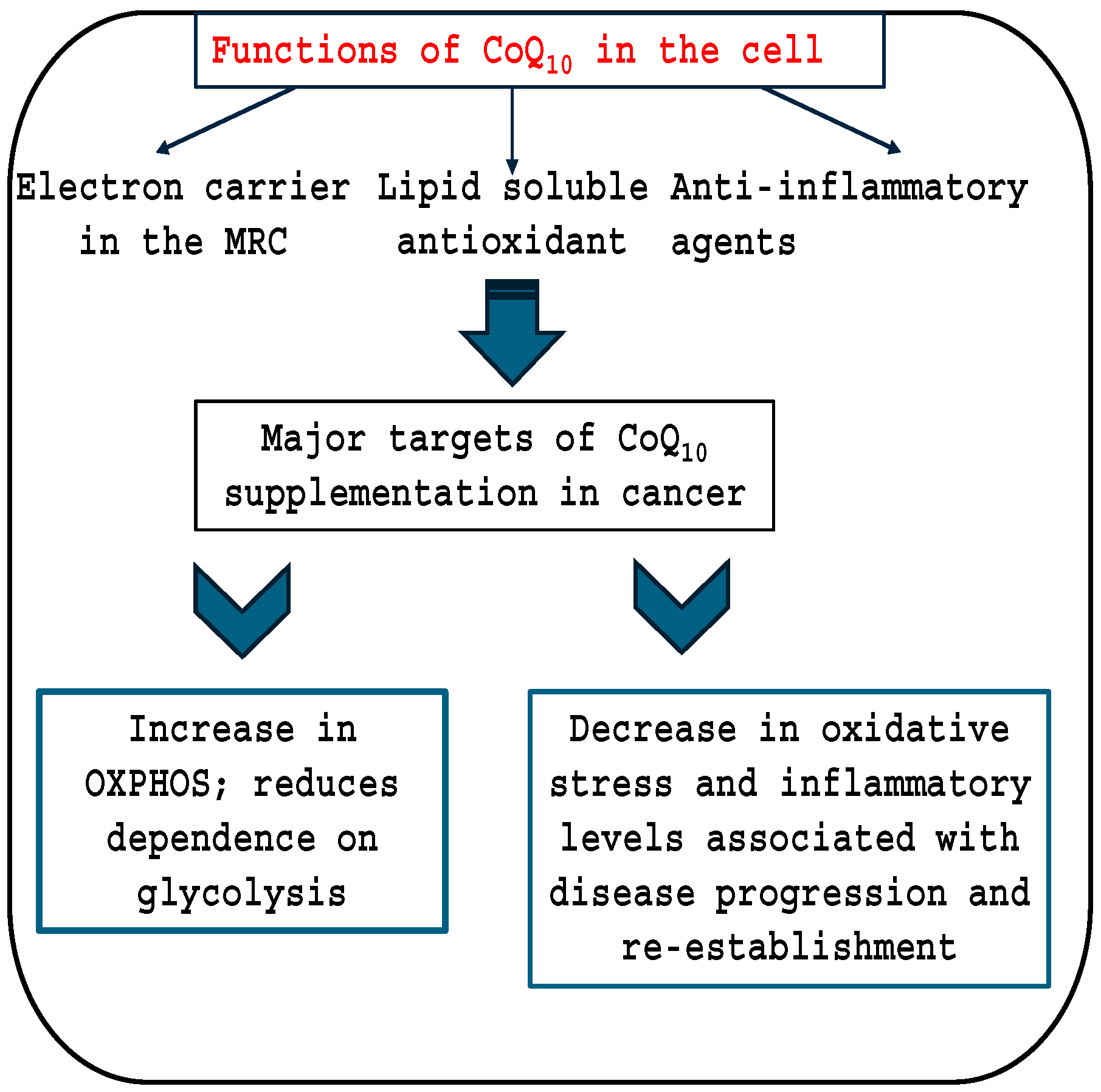
1.4. Coenzyme Q10 and Cancer

To date, there are approximately 1000 articles relating to CoQ10 and cancer listed on Medline, and these have been reviewed in three categories as follows. The cellular functions of CoQ10 and its targets in cancer therapy are outlined in Figure 2.

2. OXPHOS; Oxidative Phosphorylation

2.1. Clinical Studies

To date, there have been no randomised controlled trials to investigate the effect of CoQ10 alone directly on tumour viability in cancer patients. Some trials have demonstrated a positive effect of the combination of different compounds together with CoQ10, mainly by reducing the side-effects of chemotherapy or other treatments, or by reducing the inflammation associated with cancer, an important factor in the progression of many cancers [37].
A randomised controlled study by Premkumar et al. [38] supplementing 84 breast cancer patients undergoing tamoxifen therapy with a combination of 100 mg of CoQ10, 50 mg of niacin, and 10 mg of riboflavin reduced the level of serum tumour markers (carcinoembryonic antigen (CEA) and carbohydrate antigen 15-3 (CA 15-3)), thereby reducing the risk of cancer recurrence and metastases. In a randomised controlled study by Liu et al. [39] comprising 41 patients with primary hepatocellular carcinoma, supplementation with CoQ10 (300 mg/day for 3 months) after surgery reduced oxidative stress and inflammation levels, important factors in the re-establishment of this disease [40]. In a randomised controlled trial comprising 59 patients with breast cancer undergoing tamoxifen therapy, supplementation with CoQ10 (100 mg/day for 8 weeks) significantly reduced blood levels of the inflammatory cytokines IL-6 and IL-8, reducing the consequences of inflammation caused by breast cancer [41]. A randomised controlled trial by Iarussi et al. [42] comprising 20 children with acute lymphoblastic leukemia or non-Hodgkin lymphoma reported supplementation with CoQ10-protected cardiac function during anthracycline therapy. A meta-analysis of randomised controlled trials found that CoQ10 supplementation significantly reduced levels of a number of inflammatory markers, together with matrix metalloproteinase activity, in breast cancer patients [43].
Several randomised controlled cancer studies reported no significant benefits following CoQ10 supplementation, but also reported no adverse effects on disease outcome. Thus, two randomised controlled trials investigated the effect of CoQ10 supplementation on fatigue and quality of life in breast cancer patients; the study by Lesser et al. [44] comprising 236 patients found that CoQ10 supplementation (300 mg/day for 6 months) had no significant effect on self-reported fatigue or quality of life, but no adverse effects on disease progression were reported. Similarly, in the study by Iwase et al. [45] comprising 57 patients undergoing chemotherapy for breast cancer, supplementation with the proprietary product Inner Power (containing CoQ10) had no significant effect on general fatigue or quality of life, but again no adverse effects on disease progression were reported. In a randomised controlled study comprising 70 patients with prostate carcinoma, supplementation with a combination of vitamin E, selenium, vitamin C, and CoQ10 had no effect on serum PSA levels, but no adverse effects of the treatment regime on the disease course (compared to placebo) were noted [46].
With regard to other types of clinical studies, Lockwood et al. [47] described partial remission in a series of 32 high-risk breast cancer patients, following supplementation with a combination of antioxidants, including CoQ10. Hertz and Lister [48] described improved survival in a series of 41 patients with end-stage cancer (breast, brain, lung, kidney, pancreatic, oesophagus, stomach, colon, prostate, ovarian, and skin) treated with coenzyme Q10 and other antioxidants (vitamin C, selenium, folic acid, and beta-carotene). In patients with lung cancer, treatment with hydrogen gas enhanced the clinical efficacy of nivolumab by increasing CoQ10 levels [49]. Rusciani et al. [50] described a 3-year study in which patients with stage I and II melanoma were administered with interferon and CoQ10 (400 mg/day) following surgery; significantly decreased rates of recurrence were noted compared to treatment with interferon only, and no adverse effects were reported.
With regard to CoQ10 deficiency, decreased blood levels of CoQ10 have been reported in patients with various types of cancer (breast cancer, myeloma, lymphoma, cervical cancer, oral cancer, and lung cancer) [51,52,53,54,55,56]. Epidemiological studies have identified reduced levels of plasma CoQ10 as a risk factor for the development or progression of several types of cancer, including breast cancer [57], lung cancer [56], and melanoma [50].
In several studies, reduced levels of CoQ10 have been associated with a worsening disease prognosis. Thus, reduced CoQ10 levels in infiltrative ductal carcinoma were reported by Portakal et al. [58]; Jolliet et al. [59] correlated the magnitude of reduced CoQ10 levels in breast carcinomas with poor prognosis in breast cancer patients. The level of CoQ10 was reduced in human astrocytoma tissues and correlated negatively with the degree of malignancy [60]. Kanda et al. [61] reported decreased expression in the gastric carcinoma tissue of prenyl diphosphate synthase subunit 2 (PDSS2), which is required for the biosynthesis of CoQ10, correlated with the reduced survival of patients with gastric cancer. However, other studies have associated high CoQ10 levels with the presence of cancer. For example, increased plasma CoQ10 levels were identified as a risk factor for breast cancer by Chai et al. [62], and El-Attar et al. [63] reported increased serum CoQ10 levels in breast cancer patients.
All of the studies summarised above are essentially positive in outcome with regard to the role of CoQ10 in the prevention or treatment of cancer, or otherwise provide evidence on the lack of adverse effects of CoQ10 on disease progression. In this regard, a study by Ambrosone et al. [64] found that dietary supplementation with antioxidants (including CoQ10) during chemotherapy increased the risk of breast cancer recurrence. This study is incorrectly listed on Medline as a randomised controlled trial, when it is actually a questionnaire-based observational study, in which the data relating to CoQ10 are of borderline significance. It is of note that Medline currently lists more than 300 randomised controlled trials supplementing CoQ10 in a variety of disorders, and in a range of doses (up to 2700 mg/day) and durations (up to 5 years); none of these studies have reported any cancer-related adverse effects.

2.2. Studies in Animal Models

Animal studies have been used to determine many aspects of CoQ10 supplementation in the treatment of cancer with radiation or chemotherapy. In many cases, CoQ10 supplementation reduces the side-effects of such anticancer treatment. For example, in rats, the administration of CoQ10 (10 mg/kg) prior to gamma irradiation reduced intestinal inflammation and fibrosis, suggesting that supplementary CoQ10 might prevent intestinal complications in patients with pelvic tumours undergoing radiotherapy [65]. Similarly, the administration of CoQ10 (10 mg/kg) in gamma-irradiated rats reduced radiation-induced damage to the kidneys, the most radiosensitive organs in the abdominal cavity, and the dose-limiting issue in cancer patients receiving abdominal or total body irradiation [66].
In other studies, the administration of CoQ10 produced a direct effect on the growth of cancer cells, without a clear mechanism of action. Abdel-Latif et al. [67] showed that CoQ10 administration protected against the development of hepatocellular carcinoma in rats, via a reduction in CD59 glycoprotein expression and phospholipase D (PLD) activity. Treatment with CoQ10 (25 mg/rat) improved the survival of Yoshida-sarcoma-implanted rats [68]. In rats with chemically induced mammary carcinoma, supplementation with CoQ10 (40 mg/kg/day) improved the chemotherapeutic action of tamoxifen in preventing cancer cell proliferation [69]. In mice with lung carcinoma, the intraperitoneal injection of CoQ10 (0.2–1.2 mg/kg) inhibited tumour growth [70]. In rats, the dietary incorporation of CoQ10 (200–400 ppm for 4 weeks) suppressed the formation of colonic pre-malignant lesions induced by azoxymethane [71]. Glioblastoma growth and infiltration were reduced following CoQ10 treatment (100 mg/kg ip for 8 weeks) in mice [72]. In addition, Sun et al. [73] found that supplementary CoQ10 improved the survival of mice with highly aggressive orthotopic C6-glioblastoma cells. In mice, decylubiquinone, a dietary analogue of CoQ10, was an effective inhibitor of pulmonary metastatic melanoma [74].
Some studies in animal models have reported the adverse effects of CoQ10 administration. In mice, supplementary CoQ10 was reported to decrease the effectiveness of radiation therapy against small-cell lung cancer [75]. Said et al. [76] pre-treated rats with CoQ10 (10 mg/kg for 2 weeks), followed by subsequent gamma irradiation. The object of the latter study was to demonstrate the protective effects of CoQ10 on testicular function (of relevance to male patients undergoing radiotherapy), but the study also demonstrated the action of CoQ10 in preventing radiation-induced apoptosis (and thus potentially aiding cancer cell survival during radiotherapy). In other studies, CoQ10 interfered with the mechanism of action used by chemotherapy compounds, as shown in a rat model of chemically induced hepatocarcinogenesis, in which the inhibition of cell proliferation induced by lovastatin treatment was reversed following CoQ10 administration [77].

2.3. Cell Culture Studies

A number of cell-culture-based studies have reported the beneficial effects of CoQ10 supplementation in various cancer cell types. Several studies have found that supplementary CoQ10 inhibits cancer cell growth, increases apoptosis, or acts as a sensitiser for radiotherapy or chemotherapy. Thus, Quiles et al. [78] found that CoQ10 reduced the cell growth of a human prostate cancer cell line, while having no effect on a corresponding non-cancerous prostate cell line. The breast cancer cell line MCF-7 showed a decreased proliferation rate following the chemically induced depletion of CoQ10 levels [79]. In cultured oestrogen receptor negative breast cancer cell lines (MDA-MB-231 and SKBr3), treatment with CoQ10 (7.5 micromole/L) increased the proportion of apoptotic MDA-MB-231 and SKBr3 cells by 12-fold and 4-fold over the control, respectively [80]. In pancreatic cancer cells, administrating the supraphysiological levels (>100 times the endogenous levels) of CoQ10-induced apoptosis in cancer cells [24]. In human glioblastoma cells, loading CoQ10 acted as a sensitiser for radiotherapy, with a twofold increase in radiation-induced DNA damage and apoptosis compared to controls [81]. Treatment with the CoQ10 precursor 4-hydroxybenzoic acid enhances the sensitivity of human breast cancer cells to adriamycin [79].
CoQ10 can also contribute to changes in mitochondrial activity due to a more respiratory phenotype that can negatively affect cell growth. Increasing oxidative phosphorylation in mitochondria produces a metabolic rewiring that disrupts intrinsic resistance to ferroptosis in colon adenocarcinoma cells, indicating that improving oxidative phosphorylation activity can impair cancer progression [82]. Interestingly, CoQ10 supplementation can increase oxidative phosphorylation and reduce the dependence of tumour cells on glycolysis with this mechanism based on cell growth, metastasis, and immune evasion [83]. CoQ10 synthesis inhibition can induce the HIF-1a stabilization that is involved in the maintenance of glycolysis in these cells [84]; for this reason, supplementation with CoQ10 could help to reduce glycolysis and block cancer cell progression.
Several studies have shown that supplementary CoQ10 mediates the metastatic capacity of cancer cells. For example, the administration of CoQ10 in a breast cancer cell line reduced the activity of matrix metalloproteinase II, a key promotor of cancer cell invasion and metastasis [85]. In cultured melanoma cells, CoQ10 demonstrated an inhibitory effect on cell proliferation and migration/invasion when used individually or in combination with vemurafenib, despite an apparent protective effect of CoQ10 in protecting melanoma cells from apoptosis induction [86]. In a rat temozolomide-resistant glioma cell line, the co-administration of CoQ10 increased the efficacy of temozolomide in reducing cell proliferation and invasiveness [87]. In a hepatocellular carcinoma cell line, Heidari-Kalvani et al. [88] reported that CoQ10 administration reduced Nrf2 (nuclear factor erythroid-2-related factor 2) levels, matrix metalloproteinase activity, and metastatic potential, but induced apoptosis. Finally, Frontiñán-Rubio et al. [72] found that CoQ10 supplementation reduced glioblastoma growth and infiltration capacity in xenografts via proteome remodelling and the inhibition of angiogenesis and inflammation.
As with the case in animal model studies, some cell culture studies have reported the adverse effects of CoQ10 administration. Jain et al. [89] showed that the compound SMIP004-7, an uncompetitive inhibitor of ubiquinone, targets drug-resistant cancer cells with stem-like features by inhibiting mitochondrial respiration complex I. Brea-Calvo et al. [90] found that induced camptothecin increased CoQ10 levels in cancer cell lines, indicating that CoQ10 could aid cancer cell survival during chemotherapy.
Conflicting results have been reported for the use of statins as chemotherapeutic adjuvants. These compounds can affect the synthesis of CoQ10, as well as the synthesis of cholesterol and derivatives (such as steroid hormones and dolichol). The exact role of the level of CoQ10 in these studies is not clear, although some studies have associated the effect of statins with the depletion of CoQ10. In fact, in multiple myeloma cells, targeting CoQ10 synthesis using the mevalonate pathway inhibitor simvastatin increased cell death induced by the proteasome inhibitor bortezomib [91]. Similarly, McGregor et al. (2020) [92] reported that simvastatin reduced CoQ10 synthesis and promoted cancer cell apoptosis in mouse pancreatic ductal adenocarcinoma tumours. Kaymak et al. [93] showed that p53-deficient cancer cells subject to metabolic stress activate the mevalonate pathway promoting CoQ10 synthesis, thereby reducing oxidative stress and supporting pyrimidine nucleotide synthesis for cell growth; the inhibition of the mevalonate pathway using statins blocked pyrimidine nucleotide biosynthesis and induced oxidative stress and apoptosis in the cancer cells.
Other cell culture studies have associated the protective effect of CoQ10 on ferroptosis as the main negative role of this compound in the treatment of cancer cells. Ferroptosis suppressor protein 1 (FSP1) promotes ferroptosis resistance in cancer by generating the antioxidant form of CoQ10 [94,95,96]. Ren et al. [97] found that zoledronic acid induces ferroptosis by decreasing ubiquinone content in an osteosarcoma cell line. Cheu et al. [98] showed that hepatocellular carcinoma cells have great reliance on the CoQ10/FSP1 system in order to overcome ferroptosis, thereby enabling cell survival.
In melanoma cells, increased CoQ10 levels promoted cell survival by preventing lipid peroxidation and cell death [99]. In a lung cancer cell line, the anti-tumour action of amitriptyline was shown to be mediated by a reduction in CoQ10 levels [100]. Cell culture studies identified that bleomycin is an anti-cancer agent that induces ROS generation. In a human bleomycin-resistant oral cancer cell line, CoQ10 levels were increased; the reduction in CoQ10 levels using 4-aminobenzoate sensitized the cancer cells to bleomycin-induced cytotoxicity [101].

2.4. CoQ10 and the Prevention of Chemotherapy Side-Effects

An important aspect of CoQ10’s therapeutic potential in cancer treatment is the protection of normal cells against the secondary effects of chemotherapy. Long ago, Karl Folkers demonstrated the cardiotoxicity of the chemotherapeutic compound doxorubicin (adriamycin) used in the treatment of breast, ovarian, sarcoma, neuroblastoma, bladder, and thyroid cancers [102]. This cardiotoxicity produces heart failure in animal models [103].
Due to mechanisms that are not completely understood, doxorubicin inhibits CoQ10 synthesis, probably by affecting the activity of some of the components of the CoQ10-synthome, since supplementation with CoQ10 prevented this secondary effect [104,105,106,107]. Amongst other forms of CoQ10 with shorter isoprene units, CoQ10 was the compound that showed the highest effectivity against toxicity using doxorubicin [107]. These studies demonstrate that CoQ10 protects cells against secondary effects affecting mitochondrial activity. In the case of doxorubicin, the use of CoQ10 in the prevention of mitochondrial-damage-associated side-effects must be considered.

2.5. Conclusions

In conclusion, two processes are important for cancer initiation and subsequent development: mutations in nuclear DNA induced by free radical oxidative damage and inflammation. Given the antioxidant and anti-inflammatory action of CoQ10, the latter would be expected to protect against cancer initiation. There is support for this scenario where reduced levels of CoQ10 have been identified as a risk factor for the development of several types of cancer, as noted above.
The potential role of CoQ10 once cancer has developed is supported by the clinical studies summarised in this article (Table 1); most of these are essentially positive in outcome with regard to the role of CoQ10 in the prevention or treatment of cancer, or otherwise provide evidence on the lack of adverse effects of CoQ10 on disease progression (Table 2). Only one clinical study was identified in which supplementary CoQ10 was reported to have a possible adverse effect in cancer; the study by Ambrosone et al. [64] found that dietary supplementation with antioxidants (including CoQ10) during chemotherapy increased the risk of breast cancer recurrence. However, this study is incorrectly listed on Medline as a randomised controlled trial, when it is actually a questionnaire-based observational study, in which the data relating to CoQ10 are of borderline significance. It is of note that Medline currently lists more than 300 randomised controlled trials supplementing CoQ10 in a variety of disorders, and in a range of doses (up to 2700 mg/day) and durations (up to 5 years); none of these studies have reported any cancer-related adverse effects.
Whilst data obtained from clinical studies are obviously of greatest importance, there are also data obtained from studies centred around animal models and cell culture. In animal model systems, nine studies were identified in which supplementary CoQ10 had a beneficial effect on disease, and three studies were identified in which CoQ10 supplementation had no beneficial effect. In cell culture studies, 9 studies were identified in which supplementary CoQ10 had a beneficial effect, and 12 studies were identified with an adverse outcome. The contradictory findings from cell culture studies are of lesser importance, since cultured cells lack the complex interaction of factors found in whole organisms. The contradictory findings from cell culture studies are of lesser importance, since cultured cells lack the complex interaction of factors found in whole organisms. In cancer patients, there is a complex reciprocal interaction between tumour cells and the host microenvironment, essential for tumour progression and metastasis, which cannot be replicated in cell culture [108].

Funding

This research received no external funding.

Conflicts of Interest

Dr. Mantle is a medical adviser to Pharma Nord (UK) Ltd. who had no role in any aspect of the preparation of this article.

References

  1. Orang, A.V.; Petersen, J.; McKinnon, R.A.; Michael, M.Z. Micromanaging aerobic respiration and glycolysis in cancer cells. Mol. Metab. 2019, 23, 98–126. [Google Scholar] [CrossRef]
  2. Hsu, P.P.; Sabatini, D.M. Cancer cell metabolism: Warburg and beyond. Cell 2008, 134, 703–707. [Google Scholar] [CrossRef] [PubMed]
  3. Zhou, D.; Duan, Z.; Li, Z.; Ge, F.; Wei, R.; Kong, L. The significance of glycolysis in tumor progression and its relationship with the tumor microenvironment. Front. Pharmacol. 2022, 13, 1091779. [Google Scholar] [CrossRef] [PubMed]
  4. Desideri, E.; Ciccarone, F.; Ciriolo, M.R. Targeting Glutathione Metabolism: Partner in Crime in Anticancer Therapy. Nutrients 2019, 11, 1926. [Google Scholar] [CrossRef] [PubMed]
  5. Yu, L.; Chen, X.; Sun, X.; Wang, L.; Chen, S. The Glycolytic Switch in Tumors: How Many Players Are Involved? J. Cancer 2017, 8, 3430–3440. [Google Scholar] [CrossRef] [PubMed]
  6. Nakagawa, T.; Lanaspa, M.A.; Millan, I.S.; Fini, M.; Rivard, C.J.; Sanchez-Lozada, L.G.; Andres-Hernando, A.; Tolan, D.R.; Johnson, R.J. Fructose contributes to the Warburg effect for cancer growth. Cancer Metab. 2020, 8, 16. [Google Scholar] [CrossRef] [PubMed]
  7. Chelakkot, C.; Chelakkot, V.S.; Shin, Y.; Song, K. Modulating glycolysis to improve cancer therapy. Int. J. Mol. Sci. 2023, 24, 2606. [Google Scholar] [CrossRef]
  8. Sun, X.; Fan, T.; Sun, G.; Zhou, Y.; Huang, Y.; Zhang, N.; Zhao, L.; Zhong, R.; Peng, Y. 2-Deoxy-D-glucose increases the sensitivity of glioblastoma cells to BCNU through the regulation of glycolysis, ROS and ERS pathways: In vitro and in vivo validation. Biochem. Pharmacol. 2022, 199, 115029. [Google Scholar] [CrossRef]
  9. Zhang, Z.; Rohweder, P.J.; Ongpipattanakul, C.; Basu, K.; Bohn, M.-F.; Dugan, E.J.; Steri, V.; Hann, B.; Shokat, K.M.; Craik, C.S. A covalent inhibitor of K-Ras(G12C) induces MHC class I presentation of haptenated peptide neoepitopes targetable by immunotherapy. Cancer Cell 2022, 40, 1060–1069.e7. [Google Scholar] [CrossRef]
  10. Harrington, J.S.; Ryter, S.W.; Plataki, M.; Price, D.R.; Choi, A.M.K. Mitochondria in health, disease, and aging. Physiol. Rev. 2023, 103, 2349–2422. [Google Scholar] [CrossRef]
  11. Patel, P.S.; Castelow, C.; Patel, D.S.; Bhattacharya, S.K.; Kuscu, C.; Kuscu, C.; Makowski, L.; Eason, J.D.; Bajwa, A. Mitochondrial role in oncogenesis and potential chemotherapeutic strategy of mitochondrial infusion in breast cancer. Int. J. Mol. Sci. 2022, 23, 12993. [Google Scholar] [CrossRef]
  12. Gao, M.; Yi, J.; Zhu, J.; Minikes, A.M.; Monian, P.; Thompson, C.B.; Jiang, X. Role of mitochondria in ferroptosis. Mol. Cell 2019, 73, 354–363.e3. [Google Scholar] [CrossRef] [PubMed]
  13. Ma, K.; Chen, G.; Li, W.; Kepp, O.; Zhu, Y.; Chen, Q. Mitophagy, mitochondrial homeostasis, and cell fate. Front. Cell Dev. Biol. 2020, 8, 467. [Google Scholar] [CrossRef]
  14. Bai, R.; Cui, J. Mitochondrial immune regulation and anti-tumor immunotherapy strategies targeting mitochondria. Cancer Lett. 2023, 564, 216223. [Google Scholar] [CrossRef] [PubMed]
  15. Bazhin, A.V. Mitochondria and cancer. Cancers 2020, 12, 2641. [Google Scholar] [CrossRef] [PubMed]
  16. Wallace, D.C. Mitochondria and cancer. Nat. Rev. Cancer 2012, 12, 685–698. [Google Scholar] [CrossRef] [PubMed]
  17. Zong, W.X.; Rabinowitz, J.D.; White, E. Mitochondria and cancer. Mol. Cell 2016, 61, 667–676. [Google Scholar] [CrossRef] [PubMed]
  18. Chen, X.; Sunkel, B.; Wang, M.; Kang, S.; Wang, T.; Gnanaprakasam, J.N.R.; Liu, L.; Cassel, T.A.; Scott, D.A.; Muñoz-Cabello, A.M.; et al. Succinate dehydrogenase/complex II is critical for metabolic and epigenetic regulation of T cell proliferation and inflammation. Sci. Immunol. 2022, 7, eabm8161. [Google Scholar] [CrossRef]
  19. Martínez-Reyes, I.; Chandel, N.S. Mitochondrial TCA cycle metabolites control physiology and disease. Nat. Commun. 2020, 11, 102. [Google Scholar] [CrossRef] [PubMed]
  20. Sabharwal, S.S.; Schumacker, P.T. Mitochondrial ROS in cancer: Initiators, amplifiers or an Achilles’ heel? Nat. Rev. Cancer 2014, 14, 709–721. [Google Scholar] [CrossRef]
  21. Druzhyna, N.M.; Wilson, G.L.; LeDoux, S.P. Mitochondrial DNA repair in aging and disease. Mech. Ageing Dev. 2008, 129, 383–390. [Google Scholar] [CrossRef] [PubMed]
  22. Yang, Y.; Karakhanova, S.; Hartwig, W.; D’Haese, J.G.; Philippov, P.P.; Werner, J.; Bazhin, A.V. Mitochondria and mitochondrial ROS in cancer: Novel targets for anticancer therapy. J. Cell. Physiol. 2016, 231, 2570–2581. [Google Scholar] [CrossRef] [PubMed]
  23. Foyer, C.H.; Wilson, M.H.; Wright, M.H. Redox regulation of cell proliferation: Bioinformatics and redox proteomics approaches to identify redox-sensitive cell cycle regulators. Free Radic. Biol. Med. 2018, 122, 137–149. [Google Scholar] [CrossRef] [PubMed]
  24. Dadali, T.; Diers, A.R.; Kazerounian, S.; Muthuswamy, S.K.; Awate, P.; Ng, R.; Mogre, S.; Spencer, C.; Krumova, K.; Rockwell, H.E.; et al. Elevated levels of mitochondrial CoQ10 induce ROS-mediated apoptosis in pancreatic cancer. Sci. Rep. 2021, 11, 5749. [Google Scholar] [CrossRef] [PubMed]
  25. Eniafe, J.; Jiang, S. The functional roles of TCA cycle metabolites in cancer. Oncogene 2021, 40, 3351–3363. [Google Scholar] [CrossRef] [PubMed]
  26. Ju, Y.S.; Alexandrov, L.B.; Gerstung, M.; Martincorena, I.; Nik-Zainal, S.; Ramakrishna, M.; Davies, H.R.; Papaemmanuil, E.; Gundem, G.; Shlien, A.; et al. Origins and functional consequences of somatic mitochondrial DNA mutations in human cancer. eLife 2014, 3, e02935. [Google Scholar] [CrossRef] [PubMed]
  27. Vasan, K.; Werner, M.; Chandel, N.S. Mitochondrial Metabolism as a Target for Cancer Therapy. Cell Metab. 2020, 32, 341–352. [Google Scholar] [CrossRef] [PubMed]
  28. Neuzil, J.; Dyason, J.C.; Freeman, R.; Dong, L.-F.; Prochazka, L.; Wang, X.-F.; Scheffler, I.; Ralph, S.J. Mitocans as anti-cancer agents targeting mitochondria: Lessons from studies with vitamin E analogues, inhibitors of complex II. J. Bioenerg. Biomembr. 2007, 39, 65–72. [Google Scholar] [CrossRef] [PubMed]
  29. Neuzil, J.; Dong, L.F.; Rohlena, J.; Truksa, J.; Ralph, S.J. Classification of mitocans, anti-cancer drugs acting on mitochondria. Mitochondrion 2013, 13, 199–208. [Google Scholar] [CrossRef]
  30. Villa-Ruano, N.; Anaya-Ruiz, M.; Villafaña-Diaz, L.; Barron-Villaverde, D.; Perez-Santos, M. Drug repurposing of Mito-Atovaquone for cancer treatment. Pharm. Pat. Anal. 2023, 12, 143–149. [Google Scholar] [CrossRef]
  31. Wheaton, W.W.; Weinberg, S.E.; Hamanaka, R.B.; Soberanes, S.; Sullivan, L.B.; Anso, E.; Glasauer, A.; Dufour, E.; Mutlu, G.M.; Budigner, G.S.; et al. Metformin inhibits mitochondrial complex I of cancer cells to reduce tumorigenesis. eLife 2014, 3, e02242. [Google Scholar] [CrossRef] [PubMed]
  32. Gao, L.; Xu, Z.; Huang, Z.; Tang, Y.; Yang, D.; Huang, J.; He, L.; Liu, M.; Chen, Z.; Teng, Y. CPI-613 rewires lipid metabolism to enhance pancreatic cancer apoptosis via the AMPK-ACC signaling. Exp. Clin. Cancer Res. 2020, 39, 73. [Google Scholar] [CrossRef] [PubMed]
  33. Zheng, P.; Zhou, C.; Lu, L.; Liu, B.; Ding, Y. Elesclomol: A copper ionophore targeting mitochondrial metabolism for cancer therapy. J. Exp. Clin. Cancer Res. 2022, 41, 271. [Google Scholar] [CrossRef]
  34. Belzacq, A.S.; El Hamel, C.; Vieira, H.L.A.; Cohen, I.; Haouzi, D.; Métivier, D.; Marchetti, P.; Brenner, C.; Kroemer, G. Adenine nucleotide translocator mediates the mitochondrial membrane permeabilization induced by lonidamine, arsenite and CD437. Oncogene 2001, 20, 7579–7587. [Google Scholar] [CrossRef]
  35. Nath, K.; Guo, L.; Nancolas, B.; Nelson, D.S.; Shestov, A.A.; Lee, S.-C.; Roman, J.; Zhou, R.; Leeper, D.B.; Halestrap, A.P.; et al. Mechanism of antineoplastic activity of lonidamine. Biochim. Biophys. Acta 2016, 1866, 151–162. [Google Scholar] [CrossRef] [PubMed]
  36. Madak, J.T.; Bankhead, A.; Cuthbertson, C.R.; Showalter, H.D.; Neamati, N. Revisiting the role of dihydroorotate dehydrogenase as a therapeutic target for cancer. Pharmacol. Ther. 2019, 195, 111–131. [Google Scholar] [CrossRef] [PubMed]
  37. Zhao, H.; Wu, L.; Yan, G.; Chen, Y.; Zhou, M.; Wu, Y.; Li, Y. Inflammation and tumor progression: Signaling pathways and targeted intervention. Signal Transduct. Target. Ther. 2021, 6, 263. [Google Scholar] [CrossRef] [PubMed]
  38. Premkumar, V.G.; Yuvaraj, S.; Vijayasarathy, K.; Gangadaran, S.G.D.; Sachdanandam, P. Effect of coenzyme Q10, riboflavin and niacin on serum CEA and CA 15-3 levels in breast cancer patients undergoing tamoxifen therapy. Biol. Pharm. Bull. 2007, 30, 367–370. [Google Scholar] [CrossRef] [PubMed]
  39. Liu, H.T.; Huang, Y.C.; Cheng, S.-B.; Huang, Y.-T.; Lin, P.-T. Effects of coenzyme Q10 supplementation on antioxidant capacity and inflammation in hepatocellular carcinoma patients after surgery: A randomized, placebo-controlled trial. Nutr. J. 2016, 15, 85. [Google Scholar] [CrossRef]
  40. Takaki, A.; Yamamoto, K. Control of oxidative stress in hepatocellular carcinoma: Helpful or harmful? World J. Hepatol. 2015, 7, 968–979. [Google Scholar] [CrossRef]
  41. Zahrooni, N.; Hosseini, S.A.; Ahmadzadeh, A.; Angali, K.A.; Assarehzadegan, M.A. The Effect of Coenzyme Q10 Supplementation on Vascular Endothelial Growth Factor and Serum Levels of Interleukin 6 and 8 in Women with Breast Cancer: A Double-Blind, Placebo-Controlled, Randomized Clinical Trial. Ther. Clin. Risk Manag. 2019, 15, 1403–1410. [Google Scholar] [CrossRef]
  42. Iarussi, D.; Auricchio, U.; Agretto, A.; Murano, A.; Giuliano, M.; Casale, F.; Indolfi, P.; Iacono, A. Protective effect of coenzyme Q10 on anthracyclines cardiotoxicity: Control study in children with acute lymphoblastic leukemia and non-Hodgkin lymphoma. Mol. Asp. Med. 1994, 15 (Suppl. S1), s207–s212. [Google Scholar] [CrossRef]
  43. Alimohammadi, M.; Rahimi, A.; Faramarzi, F.; Golpour, M.; Jafari-Shakib, R.; Alizadeh-Navaei, R.; Rafiei, A. Effects of coenzyme Q10 supplementation on inflammation, angiogenesis, and oxidative stress in breast cancer patients: A systematic review and meta-analysis of randomized controlled- trials. Inflammopharmacology 2021, 29, 579–593. [Google Scholar] [CrossRef] [PubMed]
  44. Lesser, G.J.; Case, D.; Stark, N.; Williford, S.; Giguere, J.; Garino, L.A.; Naughton, M.J.; Vitolins, M.Z.; Lively, M.O.; Shaw, E.G. A randomized, double-blind, placebo-controlled study of oral coenzyme Q10 to relieve self-reported treatment-related fatigue in newly diagnosed patients with breast cancer. J. Support. Oncol. 2013, 11, 31–42. [Google Scholar] [CrossRef] [PubMed]
  45. Iwase, S.; Kawaguchi, T.; Yotsumoto, D.; Doi, T.; Miyara, K.; Odagiri, H.; Kitamura, K.; Ariyoshi, K.; Miyaji, T.; Ishiki, H.; et al. Efficacy and safety of an amino acid jelly containing coenzyme Q10 and L-carnitine in controlling fatigue in breast cancer patients receiving chemotherapy: A multi-institutional, randomized, exploratory trial (JORTC-CAM01). Support. Care Cancer 2016, 24, 637–646. [Google Scholar] [CrossRef]
  46. Hoenjet, K.M.; Dagnelie, P.C.; Delaere, K.; Wijckmans, N.; Zambon, J.; Oosterhof, G. Effect of a nutritional supplement containing vitamin E, selenium, vitamin c and coenzyme Q10 on serum PSA in patients with hormonally untreated carcinoma of the prostate: A randomised placebo-controlled study. Eur. Urol. 2005, 47, 433–439. [Google Scholar] [CrossRef]
  47. Lockwood, K.; Moesgaard, S.; Hanioka, T.; Folkers, K. Apparent partial remission of breast cancer in ‘high risk’ patients supplemented with nutritional antioxidants, essential fatty acids and coenzyme Q10. Mol. Asp. Med. 1994, 15 (Suppl. S1), s231–s240. [Google Scholar] [CrossRef] [PubMed]
  48. Hertz, N.; Lister, R.E. Improved survival in patients with end-stage cancer treated with coenzyme Q10 and other antioxidants: A pilot study. J. Int. Med. Res. 2009, 37, 1961–1971. [Google Scholar] [CrossRef]
  49. Akagi, J.; Baba, H. Hydrogen gas activates coenzyme Q10 to restore exhausted CD8+ T cells, especially PD-1+Tim3+terminal CD8+ T cells, leading to better nivolumab outcomes in patients with lung cancer. Oncol. Lett. 2020, 20, 258. [Google Scholar] [CrossRef]
  50. Rusciani, L.; Proietti, I.; Paradisi, A.; Rusciani, A.; Guerriero, G.; Mammone, A.; De Gaetano, A.; Lippa, S. Recombinant interferon alpha-2b and coenzyme Q10 as a postsurgical adjuvant therapy for melanoma: A 3-year trial with recombinant interferon-alpha and 5-year follow-up. Melanoma Res. 2007, 17, 177–183. [Google Scholar] [CrossRef]
  51. Folkers, K. Relevance of the biosynthesis of coenzyme Q10 and of the four bases of DNA as a rationale for the molecular causes of cancer and a therapy. Biochem. Biophys. Res. Commun. 1996, 224, 358–361. [Google Scholar] [CrossRef] [PubMed]
  52. Folkers, K.; Osterborg, A.; Nylander, M.; Morita, M.; Mellstedt, H. Activities of vitamin Q10 in animal models and a serious deficiency in patients with cancer. Biochem. Biophys. Res. Commun. 1997, 234, 296–299. [Google Scholar] [CrossRef]
  53. Palan, P.R.; Mikhail, M.S.; Shaban, D.W.; Romney, S.L. Plasma concentrations of coenzyme Q10 and tocopherols in cervical intraepithelial neoplasia and cervical cancer. Eur. J. Cancer Prev. 2003, 12, 321–326. [Google Scholar] [CrossRef] [PubMed]
  54. Cobanoglu, U.; Demir, H. Lipid peroxidation, DNA damage and coenzyme Q10 in lung cancer patients--markers for risk assessment? Asian Pac. J. Cancer Prev. 2011, 12, 1399–1403. [Google Scholar] [PubMed]
  55. Chan, I.S.; Knútsdóttir, H.; Ramakrishnan, G.; Padmanaban, V.; Warrier, M.; Ramirez, J.C.; Dunworth, M.; Zhang, H.; Jaffee, E.M.; Bader, J.S.; et al. Cancer cells educate natural killer cells to a metastasis-promoting cell state. J. Cell Biol. 2020, 219, e202001134. [Google Scholar] [CrossRef] [PubMed]
  56. Shidal, C.; Yoon, H.S.; Zheng, W.; Wu, J.; Franke, A.A.; Blot, W.J.; Shu, X.; Cai, Q. Prospective study of plasma levels of coenzyme Q10 and lung cancer risk in a low-income population in the Southeastern United States. Cancer Med. 2021, 10, 1439–1447. [Google Scholar] [CrossRef]
  57. Cooney, R.V.; Dai, Q.; Gao, Y.-T.; Chow, W.-H.; Franke, A.A.; Shu, X.-O.; Li, H.; Ji, B.; Cai, Q.; Chai, W.; et al. Low plasma coenzyme Q10 levels and breast cancer risk in Chinese women. Cancer Epidemiol. Biomark. Prev. 2011, 20, 1124–1130. [Google Scholar] [CrossRef]
  58. Portakal, O.; Özkaya, Ö.; Erden Inal, M.; Bozan, B.; Koşan, M.; Sayek, I. Coenzyme Q10 concentrations and antioxidant status in tissues of breast cancer patients. Clin. Biochem. 2000, 33, 279–284. [Google Scholar] [CrossRef]
  59. Jolliet, P.; Simon, N.; Barré, J.; Pons, J.Y.; Boukef, M.; Paniel, B.J.; Tillement, J.P. Plasma coenzyme Q10 concentrations in breast cancer: Prognosis and therapeutic consequences. Int. J. Clin. Pharmacol. Ther. 1998, 36, 506–509. [Google Scholar]
  60. Yen, H.C.; Chen, B.S.; Yang, S.-L.; Wu, S.-Y.; Chang, C.-W.; Wei, K.-C.; Hsu, J.-C.; Hsu, Y.-H.; Yen, T.-H.; Lin, C.-L. Levels of Coenzyme Q10 and Several COQ Proteins in Human Astrocytoma Tissues Are Inversely Correlated with Malignancy. Biomolecules 2022, 12, 336. [Google Scholar] [CrossRef]
  61. Kanda, M.; Nomoto, S.; Oya, H.; Hashimoto, R.; Takami, H.; Shimizu, D.; Sonohara, F.; Kobayashi, D.; Tanaka, C.; Yamada, S.; et al. Decreased expression of prenyl diphosphate synthase subunit 2 correlates with reduced survival of patients with gastric cancer. J. Exp. Clin. Cancer Res. 2014, 33, 88. [Google Scholar] [CrossRef]
  62. Chai, W.; Cooney, R.V.; Franke, A.A.; Shvetsov, Y.B.; Caberto, C.P.; Wilkens, L.R.; Le Marchand, L.; Henderson, B.E.; Kolonel, L.N.; Goodman, M.T. Plasma coenzyme Q10 levels and postmenopausal breast cancer risk: The multiethnic cohort study. Cancer Epidemiol. Biomark. Prev. 2010, 19, 2351–2356. [Google Scholar] [CrossRef]
  63. El-Attar, E.; Kamel, A.; Karmouty, A.; Wehida, N.; Nassra, R.; El Nemr, M.; Kandil, N.S. Assessment of Serum CoQ10 Levels and other Antioxidant Markers in Breast Cancer. Asian Pac. J. Cancer Prev. 2020, 21, 465–471. [Google Scholar] [CrossRef]
  64. Ambrosone, C.B.; Zirpoli, G.R.; Hutson, A.D.; McCann, W.E.; McCann, S.E.; Barlow, W.E.; Kelly, K.M.; Cannioto, R.; Sucheston-Campbell, L.E.; Hershman, D.L.; et al. Dietary Supplement Use During Chemotherapy and Survival Outcomes of Patients with Breast Cancer Enrolled in a Cooperative Group Clinical Trial (SWOG S0221). J. Clin. Oncol. 2020, 38, 804–814. [Google Scholar] [CrossRef]
  65. Mohamed, H.A.; Said, R.S. Coenzyme Q10 attenuates inflammation and fibrosis implicated in radiation enteropathy through suppression of NF-kB/TGF-β/MMP-9 pathways. Int. Immunopharmacol. 2021, 92, 107347. [Google Scholar] [CrossRef]
  66. Ki, Y.; Kim, W.; Kim, Y.H.; Kim, D.; Bae, J.S.; Park, D.; Jeon, H.; Lee, J.H.; Lee, J.; Nam, J. Effect of Coenzyme Q10 on Radiation Nephropathy in Rats. J. Korean Med. Sci. 2017, 32, 757–763. [Google Scholar] [CrossRef]
  67. Abdel-Latif, M.; Saidan, S.; Morsy, B.M. Coenzyme Q10 attenuates rat hepatocarcinogenesis via the reduction of CD59 expression and phospholipase D activity. Cell Biochem. Funct. 2020, 38, 490–499. [Google Scholar] [CrossRef]
  68. Kokawa, T.; Shiota, K.; Oda, K.; Okubo, S.; Okamoto, Y.; Okubo, H. Coenzyme Q10 in cancer chemotherapy--experimental studies on augmentation of the effects of masked compounds, especially in the combined chemotherapy with immunopotentiators. Gan Kagaku Ryoho 1983, 10, 768–774. [Google Scholar]
  69. Perumal, S.S.; Shanthi, P.; Sachdanandam, P. Combined efficacy of tamoxifen and coenzyme Q10 on the status of lipid peroxidation and antioxidants in DMBA induced breast cancer. Mol. Cell. Biochem. 2005, 273, 151–160. [Google Scholar] [CrossRef]
  70. Burlaka, A.P.; Ganusevich, I.; Golotiuk, V.V.; Vovk, A.V.; Lukin, S.M. Superoxide- and NO-dependent mechanisms of antitumor and antimetastatic effect of L-arginine hydrochloride and coenzyme Q10. Exp. Oncol. 2016, 38, 31–35. [Google Scholar] [CrossRef]
  71. Sakano, K.; Takahashi, M.; Kitano, M.; Sugimura, T.; Wakabayashi, K. Suppression of azoxymethane-induced colonic premalignant lesion formation by coenzyme Q10 in rats. Asian Pac. J. Cancer Prev. 2006, 7, 599–603. [Google Scholar] [PubMed]
  72. Frontiñán-Rubio, J.; Llanos-González, E.; García-Carpintero, S.; Peinado, J.R.; Ballesteros-Yáñez, I.; Rayo, M.V.; de la Fuente, J.; Pérez-García, V.M.; Perez-Romasanta, L.A.; Malumbres, M.; et al. CoQ10 reduces glioblastoma growth and infiltration through proteome remodeling and inhibition of angiogenesis and inflammation. Cell. Oncol. 2023, 46, 65–77. [Google Scholar] [CrossRef]
  73. Sun, J.; Patel, C.B.; Jang, T.; Merchant, M.; Chen, C.; Kazerounian, S.; Diers, A.R.; Kiebish, M.A.; Vishnudas, V.K.; Gesta, S.; et al. High levels of ubidecarenone (oxidized CoQ10) delivered using a drug-lipid conjugate nanodispersion (BPM31510) differentially affect redox status and growth in malignant glioma versus non-tumor cells. Sci. Rep. 2020, 10, 13899. [Google Scholar] [CrossRef] [PubMed]
  74. Chatterjee, S.; Patra, D.; Ghosh, P.; Banerjee, S.; Mishra, S.; Chakraborty, P.; Chowdhury, K.D.; Basu, A.; Sadhukhan, G.C. Protective role of Decylubiquinone against secondary melanoma at lung in B16F10 induced mice by reducing E-cadherin expression and ameliorating ROCKII-Limk1/2-Cofiliin mediated metastasis. Cell. Signal. 2023, 101, 110486. [Google Scholar] [CrossRef] [PubMed]
  75. Lund, E.L.; Quistorff, B.; Spang-Thomsen, M.; Kristjansen, P.E.G. Effect of radiation therapy on small-cell lung cancer is reduced by ubiquinone intake. Folia Microbiol. 1998, 43, 505–506. [Google Scholar] [CrossRef]
  76. Said, R.S.; Mohamed, H.A.; Kamal, M.M. Coenzyme Q10 mitigates ionizing radiation-induced testicular damage in rats through inhibition of oxidative stress and mitochondria-mediated apoptotic cell death. Toxicol. Appl. Pharmacol. 2019, 383, 114780. [Google Scholar] [CrossRef] [PubMed]
  77. Björkhem-Bergman, L.; Acimovic, J.; Torndal, U.-B.; Parini, P.; Eriksson, L.C. Lovastatin prevents carcinogenesis in a rat model for liver cancer. Effects of ubiquinone supplementation. Anticancer. Res. 2010, 30, 1105–1112. [Google Scholar] [PubMed]
  78. Quiles, J.L.; Farquharson, A.J.; Ramírez-Tortosa, M.C.; Grant, I.; Milne, L.; Huertas, J.R.; Battino, M.; Mataix, J.; Wahle, K.W.J. Coenzyme Q differentially modulates phospholipid hydroperoxide glutathione peroxidase gene expression and free radicals’ production in malignant and non-malignant prostate cells. Biofactors 2003, 18, 265–270. [Google Scholar] [CrossRef] [PubMed]
  79. Wang, X.N.; Wang, K.Y.; Zhang, X.-S.; Yang, C.; Li, X.-Y. 4-Hydroxybenzoic acid (4-HBA) enhances the sensitivity of human breast cancer cells to adriamycin as a specific HDAC6 inhibitor by promoting HIPK2/p53 pathway. Biochem. Biophys. Res. Commun. 2018, 504, 812–819. [Google Scholar] [CrossRef]
  80. Somers-Edgar, T.J.; Rosengren, R.J. Coenzyme Q0 induces apoptosis and modulates the cell cycle in estrogen receptor negative breast cancer cells. Anti-Cancer Drugs 2009, 20, 33–40. [Google Scholar] [CrossRef]
  81. Frontiñán-Rubio, J.; Santiago-Mora, R.M.; Nieva-Velasco, C.M.; Ferrín, G.; Martínez-González, A.; Gómez, M.V.; Moreno, M.; Ariza, J.; Lozano, E.; Arjona-Gutiérrez, J.; et al. Regulation of the oxidative balance with coenzyme Q10 sensitizes human glioblastoma cells to radiation and temozolomide. Radiother. Oncol. 2018, 128, 236–244. [Google Scholar] [CrossRef] [PubMed]
  82. Gotorbe, C.; Durivault, J.; Meira, W.; Cassim, S.; Ždralević, M.; Pouysségur, J.; Vučetić, M. Metabolic Rewiring toward Oxidative Phosphorylation Disrupts Intrinsic Resistance to Ferroptosis of the Colon Adenocarcinoma Cells. Antioxidants 2022, 11, 2412. [Google Scholar] [CrossRef] [PubMed]
  83. Takahashi, H.; Kawabata-Iwakawa, R.; Ida, S.; Mito, I.; Tada, H.; Chikamatsu, K. Upregulated glycolysis correlates with tumor progression and immune evasion in head and neck squamous cell carcinoma. Sci. Rep. 2021, 11, 17789. [Google Scholar] [CrossRef] [PubMed]
  84. Liparulo, I.; Bergamini, C.; Bortolus, M.; Calonghi, N.; Gasparre, G.; Kurelac, I.; Masin, L.; Rizzardi, N.; Rugolo, M.; Wang, W.; et al. Coenzyme Q biosynthesis inhibition induces HIF-1α stabilization and metabolic switch toward glycolysis. FEBS J. 2021, 288, 1956–1974. [Google Scholar] [CrossRef] [PubMed]
  85. Bahar, M.; Khaghani, S.; Pasalar, P.; Paknejad, M.; Khorramizadeh, M.R.; Mirmiranpour, H.; Nejad, S.G. Exogenous coenzyme Q10 modulates MMP-2 activity in MCF-7 cell line as a breast cancer cellular model. Nutr. J. 2010, 9, 62. [Google Scholar] [CrossRef]
  86. Hu, C.; Huang, Y.; Luo, P.; Yang, Y. Effect of antioxidants coenzyme Q10 and β-carotene on the cytotoxicity of vemurafenib against human malignant melanoma. Oncol. Lett. 2021, 21, 208. [Google Scholar] [CrossRef] [PubMed]
  87. Burić, S.S.; Podolski-Renić, A.; Dinić, J.; Stanković, T.; Jovanović, M.; Hadžić, S.; Ayuso, J.M.; Virumbrales-Muñoz, M.; Fernández, L.J.; Ochoa, I.; et al. Modulation of Antioxidant Potential with Coenzyme Q10 Suppressed Invasion of Temozolomide-Resistant Rat Glioma In Vitro and In Vivo. Oxid. Med. Cell Longev. 2019, 2019, 3061607. [Google Scholar] [CrossRef] [PubMed]
  88. Heidari-Kalvani, N.; Alizadeh-Fanalou, S.; Yarahmadi, S.; Fallah, S.; Alipourfard, I.; Farahmandian, N.; Barjesteh, F.; Bahreini, E. Investigation of the effects of catharanthine and Q10 on Nrf2 and its association with MMP-9, MRP1, and Bcl-2 and apoptosis in a model of hepatocellular carcinoma. Naunyn Schmiedebergs Arch. Pharmacol. 2023, 397, 2507–2522. [Google Scholar] [CrossRef]
  89. Jain, S.; Hu, C.; Kluza, J.; Ke, W.; Tian, G.; Giurgiu, M.; Bleilevens, A.; Campos, A.R.; Charbono, A.; Stickeler, E.; et al. Metabolic targeting of cancer by a ubiquinone uncompetitive inhibitor of mitochondrial complex I. Cell Chem. Biol. 2022, 29, 436–450.e15. [Google Scholar] [CrossRef]
  90. Brea-Calvo, G.; Rodríguez-Hernández, A.; Fernández-Ayala, D.J.; Navas, P.; Sánchez-Alcázar, J.A. Chemotherapy induces an increase in coenzyme Q10 levels in cancer cell lines. Free Radic. Biol. Med. 2006, 40, 1293–1302. [Google Scholar] [CrossRef]
  91. Zaal, E.A.; de Grooth, H.J.; Oudaert, I.; Langerhorst, P.; Levantovsky, S.; van Slobbe, G.J.J.; Jansen, J.W.A.; Menu, E.; Wu, W.; Berkers, C.R. Targeting coenzyme Q10 synthesis overcomes bortezomib resistance in multiple myeloma. Mol. Omics 2022, 18, 19–30. [Google Scholar] [CrossRef]
  92. McGregor, G.H.; Campbell, A.D.; Fey, S.K.; Tumanov, S.; Sumpton, D.; Blanco, G.R.; Mackay, G.; Nixon, C.; Vazquez, A.; Sansom, O.J.; et al. Targeting the Metabolic Response to Statin-Mediated Oxidative Stress Produces a Synergistic Antitumor Response. Cancer Res. 2020, 80, 175–188. [Google Scholar] [CrossRef] [PubMed]
  93. Kaymak, I.; Maier, C.R.; Schmitz, W.; Campbell, A.D.; Dankworth, B.; Ade, C.P.; Walz, S.; Paauwe, M.; Kalogirou, C.; Marouf, H.; et al. Mevalonate Pathway Provides Ubiquinone to Maintain Pyrimidine Synthesis and Survival in p53-Deficient Cancer Cells Exposed to Metabolic Stress. Cancer Res. 2020, 80, 189–203. [Google Scholar] [CrossRef] [PubMed]
  94. Bersuker, K.; Hendricks, J.M.; Li, Z.; Magtanong, L.; Ford, B.; Tang, P.H.; Roberts, M.A.; Tong, B.; Maimone, T.J.; Zoncu, R.; et al. The CoQ oxidoreductase FSP1 acts parallel to GPX4 to inhibit ferroptosis. Nature 2019, 575, 688–692. [Google Scholar] [CrossRef]
  95. Li, W.; Liang, L.; Liu, S.; Yi, H.; Zhou, Y. FSP1: A key regulator of ferroptosis. Trends Mol. Med. 2023, 29, 753–764. [Google Scholar] [CrossRef] [PubMed]
  96. Lee, J.; Shin, D.; Roh, J.-L. Lipid metabolism alterations and ferroptosis in cancer: Paving the way for solving cancer resistance. Eur. J. Pharmacol. 2023, 941, 175497. [Google Scholar] [CrossRef] [PubMed]
  97. Ren, T.; Huang, J.; Sun, W.; Wang, G.; Wu, Y.; Jiang, Z.; Lv, Y.; Wu, G.; Cao, J.; Liu, M.; et al. Zoledronic acid induces ferroptosis by reducing ubiquinone and promoting HMOX1 expression in osteosarcoma cells. Front. Pharmacol. 2023, 13, 1071946. [Google Scholar] [CrossRef] [PubMed]
  98. Cheu, J.W.; Lee, D.; Li, Q.; Goh, C.C.; Bao, M.H.-R.; Yuen, V.W.-H.; Zhang, M.S.; Yang, C.; Chan, C.Y.-K.; Tse, A.P.-W.; et al. Ferroptosis Suppressor Protein 1 Inhibition Promotes Tumor Ferroptosis and Anti-tumor Immune Responses in Liver Cancer. Cell. Mol. Gastroenterol. Hepatol. 2023, 16, 133–159. [Google Scholar] [CrossRef]
  99. Arslanbaeva, L.; Tosi, G.; Ravazzolo, M.; Simonato, M.; Tucci, F.A.; Pece, S.; Cogo, P.; Santoro, M.M. UBIAD1 and CoQ10 protect melanoma cells from lipid peroxidation-mediated cell death. Redox Biol. 2022, 51, 102272. [Google Scholar] [CrossRef]
  100. Ortiz, T.; Villanueva-Paz, M.; Díaz-Parrado, E.; Illanes, M.; Fernández-Rodríguez, A.; Sánchez-Alcázar, J.A.; de Miguel, M. Amitriptyline down-regulates coenzyme Q10 biosynthesis in lung cancer cells. Eur. J. Pharmacol. 2017, 797, 75–82. [Google Scholar] [CrossRef]
  101. Yen, H.C.; Li, S.H.; Majima, H.J.; Huang, Y.-H.; Chen, C.-P.; Liu, C.-C.; Tu, Y.-C.; Chen, C.-W. Up-regulation of antioxidant enzymes and coenzyme Q(10) in a human oral cancer cell line with acquired bleomycin resistance. Free Radic. Res. 2011, 45, 707–716. [Google Scholar] [CrossRef] [PubMed]
  102. Rivankar, S. An overview of doxorubicin formulations in cancer therapy. J. Cancer Res. Ther. 2014, 10, 853–858. [Google Scholar] [CrossRef] [PubMed]
  103. Pei, Z.; Ma, L.; Li, Y.; Yang, J.; Yang, Q.; Yao, W.; Li, S. CoQ10 Improves Myocardial Damage in Doxorubicin-Induced Heart Failure in C57BL/6 Mice. Front. Biosci. 2022, 27, 244. [Google Scholar] [CrossRef] [PubMed]
  104. Folkers, K.; Liu, M.; Watanabe, T.; Porter, T.H. Inhibition by adriamycin of the mitochondrial biosynthesis of coenzyme Q10 and implication for the cardiotoxicity of adriamycin in cancer patients. Biochem. Biophys. Res. Commun. 1977, 77, 1536–1542. [Google Scholar] [CrossRef] [PubMed]
  105. Iwamoto, Y.; Hansen, I.; Porter, T.H.; Folkers, K. Inhibition of coenzyme Q10-enzymes, succinoxidase and NADH-oxidase, by adriamycin and other quinones having antitumor activity. Biochem. Biophys. Res. Commun. 1974, 58, 633–638. [Google Scholar] [CrossRef] [PubMed]
  106. Kishi, T.; Folkers, K. Prevention by coenzyme Q10 (NSC-140865) of the inhibition by adriamycin (NSC-123127) of coenzyme Q10 enzymes. Cancer Treat. Rep. 1976, 60, 223–224. [Google Scholar] [PubMed]
  107. Kishi, T.; Watanabe, T.; Folkers, K. Bioenergetics in clinical medicine: Prevention by forms of coenzyme Q of the inhibition by adriamycin of coenzyme Q10-enzymes in mitochondria of the myocardium. Proc. Natl. Acad. Sci. USA 1976, 73, 4653–4656. [Google Scholar] [CrossRef]
  108. HogenEsch, H.; Nikitin, A.Y. Challenges in pre-clinical testing of anti-cancer drugs in cell culture and in animal models. J. Control. Release 2012, 164, 183–186. [Google Scholar] [CrossRef]
Figure 1. Glucose metabolism in ‘normal’ (a) and cancer (b) cells.
Oxygen 04 00008 g001
Figure 2. Cellular functions of CoQ10 and the targets of CoQ10 supplementation in cancer.
Oxygen 04 00008 g002
Table 1. Outcomes of adjuvant CoQ10 treatments.
ReferenceCancerTreatmentOutcome
Premkumar et al., 2007 [36]Breast cancer (84 patients)CoQ10 (100 mg/day) + riboflavin (10 mg/day) + niacin (50 mg/day) + tamoxifen (20 mg/day) for three monthsReduced the serum levels of tumour markers CEA and CA 15-3.
Liu et al.,
2016 [37]
Hepatocellular cancer (41 patients)CoQ10 (300 mg/day) for three monthsDecreased the serum levels of oxidative stress and inflammation markers
Zahrooni et al., 2019 [39]Breast cancer (30 patients and 29 controls)CoQ10 (100 mg/day) for two monthsReduced the serum levels of inflammation markers IL-8 and IL-6.
Iarussi et al.,
1994 [40]
Lymphoblastic leukemia or non-Hodgkin lymphoma (20 children)CoQ10 (100 mg twice a day) for an unspecified periodDemonstrated the protective effects of CoQ on cardiac function during therapy with anthracyclines.
Alimohammadi et al.,
2021 [41]
Breast cancer (pooled data from five eligible studies consisting of nine RCTs)CoQ10 (100 mg/day) for 45–90 daysReduced the levels of inflammatory markers and matrix metalloproteinases markers.
Lesser et al.,
2013 [42]
Breast cancer (236 patients)CoQ10 (300 mg/day) combined with vitamin E (300 IU/day), divided into three daily doses, for six monthsIncreased plasma CoQ10 levels but did not improve fatigue or QoL self-reports; there were no adverse effects on primary treatment.
Iwase et al.,
2016 [43]
Breast cancer (59 patients undergoing chemotherapy)Amino acid jelly Inner Power(®) containing CoQ10 and L-carnitine for three weeksSignificant differences between intervention and control groups in the worst level of fatigue, global fatigue scores, and current feelings of fatigue; there were no severe adverse effects.
Hoenjet et al.,
2005 [44]
Prostate cancer (70 patients)CoQ10 (100 mg twice a day), vitamin C (750 mg/day), vitamin E (350 mg/day), and selenium (200 mcg/day) for 21 weeksNo effect on serum levels of PSA or hormone levels in patients with hormonally untreated carcinoma of the prostate.
Lockwood et al., 1994 [45]High-risk breast cancer (32 patients)CoQ10 (90 mg/day), vitamin C (2850 mg/day), vitamin E (2500 IU/day), beta-carotene (32.5 IU/day), selenium (387 mcg/day), as well as secondary vitamins, minerals, and essential fatty acids as adjuvant treatment for 18 monthsNone of the patients died during the study period (the expected number was four); none of the patients showed signs of further distant metastases; quality of life was improved (no weight loss and reduced use of pain killers); six patients showed apparent partial remission.
Hertz and Lister, 2009 [46]End-stage cancer, including breast, brain, lung, kidney, colon, pancreatic, skin, oesophagus, stomach, ovarian, and prostate (41 patients)Daily doses of the following (divided into two administrations): 30 mg of CoQ10, 25,000 IU of vitamin A, 5.7 g of vitamin C, 1.625 g of vitamin E, 487 mcg of selenium, 5 mg of folic acid, and 76 mg of beta-carotene (not given to lung cancer patients)Median predicted survival was 12 months; median actual survival was 17 months. Mean actual survival was 28.8 months versus 11.9 months for mean predicted survival. Treatments were very well tolerated with few adverse effects.
Akagi et al., 2020 [47]Lung cancer (56 patients)Lung cancer patients treated with nivolumab received hydrogen gas; hydrogen gas restored exhausted CD8+ T cells into active CD8+ T cells, possibly by activating mitochondriaPatients treated with hydrogen gas and nivolumab (n = 42) indicated a significantly longer overall survival compared with patients treated with nivolumab only (n = 14).
Rusciani et al.,
2007 [48]
Stage I and II melanoma (small patient sample)Low-dose recombinant interferon α-2b administered twice daily and CoQ10 (400 mg/day) for three yearsInduced significantly decreased rates of recurrence and had negligible adverse effects.
Table 2. Reduced CoQ10 levels associated with worsening prognosis.
ReferenceCancerResultsConclusions
Portakal et al.,
2000 [56]
Breast cancer (21 patients with radical mastectomy)Decreased CoQ10 concentrations in tumour tissues compared to the surrounding normal tissues; increased malondialdehyde levels in tumour tissues compared to non-cancerous tissuesIncreased oxidative stress in malignant cells may cause CoQ10 consumption. The administration of oral CoQ10 may induce the protective effect of CoQ10 on breast tissue.
Jolliet et al.,
1998 [57]
Breast cancer (80 patients)CoQ10 deficiency noted both in carcinomas (n = 80) and non-malignant lesions (n = 120)A correlation existed between the intensity of the deficiency and the bad prognosis of the breast disease. Ubiquinone supplementation in breast cancer could be relevant.
Yen et al.,
2022 [58]
Brain cancer (40 patients)CoQ10 levels were higher in nontumor controls than in all grades of astrocytoma tissuesMitochondrial abnormalities are associated with impaired CoQ10 maintenance in human astrocytoma progression.
Kanda et al.,
2014 [59]
Gastric cancer (238 patients)Decreased expression in gastric carcinoma tissue of prenyl diphosphate synthase subunit 2, which is required for the biosynthesis of CoQ10Decreased precursor expression for CoQ10 biosynthesis is associated with the reduced survival of patients with gastric cancer.
Disclaimer/Publisher’s Note: The statements, opinions and data contained in all publications are solely those of the individual author(s) and contributor(s) and not of MDPI and/or the editor(s). MDPI and/or the editor(s) disclaim responsibility for any injury to people or property resulting from any ideas, methods, instructions or products referred to in the content.

