Review

An Updated Review of the Antimicrobial Potential of Selenium Nanoparticles and Selenium-Related Toxicological Issues

by
Tainá Pereira da Silva Oliveira
1,*,
Alan Kelbis Oliveira Lima
2 and
Luís Alexandre Muehlmann
3
1
Department of Genetics and Morphology, Institute of Biological Sciences, Darcy Ribeiro University Campus, University of Brasilia, Brasília 70910-900, DF, Brazil
2
Brazilian Agricultural Research Corporation (EMBRAPA), Embrapa Agroenergy, Brasília 70770-901, DF, Brazil
3
Faculty of Ceilândia, University of Brasília, Brasília 72220-275, DF, Brazil
*
Author to whom correspondence should be addressed.
Future Pharmacol. 2025, 5(1), 3; https://doi.org/10.3390/futurepharmacol5010003
Submission received: 24 November 2024 / Revised: 4 January 2025 / Accepted: 6 January 2025 / Published: 8 January 2025

Abstract

:
Discovered in mid-1817 by Jöns Jacob Berzelius, selenium, belonging to Group 16 of the periodic table is an essential trace element for human and animal health, due to its biocompatibility and bioavailability. Additionally, it is known for having different oxidation states, which allows it to interact with distinct chemical elements to form various compounds. Selenium exhibits two forms, organic and inorganic; the latter is known for its genotoxicity. Selenium nanoparticles have been investigated as an alternative to mitigate the toxicity of this element. With antidiabetic, antiviral, chemopreventive, and antimicrobial properties, SeNPs possess significant biomedical potential and can be synthesized using chemical, physical, or green methods, offering new solutions for combating microbial resistance and other diseases. This review discusses the historical discovery of selenium, preparation methods, the versatility of combinations for synthesis, morphological characteristics, and sizes, as well as the impact of SeNP applications obtained through different approaches against medically relevant microorganisms, particularly those exhibiting resistance to conventional antimicrobials.

1. Introduction

The discovery and research of selenium began in 1817 in Sweden by the “father of Swedish chemistry”, Jöns Jacob Berzelius (1779–1848) [1,2]. The investigation commenced when Berzelius was called to examine a disease affecting workers at a Swedish sulfuric acid production plant [3,4]. Berzelius observed that the lead chambers used in the acid production process contained a red residue, initially suspected to be tellurium. The residue was isolated, and its properties were analyzed, including gas formation and reactivity with oxygen and metals [1,4,5,6]. Initially, Berzelius classified selenium as a metallic element due to its characteristic luster; however, it is now categorized as a metalloid due to its intermediate properties between metals and non-metals. Selenium, with atomic number 34 and atomic mass 78.963, belongs to group 16 of the periodic table and is notable for its ability to assume various oxidation states, allowing it to interact with different chemical elements and form new compounds [7].
In nature, selenium is found in two main forms: organic and inorganic. Organic forms are often used in biological applications due to their bioactive properties, such as antioxidant, anticancer, antimicrobial, antiviral [8,9], antidiabetic, and antidepressant effects [10], as well as being nutritional sources [11], and modulators of cardiovascular, immunological, metabolic, and thyroid functions [12,13,14,15]. Inorganic forms, although considered important in biological and biogeochemical cycles [9,11,16,17], are widely recognized for their genotoxic role in various contexts, such as compromising genomic stability through induction of DNA strand breaks and oxidative damage [7,18], and inhibiting cell growth and proliferation through cytotoxicity [7].
Nanotechnology, derived from the Greek words “nános” (dwarf), “téchne” (technology), and “lógos” (study), is the science aimed at establishing a new paradigm in the production, application, and behavior of materials such as polymers, metals, semiconductors, and composites at the nanoscale through the control and manipulation of matter [19,20,21,22]. A notable advancement in the development of nanomaterials with comprehensive applications is evidenced by progress in the research and manufacturing of nanoparticles (NPs). NPs are characterized as particles with a domain size ranging from 0.1 to 100 nanometers (nm), exhibiting distinct physicochemical properties such as a high surface-to-volume ratio, increased mobility, bioavailability, and notable efficacy when associated with other compounds. Mostly, these characteristics make them preferred options compared to bulk particles, as in the case of selenium nanoparticles (SeNPs) synthesis, which are considered more advantageous in various applications [19,23,24].
In recent decades, SeNPs have emerged as promising materials in scientific and technological fields due to their therapeutic and theranostic potential, as they exhibit better bioactivity, biocompatibility, and low toxicity, making them more effective and safer compared to bulk selenium particles and their derivatives [25,26,27]. SeNPs can perform various therapeutic functions, including reducing inflammation, improving cardiovascular dysfunction and reproductive performance, regulating blood glucose levels, and protecting the central system [28], as well as theranostic roles, performing diagnostic tasks, including associated techniques such as fluorescence and photoacoustic imaging, radiosensitization, chemotherapy sensitization, photothermal dynamics, and enhanced immunotherapy [29]. Recent studies highlight that toxic forms of selenium, particularly inorganic ones, when reduced to the nanometer scale, can be transformed into stable, bioavailable, and low-toxicity SeNPs, enhancing antimicrobial activity, such as in bacteria, by impairing the function of mitochondrial enzymes involved to the process of cellular respiration and oxidative phosphorylation [30,31].
The functionality of SeNPs has been recognized for their antidiabetic [32,33], antiviral [34], antioxidant [13], and chemopreventive properties [25], particularly notable for their antimicrobial activities against pathogenic bacteria and fungi [26,35], as well as their bioactive properties in scavenging free radicals, both in vivo and in vitro [25]. Additionally, studies demonstrate that compounds containing selenium are considered potentially detoxifying against mercury, methylmercury, cadmium, silver, lead, and other elements considered hazardous [36], as shown by [37], who demonstrated the activity of SeNPs as bioabsorbents of lead and chromium.

2. Chemistry of Selenium

Initially, Berzelius classified selenium as a metallic element due to its characteristic luster, subdividing metals into two distinct classes: those capable of forming acids and those functioning as bases, placing selenium among the acidifiable metals. Currently, selenium is recognized as a metalloid, exhibiting properties that combine metallic and non-metallic characteristics. It is located between sulfur and tellurium, belonging to the 16th group of the Periodic Table, known as the chalcogens, along with oxygen and polonium, with an atomic number of 34 and an atomic weight of 78.96 [5,6,38,39].
Selenium is recognized for its versatility and unique ability to interact with various chemical elements, consequently forming new compounds. Naturally, it occurs in six distinct and stable isotopic forms (74, 76, 77, 78, 80, and 82), resulting from variations in the number of neutrons present in the nucleus of its respective isotopes, with different abundance percentages (0.87%, 9.02%, 7.85%, 23.52%, 49.82%, and 9.19%, respectively) [5]. Furthermore, it exhibits oxidation states (selenates (+6), selenites (+4), elemental (0), and selenides (−2)), directly linked to its potential chemical and biological roles [7].
Regarding polymorphic structures, the elemental allotropes of selenium can exhibit crystalline or amorphous structures. The crystalline forms display an ordered structure, resembling needles, and are considered the most stable among the allotropes, being harmful to cellular integrity and homeostasis, whereas the amorphous forms, with surface deviations, are globular and less disruptive, impacting physical properties and other characteristics [40]. Studies emphasize that amorphous forms are recognized for spontaneously converting to the crystalline form at 70–120 °C [41]. From the perspective of the vibrational dynamics of these materials, the crystalline and amorphous forms do not exhibit differences in the vibrational density of states; however, the allotropic transition between these forms is significantly important from a biotechnological standpoint [42].
In nature, selenium manifests in two distinct forms: organic and inorganic, behaving as an ambiguous element, which determines its bioavailability, function, and toxicity. Organic selenium compounds, such as selenomethionine, a naturally modified amino acid with sulfur and selenium, and selenocysteine, resulting from the oxidation of the diselenide of the amino acid selenocysteine, are often used in biological applications due to their role in biochemical processes, including antioxidant, anticancer, antiviral, and antitumor activities [9,11,43]. Meanwhile, the inorganic forms, represented by the selenite and selenate anions, are considered important in biological and biogeochemical cycles, although they are also recognized for their genotoxic role in different contexts [9,11,16,17].

3. The Two Faces of Selenium: From Toxicity to Essentiality

Selenium is known for its ambiguous behavior, as it can provide health benefits to living organisms, but also act as a toxic element, depending on the level of consumption and absorption [44,45].

3.1. Selenium Distribution on Planet Earth

Due to the absence of natural synthesis by living organisms, selenium is obtained through natural and anthropogenic processes. Natural processes include geophysical and biological phenomena, such as its release from geological sources and the availability of compounds in aquatic and terrestrial ecosystems, which directly influence its presence in water and, consequently, in food [7,17,46,47]. Anthropogenic processes are related to its redistribution in the environment, resulting from industrial activities such as copper refining, glass production, and electronics manufacturing, as well as drainage waters, mining, and fertilizer use [5].
Due to this absence, selenium intake becomes crucial for living organisms, primarily through food sourced from the soil. The amount of selenium present in these foods can vary according to the physico-chemical conditions of the soil, such as pH and redox potential, which are determined by the cultivation environment [8,48]. In soil, the main forms of selenium are selenate and selenite, with the latter being the least bioavailable due to its strong absorption by iron oxides and hydroxides [8].

3.2. Toxicity

The inorganic form of selenium is frequently reported as one of the main causes of systemic toxicity, highlighted by its genotoxicity, leading to the compromise of genomic stability through the induction of DNA breaks and oxidative damage [7,18], as well as inhibiting cell growth and proliferation through cytotoxicity [7]. Among the toxicological impacts, especially those associated with selenium accumulation in soil and plant species, stand out, directly affecting its consumption and supplementation. Studies indicate that both high and low doses of selenium intake pose risks to human health, highlighting the existence of a broad therapeutic window [7,49].
Studies indicate that the insufficient intake of foods from selenium-poor soils (<0.13 mg/kg) and the excessive intake of foods grown in selenium-rich soils (>3.00 mg/kg) are associated with the etiology of various diseases, with a greater understanding of the effects of deficiency than of excess [48,50]. Low selenium intake in deficient soils is associated with diseases such as necrotizing cardiomyopathy, conduction disorders, peripheral myopathy, neurological changes followed by malabsorption syndromes, leading to immune problems and increased susceptibility to pathogens [17,18,47,51], increased pathogen virulence, fertility problems, autoimmune thyroid disease, and type 2 diabetes [50]. On the other hand, the excessive intake of foods from selenium-rich soils may lead to tumorigenesis, amyotrophic lateral sclerosis, and cardiovascular diseases [47], as well as selenosis, alopecia, dermatitis [50], nerve damage, paralysis, hyperglycemia, and death (in extreme cases) [52].
The etiology of diet-related diseases is directly associated with selenium toxicity in different botanical species. Classic studies have demonstrated that animals feeding on local plants suffered selenium poisoning due to the accumulation and incorporation of this element into the amino acids present in the plants [53]. Over the years, studies have shown that the absorption rate by plants can vary depending on several factors, such as: plant species (including its tissue, physiological conditions, and developmental stage), physicochemical characteristics of the soil (such as pH, moisture content, soil texture, and organic matter content) and the properties of the element itself (such as concentration and chemical form) [7,54,55].

3.3. Essentiality

Despite the consequences of selenium toxicity, more than 100 years after its discovery by Jacob Berzelius, a study by Klaus Schwarz and Calvin Foltz explored the benefits of ingesting this element. The researchers demonstrated that, when associated with balanced diets, selenium was able to attenuate and protect mice against hepatic necrosis [56]. Other studies revealed the importance of the element beyond mammals, demonstrating its remarkable activity in bacteria [57] as well as in animals [58]. Since then, numerous studies have been conducted to better understand the benefits of selenium in various fields of knowledge, particularly in the biological sciences.
The significant benefits derived from selenium in human and animal health are attributed to balanced and correct micronutrient supplementation. According to guidelines developed by the Food and Nutrition Board (FNB) at the Institute of Medicine of the National Academies in the United States, reference values used to define the recommended dietary allowance (RDA) and adequate intake (AI) vary depending on the age and sex of the individual. From birth to 6 months and from 7 to 12 months of age, regardless of gender, individuals should have a daily AI of 15 µg and 20 µg, respectively, to ensure nutritional adequacy. For individuals aged 1–3, 4–8, and 9–13 years, the recommended dietary intake (RDI) to meet the nutritional needs of 97–98% of individuals is 20, 30, and 40 µg, respectively. For individuals aged 14 years or older, the RDI is 55 µg, with variations during pregnancy (60 µg) and lactation (70 µg) [59].
Classic studies emphasize the importance of adequate and balanced selenium intake due to its positive biological effects. Studies reveal that insufficient Se levels in humans impact the production of selenoproteins, leading to a decrease in glutathione peroxidase (GSH-Px), the first identified selenoenzyme with a crucial role for human and animal health, which can result in a reduced ability of cells to withstand the harmful effects of oxidative stress, affecting the production and breakdown of nucleic acids, proteins, and enzymes, as well as compromising cell division, reproduction, immunity, and the development of metabolic diseases [60]. Other studies point out that adequate intake prevents slow growth in male rats and the development of aspermatogenesis [53]; prevents heart diseases, such as Keshan disease, a frequently fatal form of endemic cardiomyopathy in regions of China with selenium-deficient soils [53]; and protects against thyroid diseases, where selenium deficiency deregulates the synthesis, activation, and metabolism of thyroid hormones, mediated by the action of the enzyme thyroxine 5-deiodinase [61].
In the field of botany, despite the fine line regarding toxicity due to accumulation in plants, its presence is crucial for maintaining their life cycle, promoting the maintenance and fluidity of chloroplasts and plasma membranes, increasing yield, reducing oxidative stress, enhancing respiratory potential, and providing protection against insects, pathogens, and herbivores, at adequate doses. Beyond the animal and plant contexts, selenium’s application is interdisciplinary, being used in metallurgical, chemical, pigment, and electronics industries [47], in addition to its use in the manufacturing of rectifiers, photovoltaic cells, bleaching agents, pigments, lubricants, and aerospace devices [2].

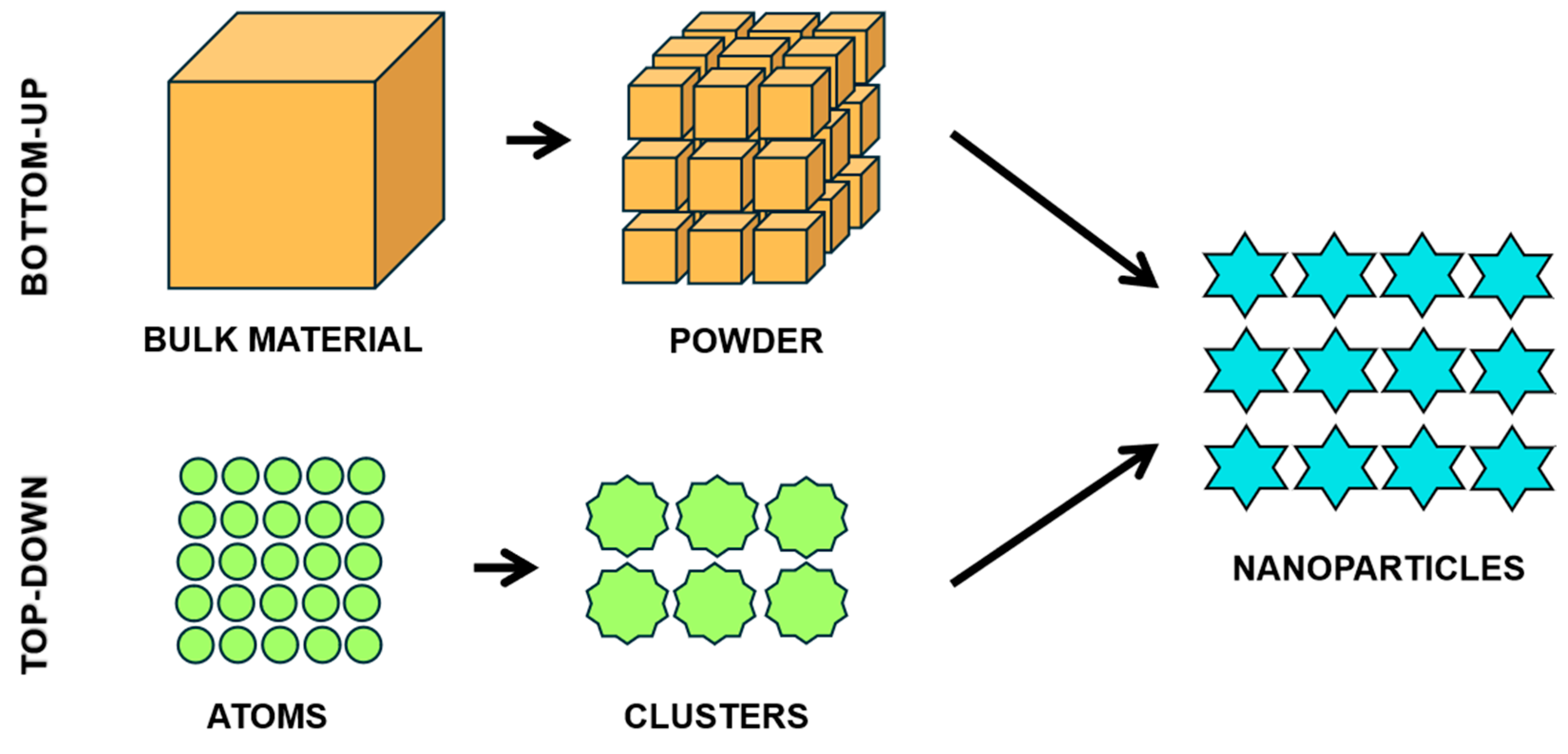
4. Nanoscience and Nanotechnology: SeNPs

Given the inherent duality of the element, especially when employed in its pure and inorganic form, there has been a growing interest in the application/production of selenium through nanotechnology. Recently, SeNPs have gained attention from the scientific community as a possible source of the element, due to their chemical stability, biocompatibility, and low toxicity when compared to other forms of the element, such as selenate and selenite ions [62]. Additionally, SeNPs are recognized for their unique physicochemical properties, which provide exceptional functionalities at the nanometric scale, such as potential as drug delivery vehicles, which may replace or enhance drug action [62,63,64]. The potentialities of the element, including to reduce toxicity, especially of the inorganic form, are related to the synthesis processes of SeNPs and, consequently, the smaller surface area per unit volume occupied by the NPs, in addition to being less interactive and releasing selenium more slowly [62].

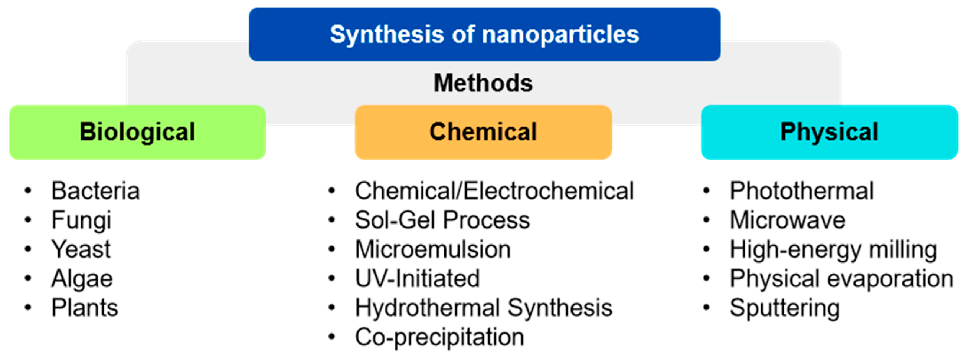
4.1. Preparation of SeNPs

The preparation of SeNPs is crucial for their efficacy in therapeutic applications, as it involves the precise control of factors such as the concentration of precursor, reducing and stabilizing agents, pH, reaction temperature, and preparation time, which determine essential properties of SeNPs such as size, polydispersity index (PDI), purity, zeta potential (ZP), surface functionality, composition, and different morphologies [62,65]. Regarding their composition, they can be classified as organic, containing some biological material, such as liposomal, micellar, and hydrogel polymeric NPs, and inorganic, containing chemical materials, such as metallic and metal oxide NPs [53]. Morphological studies reveal that SeNPs can take on different shapes, such as nanowire (NWs) [66], nanotube (NTs) [67], nanoneedle [68], nanorod (NRs) [69], nanoflowers [70], nanobelts [71], and nanospheres [72], depending on the reagents or solvents used, the distinct nucleation forms the NPs underwent, and the conditions of the medium they were subjected to during preparation [73,74]. Of these, nanospheres are the most prominent for their pharmacological and biological functionalities [74,75].
Considering the commercial potential and the wide range of application fields of NPs, another essential aspect related to synthesis is the preparation methods, aiming to ensure high yield and controlled quality [76]. Among the methods used for NP synthesis are (a) top-down approaches, which involve the reduction of material to nanostructures by physical and mechanical methods; and (b) bottom-up approaches, which involve the coalescence/construction/assembly of nanomaterials from atoms and molecules [23,77,78] (Figure 1).
Synthesis methods are frequently used to enhance the characteristics of NPs, such as size, stability, and crystallinity, and can be categorized into the following three preparation methods: (a) physical methods; (b) chemical methods; and (c) biological methods [40,75] (Figure 2). Physical and chemical methods are less prevalent due to their disadvantages compared to biological methods, which stand out for being environmentally safe and sustainable, as well as offering fast synthesis and high reproducibility [40]. However, studies present in indexed databases show the predominance of chemical methods over physical.
Physical (photothermal, microwave, high-energy milling, physical evaporation, and sputtering) and chemical (chemical/electrochemical, sol-gel, microemulsion, UV-initiated, and hydrothermal synthesis) methods, although extensively studied, exhibit significant limitations, including low energy efficiency, high costs, the use of multiple hazardous reagents, and the consequent generation of waste, making them less appealing. In contrast, biological methods stand out as an environmentally safe, non-toxic, and sustainable synthesis alternatives, offering advantages such as rapidity, high reproducibility, and the use of organisms such as bacteria, fungi, yeasts, algae, and plants in the synthesis process [40,79].

4.1.1. Chemical Synthesis Methods

Chemical synthesis methods are considered methodologically simple, as they do not require advanced technological instruments or the incubation of bio-organisms [74]. Nonetheless, they present challenges related to ecology and toxicology, as well as risks associated with the use of hazardous chemical compounds [40]. Among the chemical methods that can be applied for the synthesis of SeNPs are the sol-gel method, microemulsion, hydrothermal, polyol synthesis, vapor phase synthesis, and chemical vapor deposition, with the chemical reduction technique being the most prominent due to its simplicity of action [40,76].
Chemical reduction is the most common and widely recognized methodology in the literature, consisting of two main steps: reduction and stabilization. In the reduction step, reducing agents such as ascorbic acid (C6H8O6), acetic acid (CH3COOH), oxalic acid or ethanedioic acid (C2H2O4), glucose (C6H12O6), and L-cysteine (C3H7NO2S) transform selenium salts like sodium selenite (Na2SeO3), selenium tetrachloride (SeCl4), and sodium selenosulfate (Na2SeSO3) into SeNPs, adjusting reaction conditions to control the size and shape of the NPs [40]. In the stabilization step, stabilizers are employed to prevent NPs aggregation by creating repulsive forces that control NP growth, thereby influencing their size and final shape [80]. This step is of utmost importance, as unstabilized SeNPs tend to precipitate within a few days, losing their homogeneity and properties over time [74].

4.1.2. Physical Synthesis Methods

Physical methods, although they do not generate many chemical byproducts, demand more energy and special equipment [30,75,76]. Some of the most frequently used physical methods for generating NPs in general include electrodeposition, synthesis associated with photothermal and microwave methods [27,46], high-energy milling, physical evaporation, sputtering [76], and pulsed laser ablation (PLA) [74].
Among this set of techniques, the synthesis of SeNPs through microwaves is widely recognized as the most conventional approach for heating in laboratories, employing selenium salts in an aqueous solution. This technique offers benefits such as rapid and uniform heating and reaction, as well as higher reaction rates and reduced energy consumption [74]. Another widely used physical technique in the production of SeNPs is PLA. In this method, PLA is dissolved in deionized water, resulting in the transformation of selenium pellets into colloidal solutions. The electric charge present in the medium prevents the agglomeration of SeNPs, generating an electric charge on the surface of the particles [74].

4.1.3. Biological Synthesis Methods

Differing from chemical and physical approaches, the synthesis of SeNPs using bioagents is seen as a positive potential alternative to conventional methodologies. The green synthesis of SeNPs is achieved through the process of bioreduction and stabilization of precursor agents (sodium selenite, selenium tetrachloride, sodium selenosulfate, and others) from biomolecules present in biological organisms, avoiding the use of harmful reducing agents [40,81]. The observed reduction can be attributed to the induction caused by functional groups, such as amines and alkanes, present in metabolites like flavonoids, tannins, alkaloids, steroids, and terpenoids, resulting in red-colored SeNPs [74,79,82]. Additionally, phenolic compounds, polysaccharides, saponins, enzymes, proteins, and sugars can also play significant roles in this process [83].
Various classes of biological agents are investigated for their crucial roles in biosynthesis, including yeasts, actinobacteria, enzymes [82], fungi, protozoa [74], photosynthetic organisms such as plants and algae [83,84], and bacterial strains (anaerobic, aerobic, and anoxic) [74,83]. Additionally, studies report an increase in the use of aerobic and anaerobic bacteria due to their effectiveness in reducing inorganic selenium and rapidly forming SeNPs [36]. This capability was also investigated by Bafghi et al. (2021) [85] in pathogenic fungi, such as Aspergillus flavus and Candida albicans, highlighting the versatility of these biological processes.
The biosynthesis of SeNPs by Gram-positive and Gram-negative bacteria occurs as described in the following four phases: (1) transport of selenium oxyanions from sulfate transport proteins (permeases) into the cells; (2) reduction of selenium to amorphous red elemental selenium (Se0) by bacterial proteins, with subsequent release into the extracellular medium; (3) formation of SeNPs through the continuous reduction of selenium oxyanions to Se0; and (4) separation of SeNPs from bacterial cells and other cellular components by centrifugation and filtration. To optimize the process, it is essential to control factors such as temperature, rotation, the pH of the growth medium, incubation time, selenium salt concentration, minimum inhibitory concentration (MIC), and the reducing potential of the microorganisms [74].
When compared to fungi and bacteria, the use of plant extracts is widely addressed in the literature for the biosynthesis of SeNPs. This is due to their higher bioavailability, safety, and ease of manipulation, as well as the use of less toxic solvents and a more efficient production process [74]. The nanostructures can be prepared from leaves, shoots, fruits, seeds, or pulp of plants [83]. Another important point is the use of plant extracts derived from agricultural waste, such as fruit peels, which are increasingly valued due to their rich composition of bioactive compounds. These can include various functional groups such as lignin, proteins, lipids, hydrocarbons, and starch [37]. Additionally, this practice promotes sustainability by utilizing by-products that would otherwise be discarded, considering the abundance of such waste in agricultural countries [35,86].

5. Antimicrobial Activities of SeNPs Synthesized by Different Routes

According to the World Health Organization (WHO), the global spread of multidrug-resistant microorganisms (MDR) is considered one of the major concerns for society today. WHO estimates that, by 2050, around ten million deaths could occur annually due to infections caused by pathogenic microorganisms [87]. The main cause of the increase in antimicrobial resistance (AMR) in widely used therapies is the growing adaptation of microorganisms to the excessive use of these drugs [88].
Throughout evolution, bacteria and fungi have developed resistance to multiple antimicrobials, facilitated by both excessive use and genetic modifications, over the years [89,90]. In response to the challenge of combating microorganisms and the exponential increase in microbial resistance—considering historical changes such as enzymatic degradation, antibiotic modification, and alterations in bacterial cell wall permeability [89,91]—the concept of “human chemical evolution” emerges. In this context, humans have sought to develop safer, more effective, and more bioavailable therapeutic methods [90].
The antimicrobial activity of NPs is evidenced by several studies, highlighting their dependence on physicochemical properties such as size and surface charge [15,75,92], as the adherence of NPs to the cell walls and membranes of microorganisms can lead to the destruction of bacterial and cellular membranes, interruption of energy transfer, inhibition of enzymatic activity, and DNA synthesis [85,93].
Studies demonstrate that SeNPs can damage cellular structures, inducing the production of reactive oxygen species (ROS), and altering signal transduction mechanisms in certain biological organisms, such as fungi and bacteria, due to their physical, chemical, electrical, optical, magnetic, semiconducting, catalytic, and biological properties [85].

5.1. Biogenic SeNPs

5.1.1. Antifungal Properties

Due to their antifungal potential, recent studies have investigated the properties of green SeNPs, which utilize plants and microorganisms to reduce selenium ions to NPs, acting against various pathogenic species, and their potential is noteworthy.
A study conducted by El-Saadony et al. (2021) [94] synthesized SeNPs using Lactobacillus paracasei cells (HM1) (MW390875) (Lab-SeNPs), a Gram-positive bacterium found in human breast milk, to assess their potential against pathogenic fungi. The study compared the activity of Lab-SeNPs with chemically synthesized SeNPs (Che-SeNPs) at concentrations of 15 at 75 μg/mL. Agar Disk Diffusion tests showed that higher concentrations of Lab-SeNPs produced larger halos, with the largest reaching 29 mm. The MIC revealed that, except for C. albicans, which maintained an MIC of 55 µg/mL for both NPs, all other tested strains had a lower MIC with Lab-SeNPs than with Che-SeNPs. For Candida parapsilosis, Fusarium oxysporum, Fusarium solani, Candida krusei, Candida glabrata, and Candida tropicalis, Lab-SeNPs had MICs from 60 to 70 μg/mL, respectively, while Che-SeNPs had MICs from 65 to 80 μg/mL, respectively. To ensure complete fungal death, the minimum fungicidal concentration (MFC) was assessed, showing that Lab-SeNPs (130–180 μg/mL) caused greater mortality compared to Che-SeNPs (98–150 μg/mL). These results indicate higher antifungal efficacy of biosynthesized NPs compared to chemically synthesized ones (Table 1).
A study by Nile et al. (2023) [95] investigated the synthesis of SeNPs using Paenibacillus terreus (Pt-SeNPs), a Gram-positive bacterium, as a reducing agent, to evaluate the activity of these NPs, with or without nystatin—a widely used antifungal for topical and oral infections—against C. albicans in terms of growth inhibition, morphogenesis, and biofilm formation. The results showed that the antifungal activity of SeNPs is dose-dependent and limited, with growth inhibition below 50% at concentrations of 3.90–500 μg/mL and similar effects on biofilm formation. The activity of nystatin, both pure and in combinations, was also dose-dependent but still inferior to the antifungal activity of pure Pt-SeNPs. In contrast, Pt-SeNPs loaded with nystatin (SeNP@PVP_Nystatin) were effective at all tested concentrations, with more than 50% efficacy at the lowest concentration (3.90 μg/mL), for both antibiofilm action and antifungal activity. These results suggest that SeNP@PVP_Nystatin nanoconjugates may represent an effective strategy in combating fungal infections caused by C. albicans (Table 1).
Other studies address the synthesis of SeNPs from bacteria against C. albicans. A study conducted by Safaei et al. (2022) [15] biosynthesized SeNPs from Halomonas elongata (He-SeNPs), a Gram-negative aerobic γ-proteobacterium, by varying the incubation time and concentrations of glucose and sodium selenite (Na2SeO3), in order to evaluate their antifungal action against C. albicans using the colony-forming units (CFU) method. Nine synthesis conditions for He-SeNPs were evaluated. One of them inhibited over 70% of growth during 48 h, using the highest concentrations of He-SeNPs and glucose (0.8 and 7.5 mg/mL). The authors concluded that the positive result was due to the concentrations of He-SeNPs, glucose, and incubation time, estimating that, under ideal conditions, He-SeNPs inhibited approximately 72.01% of fungal growth (Table 1).
Bafghi et al. (2021) [85] synthesized SeNPs mediated by strains of C. albicans TIMML-1306 (Ca-SeNPs) and A. flavus TIMML-050 (Af-SeNPs), with the aim of investigating their potential and comparing them with commercial antifungals. Sodium selenate (Na2SeO4) was used for synthesis at various concentrations (0.125 to 64 µg/mL). The results showed that all tested strains were sensitive to the antifungals at concentrations ≤ 2 μg/mL, except for A. flavus, which was 100% resistant to itraconazole (ITC). The Ca-SeNPs and Af-SeNPs showed a minimum inhibitory concentration starting at 0.25 µg/mL. The combination of Ca-SeNPs + ITC was also evaluated, with sensitivity detected from 1 μg/mL for C. parapsilosis and 2 μg/mL for C. albicans. However, using 100% Ca-SeNPs, sensitivity was observed at the lowest concentration tested, 0.125 μg/mL (Table 2).
In the same year, the same authors [96] biosynthesized SeNPs and silver nanoparticles (AgNPs) using Nepeta extract (Ne-SeNPs) and Berberine (Be-AgNPs), respectively, and combined these NPs with the commercial antifungals ITC, amphotericin B (AMB), and anidulafungin (AFG), testing them against the same strains. To evaluate susceptibility, concentrations ranging from 0.125 to 64 µg/mL were tested for both NPs and medications. For C. albicans TIMML-1306, Ne-SeNPs (1.0 µg/mL) were more efficient at inhibiting growth compared to pure ITC (64 µg/mL), while the strains Aspergillus fumigatus TIMML-025 (A. fumigatus), C. parapsilosis ATCC-2201, C. albicans TIMML-491, and A. flavus TIMML-050 were sensitive to AMB and AFG at concentrations of 1 and 4 µg/mL, respectively; meanwhile, Be-AgNPs and Ne-SeNPs were effective at lower concentrations, namely 0.125 and 0.5 µg/mL, respectively. The combination of antifungals with ITC was evaluated, revealing that Be-AgNPs and Ne-SeNPs have similar activity with or without itraconazole, and that Ne-SeNPs + ITC and Be-NP + ITC were effective against A. flavus TIMML-050 at concentrations of 1 and 2 µg/mL, respectively, showing similar efficacy when used alone (Table 2).
Hashem et al. (2022) [97] synthesized SeNPs from the extract of Prickly Pear peel—Indian fig (PPPW-SeNPs)—and applied them against C. albicans and Cryptococcus neoformans. The antifungal capacity of PPPW-SeNPs was evaluated at different concentrations (1.95–2000 µg/mL) using the agar diffusion method, showing a dose-dependent relationship, with the highest concentration producing the largest inhibition halo, resulting in zones of 59.5 ± 0.7 mm and 50.2 ± 1.1 mm for C. albicans and C. neoformans, respectively. The minimum inhibitory concentration (MIC) test was performed with PPPW-SeNPs, isolated extract, and the combination of ampicillin/sulbactam/amphotericin B. Only the extract and the antibiotic combination did not demonstrate satisfactory efficacy, highlighting the superiority of SeNPs over these conventional treatments (Table 2).
Table 2. The antifungal activity of selenium nanoparticles (SeNPs) synthesized using the biogenic method using fungi and plants.
NPsSpeciesNP Size
(nm)
DLSColor NP
Solution
Evaluated
Microorganisms
MIC_SeNPsRef.
FUNGICa-SeNPsC. albicans TIMML-1306(XRD)
38
(PZ)
−19 mV
RedC. glabrata TIMML-1316
C. krusei TIMML-1321
C. glabrata TIMML-368
0.25–0.5 µg/mL[85]
C. albicans TIMML-1306
C. albicans TIMML-183
C. albicans TIMML-491
C. albicans TIMML-1291
<1 µg/mL
0.25 µg/mL
0.5 µg/mL
0.125–64 µg/mL
C. parapsilosis ATCC-22010.125–64 µg/mL
Af-SeNPsA. flavus TIMML-050(XRD)
37
(PZ)
−21 mV
RedA. fumigatus TIMML-025
A. flavus TIMML-050
1 µg/mL
PLANTSNe-SeNPsNepeta
(gender)
(SEM)
37–46
-RedC. albicans TIMML-1306
C. albicans TIMML-1291
C. albicans TIMML-491
1 µg/mL
1 µg/mL
0.5 µg/mL
[96]
A. flavus TIMML-050
C. tropicalis TIMML-1316
C. krusei TIMML-1321
0.125 µg/mL
0.125 µg/mL
0.125 µg/mL
PPPW-SeNPsPrickly Pear Peel Waste
(PPPW)
(TEM)
50–150
-ReddishC. albicans
C. neoformans
3.9 µg/mL
7.81 µg/mL
[97]
Dynamic light scattering (DLS); zeta potential (ZP); nanometer (nm); X-ray diffraction (XRD); transmission electron microscopy (TEM); scanning electron microscopy (SEM); minimum inhibitory concentration (MIC).

5.1.2. Antibacterial Properties

The potential of green SeNPs, derived from plants and bacterial species beneficial for human health, is also widely discussed, regarding their antibacterial activities against medically important bacteria (Table 3).
Abbas et al. (2021) [98] synthesized Sp-SeNPs (5 and 10 mM) from algae derived from Spirulina platensis, which were applied against Gram-negative bacteria—Salmonella abony, Escherichia coli and ten clinical isolates of Klebsiella pneumoniae. The results showed that the MICs of the SeNPs ranged from 25 to 200 µg/mL, with significant efficacy at 25 µg/mL. The most susceptible strains were S. albony NCTC 6017, followed by K. pneumoniae and E. coli, for 10 mM SeNPs. Among the K. pneumoniae isolates, 50% did not exhibit an inhibition zone for SeNPs at any sodium selenite concentration.
Bacteria beneficial for human health, such as probiotics, are also being investigated for the synthesis of SeNPs. An investigation of the production capacity of SeNPs using the probiotic Bacillus subtilis BSN313 (Bs-SeNPs) was carried out by Ullah et al. (2021) [36] at different concentrations (100, 150, and 200 µg/mL) and against both Gram-negative bacteria, namely E. coli ATCC 8739 and Pseudomonas aeruginosa ATCC 9027, and Gram-positive bacteria, namely Staphylococcus aureus ATCC 25923. The study indicated that Bs-SeNPs showed the formation of inhibition zones at the highest tested concentration after 24 h of incubation at 35 °C.
In another study, conducted by Alam et al. (2020) [99], strains of E. coli, P. aeruginosa, S. aureus, and K. pneumoniae were treated with SeNPs mediated by the probiotic bacterium Lactobacillus acidophilus (La-SeNPs) to assess their efficacy on planktonic cells and pre-formed biofilms. The activity of La-SeNPs was compared with gentamicin, a widely used antimicrobial agent. The results indicate that gentamicin has a lower MIC than La-SeNPs for E. coli and B. subtilis, whereas, for S. aureus, La-SeNPs (1.2 µg/mL) exhibited a lower MIC than gentamicin (0.5–2.0 µg/mL). For K. pneumoniae and P. aeruginosa, La-SeNPs demonstrated high efficiency, with a lower MIC (6.5 µg/mL and 4 µg/mL, respectively) compared to gentamicin (≥120 µg/mL and ≥180 µg/mL, respectively). The results for SeNPs against biofilms were concentration-dependent, showing especially high effectiveness against E. coli, S. aureus, and P. aeruginosa.
The antibacterial activity of SeNPs derived from fruits and agricultural residues is well established. Abou and Abbas (2023) [100] produced biogenic SeNPs from the aqueous extract of pomegranate peel (PPAE), with and without copper sulfate coating (PPAE-SeNPs@CuO and PPAE-SeNPs, respectively), against ten strains of Helicobacter pylori. Antibacterial activity was assessed using the agar diffusion test, with wells filled with 100 μg/mL of NPs in pre-inoculated media. The results showed that the inhibition zone of PPAE-SeNPs@CuO (11–15 mm) was larger than that of PPAE-SeNPs (0–9 mm). The MIC test demonstrated that all isolates were resistant to metronidazole (5 μg), clarithromycin (15 μg), levofloxacin (5 μg), amoxicillin/clavulanic acid (20/10 μg), tetracycline (30 μg), and amoxicillin (10 μg), whereas PPAE-CuO@SeNPs (8 μg/mL) was able to inhibit 100% of the multidrug-resistant strains.
A study by Khudier et al. (2023) [101] synthesized SeNPs from the fruit of Vaccinium arctostaphylos (L.) (Va-SeNPs) at different concentrations (500, 250, 125, 62.5, and 31.25 µg/mL) against S. aureus ATCC 29213, E. coli ATCC 25922, and Corynebacterium diphtheriae ATCC 13812. The analysis compared the use of Va-SeNPs with the pure extract and the antibiotic ciprofloxacin (positive control). The results showed that the actions of Va-SeNPs are dose-dependent, with inhibition zones exceeding 12 mm, larger at higher concentrations (500 µg/mL). Compared to the positive control, Va-SeNPs were more effective only at the concentration of 500 µg/mL for S. aureus, while the pure extract showed inhibition zones smaller than 12 mm.
PPPW-SeNPs were also used to evaluate antibacterial efficacy against E. coli, P. aeruginosa, B. subtilis, and S. aureus, at varying concentrations (1.95–2000 µg/mL), by Hashem et al. (2022) [97]. The activity of the PPPW-SeNPs was dose-dependent, with varying MICs for the tested strains, as follows: E. coli and P. aeruginosa (125 µg/mL), B. subtilis (62.5 µg/mL), and S. aureus (15.62 µg/mL). At 2000 µg/mL, PPPW-SeNPs showed inhibition zones of 26.5 ± 0.70 mm for E. coli, 24.4 ± 0.85 mm for P. aeruginosa, 30.7 ± 0.53 mm for B. subtilis, and 48.7 ± 1.06 mm for S. aureus. Tests with the pure extract and with ampicillin/sulbactam/amphotericin B (SAM/AMB) showed that the extract had no antibacterial activity, while SAM/AMB inhibited all strains except E. coli, with inhibition zones of 14.5 ± 0.5 mm for P. aeruginosa, 15.6 ± 0.4 mm for B. subtilis, and 10.1 ± 0.9 mm for S. aureus.
Table 3. The antibacterial activity of selenium nanoparticles (SeNPs) synthesized using the biogenic method using fungi and plants.
NPsSpeciesNP Size
(nm)
DLSColor NP
Solution
Evaluated
Microorganisms
MIC_SeNPsRef.
BACTERIABs-SeNPB. subtilis BSN313(DLS)
530
(TEM)
280–630
(PZ)
−26.9 mV
RedE. coli ATCC 8739
Staphylococcus aureus ATCC 9027
Pseudomonas aeruginosa ATCC 25923
200 µg/mL[36]
La-SeNPL. acidophilus(DLS)
34.13
(TEM)
2–15
(PDI)
0.28
(PZ)
+37.86 mV
Reddish-brownE. coli
S. aureus
B. subtilis
P. aeruginosa
K. pneumoniae
9.4 µg/mL
1.2 µg/mL
3.5 µg/mL
6.5 µg/mL
4 µg/mL
[99]
ALGAESp-SeNPSpirulina platensis(TEM)
79.40 ± 44.26
>100 nm (53.33%)
(PZ)
−32.9 ± 8.12 mV
Red orangeS. abony NCTC 6017
E. coli ATCC 8739
K. pneumonia ATCC700603
25 µg/mL[98]
PLANTSPPPW-SeNPPrickly Pear Peel Waste
(PPPW)
(TEM)
50–150
-ReddishE. coli
P. aeruginosa
125 µg/mL[97]
B. subtilis
S. aureus
62.5 µg/mL
15.62 µg/mL
PPAE-SeNPs
PPAE-SeNPs@CuO
Punica granatum(HRTEM)
1.97–10.56
(HRTEM)
92.18
-Red
Orange
H. pylori8 µg/mL[100]
Va-SeNPV. Arctostaphylos (L.)(DLS)
246.2 ± 4.51
(FESEM)
50 ± 1.23
(PDI)
0.267
(PZ)
−11.5 mV ± 1.24
RedS. aureus
E. coli
C. diphtheriae
500—31.25 µg/mL[101]
Dynamic light scattering (DLS); zeta potential (ZP); nanometer (nm); transmission electron microscopy (TEM); field emission scanning electron microscopy (FESEM); high-resolution transmission electron microscopy (HRTEM); minimum inhibitory concentration (MIC).
These studies confirm that SeNPs, synthesized from various natural reducing agents such as bacteria, fungi, fruit extracts, and agricultural residues, exhibit significant antifungal activity against several pathogenic species, and are often more effective than commercial antifungals, especially when used in combination.

5.2. Chemical SeNPs

Dorazilová et al. (2020) [102] demonstrate the development of polymeric scaffolds based on collagen–chitosan with SeNPs for application in infected and chronic wounds, acting against S. aureus and methicillin-resistant S. aureus (MRSA) and Staphylococcus epidermidis. In this study, the two SeNPs were prepared from the reduction of sodium selenite (Na₂SeO₃): one with carboxymethyl cellulose (SeCE), using mercaptopropionic acid in the presence of carboxymethyl cellulose, and the other with chitosan (SeCH), using mercaptopropionic acid in the presence of chitosan. The following three concentrations were evaluated: 1, 5, and 10 ppm. The study results demonstrate that the scaffolds prepared with SeNPs (antibacterial agents) increased the material’s potential, showing that, even at low concentrations of SeNPs, such as 5 ppm, they showed considerable inhibition activity against the bacterial strains (Table 4).
Antibacterial activity in wounds was also evaluated by Golmohammadi et al. (2020) [103]. The authors developed a selenium–chitosan–mupirocin nanohybrid system (M-SeNPs-CCH) against S. aureus infection in diabetic rats. Wound healing evaluation was performed macroscopically and microscopically. Macroscopically, the authors observed that wound size contraction decreased with more significance in treatments with the presence of SeNPs over 21 days. A microscopy analysis of the wound tissue confirmed that the treatment with nanoparticles led to an increase in collagenization and epidermization (Table 4).
A study by Huang et al. (2020) [104] evaluated the multimodal capability of SeNPs coated with the antimicrobial polypeptide ε-poly-l-lysine (SeNP-ε-PL) against Gram-negative and Gram-positive bacteria. The SeNPs were synthesized from the precursor Na2SeO3. The MIC and minimum bactericidal concentration (MBC) evaluations compared the use of SeNP-ε-PL with pure SeNPs and ε-PL. For Gram-positive bacteria, the authors observed that SeNP-ε-PL exhibited similar or lower MICs compared to the treatment with pure isolates, while Gram-negative bacteria showed a significant reduction in MIC, like the use of pure ε-PL. MBC results, assessed via CFU tests, demonstrated that SeNPs displayed greater activity compared to the use of pure ε-PL, whereas the opposite was observed for Gram-negatives. SeNP-ε-PL exhibited strong antibacterial properties against all strains (Table 4).

5.3. Physical SeNPs

Geoffrion et al. (2020) [105] used the pulsed laser ablation in liquid (PLAL) technique to synthesize SeNPs. In this method, selenium pellets are irradiated with a laser while immersed in deionized water, followed by an ice bath to control the size and aggregation of the particles. The SeNPs were tested against Gram-negative bacterial strains: multidrug-resistant E. coli (MDR-EC) and P. aeruginosa, as well as Gram-positive strains: MRSA and S. epidermidis. The results demonstrated that SeNPs exhibited antibacterial properties in the concentration range of 0.05 to 25 ppm, with more pronounced inhibition in Gram-negative bacteria and higher MICs for Gram-positive bacteria. The results also indicated that the inhibition of MDR-EC was dose-dependent, with a concentration of 1 ppm being sufficient to cause significant inhibition, while at the concentration of 25 ppm, a strong bacterial inhibition was observed in all tested strains (Table 4).
Menazea et al. (2020) [106] used the laser ablation method to synthesize SeNPs from polyvinyl alcohol (PVA) and chitosan (PVA/chitosan/SeNP), aiming to evaluate the antibacterial activity of PVA/chitosan/SeNP and PVA/chitosan on agar plates against E. coli, P. aeruginosa, S. aureus, and B. subtilis. The PVA/chitosan/SeNP samples were subjected to ablation for 10 and 30 min, while the other compounds were not exposed. The results demonstrated that the NPs exposed to ablation exhibited higher antibacterial activity compared to the PVA/chitosan mixture, with longer exposure times (30 min) showing greater activity against all tested strains (Table 4).

6. Antimicrobial Success of SeNPs: Mechanism of Action

The success of the antimicrobial activity of SeNPs, whether isolated or combined with widely used medical drugs, regardless of the synthesis method, against different species of fungi and bacteria, is associated with the antimicrobial mechanisms of action of these particles. Although few studies have thoroughly explored these mechanisms, the following are generally highlighted: (1) cell wall and membrane damage; (2) intracellular penetration and damage; and (3) induction of oxidative stress [107,108].
The damage to the cell wall and membrane by SeNPs is considered the main antimicrobial mechanism of action [107]. The cell wall, as well as the plasma membrane, are recognized as protective barriers against environmental threats, maintaining homeostatic balance and allowing intracellular nutrient transport, which is crucial for the antimicrobial action of nanoparticles [107,109,110]. Studies report that the interaction of SeNPs with the cell wall and membrane begins with the adhesion of the particles, through the electrostatic attraction between the negatively charged microbial membrane and the positively or less negatively charged SeNPs. This interaction causes structural and morphological changes in the membrane, resulting in depolarization, the rupture of cellular integrity, compromised permeability, the disruption of respiratory functions, rupture of the cell wall, and, consequently, cell death [107,111,112,113].
Upon contact with the plasma membrane, SeNPs also affect metabolic functions, which can be enhanced when DNA and protein damage occurs [107]. The interaction of SeNPs with the surface of microorganisms can also trigger cellular oxidative stress upon absorption. Microorganisms attempt to overcome this stress through defense mechanisms; however, if the stress is relatively high, damage caused by reactive oxygen species (ROS) and free radicals can affect the cell wall and biomolecules present in the organism, such as proteins, lipids, and DNA. Studies show that ROS production is associated with cell death and, when combined with selenium in the form of nanoparticles, this action can be enhanced, explaining the high antimicrobial capacity of SeNPs [107].

7. Conclusions

During the construction of the review for the search of studies on SeNPs applications against fungi and bacteria, a large number of studies were observed, addressing that, among the various synthesis methods, the biogenic route is the most discussed in the literature. This is probably due to the higher activity of SeNPs mediated by biological organisms (plants, seeds, algae, bacteria, and fungi) against microorganisms of therapeutic interest, in addition to the fact that the use of biological agents during the synthesis enables the reduction of toxicity levels in applications and an increase in efficiency compared to other synthesis methods. The information presented in this review will serve as a basis for future research and the development of new applications of SeNPs in various microbiological contexts, facilitating the investigation of antimicrobial activities, as well as the knowledge about selenium and nanoparticles.

Author Contributions

Conceptualization, T.P.d.S.O., A.K.O.L. and L.A.M.; methodology, T.P.d.S.O., A.K.O.L. and L.A.M.; formal analysis and investigation, T.P.d.S.O.; resources, L.A.M.; data curation, A.K.O.L. and L.A.M.; writing—original draft preparation, T.P.d.S.O.; writing—review and editing, T.P.d.S.O., A.K.O.L. and L.A.M.; and funding acquisition, L.A.M. All authors have read and agreed to the published version of the manuscript.

Funding

This research received no external funding.

Informed Consent Statement

Not applicable.

Data Availability Statement

The data presented in this manuscript were derived from published studies/results and are publicly available.

Acknowledgments

This study is based on the master’s research of the first author of this review manuscript and was supported financially by the Coordination for the Improvement of Higher Education Personnel (CAPES), the National Council for Scientific and Technological Development (CNPq), and by the infrastructure provided by the University of Brasília (UnB). The authors are grateful to the Brazilian research institutions that contributed directly and indirectly to the development of this review.

Conflicts of Interest

The author Alan Kelbis Oliveira Lima is an employee of the company Brazilian Agricultural Research Corporation (EMBRAPA). The remaining authors declare that the research was conducted in the absence of any commercial or financial relationships that could be construed as a potential conflict of interest.

References

  1. Trofast, J. Berzelius’ discovery of selenium. Chem. Int. 2011, 33, 16. [Google Scholar]
  2. Murugesan, G.; Nagaraj, K.; Sunmathi, D.; Subramani, K. Methods involved in the synthesis of selenium nanoparticles and their different applications-a review. Eur. J. Biomed. 2019, 6, 189–194. [Google Scholar]
  3. Wisniak, J. Jöns Jacob Berzelius a guide to the perplexed chemist. Chem. Educ. 2000, 5, 343–350. [Google Scholar] [CrossRef]
  4. Reich, H.J.; Hondal, R.J. Why nature chose selenium. ACS Chem. Biol. 2016, 11, 821–841. [Google Scholar] [CrossRef]
  5. Seixas, T.G.; do AKehrig, H. O selênio no meio ambiente. Oecologia Bras. 2007, 11, 264–276. [Google Scholar] [CrossRef]
  6. Boyd, R. Selenium stories. Nat. Chem. 2011, 3, 570. [Google Scholar] [CrossRef]
  7. Garousi, F. The toxicity of different selenium forms and compounds—Review. Acta Agrar. Debreceniensis 2015, 64, 33–38. [Google Scholar] [CrossRef]
  8. Perrone, D.; Monteiro, M.; Nunes, J.C. The Chemistry of Selenium; Royal Society of Chemistry: London, UK, 2015. [Google Scholar] [CrossRef]
  9. Gandin, V.; Khalkar, P.; Braude, J.; Fernandes, A.P. Organic selenium compounds as potential chemotherapeutic agents for improved cancer treatment. Free Radic. Biol. Med. 2018, 127, 80–97. [Google Scholar] [CrossRef]
  10. Wang, K.; Fang, Q.; He, P.; Tu, Y.; Liu, Z.; Li, B. Unveiling the potential of selenium-enriched tea: Compositional profiles, physiological activities, and health benefits. Trends Food Sci. Technol. 2024, 145, 104356. [Google Scholar] [CrossRef]
  11. Bisht, N.; Phalswal, P.; Khanna, P.K. Selenium nanoparticles: A review on synthesis and biomedical applications. Mater. Adv. 2022, 3, 1415–1431. [Google Scholar] [CrossRef]
  12. Handy, D.E.; Joseph, J.; Loscalzo, J. Selenium, a micronutrient that modulates cardiovascular health via redox enzymology. Nutrients 2021, 13, 3238. [Google Scholar] [CrossRef] [PubMed]
  13. Abdel-Moneim AM, E.; El-Saadony, M.T.; Shehata, A.M.; Saad, A.M.; Aldhumri, S.A.; Ouda, S.M.; Mesalam, N.M. Antioxidant and antimicrobial activities of Spirulina platensis extracts and biogenic selenium nanoparticles against selected pathogenic bacteria and fungi. Saudi J. Biol. Sci. 2022, 29, 1197–1209. [Google Scholar] [CrossRef] [PubMed]
  14. Rosaiah, G.; Mangamuri, U.K.; Sikharam, A.S.; Devaraj, K.; Kalagatur, N.K.; Kadirvelu, K. Biosynthesis of selenium nanoparticles from Annona muricata fruit aqueous extract and investigation of their antioxidant and antimicrobial potentials. Curr. Trends Biotechnol. Pharm. 2022, 16, 101–107. [Google Scholar]
  15. Safaei, M.; Mozaffari, H.R.; Moradpoor, H.; Imani, M.M.; Sharifi, R.; Golshah, A. Optimization of green synthesis of selenium nanoparticles and evaluation of their antifungal activity against oral Candida albicans infection. Adv. Mater. Sci. Eng. 2022, 2022, 1376998. [Google Scholar] [CrossRef]
  16. Speckmann, B.; Grune, T. Epigenetic effects of selenium and their implications for health. Epigenetics 2015, 10, 179–190. [Google Scholar] [CrossRef]
  17. Bjørklund, G.; Shanaida, M.; Lysiuk, R.; Antonyak, H.; Klishch, I.; Shanaida, V.; Peana, M. Selenium: An antioxidant with a critical role in anti-aging. Molecules 2022, 27, 6613. [Google Scholar] [CrossRef]
  18. Bhattacharjee, A.; Basu, A.; Bhattacharya, S. Selenium nanoparticles are less toxic than inorganic and organic selenium to mice in vivo. Nucleus 2019, 62, 259–268. [Google Scholar] [CrossRef]
  19. Durán, N.; Mattoso, L.H.C.; Morais, P.C.d. Textbook of Nanotecnologia: Introdução, Preparação e Caracterização de Nanomateriais e Exemplos de Aplicação; Artliber: São Paulo, Brazil, 2006; pp. 87–92. [Google Scholar]
  20. Bayda, S.; Adeel, M.; Tuccinardi, T.; Cordani, M.; Rizzolio, F. The history of nanoscience and nanotechnology: From chemical–physical applications to nanomedicine. Molecules 2019, 25, 112. [Google Scholar] [CrossRef]
  21. Madkour, L.H.; Madkour, L.H. Introduction to nanotechnology (NT) and nanomaterials (NMs). In Nanoelectronic Materials: Fundamentals and Applications; Springer: Cham, Switzerland, 2019; pp. 1–47. [Google Scholar] [CrossRef]
  22. Ojha, B.; Jain, V.K.; Mehra, N.K.; Jain, K. Nanotechnology: Introduction and basic concepts. In Dendrimers in Nanomedicine; CRC Press: Boca Raton, FL, USA, 2021; pp. 1–17. [Google Scholar]
  23. Selmani, A.; Kovačević, D.; Bohinc, K. Nanoparticles: From synthesis to applications and beyond. Adv. Colloid Interface Sci. 2022, 303, 102640. [Google Scholar] [CrossRef]
  24. Khalil, H.S.; Maulu, S.; Verdegem, M.; Abdel-Tawwab, M. Embracing nanotechnology for selenium application in aquafeeds. Rev. Aquac. 2023, 15, 112–129. [Google Scholar] [CrossRef]
  25. Gunti, L.; Dass, R.S.; Kalagatur, N.K. Phytofabrication of selenium nanoparticles from Emblica officinalis fruit extract and exploring its biopotential applications: Antioxidant, antimicrobial, and biocompatibility. Front. Microbiol. 2019, 10, 931. [Google Scholar] [CrossRef] [PubMed]
  26. Salem, M.F.; Abd-Elraoof, W.A.; Tayel, A.A.; Alzuaibr, F.M.; Abonama, O.M. Antifungal application of biosynthesized selenium nanoparticles with pomegranate peels and nanochitosan as edible coatings for citrus green mold protection. J. Nanobiotechnol. 2022, 20, 182. [Google Scholar] [CrossRef] [PubMed]
  27. Salem, S.S.; Hammad, E.N.; Mohamed, A.A.; El-Dougdoug, W. A comprehensive review of nanomaterials: Types, synthesis, characterization, and applications. Biointerface Res. Appl. Chem. 2022, 13, 41. [Google Scholar] [CrossRef]
  28. Xiao, X.; Deng, H.; Lin, X.; Ali AS, M.; Viscardi, A.; Guo, Z.; Qiao, L.; He, Y.; Han, J. Selenium nanoparticles: Properties, preparation methods, and therapeutic applications. Chem.-Biol. Interact. 2023, 378, 110483. [Google Scholar] [CrossRef] [PubMed]
  29. Liu, S.; Wei, W.; Wang, J.; Chen, T. Theranostic applications of selenium nanomedicines against lung cancer. J. Nanobiotechnol. 2023, 21, 96. [Google Scholar] [CrossRef]
  30. Chen, N.; Yao, P.; Zhang, W.; Zhang, Y.; Xin, N.; Wei, H.; Zhang, T.; Zhao, C. Selenium nanoparticles: Enhanced nutrition and beyond. Crit. Rev. Food Sci. Nutr. 2023, 63, 12360–12371. [Google Scholar] [CrossRef]
  31. Niranjan, R.; Zafar, S.; Lochab, B.; Priyadarshini, R. Synthesis and characterization of sulfur and sulfur-selenium nanoparticles loaded on reduced graphene oxide and their antibacterial activity against gram-positive pathogens. Nanomaterials 2022, 12, 191. [Google Scholar] [CrossRef]
  32. Galan-Chilet, I.; Grau-Perez, M.; De Marco, G.; Guallar, E.; Martin-Escudero, J.C.; Dominguez-Lucas, A.; Gonzalez-Manzano, I.; Lopez-Izquierdo, R.; Briongos-Figuero, L.S.; Redon, J.; et al. A gene-environment interaction analysis of plasma selenium with prevalent and incident diabetes: The Hortega study. Redox Biol. 2017, 12, 798–805. [Google Scholar] [CrossRef]
  33. Maiyo, F.; Singh, M. Selenium nanoparticles: Potential in cancer gene and drug delivery. Nanomedicine 2017, 12, 1075–1089. [Google Scholar] [CrossRef]
  34. Kopel, J.; Fralick, J.; Reid, T.W. The potential antiviral effects of selenium nanoparticles and coated surfaces. Antibiotics 2022, 11, 1683. [Google Scholar] [CrossRef]
  35. Iqbal, M.S.; Abbas, K.; Qadir, M.I. Synthesis, characterization and evaluation of biological properties of selenium nanoparticles from Solanum lycopersicum. Arab. J. Chem. 2022, 15, 103901. [Google Scholar] [CrossRef]
  36. Ullah, A.; Yin, X.; Wang, F.; Xu, B.; Mirani, Z.A.; Xu, B.; Chan, M.W.H.; Ali, A.; Usman, M.; Ali, N.; et al. Biosynthesis of selenium nanoparticles (via Bacillus subtilis BSN313), and their isolation, characterization, and bioactivities. Molecules 2021, 26, 5559. [Google Scholar] [CrossRef] [PubMed]
  37. Aziz, M.; Aziz, R.; Rafiq, M.T.; Abbasi, M.; Taneez, M.; Azhar, M.U.; El Askary, A.; Elesawy, B.H.; Eed, E.M.; Khalifa, A.S.; et al. Qayyum, A. Efficient Removal of Lead and Chromium from Aqueous Media Using Selenium Based Nanocomposite Supported by Orange Peel. Front. Environ. Sci. 2022, 10, 94. [Google Scholar] [CrossRef]
  38. Hardy, G.; Hardy, I.; Manzanares, W. Selenium supplementation in the critically ill. Nutr. Clin. Pract. 2012, 27, 21–33. [Google Scholar] [CrossRef]
  39. Kieliszek, M.; Błażejak, S. Selenium: Significance, and outlook for supplementation. Nutrition 2013, 29, 713–718. [Google Scholar] [CrossRef]
  40. Ruiz-Fresneda, M.A.; Staicu, L.C.; Lazuén-López, G.; Merroun, M.L. Allotropy of selenium nanoparticles: Colourful transition, synthesis, and biotechnological applications. Microb. Biotechnol. 2023, 16, 877–892. [Google Scholar] [CrossRef]
  41. Bouroushian, M.; Bouroushian, M. Chalcogens and metal chalcogenides. In Electrochemistry of Metal Chalcogenides; Springer: Berlin/Heidelberg, Germany, 2010; pp. 1–56. [Google Scholar] [CrossRef]
  42. Scopigno, T.; Steurer, W.; Yannopoulos, S.N.; Chrissanthopoulos, A.; Krisch, M.; Ruocco, G.; Wagner, T. Vibrational dynamics and surface structure of amorphous selenium. Nat. Commun. 2011, 2, 195. [Google Scholar] [CrossRef]
  43. Kuršvietienė, L.; Mongirdienė, A.; Bernatonienė, J.; Šulinskienė, J.; Stanevičienė, I. Selenium Anticancer Properties and Impact on Cellular Redox Status. Antioxidants 2020, 9, 80. [Google Scholar] [CrossRef]
  44. Oldfield, J.E. The two faces of selenium. J. Nutr. 2015, 117, 2002–2008. [Google Scholar] [CrossRef]
  45. Pecoraro, B.M.; Leal, D.F.; Frias-De-Diego, A.; Browning, M.; Odle, J.; Crisci, E. The health benefits of selenium in food animals: A review. J. Anim. Sci. Biotechnol. 2022, 13, 58. [Google Scholar] [CrossRef]
  46. Tan, J.A.; Zhu, W.; Wang, W.; Li, R.; Hou, S.; Wang, D.; Yang, L. Selenium in soil and endemic diseases in China. Sci. Total Environ. 2002, 284, 227–235. [Google Scholar] [CrossRef] [PubMed]
  47. Kushwaha, A.; Goswami, L.; Lee, J.; Sonne, C.; Brown, R.J.; Kim, K.H. Selenium in soil-microbe-plant systems: Sources, distribution, toxicity, tolerance, and detoxification. Crit. Rev. Environ. Sci. Technol. 2022, 52, 2383–2420. [Google Scholar] [CrossRef]
  48. Sun, Y.; Wang, Z.; Gong, P.; Yao, W.; Ba, Q.; Wang, H. Review on the health-promoting effect of adequate selenium status. Front. Nutr. 2023, 10, 1136458. [Google Scholar] [CrossRef] [PubMed]
  49. Coppinger, R.J.; Diamond, A.M. Selenium deficiency and human disease. In Selenium: Its Molecular Biology and Role in Human Health; Springer US: Boston, MA, USA, 2001; pp. 219–233. [Google Scholar] [CrossRef]
  50. Rayman, M.P. Selenium intake, status, and health: A complex relationship. Hormones 2020, 19, 9–14. [Google Scholar] [CrossRef]
  51. Zwolak, I.; Zaporowska, H. Selenium interactions and toxicity: A review: Selenium interactions and toxicity. Cell Biol. Toxicol. 2012, 28, 31–46. [Google Scholar] [CrossRef]
  52. Chen, N.; Zhao, C.; Zhang, T. Selenium transformation and selenium-rich foods. Food Biosci. 2021, 40, 100875. [Google Scholar] [CrossRef]
  53. Burk, R.F. (Ed.) Selenium in Biology and Human Health; Springer: New York, NY, USA, 1994; pp. 14–221. [Google Scholar]
  54. Terry, N.; Zayed, A.M.; De Souza, M.P.; Tarun, A.S. Selenium in higher plants. Annu. Rev. Plant Biol. 2000, 51, 401–432. [Google Scholar] [CrossRef]
  55. Imran, M.; Chen, Z.; Mehmood, A.; Rukh, S.; Weixie, W.; Asghar, W.; Iftikhar, F. Distribution of selenium in soils and human health. In Selenium and Human Health; IntechOpen: London, UK, 2023. [Google Scholar]
  56. Schwarz, K.; Foltz, C.M. Selenium as an integral part of factor 3 against dietary necrotic liver degeneration. Nutrition 1999, 15, 255. [Google Scholar] [CrossRef]
  57. Pinsent, J. The need for selenite and molybdate in the formation of formic dehydrogenase by members of the coli-aerogenes group of bacteria. Biochem. J. 1954, 57, 10. [Google Scholar] [CrossRef]
  58. Patterson, E.L.; Milstrey, R.; Stokstad EL, R. Effect of selenium in preventing exudative diathesis in chicks. Proc. Soc. Exp. Biol. Med. 1957, 95, 617–620. [Google Scholar] [CrossRef]
  59. Institute of Medicine. Dietary Reference Intakes for Vitamin C, Vitamin E, Selenium, and Carotenoids; The National Academies Press: Washington, DC, USA, 2000. [Google Scholar] [CrossRef]
  60. Senthamarai, M.D.; Hillary, V.E.; Rajan, M.R.; Ceasar, S.A. Biosynthesis of selenium nanoparticles and its biological applications: A systematic review. Nano Struct. Nano Objects 2024, 39, 101261. [Google Scholar] [CrossRef]
  61. Kieliszek, M. Selenium–fascinating microelement, properties and sources in food. Molecules 2019, 24, 1298. [Google Scholar] [CrossRef] [PubMed]
  62. Skalickova, S.; Milosavljevic, V.; Cihalova, K.; Horky, P.; Richtera, L.; Adam, V. Selenium nanoparticles as a nutritional supplement. Nutrition 2017, 33, 83–90. [Google Scholar] [CrossRef]
  63. Karthik, K.K.; Cheriyan, B.V.; Rajeshkumar, S.; Gopalakrishnan, M. A review on selenium nanoparticles and their biomedical applications. Biomed. Technol. 2024, 6, 61–74. [Google Scholar] [CrossRef]
  64. Wang, H.; Zhang, J.; Yu, H. Elemental selenium at nano size possesses lower toxicity without compromising the fundamental effect on selenoenzymes: Comparison with selenomethionine in mice. Free Radic. Biol. Med. 2007, 42, 1524–1533. [Google Scholar] [CrossRef]
  65. Wong, C.W.; Chan, Y.S.; Jeevanandam, J.; Pal, K.; Bechelany, M.; Abd Elkodous, M.; El-Sayyad, G.S. Response surface methodology optimization of mono-dispersed MgO nanoparticles fabricated by ultrasonic-assisted sol–gel method for outstanding antimicrobial and antibiofilm activities. J. Clust. Sci. 2020, 31, 367–389. [Google Scholar] [CrossRef]
  66. Agilan, S.; Velauthapillai, D.; Muthukumarasamy, N.; Thambidurai, M.; Senthil, T.S.; Balasundaraprabhu, R. Synthesis and characterization of selenium nanowires. Int. Sch. Res. Not. 2011, 2011, 589073. [Google Scholar] [CrossRef]
  67. Xi, G.; Xiong, K.; Zhao, Q.; Zhang, R.; Zhang, H.; Qian, Y. Nucleation− dissolution− recrystallization: A new growth mechanism for t-selenium nanotubes. Cryst. Growth Des. 2006, 6, 577–582. [Google Scholar] [CrossRef]
  68. Nagar, D.N.; Ghosh, N.N.; Braganca, J.M. Green synthesis of selenium nanospheres and nanoneedles by halophilic archaea. Appl. Nanosci. 2022, 12, 3983–3994. [Google Scholar] [CrossRef]
  69. Sawant, V.J.; Sawant, V.J. Biogenic capped selenium nano rods as naked eye and selective hydrogen peroxide spectrometric sensor. Sens. Bio Sens. Res. 2020, 27, 100314. [Google Scholar] [CrossRef]
  70. Sowmiya, P.; Dhas, T.S.; Inbakandan, D.; Mani, R.; Natarajan, A.; Dharani, G.; Govindaraju, K.; Kannan, M.; Velu, K.; Kumar, C.M.V. Antagonistic activity of a novel chitosan-selenium nanoflower against common aquaculture pathogen Aeromonas caviae. Aquac. Int. 2023, 31, 3109–3123. [Google Scholar] [CrossRef]
  71. Shanthappa, R.; Ankinapalli, O.R.; Kakarla, A.K.; Narsimulu, D.; Bandi, H.; Syed, W.A.; Yu, J.S. Selenium incorporated sodium vanadate nanobelts as high-performance electrode material for long-lasting aqueous zinc-ion batteries and supercapacitors. Chem. Eng. J. 2023, 476, 146777. [Google Scholar] [CrossRef]
  72. Shah, V.; Medina-Cruz, D.; Vernet-Crua, A.; Truong, L.B.; Sotelo, E.; Mostafavi, E.; González, M.U.; García-Martín, J.M.; Cholula-Díaz, J.L.; Webster, T.J. Pepper-Mediated Green Synthesis of Selenium and Tellurium Nanoparticles with Antibacterial and Anticancer Potential. J. Funct. Biomater. 2022, 14, 24. [Google Scholar] [CrossRef] [PubMed]
  73. Yang, Z.; Zuo, Y.; Dai, L.; Zhang, L.; Yu, Y.; Zhou, L. Effect of ultrasonic-induced selenium crystallization behavior during selenium reduction. Ultrason. Sonochem. 2023, 95, 106392. [Google Scholar] [CrossRef]
  74. Ferro, C.; Florindo, H.F.; Santos, H.A. Selenium nanoparticles for biomedical applications: From development and characterization to therapeutics. Adv. Healthc. Mater. 2021, 10, 2100598. [Google Scholar] [CrossRef]
  75. Ashraf, H.; Cossu, D.; Ruberto, S.; Noli, M.; Jasemi, S.; Simula, E.R.; Sechi, L.A. Latent Potential of Multifunctional Selenium Nanoparticles in Neurological Diseases and Altered Gut Microbiota. Materials 2023, 16, 699. [Google Scholar] [CrossRef]
  76. Dhand, C.; Dwivedi, N.; Loh, X.J.; Ying AN, J.; Verma, N.K.; Beuerman, R.W.; Lakshminarayanan, R.; Ramakrishna, S. Methods and strategies for the synthesis of diverse nanoparticles and their applications: A comprehensive overview. RSC Adv. 2015, 5, 105003–105037. [Google Scholar] [CrossRef]
  77. Shnoudeh, A.J.; Hamad, I.; Abdo, R.W.; Qadumii, L.; Jaber, A.Y.; Surchi, H.S.; Alkelany, S.Z. Synthesis, characterization, and applications of metal nanoparticles. In Biomaterials and Bionanotechnology; Academic Press: Cambridge, MA, USA, 2019; pp. 527–612. [Google Scholar] [CrossRef]
  78. Sarkar, J.; Mridha, D.; Davoodbasha, M.A.; Banerjee, J.; Chanda, S.; Ray, K.; Roychowdhury, T.; Acharya, K.; Sarkar, J. A state-of-the-art systemic review on selenium nanoparticles: Mechanisms and factors influencing biogenesis and its potential applications. Biol. Trace Elem. Res. 2023, 201, 5000–5036. [Google Scholar] [CrossRef]
  79. Ikram, M.; Javed, B.; Raja, N.I.; Mashwani, Z.U.R. Biomedical Potential of Plant-Based Selenium Nanoparticles: A Comprehensive Review on Therapeutic and Mechanistic Aspects. Int. J. Nanomed. 2021, 16, 249–268. [Google Scholar] [CrossRef]
  80. Song, X.; Chen, Y.; Sun, H.; Liu, X.; Leng, X. Physicochemical stability and functional properties of selenium nanoparticles stabilized by chitosan, carrageenan, and gum Arabic. Carbohydr. Polym. 2021, 255, 117379. [Google Scholar] [CrossRef]
  81. Indhira, D.; Aruna, A.; Manikandan, K.; Albeshr, M.F.; Alrefaei, A.F.; Vinayagam, R.; Kathirvel, A.; Priyan, S.R.; Kumar, G.S.; Srinivasan, R. Antimicrobial and Photocatalytic Activities of Selenium Nanoparticles Synthesized from Elaeagnus indica Leaf Extract. Processes 2023, 11, 1107. [Google Scholar] [CrossRef]
  82. Yilmaz, M.T.; Ispirli, H.; Taylan, O.; Dertli, E. A green nano-biosynthesis of selenium nanoparticles with Tarragon extract: Structural, thermal, and antimicrobial characterization. LWT 2021, 141, 110969. [Google Scholar] [CrossRef]
  83. Pyrzynska, K.; Sentkowska, A. Biosynthesis of selenium nanoparticles using plant extracts. J. Nanostruct. Chem. 2021, 12, 467–480. [Google Scholar] [CrossRef]
  84. Mikhailova, E.O. Selenium Nanoparticles: Green Synthesis and Biomedical Application. Molecules 2023, 28, 8125. [Google Scholar] [CrossRef] [PubMed]
  85. Bafghi, M.H.; Darroudi, M.; Zargar, M.; Zarrinfar, H.; Nazari, R. Biosynthesis of selenium nanoparticles by Aspergillus flavus and Candida albicans for antifungal applications. Micro Nano Lett. 2021, 16, 656–669. [Google Scholar] [CrossRef]
  86. Chaudhary, S.; Jain, V.P.; Sharma, D.; Jaiswar, G. Implementation of agriculture waste for the synthesis of metal oxide nanoparticles: Its management, future opportunities and challenges. J. Mater. Cycles Waste Manag. 2023, 25, 3144. [Google Scholar] [CrossRef]
  87. Kobayashi, R.K.T.; Nakazato, G. Editorial: Nanotechnology for Antimicrobials. Front. Microbiol. 2020, 11, 1421. [Google Scholar] [CrossRef]
  88. Weldick, P.J.; Wang, A.; Halbus, A.F.; Paunov, V.N. Emerging nanotechnologies for targeting antimicrobial resistance. Nanoscale 2022, 14, 4018–4041. [Google Scholar] [CrossRef]
  89. Sekyere, J.O.; Asante, J. Emerging Mechanisms of Antimicrobial Resistance in Bacteria and Fungi: Advances in the Era of Genomics. Future Microbiol. 2018, 13, 241–262. [Google Scholar] [CrossRef]
  90. Wright, P.M.; Seiple, I.B.; Myers, A.G. The evolving role of chemical synthesis in antibacterial drug discovery. Angew. Chem. Int. Ed. 2014, 53, 8840–8869. [Google Scholar] [CrossRef]
  91. Ojemaye, M.O.; Adefisoye, M.A.; Okoh, A.I. Nanotechnology as a viable alternative for the removal of antimicrobial resistance determinants from discharged municipal effluents and associated watersheds: A review. J. Environ. Manag. 2020, 275, 111234. [Google Scholar] [CrossRef] [PubMed]
  92. Li, F.; Huang, T.; Pasic, P.; Easton, C.D.; Voelcker, N.H.; Heath, D.E.; O’Brien-Simpson, N.M.; O’Connor, A.J.; Thissen, H. One step antimicrobial coatings for medical device applications based on low fouling polymers containing selenium nanoparticles. Chem. Eng. J. 2023, 467, 143546. [Google Scholar] [CrossRef]
  93. Al-Awsi, G.R.L.; Alameri, A.A.; Al-Dhalimy, A.M.B.; Gabr, G.A.; Kianfar, E. Application of nano-antibiotics in the diagnosis and treatment of infectious diseases. Braz. J. Biol. 2023, 84, e264946. [Google Scholar] [CrossRef] [PubMed]
  94. El-Saadony, M.T.; Saad, A.M.; Taha, T.F.; Najjar, A.A.; Zabermawi, N.M.; Nader, M.M.; AbuQamar, S.F.; El-Tarabily, K.A.; Salama, A. Selenium nanoparticles from Lactobacillus paracasei HM1 capable of antagonizing animal pathogenic fungi as a new source from human breast milk. Saudi J. Biol. Sci. 2021, 28, 6782–6794. [Google Scholar] [CrossRef]
  95. Nile, S.H.; Thombre, D.; Shelar, A.; Gosavi, K.; Sangshetti, J.; Zhang, W.; Sieniawska, E.; Patil, R.; Kai, G. Antifungal properties of biogenic selenium nanoparticles functionalized with nystatin for the inhibition of Candida albicans biofilm formation. Molecules 2023, 28, 1836. [Google Scholar] [CrossRef]
  96. Hosseini Bafghi, M.; Safdari, H.; Nazari, R.; Darroudi, M.; Sabouri, Z.; Zargar, M.; Zarrinfar, H. Evaluation and comparison of the effects of biosynthesized selenium and silver nanoparticles using plant extracts with antifungal drugs on the growth of Aspergillus and Candida species. Rendiconti Lincei. Sci. Fis. Nat. 2021, 32, 791–803. [Google Scholar] [CrossRef]
  97. Hashem, A.H.; Selim, T.A.; Alruhaili, M.H.; Selim, S.; Alkhalifah DH, M.; Al Jaouni, S.K.; Salem, S.S. Unveiling antimicrobial and insecticidal activities of biosynthesized selenium nanoparticles using prickly pear peel waste. J. Funct. Biomater. 2022, 13, 112. [Google Scholar] [CrossRef]
  98. Abbas, H.S.; Abou Baker, D.H.; Ahmed, E.A. Cytotoxicity and antimicrobial efficiency of selenium nanoparticles biosynthesized by Spirulina platensis. Arch. Microbiol. 2021, 203, 523–532. [Google Scholar] [CrossRef]
  99. Alam, H.; Khatoon, N.; Khan, M.A.; Husain, S.A.; Saravanan, M.; Sardar, M. Synthesis of selenium nanoparticles using probiotic bacteria Lactobacillus acidophilus and their enhanced antimicrobial activity against resistant bacteria. J. Clust. Sci. 2020, 31, 1003–1011. [Google Scholar] [CrossRef]
  100. Abou Baker, D.H.; Abbas, H.S. Antimicrobial activity of Biosynthesized Cuo/Se nanocomposite against Helicobacter pylori. Arab. J. Chem. 2023, 16, 105095. [Google Scholar] [CrossRef]
  101. Khudier, M.A.; Hammadi, H.A.; Atyia, H.T.; Al-Karagoly, H.; Albukhaty, S.; Sulaiman, G.M.; Dewir, Y.H.; Mahood, H.B. Antibacterial activity of green synthesized selenium nanoparticles using Vaccinium arctostaphylos (L.) fruit extract. Cogent Food Agric. 2023, 9, 2245612. [Google Scholar] [CrossRef]
  102. Dorazilová, J.; Muchová, J.; Šmerková, K.; Kočiová, S.; Diviš, P.; Kopel, P.; Veselý, R.; Pavliňáková, V.; Adam, V.; Vojtová, L. Synergistic effect of chitosan and selenium nanoparticles on biodegradation and antibacterial properties of collagenous scaffolds designed for infected burn wounds. Nanomaterials 2020, 10, 1971. [Google Scholar] [CrossRef]
  103. Golmohammadi, R.; Najar-Peerayeh, S.; Tohidi Moghadam, T.; Hosseini, S.M.J. Synergistic antibacterial activity and wound healing properties of selenium-chitosan-mupirocin nanohybrid system: An in vivo study on rat diabetic staphylococcus aureus wound infection model. Sci. Rep. 2020, 10, 2854. [Google Scholar] [CrossRef] [PubMed]
  104. Huang, T.; Holden, J.A.; Reynolds, E.C.; Heath, D.E.; O’Brien-Simpson, N.M.; O’Connor, A.J. Multifunctional antimicrobial polypeptide-selenium nanoparticles combat drug-resistant bacteria. ACS Appl. Mater. Interfaces 2020, 12, 55696–55709. [Google Scholar] [CrossRef] [PubMed]
  105. Geoffrion, L.D.; Hesabizadeh, T.; Medina-Cruz, D.; Kusper, M.; Taylor, P.; Vernet-Crua, A.; Chen, J.; Ajo, A.; Webster, T.J.; Guisbiers, G. Naked selenium nanoparticles for antibacterial and anticancer treatments. ACS Omega 2020, 5, 2660–2669. [Google Scholar] [CrossRef]
  106. Menazea, A.A.; Ismail, A.M.; Awwad, N.S.; Ibrahium, H.A. Physical characterization and antibacterial activity of PVA/Chitosan matrix doped by selenium nanoparticles prepared via one-pot laser ablation route. J. Mater. Res. Technol. 2020, 9, 9598–9606. [Google Scholar] [CrossRef]
  107. Escobar-Ramírez, M.C.; Castañeda-Ovando, A.; Pérez-Escalante, E.; Rodríguez-Serrano, G.M.; Ramírez-Moreno, E.; Quintero-Lira, A.; Contreras-López, E.; Añorve-Morga, J.; Jaimez-Ordaz, J.; González-Olivares, L.G. Antimicrobial activity of Se-nanoparticles from bacterial biotransformation. Fermentation 2021, 7, 130. [Google Scholar] [CrossRef]
  108. Sánchez-López, E.; Gomes, D.; Esteruelas, G.; Bonilla, L.; Lopez-Machado, A.L.; Galindo, R.; Cano, A.; Espina, M.; Ettcheto, M.; Camins, A.; et al. Metal-based nanoparticles as antimicrobial agents: An overview. Nanomaterials 2020, 10, 292. [Google Scholar] [CrossRef]
  109. Martínez, B.; Rodríguez, A.; Kulakauskas, S.; Chapot-Chartier, M.P. Cell wall homeostasis in lactic acid bacteria: Threats and defences. FEMS Microbiol. Rev. 2020, 44, 538–564. [Google Scholar] [CrossRef]
  110. Zhou, J.; Cai, Y.; Liu, Y.; An, H.; Deng, K.; Ashraf, M.A.; Zou, L.; Wang, J. Breaking down the cell wall: Still an attractive antibacterial strategy. Front. Microbiol. 2022, 13, 952633. [Google Scholar] [CrossRef]
  111. Huang, X.; Chen, X.; Chen, Q.; Yu, Q.; Sun, D.; Liu, J. Investigation of functional selenium nanoparticles as potent antimicrobial agents against superbugs. Acta Biomater. 2016, 30, 397–407. [Google Scholar] [CrossRef] [PubMed]
  112. Zhang, H.; Li, Z.; Dai, C.; Wang, P.; Fan, S.; Yu, B.; Qu, Y. Antibacterial properties and mechanism of selenium nanoparticles synthesized by Providencia sp. DCX. Environ. Res. 2021, 194, 110630. [Google Scholar] [CrossRef] [PubMed]
  113. Chandramohan, S.; Sundar, K.; Muthukumaran, A. Reducing agents influence the shapes of selenium nanoparticles (SeNPs) and subsequently their antibacterial and antioxidant activity. Mater. Res. Express 2019, 6, 0850i2. [Google Scholar] [CrossRef]
Figure 1. Scheme of nanoparticle preparation methods: bottom-up and top-down.
Futurepharmacol 05 00003 g001
Figure 2. Scheme of synthesis methodologies using biological, chemical, and physical methods.
Futurepharmacol 05 00003 g002
Table 1. The antifungal activity of selenium nanoparticles (SeNPs) synthesized by the biogenic method using bacteria.
NPsSpeciesNP Size
(nm)
DLSColor NP
Solution
Evaluated
Microorganisms
Antifungal
Parameters SeNPs
Ref.
BACTERIAHe-SeNPsHalomonas elongata(SEM)
11–50
(TEM)
5–25
--C. albicans24 h: 55.14–66.17%
48 h: 60.96–70.86%
72 h: 58.31–61.52%
[15]
Lab-SeNPsLactobacillus paracasei (HMI) (MW390875)(DLS)
56.91 ± 1.8
(TEM)
3–50
(ZP)
20.1 ± 0.6 mV
RedC. albicans ATCC 4862
C. parapsilosis ATCC 22019
F. oxysporum ATCC 62506
F. solani ATCC 38341
C. krusei ATCC 14243
C. glabrata ATCC 64677
C. tropicalis ATCC 66029
MIC
55–70 μg/mL
MFC
80–130 μg/mL
[94]
Pt-SeNPsPaenibacillus terreus(DLS)
200–220
(FESEM)
220–240
(PDI)
0.265
(ZP)
−37.77 mV
-C. albicansMIC
3.90–500 μg/mL
[95]
Dynamic light scattering (DLS); polydispersity index (PDI); zeta potential (ZP); nanometer (nm); transmission electron microscopy (TEM); scanning electron microscopy (SEM); field emission scanning electron microscopy (FESEM); minimum inhibitory concentration (MIC); and minimum fungicidal concentration (MFC).
Table 4. The antibacterial activity of selenium nanoparticles (SeNPs) synthesized by chemical and physical methods.
NPsPrecursor
Agent
Reducing
Agent
Stabilizing
Agent
NP Size
(nm)
Tested
Bacteria
Antibacterial ParametersRef.
CHEMICALSeNPs(Na2SeO3)
Sodium selenite
Mercaptopropionic acidBiopolymer50–300S. aureus
MRSA
S. epidermidis
5 ppm[102]
SeNPs(Na2SeO3)
Sodium selenite pentahydrate
C6H8O6
Ascorbic acid
-(DLS)
66 ± 8
(TEM)
61 ± 7
MRSAMIC
32 μg/mL
[103]
SeNP-ε-PL(SeO2)
Selenium dioxide
Na2S2O3
Sodium thiosulfate
PVA/ε-PL(TEM)
80
S. aureus
MRSA
E. faecalis
E. coli
A. baumannii
P. aeruginosa
K. pneumoniae
K. pneumoniae (MDR)
MIC
6.0–26.2 μg/mL
MBC
12.6–63 μg/mL
[104]
PHYSICALSeNPsBulk Se pellets (target)--(DLS)
144 ± 46
MDR-EC
P. aeruginosa
S. Aureus
MRSA
MIC: 2.35 ppm
MIC: 4.45 ppm
MIC: 12.77 ppm
MIC: 14.26 ppm
[105]
PVA/CS/SeNPsSelenium plate--(XRD)
17
E. coli
P. aeruginosa
S. aureus
B. subtilis
10 min
33–56% *
30 min
44–69% *
[106]
Dynamic light scattering (DLS); transmission electron microscopy (TEM); X-ray diffraction (XRD); minimum inhibitory concentration (MIC); and minimum bactericidal concentration (MBC). * Diameter of inhibition zone and activity index of tested samples.
Disclaimer/Publisher’s Note: The statements, opinions and data contained in all publications are solely those of the individual author(s) and contributor(s) and not of MDPI and/or the editor(s). MDPI and/or the editor(s) disclaim responsibility for any injury to people or property resulting from any ideas, methods, instructions or products referred to in the content.

