Article

A Mathematical Model for Collective Behaviors and Emergent Patterns Driven by Multiple Distinct Stimuli Produced by Multiple Species

1
School of Medicine, Duke University, Durham, NC 27710, USA
2
University of Chicago, Chicago, IL 60637, USA
3
Department of Mathematics, Dartmouth College, Hanover, NH 03755, USA
*
Author to whom correspondence should be addressed.
These authors contributed equally to this work.
AppliedMath 2024, 4(4), 1453-1470; https://doi.org/10.3390/appliedmath4040077
Submission received: 17 September 2024 / Revised: 14 November 2024 / Accepted: 18 November 2024 / Published: 25 November 2024

Abstract

:
Collective migration underlies key developmental and disease processes in vertebrates. Mathematical models describing collective migration can shed light on emergent patterns arising from simple mechanisms. In this paper, a mathematical model for collective migration is given for arbitrary numbers and types of individuals using principles outlined as a set of assumptions, such as the assumed preference for individuals to be “close but not too close" to others. The model is then specified to the case of two species with arbitrary numbers of individuals in each species. A particular form of signal response is used that may be parameterized based on experiments involving two or three agents. In its simplest form, the model describes two species of individuals that emit distinct signals, distinguishes between them, and exhibits responses unique to the type by moving according to signal gradients in various planar regions, a situation described as "mixotaxis". Beyond this simple form, initial conditions and boundary conditions are altered to simulate specific, additional in vitro as well as in vivo dynamics. The behaviors that were specifically accounted for include motility, directed migration, and a functional response to a signal. Ultimately, the paper’s results highlight the ability of a single framework for signal and response to account for patterns seen in multi-species systems, in particular the emergent self-organization seen in the embryonic development of placodal cells, which display chase-and-run behavior, flocking behavior, herding behavior, and the splitting of a herd, depending on initial conditions. Numerical experiments focus around the primary example of neural crest and placodal cell “chase-and-run” and “flocking” behaviors; the model reproduces the separation of placodal cells into distinct clumps, as described in the literature for neural crest and placodal cell development. This model was developed to describe a heterogeneous environment and can be expanded to capture other biological systems with one or more distinct species.

1. Introduction

Agent-based models date back to the 1970s, with a Google Scholar search revealing over seven million articles. Relatively recent reviews of agent-based models by Cardoso and Ferrando [1] and Tang and Bennett [2] summarize a representative collection of these. Consequential swarm intelligence (SI) models derived from agent-based models are a newer development, reviewed by Monga et al. [3], Chakraborty and Kar [4], and Nayyar and Nguyen [5]. Together, these suffice to obtain a current picture of the field.
Cardoso and Ferrando [1] sort fifteen recent models into classes based on their structure. One such class is “belief, desire, intention” models, in which the “belief” of an agent about its environment together with its “desire” creates “intention” that drives change. This overarching structure is a reasonable description of the model proposed here, in which “belief” is given as a signal strength representing knowledge about the environment, “desire” is given by the functional response to the signal which dictates the preferred signal strength, and “intention” is instantiated in movement along a linear combination of gradients for each signal perceived. Tang and Bennett [2] distinguish among models through four components: internal states, external factors, motion capacities, and navigation capacities. The model given here has a different functional response to signals of various kinds, representing the “internal state” for each species, but within the species all individuals are the same. External factors are given as signal levels for various kinds of signals emitted by each species represented. Motion capacities are driven by signal levels and navigation is along a linear combination of signal gradients for each signal type. The linear combination chosen may be different for each species modeled.
Necessarily, as computers have improved, the reach and utility of these models has increased, with multiple languages and platforms available [1]. Cardoso and Ferrando point out that the need to learn new programming languages is an obstacle to implementation. The model offered here is implemented as a system of ordinary differential equations integrated using MATLAB software, v. 9.10which is within the reach of most users [6]. Cardoso and Ferrando also state the need for more real-world applications, such as the example given here of early fetal development of neural crest and placodal cells [1].
The emergent properties of these agent-based models, in which there are many essentially similar agents, are called “swarm intelligence” and are considered a part of artificial intelligence (AI) when applied to user-made optimization problems [4,5]. Chakraborty and Kumar [4] review computational algorithms inspired by insects and animals, which are then modified to solve optimization problems unrelated to the original examples, emphasizing the desirability of creating further bio-inspired algorithms. Nayyar and Nguyen [5] list principles that drive these types of models, most of which apply to the model developed here. Monga et al. [3] review numerous SI models inspired by animal behavior and state that insect and animal models are the basis of SI. No models are listed based on the behavior of cells responding to chemical signals. The model described here offers one such possibility due to its generality. The individuals in the model act through motion to optimize a linear combination of signal types, described as “mixotaxis” in the context of neural crest development by Barriga and Theveneau [7], to the preferred signal strength of each individual. The optimization goal could be changed.
DeAngelis and Diaz [8] give an overview of the individual decision-making processes in ecological agent-based models. The authors describe increasingly complex models where migrating individuals must decide both when to move and where to move. In the model presented here, individuals must make similar decisions—the speed of the signal is dependent on signal strength, and the direction is dependent on the balance of two signal types given as gradients. Furthermore, in our model, each individual agent only has local information of signal strengths. There is little complexity in the individual decision but much complexity in the changing state of signal strengths, as determined by the locations of the other agents. This differs from models of increased decision complexity, which include cost/benefit analyses of an agent such as expected survival or increased fitness, or other computations that are assumed to be intuited somehow by an organism [8].
It is important to recognize the level of complexity of the environment under which individuals migrate. Recent findings suggest the influence of “mixotaxis” on the directed collective cell migration of neural crest and placodal cells in vivo, as one example. Barriga and Theveneau define mixotaxis as the integrated contributions of physical factors, including rigidity gradients (durotaxis), the topographical organization of sub-strates (ratchetaxis), and electric fields (galvanotaxis), in directed cell migration [7]. According to Barriga and Theveneau, the existence of these additional factors demonstrates that no factor is a single cause of neural cell migration. Rather, a combination of effects from various external cues influences observed biological outcomes. These cues may be chemical, olfactory, auditory, visual, or other, reflecting the umwelt, or sense-scape, of an individual and creating a “sensory collective” from the interaction between individual perception and group behavior [9].
To navigate this complexity, we first offer a set of assumptions that drive the construction of the model, followed by model equations. For simplicity, we consider the case of two species, each of which senses a collective signal given by its own and the other species. It is straightforward to see how to generalize this scenario to more than two species, signals, or spatial dimensions. Numerical experiments are described. The results are separated into theoretical results and the specific example of neural crest and placodal cells, elaborated and compared to observations from the literature.
Mathematical models have been previously constructed which accurately describe collective motion in a variety of biological systems, including cell migration [10,11,12,13,14], flocking and herding behaviors [15,16], schooling behaviors [17,18], and chase-and-run behaviors [19]. Here is given a framework for an arbitrary number of species in the presence of mixotaxis, each of which react to the presence of the other species through a signal representing an individual’s cumulative awareness of many individuals in a given species. Chase-and-run behavior, flocking behavior, and herding behavior are produced by a single set of equations and parameters, depending on whether one or more species are present and on the initial spatial distribution of individuals.
Drogoul et al. describe the process of creating an agent-based model as three categories of work. The “thematician” clarifies the assumptions behind the model and determines the immediate domain of application, examined here in the Introduction. The thematician also poses research questions. Here, the summary research questions are as follows: 1. Can the model be built with sufficient generality to increase the number of species, types of signals, and dimension of motion? 2. Can the two-species version in two dimensions produce results qualitatively similar to the self-organization seen in neural crest/placodal cell dynamics, in particular the separation of the placode into distinct regions? The second category is described as the “modeler”, referring to the task of formalizing the assumptions of the thematician. The model given here is formalized as a system of ordinary differential equations (ODEs), highly coupled, which describe the coordinates of the agents in “space”. Drogoul et al. consider this task intermediary between that of the thematician and the computer coding task. In the case of this model, the coding task was made relatively straightforward by relying on MATLAB ’s ODE solver [6].
In summary, the aim of this paper is to present a model based on the idea of mixotaxis, which can be generalized to describe collective migration in various biological systems with an arbitrary number of species, where chase-and-run dynamics are observed as well as flocking behaviors, and where the information available to an individual is local and dynamic. The purpose of this paper is to model and reproduce qualitative biological observations. We do not suggest an underlying physical cause of these observations but, rather, propose a general set of principles. As an example, the neural crest and placodal cell interactions are modeled by creating initial conditions and boundary conditions comparable to observed data, and the resulting distribution is compared with observations described in the literature.

2. Underlying Assumptions Used for Collective Migration

An et al. [20,21] challenge agent-based models to identify the general principles underlying a model and to make model design “coherent”. All models require some assumptions and simplifications of reality. For transparency, the six major assumptions of the general model are listed here, with commentary. Let A ( 1 ) , . . . , A ( n ) represent n species, with subscripts reserved to denote individual cells of a single species.
  • Unique signal for each type of species. Each individual produces a positive signal unique to its type, centered at its location. In the model, this is denoted by S A i ( j ) ( x i , y i ) for the ith cell of type j moving in the ( x , y ) plane. Note that this signal depends only on the present location of the cell, reflecting the assumption that diffusion of a signal is relatively fast compared to motion and, therefore, past signal emission is ignored. For example, visual signals only tell the present location of the individual being observed, while chemical signals may persist in a region where the individual has left. Marking behaviors leave a long-lasting signal. The example developed here assumes fast dispersion of the signals used. Note also that organisms may produce more than one type of signal which, taken together, are assumed to be a single signal in this paradigm.
  • Signal distribution function for a given type. The signal distribution at a given time for the overall signal produced by m individuals of type j is the sum of individual signals. In the model, this is denoted by
    S A ( j ) = Σ i = 1 m S A i ( j ) ( x i , y i ) .
    In the example developed in this study, signals are represented as two-dimensional Gaussians centered at the location of the individual emitting the signal, although it would be easy to extend the problem to three dimensions. Summing the signals therefore gives greater weight to those near the observing individual. The use of Gaussians approximates the distribution around a point source of a chemical, and makes more sense for cells in water or for olfactory signals. For a visual signal, the Gaussian gives a measure of distance, which can be determined visually, but it is probably not the best representation.
  • Close but not too close. Each individual of a given type, k, has a preferred signal strength, S A ( j ) , for each species (j). In the case of two individuals, this preferred strength determines their distance from each other at equilibrium, a preferred distance reflecting the desire to be close, but not too close. In the model setup, this preferred signal strength is denoted w. In some cases, such as prey, this assumption could be weakened although it is probably sufficient to make the preferred distance large enough. The response function is constructed to be positive when the signal is less than preferred and negative when the signal is stronger than preferred.
  • Motion along the gradient. In the case of a single species, motion is along the signal gradient at the location of the observing individual, until the preferred signal strength is obtained. It is possible, given some boundary conditions and parameters, that equilibrium is never obtained. Anything other than motion along the gradient would imply some kind of strategy on the part of the individual, which is not represented in this model.
  • Each individual can distinguish between signals of each species. Most importantly, an individual can distinguish between its own species and other species. Smell, for example, distinguishes prey from predator. Neural crest cells move toward placodal cells because they can distinguish a molecular signal given by those cells. For some examples, this assumption could be weakened, which can be resolved with the model by setting response functions to be the same to both species.
  • Motion in the presence of multiple signal types. In this case, the motion of an individual is in the direction of some linear combination of signal gradients for each of the species. Gradients will be weighted in a manner particular to the species of that individual. For example, the urge to be with one’s own kind is balanced against the urge to be with the other species, and that balance is assumed to be different for each species.

3. Model Equations

A detailed description is given of the model for two species, labeled N and P in anticipation of the main example. A general description for multiple species is included at the end of the section.

3.1. Equations of the Motion of Two Types of Cell, N and P

We consider a population of individuals n N ( N i ) and p P ( P j ), with each individual located at ( XN i , YN i ) and ( XP j , YP j ), respectively. Let each individual emit a signal described by two-dimensional distributions ( SN i (x,y)) and ( SP j (x,y)), respectively. A given individual will detect the gradient of the sum of all signals, differentiating between those of N and those of P. The cell will respond by moving along a linear combination of two response vectors, one for each type of signal. These response vectors have direction determined by the gradient of the signal and respond with magnitude determined by the overall signal concentration. These scalar-valued response–magnitude functions are generically denoted ( R A B ( S B ) ) for the response of species A to the signal of species B.
When an individual responds to signals from cells of the same type as itself, the individual receives its own signal in addition to those of the others. Overall, the function for the two signal strengths is given by
S N ( x , y ) = i = 1 n S N i ( x , y ) ,
S P ( x , y ) = j = 1 p S P j ( x , y ) .
System equations may then be summarized as the following, for k = 1, …, n and m = 1, …, p:
d d t ( X N k , Y N k ) = ( R N N ( S N k ( x , y ) ) × ( S N ( x , y ) ) + ( R N P ( S P ( x , y ) ) × ( S P ( x , y ) ) ,
d d t ( X P m , Y P m ) = ( R P P ( S P m ( x , y ) ) × ( S P ( x , y ) ) + ( R P N ( S N ( x , y ) ) × ( S N ( x , y ) ) .
Both Equations (1) and (2) are represented in Figure 1.

3.2. Signal Distribution

As signals are assumed to diffuse quickly relative to motion, this paper makes a simplifying assumption that signal distribution only depends on the current location of the cell.
In particular, it is assumed that signal distributions ( SN i (x,y)) and ( SP j (x,y)) are Gaussian distributions centered at the location of a cell ( XN i , YN i ) and ( XP j , YP j ), respectively. In our experiments, parameters a and c represent, respectively, the maximum magnitude of the signal and twice the variance of the signal.

3.3. Response–Magnitude Functions

Cells distinguish among different signals and respond to specific signals through response–magnitude functions RNN(SN), RNP(SP), RPN(SN), and RPP(SP), which may be described by a generic form R A B ( S B ) for the response of cell-type A to the signal of cell-type B. This generic form is defined by a single boundary constant qv, where v is the maximum speed of cellular motion for generic cell-type A, and q is the relative, weighted influence of mixotaxis from the signal SB of cell-type B compared to other signals affecting cell-type A. Note that for any cell-type A, each defined q corresponds to a signal S that is included in a respective response–magnitude function, and all defined values of q are normalized and sum to 1 with 0 < q B < 1 for each generic cell-type B.
For a given response–magnitude function, when the signal is below a threshold w, the response is positive; when the signal is above w, the response is negative and bounded by the same constant qv. Response functions should be zero when the S = 0 and S = w.
These considerations lead to the following form of the response functions used in the example. A response function (generically R) of a cell type (generically A) to a another cell type (generically B) with a signal (generically SB), at some location, is given by
R A B ( S B ) = R ( S ) = q v S ( w S ) ( S 2 + h ) .
Here, h is a half saturation constant controlling the speed with which R rises to its maximum or declines to its minimum. Since the maximum magnitude of a cell’s response when moving in either direction is assumed to be q v , h is limited to a single value:
h = w 2 / 8 .
This leads us to the following form of the response function:
R ( S ) = q v S ( w S ) ( S 2 + w 2 / 8 ) .

3.4. Parametrization

With two cell types, there are two choices for maximum velocity v ( v N , v P ) and two choices for q ( q N , q P ). Two signals are produced, leading to four choices for w depending on which type of cell is responding to which type of signal. Generically, w X Y is the threshold for the response of X to the signal from Y. Note that response parameters can be measured by observing experiments on two individuals.

3.5. General Framework for Multiple Cell Types in Two or More Spatial Dimensions

Beyond the two-species system, this framework gives a general model which can be extended to any theoretical or real system. For instance, let a system be given for n species of cells, indexed by j, with u j cells of generic cell type j, each producing distinct chemotactic stimuli. In this case, each cell type’s magnitude of total signal can be described by the following equation:
S A ( j ) ( x , y ) = i = 1 u n S A i ( j ) ( x i , y i ) .
Let j and k index the species and i index a given cell in species n. Let the response of species A ( j ) to the signal of species A ( k ) be given as R A ( j ) A ( k ) ( S A ( k ) ( x , y ) ) .
Moreover, response functions for this system can be represented as they are for the neural crest in generic Equation (5), with n choices for maximum velocity, n choices for q, and n 2 choices for w. The weighting of the various signal gradients is assumed to be included in the response function. This weighting can be inferred from experiments on three or more individuals.
System equations are then given as
d d t ( X A i ( j ) , Y A i ( j ) = k = 1 n ( R A ( j ) A ( k ) ( S A ( k ) ( x , y ) ) × ( S A ( k ) ( x , y ) ) .
Note that an additional spatial variable may be added, ( x i ) , with no change to the model.

4. Numerical Experiments

A series of numerical experiments designed to imitate specific published experimental results were carried out using MATLAB software [6]. Except as noted, simulations were run to approximate equilibrium. Code is available at https://github.com/bradleyqfox/Collective-Cell-Migration-Model/tree/main Parameters are for the example of neural crest and placodal cell interactions, visualized in Figure 2.

4.1. Two Cells of Different Types Exhibiting Chase-And-Run Behavior

A model based on two cells moving in a plane was tested to identify parameters that gave convenient spatial and time ranges while exhibiting chase-and-run behavior. Both unbounded and bounded planar systems were simulated. For each simulation, the system was run to t = 500 and t = 5000 and evaluated for stationary equilibrium. Results for unbounded and bounded scenarios at t = 500 are shown in Figure 3.

4.2. Multiple Cells of a Single Type in a Disc Congregating in a “Close but Not Too Close” Fashion

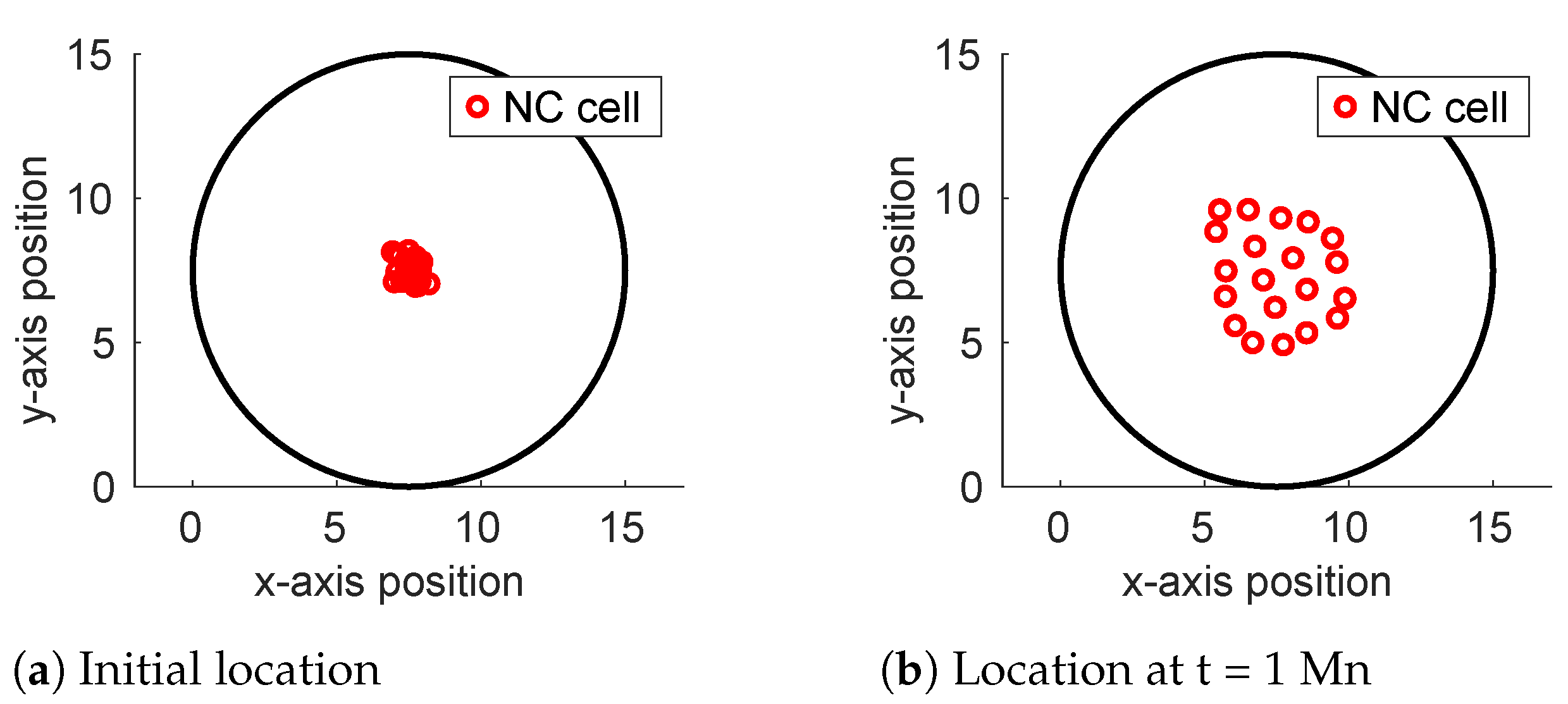
Theveneau et al. track the motion of a culture of NC cells in a bounded rectangular region [22]. One numerical run in the study shows NC cells only, starting in a tight clump and spreading out. This scenario was simulated with 20 NC cells in a disc of radius 5, shown in Figure 4. Initial conditions were set by choosing NC cells to have random coordinates in a circular region of radius 1 in the center of the disc, shown in Figure 4a. The system was run to t = 1,000,000 to produce Figure 4b. Default parameters were used with a set to 0.01.

4.3. A Heterogeneous Cell Environment in a Rectangular Region Produces Herding and Herd Splitting

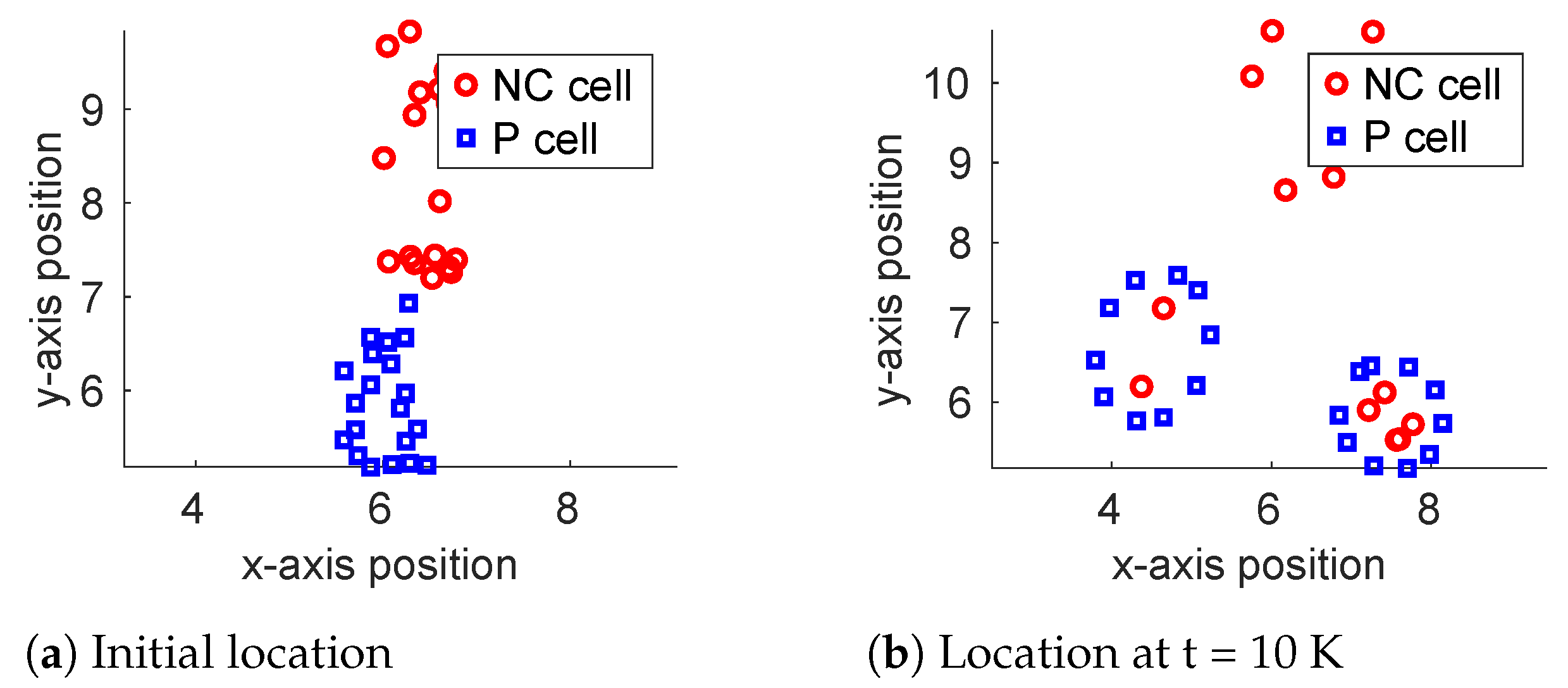
The same study by Theveneau et al. [22] tracks the motion of a co-culture of NC and P cells in a bounded rectangular region. This scenario was simulated with 20 cells of each type in the rectangle [0,8] × [0,11]. Signal response between like cells was eliminated by setting wN = wP = 1, so P cells only respond to the signal from N and vice versa. Initial conditions were set by choosing NC cells to have random coordinates in the region [6,7] × [7,10], and P cells with random coordinates in [5.5,6.5] × [5,7]. This choice places the initial positions shown in Figure 5a, with NC cells slightly above and to the right of P cells, in imitation of the figure in Theveneau et al. [22]. The system was run to t = 10,000 to produce the final position in Figure 5b.

4.4. Heterogeneous Cell Environment on a Disc in Imitation of Neural Crest and Placodal Cell Behavior

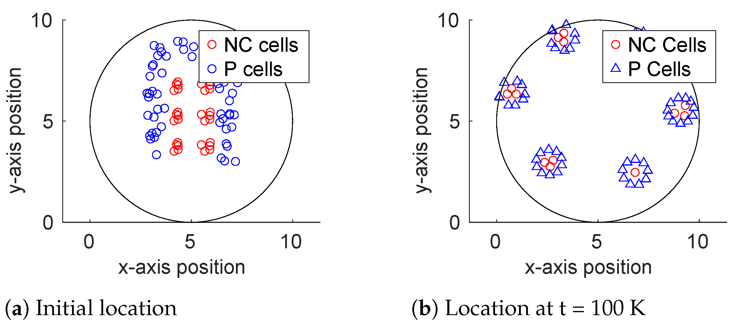
Neural crest streaming is a phenomenon by which differentiated cells, spaced unevenly along the dorsal midline, split it into spatially distinct groups that migrate separately throughout the embryo, while placodal cells began as an inverted U-shape around the perimeter [7,23]. This scenario was simulated with six groups of 20 NC cells and three groups of 20 P cells in a disc of diameter 15, shown in Figure 6a. Five NC cells were randomly activated for time increments of 5 until all cells were active, reflecting continuous EMTs observed in vivo [7]. The system was run to t=100,000 to produce Figure 6b.

5. A Few Theoretical Results

Two Individuals of Different Types

The simulation of a two-cell system in the unbounded plane showed both cells moving in a straight line in a steady state, with no stationary equilibrium, as shown in Figure 3a. Theorem 1 confirms this behavior. In a bounded region, motion stopped only when the “runner” hit the boundary as shown in Figure 3b. The line between the two cells was perpendicular to the boundary at equilibrium.
Theorem 2 confirms this observed behavior.
Theorem 1. 
Let X’=F(X) be the four-dimensional system describing two individuals, N and P, moving in the plane according to Equation (2) and let w N P ≠ w P N. Let initial conditions be given so that ( XN 0 , YN 0 ) ≠ ( XP 0 , YP 0 ). Let the signals from both cells have identical Gaussian distributions. Then, the system has no equilibrium.
Proof. 
The system equations from Equation (2) in this case simplify to
d d t ( X N , Y N ) = R N P ( S P ( x , y ) ) × ( S P ( x , y ) ) .
d d t ( X P , Y P ) = R P N ( S N ( x , y ) ) × ( S N ( x , y ) ) .
Because the initial conditions do not coincide and because RNP and RPN are negative for high signal values, the two cells do not come arbitrarily close to each other. Therefore, the gradient is never zero. If the cells come to equilibrium, it must therefore be that RNP and RPN both equal zero. The assumption that both signals have identical Gaussian distributions implies that both cells respond to the same signal strength. By the assumptions that w N P ≠ w P N, both RNP and RPN cannot equal zero for the same signal value. Therefore, there is no equilibrium. □
Theorem 2. 
Let the hypotheses be as in Theorem 1 except for constraining the motion to a bounded region with a differentiable boundary. Then, equilibrium is possible only if P is at the boundary, the line NP is perpendicular to the boundary, and N is at the point where SP = w N P.
Proof. 
As neither of the derivatives in Equation (2) can be zero, if motion stops it is due to the boundary. Let w P N w N P . If N touches the boundary, P will approach a location where the signal strength to which it responds is equal to w P N. As this is less than w N P, N will move away from the boundary towards P. So, there is not an equilibrium with N on the boundary. If P touches the boundary, it will move against the gradient of SN. If that gradient is perpendicular to the boundary, no motion is possible for P and N will cease motion when SP = w N P. □
Generally, the large system given by (2) will have many equilibrium configurations in addition to those produced when either response functions or gradients are set to zero. For this reason, investigations of larger populations are numerical.

6. Numerical Results: The Example of Neural Crest and Placodal Cells

Collective cell migration involves coordinated movement within or between cell populations and occurs in various biological species during development. In C. elegans, for example, hypodermal cells coordinate to form “epithelial sheets” enabling ventral enclosure [24,25]. In D. melanogaster, clusters of border cells move towards a developing oocyte by engaging in group motion, migrating together and in between the follicular epithelium of a developing egg chamber [10,26]. In vertebrates, neural crest and placodal cells exhibit chase-and-run migration as they emerge from the neural tube and ectoderm, proliferating outward to surrounding tissue and differentiating to facilitate organogenesis [19,27,28]. Collective cell migration is critical to life; characterizing the mechanisms that govern this movement can shed light on processes arising from faulty mechanisms, including cancer-associated angiogenesis and metastasis.
In particular, recent research has identified and used the neural crest as a model system to characterize broader cell population movement across vertebrate species [29,30,31]. Furthermore, in its simplest form, our paper describes a system with two coordinating cell types, consistent with the movement of the neural crest during embryogenesis. Therefore, the model developed here uses neural crest (“N”) and placodal (“P”) collective cell migration as a working example.
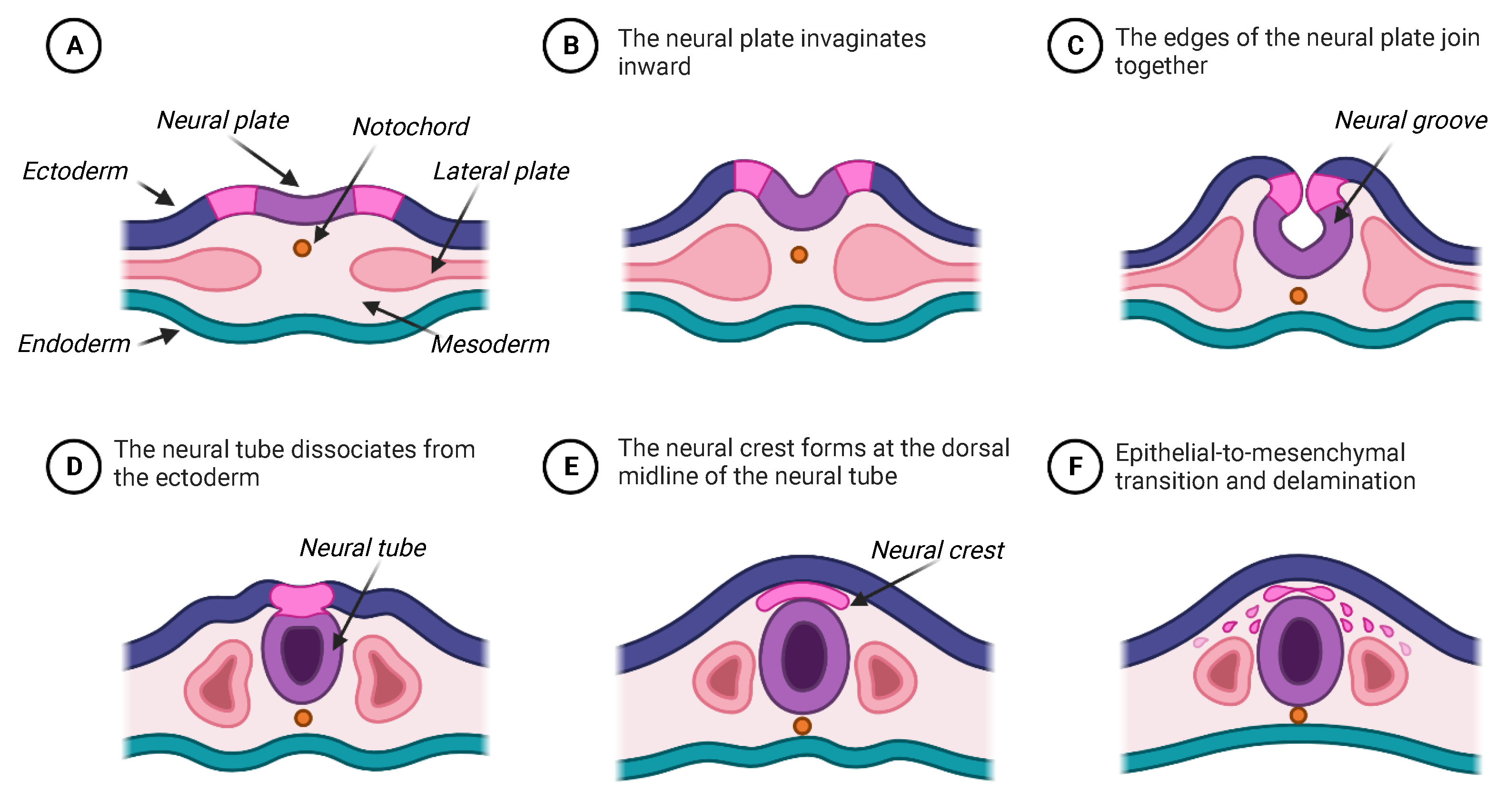
N and P cell formation can be understood in the context of biological processes which follow gastrulation. Gastrulation is the process by which a hollow sphere of cells known as the blastula rearranges to form distinct endoderm (“inner”), mesoderm (“middle”), and ectoderm (“outer”) layers. Soon after gastrulation, the notochord, a cluster of mesodermal cells, induces a subset of ventral ectodermal cells to thicken and form into a cohesive epithelial structure called the neural plate [32]. Neurulation is the subsequent process by which this neural plate invaginates inward; the edges of the plate join together, resulting in a neural tube located between the notochord and the ectoderm. The neural crest (the “group of NC cells”) is a component of this neural tube and lies at its dorsal midline. The neural crest undergoes an epithelial-to-mesenchymal transition (EMT) and disaggregates from the neural tube in a process called delamination, at which point NC cells have emerged and begin migration [32,33,34]. Concomitantly, placodal cells emerge from epithelial tissue adjacent to the neural plate, which remains attached to the ectoderm throughout neurulation [22,35,36]. This process is summarized in Figure 2.
By the end of migration, NC and P cells travel to diverse areas of the embryo, giving rise to several important derivatives. To illustrate, the neural crest comprises four domains—the cranial, trunk, vagral and sacral, and cardiac neural crest—which delineate physiological influence. These domains represent the influence of NC cells on the formation of various structures, including cartilage, middle ear and jaw bones, vertebral cartilage, sensor neurons, and melanocytes [37,38] Meanwhile, P cells have been shown to contribute to the formation of sensory ganglia, the olfactory apparatus, the pituitary gland, and other aspects of sensory and endocrine organ development [36,39,40]. Researchers have demonstrated that the disruption or aberration of NC and P migration leads to the development of diseases, including cancers and defective craniofacial and cardiac structures [41,42,43].

6.1. Mechanisms for Neural Crest Cell Motility and Directed Migration

Cell movement can be described by mechanisms which describe overall motility and mechanisms which describe directed migration. For example, NC cell motility is characterized by repulsive and attractive forces between individual NC cells [30]. Repulsive forces occur between NC cells via contact inhibition of locomotion (CIL). CIL involves the buildup of proteins including N-Cadherin and RhoA at points of cell-to-cell contact. This leads to Rac1 activity at the cells’ edges, causing movement away from each other [30,44,45]. Additionally, migratory NC cells express both the protein C3a and its receptor (C3aR). C3a-C3aR binding acts as an attractive force between neighboring NC cells [30,46]. Finally, NC cells will also experience alignment behaviors, wherein any two NC cells may move alongside each other for a brief moment during initial cell-to-cell contact [30].
Repulsive and attractive forces influence directed cell migration between NC and P cells. Both cell types experience CIL upon cell–cell contact [22,45], whereas attractive forces are influenced via the action of stromal cell-derived factor-1 (SDF1). SDF1 is a known chemoattractant that binds to the chemokine (CXC motif) receptor 4 (CXCR4) on the extracellular matrix of NC cells. Produced by P cells, SDF1 stabilizes NC cell polarity and affects intracellular Rac1 activity, leading to directed NC cell movement towards the P cells [47,48]. Ultimately, SDF1-mediated chemo-attraction and CIL are represented as a “chase” and “run” behavior of migrating NC and P cells, respectively [22]. Moreover, during the “chase,” SDF1 typically binds to the NC cells at the front of a migrating group; because NC cells across the group are mechanically bounded, binding at the front causes contraction at the rear. This leads to forward cellular intercalation and long-range group motion, a phenomenon referred to as “supracellular”. We refer to Shellard et al. for a more detailed description and depiction of these dynamics [30].
All of this complexity is abbreviated using the “close but not too close” assumption that leads to a single response function for a given type of cell to all of the signals given by other cells of some given type. The choice of response function requires the identification of only a few parameters. These were selected with a view of producing results visually similar to those pictured in key papers associated with numerical experiments [7,22,23]. Initial conditions were set in imitation of Kulesa et al. [23]. Default parameters are given in Table 1. All simulations were performed with MATLAB [6].
These dimensionless parameters are chosen specifically to reproduce observed behaviors and convenient run times. For example, the parameter w N N was chosen to reproduce the observed sparse clumps of neural crest cells at equilibrium, and w P P was chosen correspondingly smaller to create the observed tight clumps of placodal cells. Note that a was adjusted to 0.01 to reproduce the behaviors in Figure 4, Figure 5 and Figure 6.

6.2. Two Individuals in Engaged in “Chase-And-Run” Behavior

Figure 3 describes one NC cell and one P cell interacting on an unbounded plane and in a bounded rectangle, with the X coordinate position shown in a time series in Figure 3b (top and bottom). Because the preferred distance for N is closer than that of P, a chase ensues which does not reach equilibrium in the unbounded domain, as shown in Theorem 1, but reaches equilibrium in the bounded domain when the gradient points toward the boundary.

6.3. Multiple Cells of a Single Type Flocking or Congregating in a Disc

Figure 4 shows the results of multiple NC cells interacting only with their own signals in a disc. Initial conditions were chosen in imitation of an experiment in Theveneau [22], with NC cells clustered tightly together, seen in Figure 4a. The system achieves approximate equilibrium, with final N cell positions shown in Figure 4b, which depicts the paths of cell motion during the run. NC cells (red) disperse until the signal strengths detected by each cell are at the root of the response function.

6.4. Heterogeneous Cell Environment in a Rectangular Region Exhibits Herding and Herd Splitting

Figure 5 shows the results of multiple NC and P cells interacting only to non-like signals in a rectangle. Initial conditions were chosen in imitation of Theveneau et al. [22], with NC cells slightly above and to the right of P cells, seen in Figure 5a The system does not achieve equilibrium during the simulation, with final cell positions shown in Figure 5b. NC cells (red) split into two groups, with one group moving toward P cells in a coordinated manner and the other group not moving at all, splitting the herd of N cells. Some of the P cells retreat downward to the final configuration, while lower ones rise as all are arranged in the circles seen in Figure 5b, which is also an example of NC cells splitting the herd of P cells. Note that there is no cell that arrives at an equilibrium position during the simulation, including when the simulation was run until t = 250,000.

6.5. Heterogeneous Cell Environment on a Disc in Imitation of Neural Crest and Placodal Behavior

Figure 6 shows the results of multiple NC and P cells interacting with both signals on a disc. Initial conditions were chosen in imitation of Barriga et al. [7], with NC cells in clusters spaced along the dorsal midline and surrounded by P cells in an inverted U-shape, seen in Figure 6a. The system does not achieve equilibrium in the x-axis or y-axis position, respectively. However, as shown in Figure 6b, the final cell positions remain in clusters. NC cells (red) split into clusters and are activated continuously at time 0; the groups move toward surrounding P cells in a coordinated manner. Note that all clusters, including both types of cells, approach the boundary of the region by time 100,000.

7. Discussion

During embryogenesis, neural crest cell populations exhibit cellular motility, wherein adjacent cells experience attractive, repulsive, and alignment forces between each other. Neural crest cells also undergo directional migration, influenced by chase-and-run chemotaxis and supracellular dynamics. This paper presents a model that demonstrates both cellular motility and directional migration. Its framework can be expanded to account for cell-to-cell behaviors in other biological systems with two or more distinct stimuli.

7.1. Modeling of Neural Crest and Placodal Collective Cell Migration

The collective migration of NC and P cells has been modeled extensively. Theveneau et al. [32] and Colombi et al. [19] offer a broad and additive review of the current literature. At a general level, Giniunaite et al. review individual-based, partial differential equation (PDE)-based, and integro-PDE models as broad examples of a methodology. [31]. McLennan et al. offer a model of neural crest motion in the presence of a fixed or changing spatial gradient [49,50] but without placodal cells represented. In summary, this paper adds to a growing list of models that seek to characterize experimental results.
The model presented here is closest in spirit to that of Colombi et al., in that it models both attractive and repulsive forces between NC and P cells as functions of distance, which are then summed to determine the rate of motion [19]. In this study, a Gaussian distribution of two signal types replaces the role of distance. Both attraction and repulsion of a given cell type to a given signal are incorporated into a single response function for each pair of cell types, resulting in two responses to two different signals for a given cell, combined in a weighted sum. The model in Colombi et al. achieves chase-and-run dynamics in the plane that ultimately ceases motion. The general framework presented here also produces chase-and-run dynamics, as well as the herding behavior necessary to separate the placode into distinct regions.
While extensive evidence exists for Sdf1 as a chemoattractant produced by P cells [19,32,47,48], the literature does not suggest a similar chemoattractant produced by NC cells and specifies CIL as a non-chemical, multifaceted force that acts between the migrating “chaser” NC and “runner” P cells [7,19,44]. For this paper, CIL is framed as one aspect of a “signal” that moderates attractive and repulsive forces between proximal NC cells as described by the Gaussian and response functions. This interpretation served as a helpful simplifying assumption that eased computation while still producing results consistent with observations of real NC and P interactions.

7.2. Collective NC Cell Motility

Figure 4, Figure 5 and Figure 6 are each consistent with aspects of NC cell motility. In Figure 4, NC cells are simulated traveling in clumps, with each clump moving apart from another. As they become arbitrarily distant, these clumps themselves disperse, producing the evenly distributed array of cells in Figure 4b. This motion is consistent with Shellard’s principles of NC cell attraction, repulsion, and alignment [30]. This motion also closely resembles Theveneau et al.’s NC cell culturing [22], which shows NC cells moving in transient clumps. Overall, the observation of NC motility is expected and unsurprising given the consistent transmission of signals between cells within numerical experiments.

7.3. Directional NC Cell Migration and Emergent Patterns, Herding, and Herd Splitting

Figure 5 demonstrates chase-and-run directional migration between NC and P cell populations. In the literature, directional migration is described by Shellard and Mayor [30] and observed in vitro by Theveneau et al. [22]. Specifically, Shellard and Mayor describe the molecular underpinnings and physical manifestation of chase-and-run dynamics by comparing the influence of SDF1 and CIL on directed cell motion. These qualitative descriptions complement Theveneau et al.’s experimental observations in which NC cells chase P cells in a collective pack. In this simulation, NC and P cells were allowed to sense only those cells of the other type, providing an exciting illustration of how clumping of like cells may occur even with no attracting force between them. The NC cells appear to form a clump not because of their own cell–cell interaction, which is turned off, but because they are all pursuing the other species. This observation raises the possibility that when like-cell clumping is observed in an experiment, it need not necessarily be because of like-to-like cell affinity.
Figure 5 also illustrates how a pattern of NC clumps surrounded by circles of P emerges from the collection of individuals before the NC cells chase the P cells all the way to the boundary of the bounded set. This phenomenon occurs because there are many cells, and this stands in direct contrast to the theorems for the two-cell case. The theorems show that equilibrium away from the boundary is not possible with only two cells that have response functions with different roots. Simulations show that equilibrium is possible with a larger cell population. It is easy to invent equilibrium configurations with as few as one NC and two P cells just by placing them in a collinear configuration with the NC cell in the middle. The gradient of the signal received by the NC cell will then be zero and the P cells can move to the distance that produces a response of zero. There are many other such configurations possible with small numbers of cells, suggesting that equilibrium, or quasi-equilibrium, configurations away from the boundary, such as those seen in Figure 5 and Figure 6, are not rare.
In Figure 5, one also sees the NC cells split into two groups, or “herds”. One of these herds is essentially stationary from t = 10,000 (when it is initially formed) until above t = 250,000. The other pursues the P cells downward in the rectangle. All of these NC cells are experiencing a signal strength that would draw them toward the P cells, but the ones farther away experience a weak signal that produces a prolonged, imperceptible motion. As the P cells move downward, this signal becomes even weaker and motion slower. The distant NC cells do, in time, join the others; the time frame is very long, and the result is a cluster of NC and P cells that is distinct from the initial clusters.

7.4. Emergent Self-Organization

Neural crest and placodal cells interact using chase-and-run migration dynamics to separate placodal cells into groups that will differentiate into distinct organs [19,27,28].
In Figure 6b, NC cells are shown in distinct packs surrounded by clusters of P cells. Figure 6 simulates the spontaneous self-organizing behavior that allows differentiation to occur. In this simulation, all cells respond to both cell types, with a different response to each cell type determined by both signaling and receiving species, as would be expected with mixotaxis [7]. No background signal is required to achieve the geometric distribution that qualitatively mirrors diagrams in Barriga et al. [7]. Rather, the arrangement arises from mixotaxis acting on the initial conditions shown in Figure 6a that are qualitatively similar to the arrangements described in the literature, with NC cells arranged at intervals along the vertical diameter and P cells in a surrounding inverted horseshoe shape [7,23].
Observations of NC cells show streaming behaviors in addition to clumping behaviors [7]. In Figure 6 one sees clumps of NC cells moving together outward from their initial placement in clumps along the central line. They do not appear to stream. The streaming behavior observed in Kulesa et al. [23] can be explained by the continuous nature of biological EMT, which activates NC cells to become mobile. In our experiment, this transition occurs through incremental moments and is not a continuous phenomenon, which may explain why streaming is not seen in Figure 6. Also, because there is no stream of NC cells, the P cells are able to close in behind clumps of NC cells and surround them, rather than being pierced by the streams as pictured in Barriga et al.’s diagrams [7].

7.5. The Functional Response to a Signal

The design choice to summarize the response of one species to all chemical signals detected from a given (same or other) species has certain advantages. Kulesa et al., for example, construct a model of neural crest motion in the presence of diffusing signals, but omit the secondary species of placodal cells and their motion [51]. In doing so, the model sacrifices one type of complexity for another. Without knowing all of the possible chemicals and receptors in play, one could in principle conduct experiments with a time-stamped video of pairs of individual cells. The two-cell model with few parameters could be fit to the resulting time series for intercellular distance. As currently given, the response function is based on the simple idea that cells want to be close, but not too close, to each other. A form with few parameters was chosen that could be adjusted and improved based on these experimental data. A second advantage of this approach is the ability to prove theorems for the two-cell case. Kulesa et al., for example, construct a model of neural crest motion in the presence of diffusing signals, but omit the secondary species of placodal cells and their motion [51,52].

7.6. Expanded Framework for Other Biological Systems

A variety of biological systems may be described using the framework offered for an arbitrary number of cell types. Consider the migration of border cells during D. melanogaster egg development. In this case, clusters containing follicle and polar cells participate in growth factor-mediated chemotaxis, collectively migrating from the anterior tip of a developing egg chamber, in between Nurse cells, and towards the oocyte border [10]. Border cell migration constitutes a three-species system where each species can be modeled as emitting and responding to signals using a generic Equation (6), a generic system Equation (7) and a generic response function (5). We suggest the development and analysis of this model as one direction for future work.

7.7. Limitations of the Study

All models have limitations. The one presented here involves signals released by individuals that are characterized by a simple response function by the observing individual. This simplicity suggests easy ways to measure the resulting parameters in simple experiments, without regard to the biology behind the signal production or the observer’s sensory organs. However, it is certainly possible that one species may emit multiple signals, or that they may be detected by the observer in multiple ways.
The complex behaviors that result from relatively simple assumptions are not due to subtle biological phenomena, but rather to the complexity of large numbers of individuals responding to signals without other types of communication. Differences in output come from the initial positions of these individuals. So, this model does not include communication between individuals (other than sensory) or learning, strategy, or population growth. All of these extra features may be necessary, depending on the organism chosen as an example. The perception of signal in this study is omni-directional, without regard to possible asymmetric placement of sensory organs (such as eyes).

7.8. Future Work

We suggest some additional avenues for future work, including increasing the number of species in the model.
It has been observed that, even at near equilibrium, NC cells and many other organisms exhibit some random motion [22]. Such displacements could disrupt equilibrium patterns if too large. Adding random motion to the model would elucidate this, and of course, there is more than one way to conduct this.
NC cells are observed to “stream” outward from their starting locations [23]. This may be due to the ongoing activation of NC cells, which is not yet in the model and could be added. If experiments giving time series for small numbers of cells become available so that parameterization becomes possible, it would be very interesting to model large data sets specifically to find features that this model fails to explain.
All individuals are detecting and responding to all signals at levels that depend on biological features, such as receptor proteins, that are particular to a given species. So, how many types of behavior might one see by adjusting both the number of species and the parameters assigned to each? This question begs a sensitivity analysis and opens the door to modeling future, more complex processes.

8. Conclusions

This study develops a model, based on the phenomenon of mixotaxis described for cell motion, for any biological system with multiple individuals of two or more species that coordinate using multiple distinct stimuli arising from each species. The model is based on explicit intuitive assumptions and may be parameterized by experiments involving two or three individuals. The two-species system is analyzed numerically, demonstrating that it produces chase-and-run behavior (two individuals, Figure 3), herding of individuals of the same species (Figure 4), and herd separation (Figure 5). A single two-dimensional instantiation of the model produces all of these behaviors depending on the initial conditions for number and the location of the individuals.
The example includes two cell types that emit distinct signals, distinguish between them, and exhibit unique responses. The model qualitatively describes the basic characteristics of neural crest cell motility and reproduces images qualitatively similar to those in the literature for neural crest and placodal cell interactions and patterns. Starting with neural crest cells aligned on the central chord and placodal cells arranged in a U-shape around them, the system evolves to produce spatially distinct clumps of placodal cells associated with clumps of neural crest cells (Figure 6).

Author Contributions

Conceptualization, B.Q.F.; methodology, B.Q.F. and D.W.; software, S.M.; validation, B.Q.F., S.M.s and D.W.; formal analysis, D.W.; investigation, S.M.; writing—original draft preparation; B.Q.F.; writing—review and editing, S.M. and D.W.; visualization, S.M.; supervision, D.W. All authors have read and agreed to the published version of the manuscript.

Funding

This research did not receive any specific grant from funding agencies in the public, commercial, or not-for-profit sectors.

Institutional Review Board Statement

Not applicable.

Informed Consent Statement

Not applicable.

Data Availability Statement

Conflicts of Interest

The authors declare no conflicts of interest.

References

  1. Cardoso, R.C.; Ferrando, A. A review of agent-based programming for multi-agent systems. Computers 2021, 10, 16. [Google Scholar] [CrossRef]
  2. Tang, W.; Bennett, D.A. Agent-based modeling of animal movement: A review. Geogr. Compass 2010, 4, 682–700. [Google Scholar] [CrossRef]
  3. Monga, P.; Sharma, M.; Sharma, S.K. A comprehensive meta-analysis of emerging swarm intelligent computing techniques and their research trend. J. King Saud-Univ.-Comput. Inf. Sci. 2022, 34, 9622–9643. [Google Scholar] [CrossRef]
  4. Chakraborty, A.; Kar, A.K. Swarm intelligence: A review of algorithms. In Nature-Inspired Computing and Optimization: Theory and Applications; Springer: Berlin/Heidelberg, Germany, 2017; pp. 475–494. [Google Scholar]
  5. Nayyar, A.; Nguyen, N.G. Introduction to swarm intelligence. In Advances in Swarm Intelligence for Optimizing Problems in Computer Science; Chapman and Hall/CRC: Boca Raton, FL, USA, 2018; pp. 53–78. [Google Scholar]
  6. Matlab. version 9.10 (R2021a); The MathWorks Inc.: Natick, MA, USA, 2021. [Google Scholar]
  7. Barriga, E.H.; Theveneau, E. In vivo Neural Crest Cell Migration Is Controlled by “Mixotaxis”. Front. Physiol. 2020, 11, 586432. [Google Scholar] [CrossRef] [PubMed]
  8. DeAngelis, D.L.; Diaz, S.G. Decision-making in agent-based modeling: A current review and future prospectus. Front. Ecol. Evol. 2019, 6, 237. [Google Scholar] [CrossRef]
  9. Williams, H.J.; Sridhar, V.H.; Hurme, E.; Gall, G.E.; Borrego, N.; Finerty, G.E.; Couzin, I.D.; Galizia, C.G.; Dominy, N.J.; Rowland, H.M.; et al. Sensory collectives in natural systems. Elife 2023, 12, e88028. [Google Scholar] [CrossRef]
  10. Stonko, D.P.; Manning, L.; Starz-Gaiano, M.; Peercy, B.E. A mathematical model of collective cell migration in a three-dimensional, heterogeneous environment. PLoS ONE 2015, 10, e0122799. [Google Scholar] [CrossRef]
  11. Peercy, B.E.; Starz-Gaiano, M. Clustered cell migration: Modeling the model system of Drosophila border cells. Semin. Cell Dev. Biol. 2020, 100, 167–176, Place: England. [Google Scholar] [CrossRef]
  12. Berez, A.; Peercy, B.E.; Starz-Gaiano, M. Development and Analysis of a Quantitative Mathematical Model of Bistability in the Cross Repression System Between APT and SLBO Within the JAK/STAT Signaling Pathway. Front. Physiol. 2020, 11, 803. [Google Scholar] [CrossRef]
  13. Boyd, W.A.; Smith, M.V.; Kissling, G.E.; Rice, J.R.; Snyder, D.W.; Portier, C.J.; Freedman, J.H. Application of a mathematical model to describe the effects of chlorpyrifos on Caenorhabditis elegans development. PLoS ONE 2009, 4, e7024. [Google Scholar] [CrossRef]
  14. Hench, J.; Henriksson, J.; Lüppert, M.; Bürglin, T.R. Spatio-temporal reference model of Caenorhabditis elegans embryogenesis with cell contact maps. Dev. Biol. 2009, 333, 1–13. [Google Scholar] [CrossRef] [PubMed]
  15. Wood, A.J.; Ackland, G.J. Evolving the selfish herd: Emergence of distinct aggregating strategies in an individual-based model. Proc. R. Soc. Biol. Sci. 2007, 274, 1637–1642. [Google Scholar] [CrossRef] [PubMed]
  16. Setekera, R.; Klar, A. Numerical Simulation for Herding and Flocking Models. LAP LAMBERT Academic Publishing: London, UK, 2010. [Google Scholar]
  17. Lopez, U.; Gautrais, J.; Couzin, I.D.; Theraulaz, G. From behavioural analyses to models of collective motion in fish schools. Interface Focus 2012, 2, 693–707. [Google Scholar] [CrossRef] [PubMed]
  18. Katz, Y.; Tunstrøm, K.; Ioannou, C.C.; Huepe, C.; Couzin, I.D. Inferring the structure and dynamics of interactions in schooling fish. Proc. Natl. Acad. Sci. USA 2011, 108, 18720–18725. [Google Scholar] [CrossRef] [PubMed]
  19. Colombi, A.; Scianna, M.; Painter, K.J.; Preziosi, L. Modelling chase-and-run migration in heterogeneous populations. J. Math. Biol. 2020, 80, 423–456. [Google Scholar] [CrossRef]
  20. An, L.; Grimm, V.; Turner II, B.L. Meeting grand challenges in agent-based models. J. Artif. Soc. Soc. Simul. 2020, 23. [Google Scholar] [CrossRef]
  21. An, L.; Grimm, V.; Sullivan, A.; Turner Ii, B.; Malleson, N.; Heppenstall, A.; Vincenot, C.; Robinson, D.; Ye, X.; Liu, J.; et al. Challenges, tasks, and opportunities in modeling agent-based complex systems. Ecol. Model. 2021, 457, 109685. [Google Scholar] [CrossRef]
  22. Theveneau, E.; Steventon, B.; Scarpa, E.; Garcia, S.; Trepat, X.; Streit, A.; Mayor, R. Chase-and-run between adjacent cell populations promotes directional collective migration. Nat. Cell Biol. 2013, 15, 763–772. [Google Scholar] [CrossRef]
  23. Kulesa, P.M.; Fraser, S.E. Neural crest cell dynamics revealed by time-lapse video microscopy of whole embryo chick explant cultures. Dev. Biol. 1998, 204, 327–344. [Google Scholar] [CrossRef]
  24. Martin, P.; Parkhurst, S.M. Parallels between tissue repair and embryo morphogenesis. Development 2004, 131, 3021–3034. [Google Scholar] [CrossRef]
  25. Olson, H.M.; Nechiporuk, A.V. Using Zebrafish to Study Collective Cell Migration in Development and Disease. Front. Cell Dev. Biol. 2018, 6, 83. [Google Scholar] [CrossRef] [PubMed]
  26. Prasad, N.G.; Joshi, A. Remarks on the article on life-history traits in Drosophila populations selected for rapid development by Yadav and Sharma. J. Exp. Biol. 2015, 218, 326–327. [Google Scholar] [CrossRef] [PubMed]
  27. Hintze, M.; Prajapati, R.S.; Tambalo, M.; Christophorou, N.A.D.; Anwar, M.; Grocott, T.; Streit, A. Cell interactions, signals and transcriptional hierarchy governing placode progenitor induction. Development 2017, 144, 2810–2823. [Google Scholar] [CrossRef] [PubMed]
  28. Steventon, B.; Mayor, R.; Streit, A. Directional cell movements downstream of Gbx2 and Otx2 control the assembly of sensory placodes. Biol. Open 2016, 5, 1620–1624. [Google Scholar] [CrossRef] [PubMed]
  29. Theveneau, E.; Mayor, R. Collective cell migration of the cephalic neural crest: The art of integrating information. Genesis 2011, 49, 164–176. [Google Scholar] [CrossRef]
  30. Shellard, A.; Mayor, R. Rules of collective migration: From the wildebeest to the neural crest. Philos. Trans. R Soc. Lond. B Biol. Sci. 2020, 375, 20190387. [Google Scholar] [CrossRef]
  31. Giniūnaitė, R.; Baker, R.E.; Kulesa, P.M.; Maini, P.K. Modelling collective cell migration: Neural crest as a model paradigm. J. Math. Biol. 2020, 80, 481–504. [Google Scholar] [CrossRef]
  32. Theveneau, E.; Mayor, R. Neural crest delamination and migration: From epithelium-to-mesenchyme transition to collective cell migration. Dev. Biol. 2012, 366, 34–54. [Google Scholar] [CrossRef]
  33. Purves, D.; Augustine, G.; Fitzpatrick, D. The Initial Formation of the Nervous System: Gastrulation and Neurulation. In Neuroscience, 2nd ed.; Sinauer Associates: Sunderland, MA, USA, 2001. [Google Scholar]
  34. DeSesso, J.M.; Williams, A.L. Chapter 10—Periods of Susceptibility: Interspecies Comparison of Developmental Milestones During Ontogenesis of the Central Nervous System. In Handbook of Developmental Neurotoxicology (Second Edition), 2nd ed.; Slikker, W., Paule, M.G., Wang, C., Eds.; Academic Press: Cambridge, MA, USA, 2018; pp. 113–125. [Google Scholar] [CrossRef]
  35. York, J.R.; Yuan, T.; McCauley, D.W. Evolutionary and Developmental Associations of Neural Crest and Placodes in the Vertebrate Head: Insights From Jawless Vertebrates. Front. Physiol. 2020, 11, 986. [Google Scholar] [CrossRef]
  36. Koontz, A. Neural Crest and Placodal Cells Contributions to Cranial Sensory Development. Ph.D Thesis, California Institute of Technology, Pasadena, CA, USA, 2021. [Google Scholar]
  37. Bronner, M.E. Formation and migration of neural crest cells in the vertebrate embryo. Histochem. Cell Biol. 2012, 138, 179–186. [Google Scholar] [CrossRef]
  38. Gilbert, S. The Neural Crest. In Developmental Biology, 6th ed.; Sinauer Associates: Sunderland, MA, USA, 2000. [Google Scholar]
  39. Dincer, Z.; Piao, J.; Niu, L.; Ganat, Y.; Kriks, S.; Zimmer, B.; Shi, S.H.; Tabar, V.; Studer, L. Specification of functional cranial placode derivatives from human pluripotent stem cells. Cell Rep. 2013, 5, 1387–1402. [Google Scholar] [CrossRef] [PubMed]
  40. Katoh, H.; Shibata, S.; Fukuda, K.; Sato, M.; Satoh, E.; Nagoshi, N.; Minematsu, T.; Matsuzaki, Y.; Akazawa, C.; Toyama, Y.; et al. The dual origin of the peripheral olfactory system: Placode and neural crest. Mol. Brain 2011, 4, 34. [Google Scholar] [CrossRef] [PubMed]
  41. Inman, K.E.; Caiaffa, C.D.; Melton, K.R.; Sandell, L.L.; Achilleos, A.; Kume, T.; Trainor, P.A. Foxc2 is required for proper cardiac neural crest cell migration, outflow tract septation, and ventricle expansion. Dev. Dyn. 2018, 247, 1286–1296. [Google Scholar] [CrossRef] [PubMed]
  42. Johnsen, J.I.; Dyberg, C.; Wickström, M. Neuroblastoma-A Neural Crest Derived Embryonal Malignancy. Front. Mol. Neurosci. 2019, 12, 9. [Google Scholar] [CrossRef] [PubMed]
  43. Li, L.; He, Y.; Zhao, M.; Jiang, J. Collective cell migration: Implications for wound healing and cancer invasion. Burns Trauma 2013, 1, 21–26. [Google Scholar] [CrossRef]
  44. Carmona-Fontaine, C.; Matthews, H.K.; Kuriyama, S.; Moreno, M.; Dunn, G.A.; Parsons, M.; Stern, C.D.; Mayor, R. Contact inhibition of locomotion in vivo controls neural crest directional migration. Nature 2008, 456, 957–961. [Google Scholar] [CrossRef]
  45. Roycroft, A.; Mayor, R. Molecular basis of contact inhibition of locomotion. Cell. Mol. Life Sci. 2016, 73, 1119–1130. [Google Scholar] [CrossRef]
  46. Carmona-Fontaine, C.; Theveneau, E.; Tzekou, A.; Tada, M.; Woods, M.; Page, K.M.; Parsons, M.; Lambris, J.D.; Mayor, R. Complement fragment C3a controls mutual cell attraction during collective cell migration. Dev. Cell 2011, 21, 1026–1037. [Google Scholar] [CrossRef]
  47. Belmadani, A.; Tran, P.B.; Ren, D.; Assimacopoulos, S.; Grove, E.A.; Miller, R.J. The chemokine stromal cell-derived factor-1 regulates the migration of sensory neuron progenitors. J. Neurosci. 2005, 25, 3995–4003. [Google Scholar] [CrossRef]
  48. Theveneau, E.; Marchant, L.; Kuriyama, S.; Gull, M.; Moepps, B.; Parsons, M.; Mayor, R. Collective chemotaxis requires contact-dependent cell polarity. Dev. Cell 2010, 19, 39–53. [Google Scholar] [CrossRef]
  49. McLennan, R.; Schumacher, L.J.; Morrison, J.A.; Teddy, J.M.; Ridenour, D.A.; Box, A.C.; Semerad, C.L.; Li, H.; McDowell, W.; Kay, D.; et al. Neural crest migration is driven by a few trailblazer cells with a unique molecular signature narrowly confined to the invasive front. Development 2015, 142, 2014–2025. [Google Scholar] [CrossRef] [PubMed]
  50. McLennan, R.; Dyson, L.; Prather, K.W.; Morrison, J.A.; Baker, R.E.; Maini, P.K.; Kulesa, P.M. Multiscale mechanisms of cell migration during development: Theory and experiment. Development 2012, 139, 2935–2944. [Google Scholar] [CrossRef] [PubMed]
  51. Kulesa, P.M.; Teddy, J.M.; Stark, D.A.; Smith, S.E.; McLennan, R. Neural crest invasion is a spatially-ordered progression into the head with higher cell proliferation at the migratory front as revealed by the photoactivatable protein, KikGR. Dev. Biol. 2008, 316, 275–287. [Google Scholar] [CrossRef] [PubMed]
  52. Dyson, L.; Holmes, A.; Li, A.; Kulesa, P.M. A chemotactic model of trunk neural crest cell migration. Genesis 2018, 56, e23239. [Google Scholar] [CrossRef]
Figure 1. General framework for two-signal mixotaxis with two cell populations of arbitrary size. Two species of cells of arbitrary size (NC or “N” in red and P in blue) are represented producing signals SN and SP with respective magnitudes given by Equation (1). As described by (2), the rate of motion equals a linear combination of gradients for both signals at the cell’s location, with coefficients determined by response functions for each cell type to each signal.
Appliedmath 04 00077 g001
Figure 2. Initiation of neural crest cell migration in vertebrates. Neural crest cell migration occurs after the formation of the gastrula during embryogenesis. (A) The notochord induces adjacent ectodermal cells, which thicken, forming the neural plate. (BD) The neural plate invaginates inward, forming the neural tube. (E) The neural crest forms at the dorsal midline of the neural tube. (F) The neural crest undergoes EMT; individual neural crest cells delaminate and begin “chasing” placodal cells (not shown). Created with BioRender.com.
Appliedmath 04 00077 g002
Figure 3. Simulations showing two cells of different types, NC and P, moving in a plane according to the framework for signal and response presented in Figure 1. (a) Stationary equilibrium is not observed for NC and P cell two-signal mixotaxis in an unbounded system, as demonstrated by (top) motion tracks and (middle) y-axis position. (b) Equilibrium achieved for NC and P cell two-signal mixotaxis in bounded square planar region, as demonstrated by (bottom) motion tracks and y-axis position. For each simulation, default parameters from Table 1 were used. All simulations were completed in MATLAB.
Appliedmath 04 00077 g003
Figure 4. Simulation showing cells of a single type (red) reacting only to their own signal in accordance with the framework for signal response presented in Figure 1 and under default parameters with a set to 0.01. Initial (a) and final (b) locations of cells. Simulation completed in MATLAB.
Appliedmath 04 00077 g004
Figure 5. Simulations showing displacement in a rectangle for NC (red) and P (blue) cells with no intra-species signal response. Parameter a adjusted to 0.01 and time run to t = 10,000. Initial (a) and final (b) locations are at t = 0 and t = 10,000, respectively. All simulations completed in MATLAB.
Appliedmath 04 00077 g005
Figure 6. Simulations showing streaming in a disc for NC (red) and P (blue) cells using default parameters with parameter a adjusted to 0.01 and time run to t = 100,000. Initial (a) and final (b) locations of cells show displacement of NC cells in clusters surrounded by P cells. Position graphs (not shown) suggest that equilibrium is not reached. All simulations completed with MATLAB.
Appliedmath 04 00077 g006
Table 1. Default parameters of the model specified by Figure 1 and equations in two-cell mixotaxis.
Parameter SymbolDefault ValueDescription
a2Maximum magnitude of Gaussian signal
c1Twice the variance of signal
q N 0.8Weight given by N to gradient of signal from P
1 q N 0.2Weight given by N to gradient of signal from N
q P 0.8Weight given by P to gradient of signal from N
1 q P 0.2Weight given by P to gradient of signal from P
w N P 0.08Root of response of N to signal from P
w N N 0.01Root of response of N to signal from N
w P N 0.04Root of response of P to signal from N
w P P 0.005Root of response of P to signal from P
v N 1Maximum speed of N
v P 1Maximum speed of P
Disclaimer/Publisher’s Note: The statements, opinions and data contained in all publications are solely those of the individual author(s) and contributor(s) and not of MDPI and/or the editor(s). MDPI and/or the editor(s) disclaim responsibility for any injury to people or property resulting from any ideas, methods, instructions or products referred to in the content.