Proceeding Paper

An In Silico Approach to Evaluate the Diabetic Wound Healing Potential of Phenylethanoid Glycoside in Inhibiting the Receptor for Advanced Glycation End Products (RAGE) †

Department of Pharmaceutical Sciences and Technology, Birla Institute of Technology, Mesra, Ranchi 835215, India
*
Author to whom correspondence should be addressed.
Presented at the 2nd International Electronic Conference on Biomedicines, 1–31 March 2023; Available online: https://ecb2023.sciforum.net/.
Med. Sci. Forum 2023, 21(1), 24; https://doi.org/10.3390/ECB2023-14137
Published: 8 March 2023
(This article belongs to the Proceedings of The 2nd International Electronic Conference on Biomedicines)

Abstract

:
Diabetes mellitus (DM) is a chronic metabolic disorder and is associated with impaired wound healing. Non-healing leg and foot ulcers are a frequent significant consequence of diabetes and are caused by a combination of inadequate tissue perfusion, suppression of re-epithelialization, and poor collagen production. Receptor for Advanced Glycation End Products (RAGE) is a multiligand cell surface molecule that belongs to the immunoglobulin superfamily and is crucial in the pathophysiology of poor wound healing in diabetics. By inhibiting RAGE, a chronic non-healing wound is more likely to undergo angiogenesis, enhance blood supply to hypoxic areas of the wound, and decrease the pro-inflammatory reaction and pro-apoptotic signaling. Phenylethanoid glycosides (PhGs) are a class of natural glycosides that possess anti-diabetic, wound-healing, antimicrobial, anti-inflammatory, and antioxidant properties. Echinacoside, a phenylethanoid glycoside, has a promising role in wound healing by enhancing angiogenesis, promoting keratinocyte migration and proliferation, and enhancing neutrophil and macrophage activity. Consequently, molecular docking was performed to assess the interaction between Echinacoside and the RAGE receptor (PDB ID: 6VXG). The ligand and receptor had a strong binding interaction, as indicated by the lowest binding energy, which was found to be −6.1 kcal/mol. To further assess the activity of Echinacoside in diabetic wound healing, in vitro and in vivo studies are needed.

1. Introduction

Diabetes is correlated with impaired wound healing, which causes considerable financial and healthcare problems [1,2]. Non-healing leg and foot ulcers are a frequent significant consequence of diabetes and are caused by a combination of inadequate tissue perfusion, suppression of re-epithelialization, poor collagen production, peripheral neuropathy, changed red blood cell rheology, and decreased host immunity (Figure 1A) [3,4]. Regardless of the specific etiological reason, it is known that diabetes impairs effective reparative reactions, which results in the formation of chronic, non-healing wounds [1].
The phases of wound healing include hemostasis, inflammation, proliferation, and remodeling. They reflect a dynamic chain of occurrences that turn an open wound into newly created, well-vascularized granulation tissue with overlaying skin that is rich in collagen and other structural components of the extracellular matrix [5]. There is substantial evidence that the stages of wound healing are aberrant in diabetes. The diminished availability of components necessary for efficient wound repair occurs in diabetes due to reduced chemotaxis of inflammatory cells into the wound, accompanied by impaired phagocytosis and intracellular death [6].
Receptor for Advanced Glycation End Products (RAGE) is a multiligand cell surface molecule that belongs to the immunoglobulin superfamily and is crucial in the pathophysiology of poor wound healing in diabetics. When RAGE binds to its ligands, procoagulant initiator tissue factors, including cytokines, such as tumor necrosis factor-α (TNF-α), interleukin (IL)-6, IL-1, and cell adhesion molecules, are produced. These cytokines have an impact on the immune system, extending the amount of time a wound is exposed and making the wound more vulnerable to bacterial infection. In diabetes, failure of the angiogenic response to tissue hypoxia is caused by RAGE, and angiogenesis failure is a significant factor in the loss of tissue viability [7]. RAGE activation on fibroblasts also causes a decrease in collagen production [8].
The blockade of RAGE enhances angiogenesis, increases the flow of blood to hypoxic sites, and decreases pro-apoptotic signaling [7]. It also increases neovascularization and granulation tissue formation, and increases functional disorders of macrophages as a result enhances diabetic wound healing [2,9]. Furthermore, it increases the number of neutrophils phagocytized by macrophages, and decreases the levels of cytokines such as TNF-α, IL-6, and MMPs-2, -3, and -9 (Figure 1B) [10,11,12]. These findings point to the critical function of RAGE in disrupted wound healing related to diabetes and raise the possibility that blocking this receptor could offer a focused approach to regaining effective wound repair [1].
Phenylethanoid glycosides (PhGs) are a class of naturally occurring glycosides with phenylethyl alcohol and glycosyl components (Figure 2A). PhGs are obtained from a variety of sources and exhibit enhanced biological and pharmacological activities, such as anti-diabetic, wound-healing, antimicrobial, anti-inflammatory, and antioxidant properties. Polyphenols inhibit advanced glycation end product formation in hyperglycemic conditions. Many phenolic hydroxyl groups that are weakly acidic are present in PhG molecules. These substances are easily extracted and separated using conventional techniques, since the core structure contains at least one glycosyl moiety and is water soluble. In addition, the presence of a phenolic hydroxyl group increases the compounds’ antioxidant activity [13,14].
Echinacoside, a phenylethanoid glycoside has a promising role in wound healing by enhancing angiogenesis, promoting keratinocyte migration and proliferation, and enhancing neutrophil and macrophage activity [15]. The molecular structure of ECH is shown in Figure 2B. Echinacoside decreases the elevated levels of inflammatory cytokines, and shows good antioxidant and free radical scavenging properties [16]. Additionally, ECH inhibits the elevation in postprandial blood glucose levels, and considerably reduces the reactive oxygen species levels [17,18].

2. Materials and Methods

2.1. Protein and Ligand Preparation

Molecular docking is an in silico method for determining the binding free energy of a structure to a protein’s active site. AutoDock Vina, a web-based server for docking, was used for the docking study of Echinacoside into the binding site of the PDB protein [19]. The three-dimensional structure of RAGE (PDB ID: 6VXG) and Echinacoside were obtained from the RCSB Protein Data Bank in PDB format and the PubChem database in MOL SDF format [20,21,22]. Ligand and water molecules were removed while polar hydrogen and Gasteiger charge were added to RAGE to convert it into PDBQT format.

2.2. Active Binding Site Selection

The polyphenol binding site was examined using PyMOL software (The PyMOL Molecular Graphics System, Version 2.0, Schrödinger, LLC., New York, NY, USA) to determine the active binding site [20].

2.3. Assessment of Binding Affinities and Interactions

The interaction between Echinacoside (ligand) and RAGE (protein) was examined using the computational ligand–protein docking method. On the protein’s chosen active binding site, a grid point was assigned. Then, by using AutoDock Vina (AutoDock Vina 1.2.0, Scripps Research Institute, San Diego, CA, USA), molecular docking of the compounds was done to assess the binding energy. PyMOL software was used to visualize the docking data to show the binding interactions more clearly between the ligand and protein. Additionally, this software aids in determining the separation between the ligand and the interfacing amino acids. Analysis of the ligand’s various poses in the protein’s binding pocket was carried out.

3. Results and Discussion

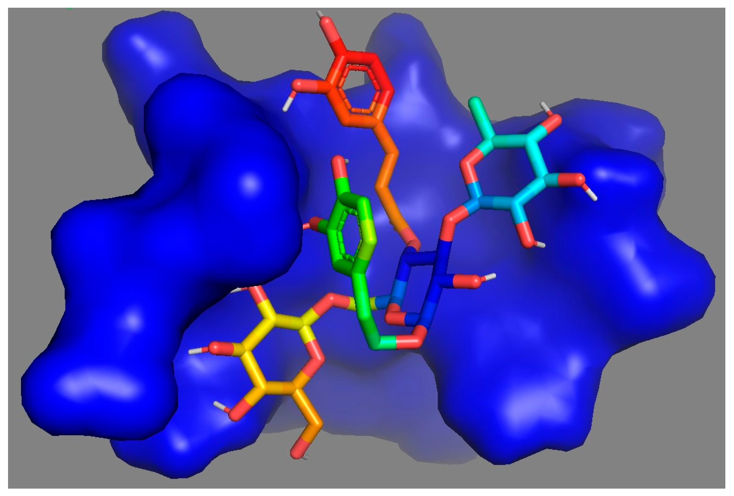
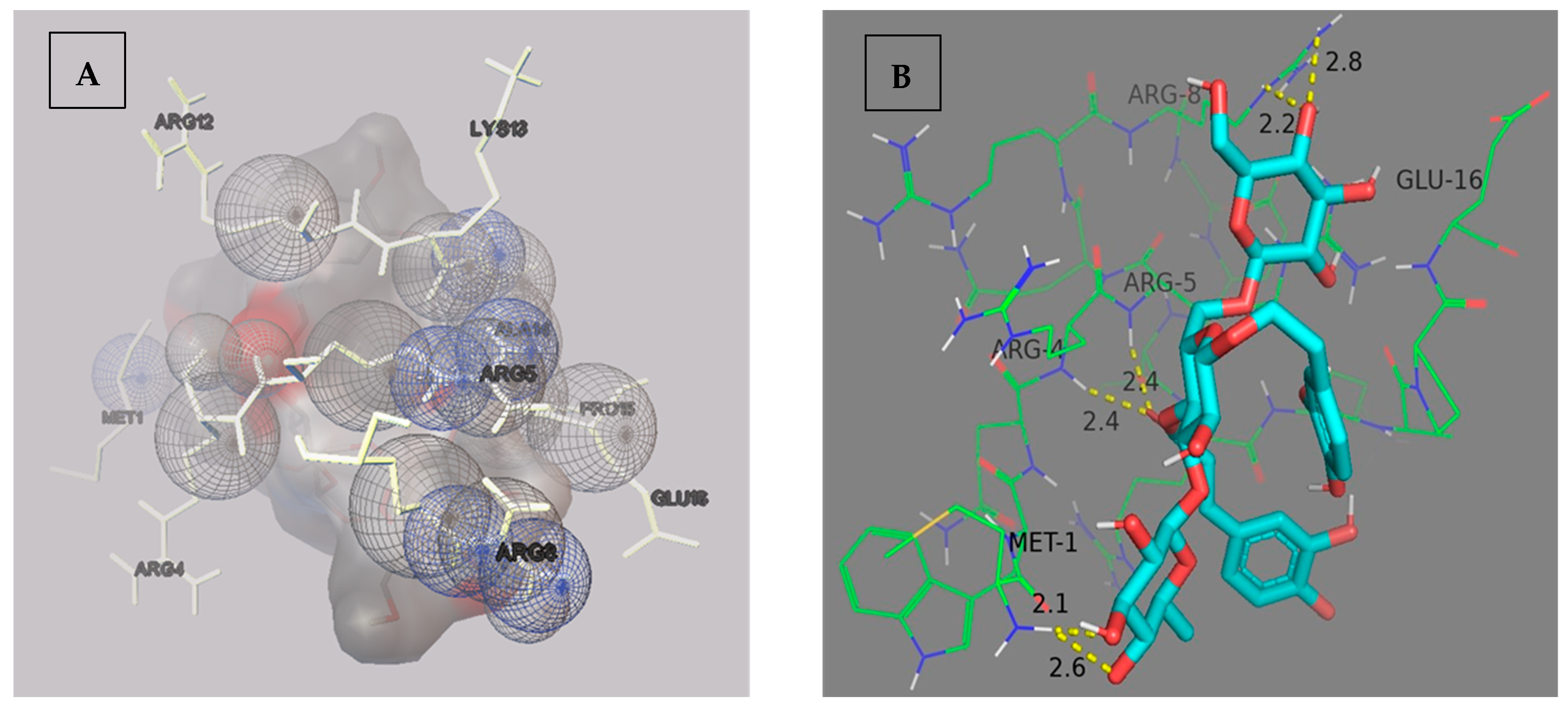
The molecular docking results of the echinacoside (ECH) ligand and RAGE protein are presented in Table 1. The ligand–protein interaction complex was arranged in ascending order of binding energy. The docking pose 1 of the ligand has the most negative binding affinity, with a binding energy of −6.1 kcal/mol, followed by docking pose 2 and 3 with binding energies of −6.0 kcal/mol and −5.9 kcal/mol, respectively. The higher the negative binding affinity, the stronger the anticipated binding to that target protein [23]. Using PyMOL software, the ligand binding pocket for echinacoside in RAGE protein was visualized (Figure 3). The interacting amino acids of the binding site with the ECH molecule were visualized using AutoDock software (Autodock 4.2.6, Scripps Research Institute, San Diego, CA, USA) (Figure 4A). Using PyMOL software, the polar interactions between ECH and the amino acids of the binding site were obtained. Close interaction (2.1–2.8 Å) was observed between the ligand and the protein (Figure 4B).

4. Conclusions

A strong binding relationship between the protein and the ligand is demonstrated through a molecular docking study of echinacoside (ECH) with RAGE. ECH’s potential for diabetic wound healing has not yet been investigated. Therefore, using innovative topical formulations of phenylethanoid glycosides such as ECH can aid in the efficient delivery of drugs. The effectiveness and toxicity of diabetic wound care must be determined by extensive in vitro and in vivo investigations.

Author Contributions

Conceptualization: R.B. and B.S.; methodology: R.B.; software: R.B.; validation: R.B. and B.S.; formal analysis: R.B. and B.S.; investigation: R.B.; resources: R.B.; data curation: R.B.; writing—original draft preparation: R.B.; writing—review and editing: R.B. and B.S.; visualization: R.B.; supervision: B.S.; project administration: R.B.; funding acquisition: R.B. and B.S. All authors have read and agreed to the published version of the manuscript.

Funding

This research was funded by Department of Science & Technology, Government of India, Science and Engineering Research Board, Empowerment and Equity Opportunities for Excellence in Science (DST-SERB-EMEQ), file no. EEQ/2021/000850.

Institutional Review Board Statement

Not applicable.

Informed Consent Statement

Not applicable.

Data Availability Statement

Not applicable.

Conflicts of Interest

The authors declare no conflict of interest.

References

  1. Goova, M.T.; Li, J.; Kislinger, T.; Qu, W.; Lu, Y.; Bucciarelli, L.G.; Nowygrod, S.; Wolf, B.M.; Caliste, X.; Yan, S.F.; et al. Blockade of receptor for advanced glycation end-products restores effective wound healing in diabetic mice. Am. J. Pathol. 2001, 159, 513–525. [Google Scholar] [CrossRef] [PubMed]
  2. Wang, Q.; Cao, X.; Zhu, G.; Xie, T.; Ge, K.; Niu, Y. Blockade of receptor for advanced glycation end products improved essential response of inflammation in diabetic wound healing. Int. J. Diabetes Dev. Ctries. 2020, 40, 283–289. [Google Scholar] [CrossRef]
  3. Wang, C.C.; Blomster, J.I.; Heizer, G.; Berger, J.S.; Baumgartner, I.; Fowkes, F.G.; Held, P.; Katona, B.G.; Norgren, L.; Jones, W.S.; et al. Cardiovascular and limb outcomes in patients with diabetes and peripheral artery disease: The EUCLID trial. J. Am. Coll. Cardiol. 2018, 72, 3274–3284. [Google Scholar] [CrossRef] [PubMed]
  4. Goodson, W.H., III; Hunt, T.K. Studies of wound healing in experimental diabetes mellitus. J. Surg. Res. 1977, 22, 221–227. [Google Scholar] [CrossRef] [PubMed]
  5. Ehrlich, H.P. The physiology of wound healing: A summary of normal and abnormal wound healing processes. Adv. Ski. Wound Care 1998, 11, 326–328. [Google Scholar]
  6. Morain, W.D.; Colen, L.B. Wound healing in diabetes mellitus. Clin. Plast. Surg. 1990, 17, 493–501. [Google Scholar] [CrossRef] [PubMed]
  7. Shoji, T.; Koyama, H.; Morioka, T.; Tanaka, S.; Kizu, A.; Motoyama, K.; Mori, K.; Fukumoto, S.; Shioi, A.; Shimogaito, N.; et al. Receptor for advanced glycation end products is involved in impaired angiogenic response in diabetes. Diabetes 2006, 55, 2245–2255. [Google Scholar] [CrossRef] [PubMed]
  8. Owen, W.F., Jr.; Hou, F.; Stuart, R.O.; Kay, J.; Boyce, J.; Chertow, G.M.; Schmidt, A.M. β2-Microglobulin modified with advanced glycation end products modulates collagen synthesis by human fibroblasts. Kidney Int. 1998, 53, 1365–1373. [Google Scholar] [CrossRef] [PubMed]
  9. Wang, Q.; Zhu, G.; Cao, X.; Dong, J.; Song, F.; Niu, Y. Blocking AGE-RAGE signaling improved functional disorders of macrophages in diabetic wound. J. Diabetes Res. 2017, 2017, 1428537. [Google Scholar] [CrossRef] [PubMed]
  10. Wicks, K.; Torbica, T.; Mace, K.A. Myeloid cell dysfunction and the pathogenesis of the diabetic chronic wound. In Seminars in Immunology; Academic Press: Cambridge, MA, USA, 2014; Volume 26, pp. 341–353. [Google Scholar]
  11. Embil, J.M.; Papp, K.; Sibbald, G.; Tousignant, J.; Smiell, J.M.; Wong, B.; Lau, C.Y.; Canadian Becaplermin Study Group. Recombinant human platelet-derived growth factor-BB (becaplermin) for healing chronic lower extremity diabetic ulcers: An open-label clinical evaluation of efficacy. Wound Repair Regen. 2000, 8, 162–168. [Google Scholar] [CrossRef] [PubMed]
  12. Wetzler, C.; Kämpfer, H.; Stallmeyer, B.; Pfeilschifter, J.; Frank, S. Large and sustained induction of chemokines during impaired wound healing in the genetically diabetic mouse: Prolonged persistence of neutrophils and macrophages during the late phase of repair. J. Investig. Dermatol. 2000, 115, 245–253. [Google Scholar] [CrossRef] [PubMed]
  13. Tian, X.Y.; Li, M.X.; Lin, T.; Qiu, Y.; Zhu, Y.T.; Li, X.L.; Tao, W.D.; Wang, P.; Ren, X.X.; Chen, L.P. A review on the structure and pharmacological activity of phenylethanoid glycosides. Eur. J. Med. Chem. 2021, 209, 112563. [Google Scholar] [CrossRef] [PubMed]
  14. Khangholi, S.; Majid, F.A.; Berwary, N.J.; Ahmad, F.; Abd Aziz, R.B. The mechanisms of inhibition of advanced glycation end products formation through polyphenols in hyperglycemic condition. Planta Med. 2016, 82, 32–45. [Google Scholar] [CrossRef] [PubMed]
  15. Huang, J.; Zhao, D.; Cui, C.; Hao, J.; Zhang, Z.; Guo, L. Research progress and trends of phenylethanoid glycoside delivery systems. Foods 2022, 11, 769. [Google Scholar] [CrossRef] [PubMed]
  16. Li, X.; Gou, C.; Yang, H.; Qiu, J.; Gu, T.; Wen, T. Echinacoside ameliorates D-galactosamine plus lipopolysaccharide-induced acute liver injury in mice via inhibition of apoptosis and inflammation. Scand. J. Gastroenterol. 2014, 49, 993–1000. [Google Scholar] [CrossRef] [PubMed]
  17. Morikawa, T.; Ninomiya, K.; Imamura, M.; Akaki, J.; Fujikura, S.; Pan, Y.; Yuan, D.; Yoshikawa, M.; Jia, X.; Li, Z.; et al. Acylated phenylethanoid glycosides, echinacoside and acteoside from Cistanche tubulosa, improve glucose tolerance in mice. J. Nat. Med. 2014, 68, 561–566. [Google Scholar] [CrossRef] [PubMed]
  18. Chen, W.; Lin, H.R.; Wei, C.M.; Luo, X.H.; Sun, M.L.; Yang, Z.Z.; Chen, X.Y.; Wang, H.B. Echinacoside, a phenylethanoid glycoside from Cistanche deserticola, extends lifespan of Caenorhabditis elegans and protects from Aβ-induced toxicity. Biogerontology 2018, 19, 47–65. [Google Scholar] [CrossRef] [PubMed]
  19. Grosdidier, A.; Zoete, V.; Michielin, O. SwissDock, a protein-small molecule docking web service based on EADock DSS. Nucleic Acids Res. 2011, 39 (Suppl. 2), W270–W277. [Google Scholar] [CrossRef] [PubMed]
  20. Singh, H.; Agrawal, D.K. Therapeutic Potential of Targeting the HMGB1/RAGE Axis in Inflammatory Diseases. Molecules 2022, 27, 7311. [Google Scholar] [CrossRef] [PubMed]
  21. RCSB PDB. Available online: https://www.rcsb.org/structure/6VXG (accessed on 4 January 2023).
  22. National Institutes of Health (NIH). Available online: https://pubchem.ncbi.nlm.nih.gov/compound/Echinacoside (accessed on 4 January 2023).
  23. Dallakyan, S.; Olson, A.J. Small-molecule library screening by docking with PyRx. Chem. Biol. Methods Protoc. 2015, 1263, 243–250. [Google Scholar]
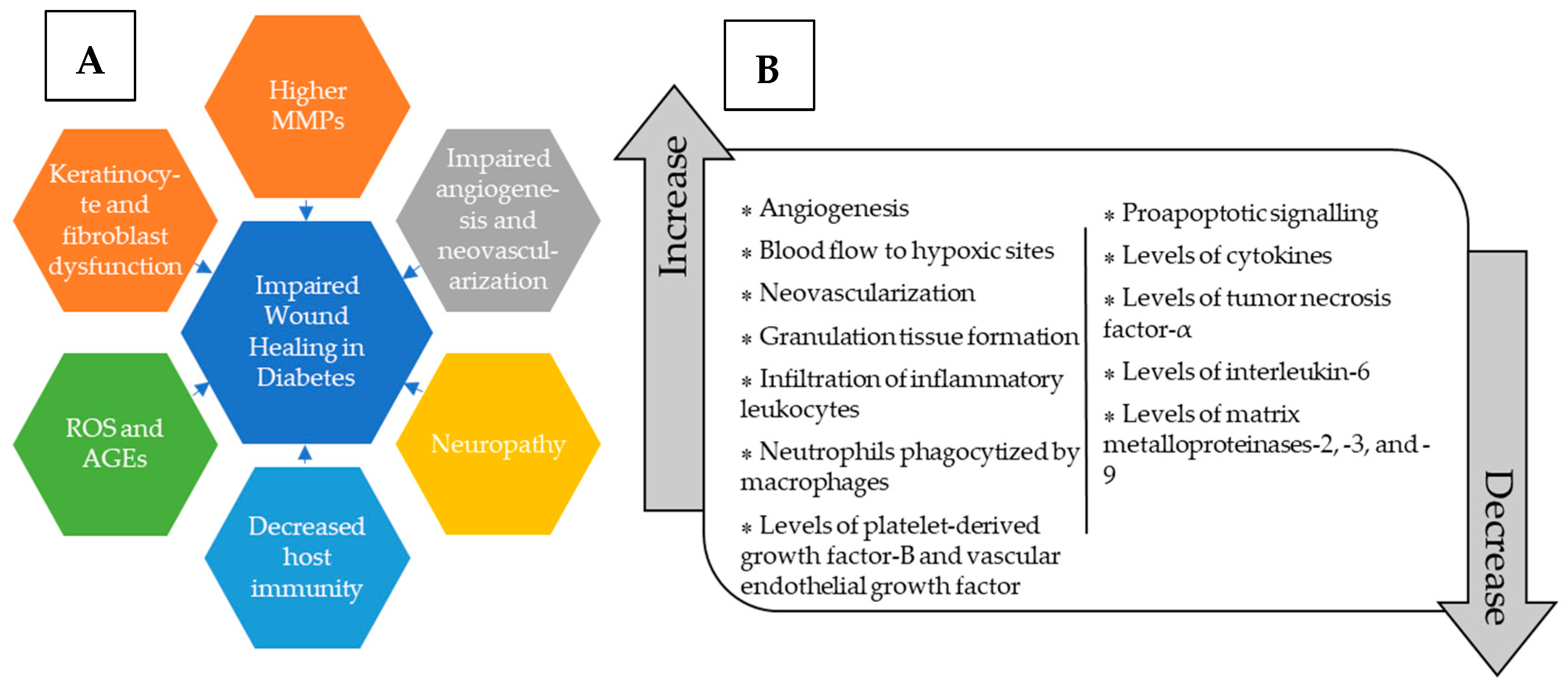
Figure 1. (A) Potential effects of diabetes on wound healing. (B) Role of blockage of RAGE in diabetic wound healing.
Msf 21 00024 g001
Figure 2. (A) Structure of PhG. (B) Structure of ECH. The structure was generated using ChemDraw Software (ChemDraw Professional, Version 15.0.0.106, Perkin Elmer, Informatics, Inc., Bend, OR, US).
Msf 21 00024 g002
Figure 3. The ligand binding pocket for echinacoside in the RAGE protein. RAGE is represented in the form of a surface diagram, and the Echinacoside as sticks.
Msf 21 00024 g003
Figure 4. (A) The interacting amino acids of the binding site (represented as hollow spheres) with the ECH molecule. (B) The polar interactions between ECH and the amino acids of the binding site.
Msf 21 00024 g004
Table 1. The binding energies obtained after molecular docking analysis of the echinacoside (ECH) ligand and RAGE protein. ECH—echinacoside; RAGE—receptor for advanced glycation end products; RMSD—root mean square deviation; L.B.—lower bound; U.B.—upper bound.
Docking PosesComplexBinding Energy (kcal/mol)RMSD L.B.RMSD U.B.
1ECH_RAGE−6.10.0000.000
2ECH_RAGE−6.02.9796.833
3ECH_RAGE−5.92.2984.816
Disclaimer/Publisher’s Note: The statements, opinions and data contained in all publications are solely those of the individual author(s) and contributor(s) and not of MDPI and/or the editor(s). MDPI and/or the editor(s) disclaim responsibility for any injury to people or property resulting from any ideas, methods, instructions or products referred to in the content.