Article

Occurrence of Picobirnavirus in Domestic and Wild Animals from Three Cities of Brazilian Amazon

by
Elaine Hellen Nunes Chagas
1,2,
Julia Rezende da Silva
2,3,
Bruno de Cássio Veloso de Barros
2,4,
José Wandilson Barbosa Duarte Júnior
1,2,
Fabiolla da Silva dos Santos
2,
Edivaldo Costa Sousa Júnior
2,
Delana Andreza Melo Bezerra
2,
Maria Inês dos Santos
1,
Helder Henrique Costa Pinheiro
4,
Yashpal Singh Malik
5 and
Joana D’Arc Pereira Mascarenhas
2,*
1
Department of Parasitology, Institute for Biomedical Sciences, University of São Paulo, São Paulo 05508-060, Brazil
2
Evandro Chagas Institute, Ministry of Health, Ananindeua 67030-000, Brazil
3
Clinical Chemistry and Toxicology Department, School of Pharmaceutical Sciences, University of Sao Paulo, FCF/USP, São Paulo 05508-090, Brazil
4
Healthy Science Institute, Faculty of Dentistry, University Federal of Pará, Belém 66075-110, Brazil
5
ICAR-Indian Veterinary Research Institute, Izatnagar, Bareilly 243122, Uttar Pradesh, India
*
Author to whom correspondence should be addressed.
Zoonotic Dis. 2024, 4(1), 74-85; https://doi.org/10.3390/zoonoticdis4010008
Submission received: 8 November 2023 / Revised: 9 January 2024 / Accepted: 11 January 2024 / Published: 20 February 2024

Abstract

:

Simple Summary

One of the factors contributing to the emergence and resurgence of diseases is the persistent disruption and inadequate management of natural ecosystems. This poses a significant threat to many wildlife species, which are considered potential reservoirs of human diseases, as well as to the biodiversity of the Brazilian Amazon. Understanding the potential of enteric viruses is of great importance due to the substantial impact on both animal and human populations. Picobirnaviruses serve as examples of pathogens that infect various animal species, including mammals and non-mammals, either symptomatically or asymptomatically. They function as emerging opportunistic agents and are regarded as potential zoonotic pathogens. This study successfully detected picobirnavirus in fecal specimens collected from companion animals (mostly cats), livestock (mostly pigs), and wild animals inhabiting areas affected by anthropogenic changes. The data underscore the importance of monitoring viruses in animals in order to identify potential points of spillover to humans.

Abstract

This study aimed to detect picobirnavirus (PBV) in the fecal samples of wild and domestic animals from 2014 to 2016 in the Amazon biome. Fecal samples from different animals, including birds (n = 41) and mammals (n = 217), were used. The PAGE test showed negativity for PBV. However, 32 samples (12.4%, 32/258) showed positive results in RT-PCR analyses. Among the positive samples, pigs and cats, both with 28.12% (9/32), registered the highest frequencies. In a phylogenetic analysis, eight sequences from positive samples were grouped in the Genogroup 1 of PBV (PBV GI). PBV occurrence was significantly related to cats and pigs but not other mammals or birds, independently of their geographical origin. A nucleotide analysis demonstrated similarity among the feline group but the absence of a defined structure between the clades. PBVs are highly widespread viruses that can affect the most diverse types of hosts in the Amazon biome, including humans.

Graphical Abstract

1. Introduction

Picobirnaviruses (PBVs) are small viruses approximately 40 nm in diameter, non-enveloped, with icosahedral symmetry and two genomic segments, a larger segment with 2.2–2.7 kbp size and a smaller segment with 1.2–1.9 kbp [1,2,3]. The larger segment (segment I) plays a role in the coding of structural proteins, and the smaller (segment II) encodes the RNA-dependent viral RNA polymerase (RdRp) and determines PBV’s classification [4].
The current PBV is classified into Genogroup I and II, based on strains 1-CHN-97 (GI) and 4-GA-91 (GII) from humans infecting vertebrate animals, and the Genogroup III, recently proposed, from strains infecting invertebrate animals [5].
PBVs are associated with gastroenteritis in both humans and animals and may present as an apparent symptom of diarrhea that may be directly linked to the virus as a single pathogen or other enteric pathogens, attributing to it the characteristic of a secondary pathogen in mixed infections [6]. These viruses are further classified as opportunistic pathogens when detected in immunocompromised patients and those who have diarrheal conditions [7,8].
PBVs are considered to be emerging, opportunistic, and suggestive of zoonotic potential [9]. The first report on PBV appeared from an outbreak of gastroenteritis in Brazil, where viruses were detected in human stool specimens and in a rat species [9]. Since then, PBVs have been described as pathogens that infect a variety of animal species, such as mammals, reptiles, birds, humans, and is also present in sewage, showing a genetic diversity among circulating strains [6,10,11]. Studies suggest that PBVs are bacteriophages of bacteria in the digestive tract of possible hosts of this pathogen [12,13].
Currently, several pathogens, including enteric viruses that affect humans and animals, have been estimated as responsible for 60% of zoonotic infections in humans [14,15,16]. Zoonoses are responsible for 75% of emerging and reemerging diseases that affect humans and cause at least a 20% loss in animal production [14]. Thus, the investigation of PBV in samples from different hosts is important in relation to public health, due to their potential for zoonotic transmission [17]. Therefore, it is imperative to monitor wild and domestic animals for the presence of different human pathogens which may trigger possible events of zoonotic transmission.
The presence of enteric viruses in various matrices highlights the anthropogenic impact on the surrounding population, indicating the potential risk of infection through the exposure of susceptible individuals. Such records enhance the tracking of fecal contamination in the environment, utilizing animals as indicators, aiming to minimize the risk of infection through exposure to different animal species or even human populations [18].
In this study, we chose three cities that represent deforestation areas where the interaction among animals and humans are provided by anthropic actions. We aimed to detect the presence of PBV in fecal specimens of wild and domestic animals collected in deforested areas of the Amazon biome.

2. Materials and Methods

2.1. Ethical Aspects
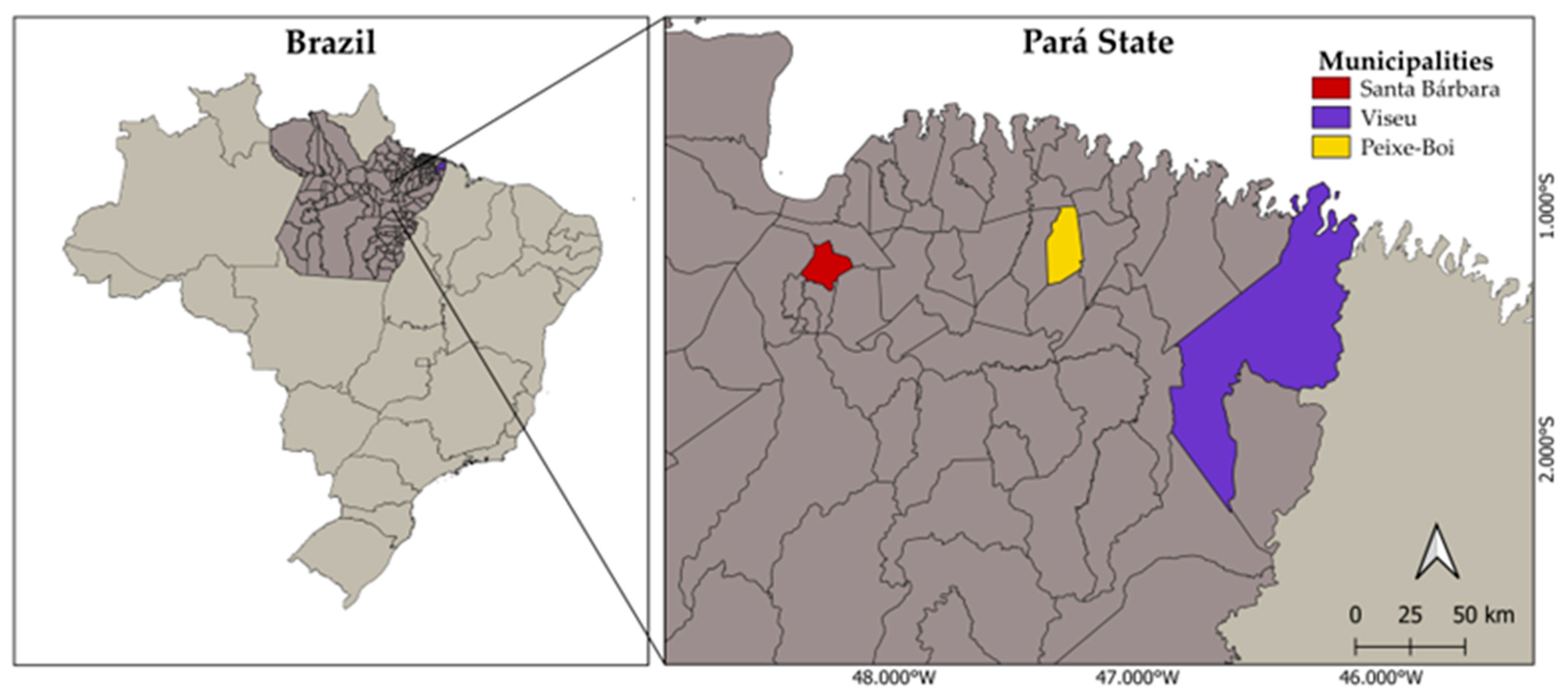
This research was approved by the National Council for Animal Control and Experimentation (CONCEA), System of Authorization and Information in Biodiversity—SISBIO/ICMBIO/Ministry of the Environment, under protocol No. 37174–1, and the Ethics Commission at the Evandro Chagas Institute (CEUA) under protocol number 35/2016.

2.2. Study Area

The study area and the collection of biological samples comprised three cities: Santa Bárbara (Expedito Ribeiro settlement), Peixe-Boi (Vila do Ananin), and Viseu (Açaiteua-Centro Alegre), located in the north of Pará state (Figure 1). The respective area of the three municipalities comprises 278.15 km2, 450.29 km2, and 4.934.54 km2. The cities are part of a portion of the Brazilian Amazon, where the strong anthropic pressure increases the interaction between humans and animals. Also, the contact and handling of animals is becoming increasingly intense due to agribusiness, which is the main source of subsistence for families living there.
These areas have already been investigated for enteric virus, rotavirus, and infected domestic and wild animals. Data concerning territorial extension and deforestation were published before [19].

2.3. Sample Collection

Sample collection was carried out from October 2014 to April 2016, comprising two annual visits to each city in the study area. The capture of wild animals (bats, birds, marsupials, and rodents) was carried out from areas of fragmented forest and adjacent urban areas, where a greater concentration of these species would be expected.
Different traps were used to capture wild animals. For birds and bats, fog nets were set up, at different times, from 4:00 a.m. to 9:00 a.m. and 6:00 p.m. to 7:00 a.m., respectively, and inspected periodically. The other wild animals were captured using Tomahawk (45 × 16 × 16 cm), Sherman (30 × 9 × 8 cm), and Pitfall traps. The baits used to attract the animals to the traps were peanut butter, sardines, bacon, and fruit (pineapple, banana, and apple).
The biological material was collected by stimulation of the rectum using urethral probe (No 10) for the no-fly wild mammals and rectal swab “Zaragatoa” for wild birds and bats (small animals) through stimulation of the rectal ampulla. In relation to companion animals (dogs, cats, and poultries) and other mammals (pigs, horses, and cattle), sample collection and necessary information about the animals were carried out with authorization and contribution from their respective owners. The consent letter given to the owners can be found in the Supplementary Materials.
All samples were stored in sterile plastic vials and properly packed (−30 °C) to maintain the quality of the material before processing in the virology laboratory.

2.4. Samples

A total of 258 fecal samples were used in this study. The sample groups were composed of mammals (flying and non-flying) and birds (domestic and wild). The non-flying mammals include bovines (n = 23), canines (n = 34), equines (n = 41), felines (n = 33), rodents (n = 8), marsupials (n = 25), and swines (n = 23). The flying mammals are the chiropters (n = 30). The bird group includes domestic birds (n = 3) and wild birds (n = 38). The sample table is available in Supplementary Material (Table S1).

2.5. Sample Preparation

Fecal suspensions were prepared at 20% (w/v) in Tris/Ca2+ and clarified by centrifugation at 5000 rpm for 10 min at 4 °C. Then, supernatants were collected and stored at −20 °C for later use in the extraction, detection, and characterization of viral genetic material.
The viral genome was extracted from the fecal suspensions using silica glass powder [20]. During the extraction process, all contamination control measures were performed, including the use of positive (PBV positive sample) and negative controls (ultrapure water). Polyacrylamide Gel Electrophoresis (PAGE) and silver staining were applied to analyze the electrophoretic profile of the samples, according to the method previously described [21].
RT-PCR was used to detected PBV, amplifying the RdRp genomic segment using the PicoB25 and PicoB43 primers for Genogroup I, and PicoB23 and PicoB24 for Genogroup II, which amplify 201 bp and 369 bp products, respectively [22]. cDNA was synthesized by adding first 4 μL of extracted RNA and 1 μL of each primer pair (20 mM), mixed and denatured at 97 °C for 5 min in a thermocycler, followed by 5 min in an ice bath. Subsequently, 2 μL of dNTPs (20 mM, Promega®, Madison, WI, USA), 0.75 μL of MgCl2 (25 mM, Promega®), 2.5 μL of buffer (5×, Promega®), 0.5 μL of Reverse Transcriptase enzyme (4U, Promega®), and 13.25 μL of RNAse/DNAse free H2O were added, up to 25 μL of final volume. The synthesis reaction was carried out at 42 °C for 1 h.
cDNA was PCR-amplified by adding 25 μL of cDNA to 3 μL of dNTP (20 mM, Promega®), 2.5 μL of buffer (5×, Promega®), 0.75 μL of MgCl2 (25 mM, Promega®), 0.25 μL of Taq Polymerase (5U, Promega®), and 18.50 μL of RNAse/DNAse free H2O (Promega®, Madison, WI, USA). The tube with 50 μL of final volume was submitted to PCR as follows: initial denaturation step at 95 °C for 2 min, 35 cycles at 95 °C for 1 min, 50 °C for 1 min, and 72 °C for 1 min, and a last extension step of 72 °C for 5 min.
Positive samples from PCR were tested to Nested-PCR. This Nested-PCR amplified gene segment-2 (RdRp gene) by using the primer pairs PBV 1.2 FP, 5′AAGGTCGGKCCRATGT3′, and PBV RP, 5′TTATCCCYTTTCATG CA3′ resulting in a ~1229 bp amplicon for the first stage (RT-PCR), according to protocols previously described [23].
Positive samples in the first Nested-PCR were then submitted to a second PCR using the primers Malik-2-FP (5’-TGG GWT GWT GGC GWG GAC ARG ARGG-3′) and Malik-2-RPreverse (5′-YSC AYT ACA TCC AC-3′TCC) which amplified a ~580 bp RdRp fragment only for the Genogroup I [18]. Conditions for this step were: initial denaturation at 95 °C for 5 min, followed by 35 cycles at 94 °C for 10 s, 48 °C for 25 s, and 72 °C for 45 s, and a last extension step of 72 °C for 10 min.
Subsequently, amplicons were purified (Promega Wizard PCR kit) and subjected to Sanger sequencing using the protocol of the Big Dye Terminator® v.3.1 kit (Applied Biosystems, Foster City, CA, USA).

2.6. Sequence Analysis

Obtained sequences were aligned and edited using the Geneious program (version 8.1.9) and compared with sequences available in the National Center for Biotechnology Information (NCBI) database (www.ncbi.nlm.nih.gov, accessed on 11 August 2020) using the BLAST algorithm (version BLAST + 2.8.0-alpha).
Phylogenetic trees were built using the maximum likelihood method (Maximum Likelihood-ML) with the FastTree program. To determine the best nucleotide replacement model, the GTR (General Time Reversible) program was used. Bootstrap analysis (1000 replicates) was used to give reliability to phylogenetic groups. The tree was visualized using the Evolview software version 2 [24].
Samples exhibiting similarities to bacteria, fungi, and other microorganisms were considered unviable. Consequently, these specific samples were intentionally excluded from the analysis to bolster the overall reliability of our results. The construction of the phylogenetic tree included PBV sequences available from GenBank, belonging to different animal hosts and samples from previous studies from the same collection region and belonging to PBV-GI.

2.7. Statistical Analysis
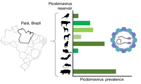
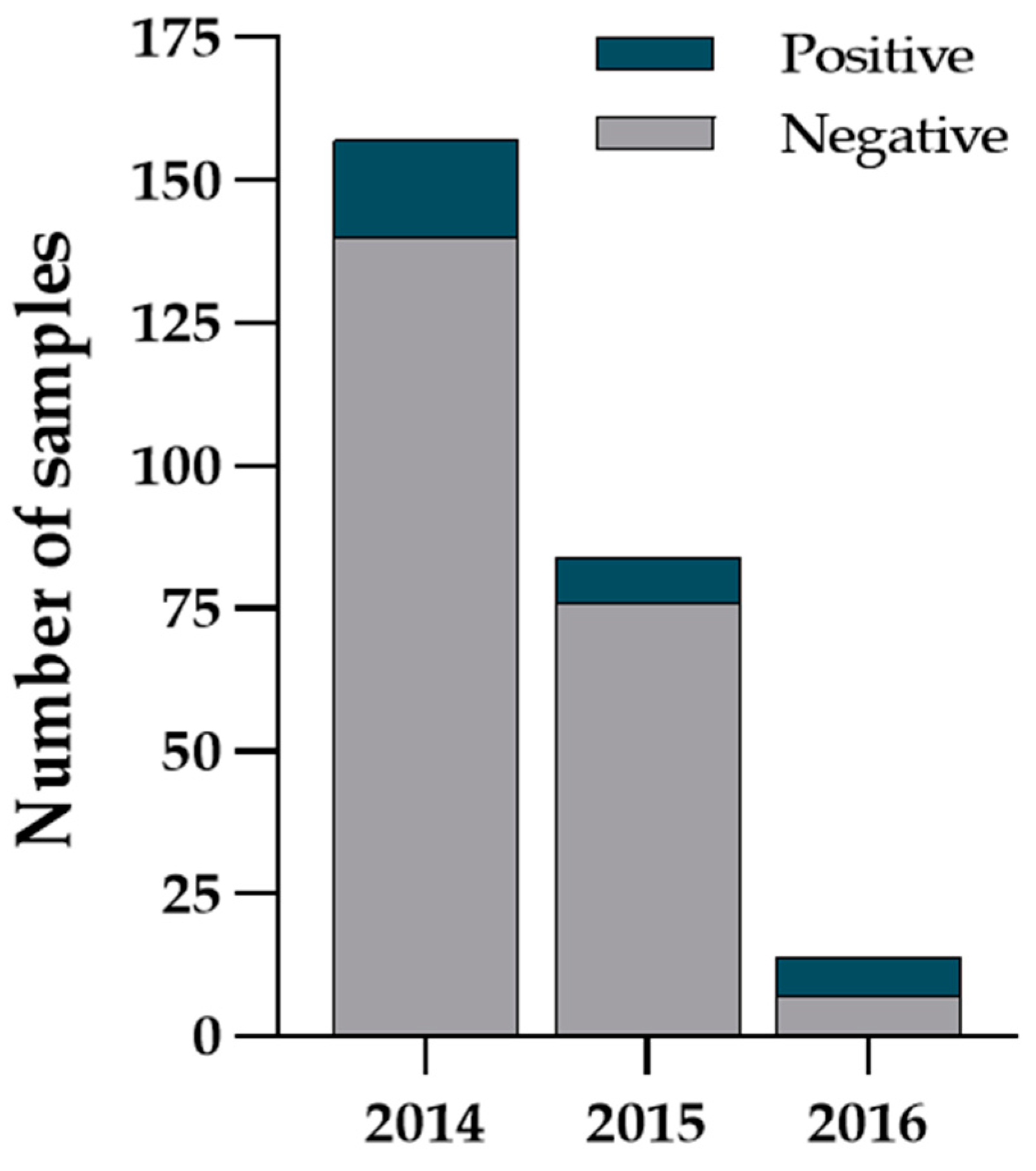
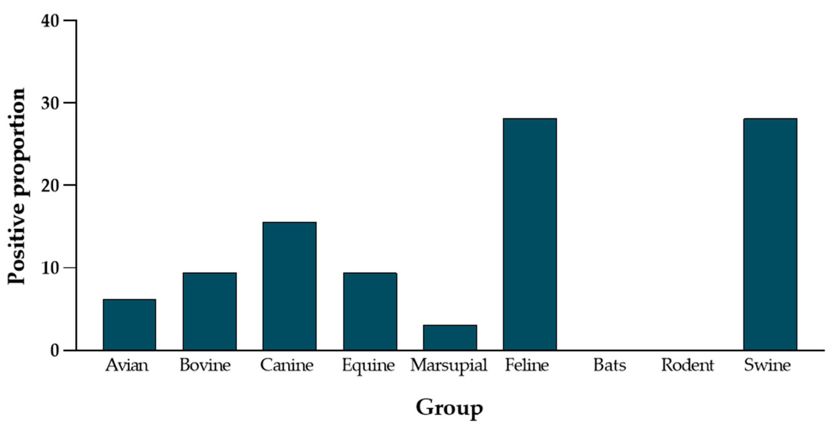
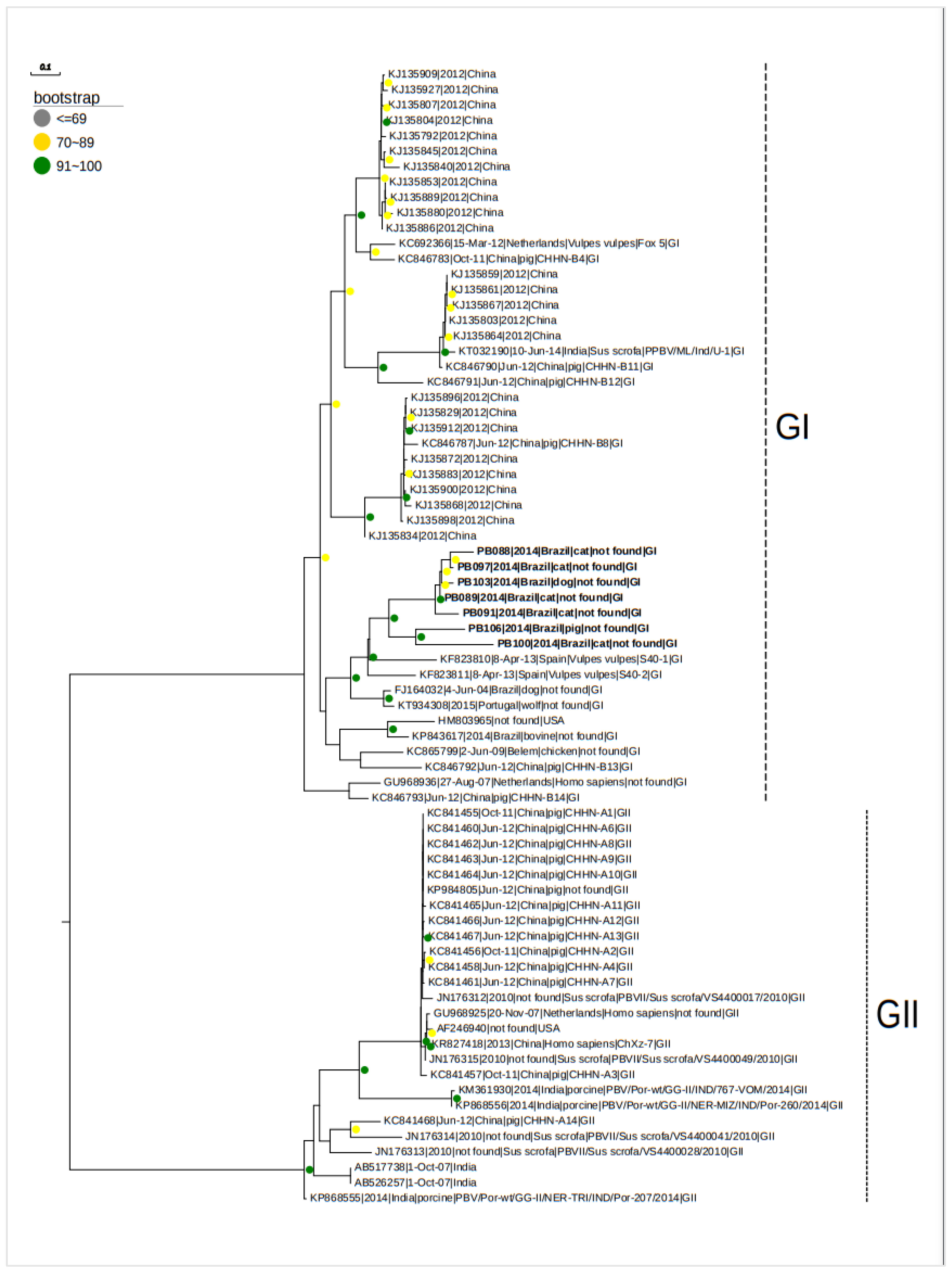
A descriptive statistical analysis of the data was performed using R Statistical Software version 4.2.1 (RVAidemamoirre package and Graphpad Prism version 8.4). Categorical variables were summarized using frequencies and percentages. G and Chi-square tests were used to identify the frequency of pathogens in the cities, the period of collection, and the categorical variables: group of animals, gender (male and female), and city from where the samples originated. The significance value in the results was α = 0.05 (error probability of 5%).

3. Results

From September 2014 to March 2016, 258 biological samples were collected from the municipalities of Santa Bárbara, Peixe-Boi, and Viseu located in the state of Pará, Brazil. Most part of the samples (157/258) were collected in the year 2004, as shown in Figure 2. The biological materials were from different groups of animals including 84.10% of mammals (217/258) and 15.90% of birds (41/258). The viral RNA of each sample was analyzed using PAGE and then RT-PCR. No viral RNA could be detected with PAGE; however, RT-PCR assays showed positive results exclusively for Genogroup I (PBV GI).
In a total of 32 samples (12.4%), segment 2 (PBV-G1) could be amplified. No sample contained PBV GII. The frequency of PBV in this study (Figure 3) was concentrated in swines (9/32), which represented 28.13% of positivity, and felines (9/32), which showed 28.13% of positivity in cats, similar to swine. The other PBV-positive samples were 6.3% of avian (2/32), 9.4% of bovine (3/32), 145.6% of canine (5/32), and 9.4% of equine (3/32) origin. Only one positive sample was from a marsupial (3.13%, 1/32). No PBV was detected in the samples from bats or rodents.
In total, 211 animal samples were gender-identified, and of these, 47.40% samples were from males (100/211) and 52.60% from females (111/211), with it not being possible to identify the sex of 47 animals. Most positive samples were identified in Viseu with 43.75% (14/32), followed by Santa Bárbara (10/32) and Peixe-Boi (8/32) (Figure 4).
In the statistical analysis, G and Chi-square tests were performed to assess the significance of positivity between the different groups of animals (birds, cattle, canines, equines, swine, felines, bats, marsupial, and rodents). The results of this analysis demonstrated that the frequency of PBV was not significant between genders (p > 0.05). The occurrence of PBV between animal groups and among the cities from the samples was significant, showing p < 0.05 in both variables. The data comparing the groups of animals, their genders (male and female), and cities from the samples (Santa Bárbara, Peixe-Boi, and Viseu) are reported in Supplementary Materials (Table S2).
For further phylogenetic analysis, 32 samples were amplified by RT-PCR for PBV GI. These samples were subjected to Nested-PCR since only seven samples (21.87%) were satisfactory for sequencing. The PBV-positive samples sequenced were from five felines, one swine, and one canine. Accession numbers in GenBank are MT872191 (PB089), MT872192 (PB097), PB100 MT872193 (PB100), PB103 MT872194 (PB103), PB106 MT872195 (PB106), PB088 MT872196 (PB088), and MT872197 (PB091).
The samples in the phylogenetic tree were grouped heterogeneously, coinciding with clades of PBV reported from different countries and a variety of species. The genotype characterization identified all the sequences as part of PBV GI, as illustrated in Figure 4.
Alignment and phylogenetic analysis showed a nucleotide identity from 70% to 89% identity among the samples in the study. All of the sequences from five felines (Felis silvestris catus) with codes PB088, PB097, PB089, PB091 and PB100, one dog (Canis lupus familiaris) with code PB103 and one swine with code PB106 remained close together in the same clade with bootstrap values in the range of 70–100%. Interestingly, a comparison of the identified RdRp gene sequences with other PBVs showed that sequences were most closely related to picobirnaviruses detected previously in red fox samples from Spain [25].

4. Discussion

Several pathogens, including enteric viruses, which affect humans and the most varied species of animals are important players of zoonotic infections in humans and also cause a loss of productivity in life stock [14].
In the first attempt, the simple but accurate PAGE technique was used to screen the electrophoretical profile of infecting viruses in diarrheal infections as successfully carried out by others [4]. In the present study, PAGE proved to exert a too low sensitivity of detection of PBV, probably due to the limited sample quantity. It is worth noting that it was not considered whether fecal samples collected were from animals displaying clinical signs of diarrhea or not. This statement may be relevant in the current study, as the reduced viral load in the animal excreta could justify the low sensitivity of PAGE, consistent with studies that did not exhibit an electrophoretic profile in positive samples [18,19]. We emphasize that even though we used 10 µL of the extracted sample, the PBV-positive samples did not exhibit an electrophoretic profile. It is known that, even in animals with PBV infections, the quantity of viral particles excreted in feces may not be detectable by PAGE. For this reason, the RT-PCR based approach was employed, given its greater sensitivity and specificity [8]. Additionally, fecal samples with lower viral amounts could be identified.
Clinical aspects of animals and fecal specimens were not considered, since detection may happen even though hosts, in many cases, remain asymptomatic [4,17]. In the present study, no specific effort was made to elucidate viral titers in samples. Previous studies carried out with fecal samples of broilers (Gallus gallus) from the northeastern region of the state of Pará, reported PAGE-based PBV detection in 15.3% [26] and 30% [27] of samples, indicating that in comparison to fowl feces.
We have documented a positivity rate of 12.4% among the samples, all of which tested positive for PBV-GI. Different primer sets were employed in the investigation, utilizing both conventional and NESTED PCR techniques to ensure heightened specificity in detecting positivity. Despite these efforts, the outcome for PBV-GII yielded negative results. Consequently, we suggest that GII might not have been in circulation within the geographical regions from which the samples were derived. Alternatively, the absence of positive findings could be attributed to the restricted sample size, possibly resulting in an underestimated prevalence of PBV in the studied animals. It is worth noting that PBV-GII is generally detected to a lesser extent when compared to its counterpart, GI.
PBV were previously reported in several different hosts, being detected in 3.63% of cats from Portugal [28], 14.28% in horses [17], 11.15% in pigs [29], and 0.73% in cattle in India [30]. In Brazil, the positivity in swine was reported in 12.45% to 43.24% of animals [31], 8.30% in cattle [32], 1.84% in canines [33], and 49.4% in broilers [26]. Until now, no PBV had been detected in marsupials (Didelphidae family) and in wild birds. The present study supports a high PBV positivity in swine in Brazil, considering that the population with the highest frequency of PBVs was in swine showing 28.13% of positivity.
The counties of origin of samples in this study were strongly deforested during 2014 to 2016, due the use of the land for subsistent agricultural and livestock production [34]. Clearly, the increase in the area of fragmentation, the alteration of the habitat and ecological niches of animals caused by anthropic pressure, and consequently, the closer contact between humans and wild animals contribute to the occurrence of infectious diseases, the spread of emerging pathogens, and new hosts [35,36,37,38]. This fact may have contributed to the wide interaction between the species, facilitated by the change in the environment where they live, and consequently higher viral spillover between species.
The dissemination of PBV among the most varied animal hosts in the present study was confirmed by phylogenetic analysis. The virus presence in canines, felines, cattle, poultry, swine, and in humans shows a lack of host specificity compared to a previous study [10], and we detected lower positive populations of PBV in our animal groups. Phylogenetic analysis revealed that the virus circulated in several species of animals, regardless of time and geographic area of detection. This fact suggesting that the sequences do cluster together can be a contributing factor from time and the geographic area of detection. The similar profile was observed in mammals, such as orangutans [39].
Rodents, birds, and bats are among the wild animals in this study. Considering a fecal–oral infection route, and that the diet of marsupial (Marmosa sp., family Didelphidae) is composed of small vertebrates (little birds, chickens), fruit, decomposing organic matter (carrion), and even invertebrates [40], the dietary and social habits of these animals could, theoretically, facilitate the spread of infection among them; however, interestingly, these same animals showed lower or no positivity for PBV. Thus, we can speculate that PBV was more prevalent in urban and peri-urban environments than in the wild.
Birds and chiropterans present great importance for public health. They are considered as potential reservoirs of pathogens that cause zoonotic diseases. Due to their ability to fly, they can act in the dispersion of viruses and thus establish new foci of emerging or re-emerging diseases along their paths. Chiropterans that participated in this study, except Desmodus rotundus species (blood-sucking habit), are frugivorous. Characteristics such as diet, the ability to fly, seasonal migration, behavioral patterns, an affinity to live in colonies, and others, make this group a potential zoonotic host. Perhaps for their foraging habits, there was no PBV positivity for bats in this study. This assumption is also supported by the higher positivity rate observed in domestic and livestock animals, possibly due to increased contact with other pets, humans, and areas where these animals interact with each other.
The strong anthropic pressure in the Amazon has been an important factor of spreading zoonotic diseases, infecting new hosts, and expanding pathogens with wide-reaching potential through the interaction between species, including enteric pathogens such as PBV and RV [19]. Pets (canines and felines) and livestock animals (cattle, horses, pigs, and birds) sharing the same space and environment as humans, circulate in forest and home areas, interacting with each other and with humans, which may increase the chances of viral transmission, especially in cases of animals infected in a phase of viral excretion.
This study is the first to characterize PBV (GI) in felines (Felis catus) in Brazil. These data suggest the possible interspecies transmission of PBV among the animals included in this study, considering that some animals were grouped with different taxa and some samples formed clusters with strains similar to strains isolated in porcine detected mainly in China. The study identified the presence of PBV in various animal species, both in proximity to human settlements and in areas within forest remnants. These locations are situated in municipalities experiencing high environmental impact due to activities such as plant extractivism, pasture formation for livestock, exploitation of natural resources, and direct disturbances to the habitats of wild animals, which may serve as reservoirs for viruses, thereby facilitating viral transmission among coexisting animal communities. In these settings, animals exhibit increased interaction with the human population, sharing common spaces and traversing between forested environments and this study’s collection areas.
Notably, poultry farming is situated in close proximity to residences, and domestic dogs and cats freely move between homes and the forest, potentially establishing direct contact with potential sources of contamination. The absence of septic tanks in residential areas is another contributing factor that may promote, or even escalate, the risk of viral dissemination in the environment, leading to infections in diverse animal populations.
The occurrence of PBV in wild bird species Glyphorynchus spirurus and Attila spadicrus has been described for the first time in Brazil. The epidemiological monitoring of infectious agents in animals, especially companion animals, is crucial for mitigating the rapid transmission, evolution, and adaptation to new environments and hosts. While PBV is not a recently discovered virus, epidemiological studies related to it are still in their early stages, and there is a significant shortage of epidemiological data and experimental models globally. In the context of the current study, we conducted an eco-surveillance to identify the circulation of PBV in domestic, wild, and peri-domestic animals. The application of molecular characterization and phylogenetic analysis emerges as an essential tool to deepen our understanding of the epidemiology, origin, evolution, and emergence of new viruses.

5. Conclusions

Further investigation picobirnavirus was detected in a variety of species including domestic and wild animals from three different cities in Brazil. The incidence of PBV G-I in this study occurred in different groups of both sylvatic and domestic animals. Investigations to better characterize transmission, pathogenicity, and evolution, and increasing surveillance of picobirnaviruses are essential to a more comprehensive understanding of their potential transmission to humans or other animals.

Supplementary Materials

The following supporting information can be downloaded at: https://www.mdpi.com/article/10.3390/zoonoticdis4010008/s1. Table S1: Samples collected and tested to Picobirnavirus, Table S2: Data referring to statistical analysis of the frequency of PBV in fecal samples of animals, according to the group, gender, and city of origin, in the period from October-2014 to April-2016, Informed Consent–IEC.

Author Contributions

Conceptualization, E.H.N.C., J.D.P.M., J.R.d.S. and Y.S.M.; Methodology, E.H.N.C., J.W.B.D.J., D.A.M.B. and B.d.C.V.d.B.; Statics Analyze, M.I.d.S. and H.H.C.P.; Phylogenetic Analyze, E.C.S.J. and F.d.S.d.S.; Writing—Original Draft, E.H.N.C.; Writing—Review and Editing, all authors; Supervision, J.D.P.M. and Y.S.M. All authors have read and agreed to the published version of the manuscript.

Funding

This study was financially supported by the Brazilian National Council for Scientific and Technological Development (CNPq), process nos. 137442/2016-5, 1666682/2017-9, and Evandro Chagas Institute, Ministry of Health, Ananindeua, Brazil. E.H.N.C., J.R.S. and J.W.B.D.J. were recipients of CNPq fellowships; B.D.C.V.B. and C.M.O.C. were recipients of CAPES fellowships; and J.D.P.M. is the recipient of CNPq fellowships. Y.S.M. acknowledges the help received from Education Division, Indian Council of Agricultural Research, New Delhi for National Fellowship.

Institutional Review Board Statement

The National Council for Animal Control and Experimentation (CONCEA), System of Authorization and Information in Biodiversity—SISBIO/ICMBIO/Ministry of the Environment approved this research under protocol No. 37174-1, and the Ethics Commission at the Evandro Chagas Institute (CEUA) under protocol number 35/2016. This document was approved on 19 September 2016.

Informed Consent Statement

The informed consent statement can be found in the Supplementary Materials.

Data Availability Statement

The data presented in the manuscript are available on request from the first author/corresponding author.

Acknowledgments

We would like to thank Ceyla Castro for her support and essential collaboration in this study.

Conflicts of Interest

The authors declare no conflicts of interest regarding this publication.

References

  1. Malik, Y.S.; Ghosh, S. Etymologia: Picobirnavirus. Emerg. Infect. Dis. 2020, 26, 89. [Google Scholar] [CrossRef]
  2. Collier, A.M.; Lyytinen, O.L.; Guo, Y.R.; Toh, Y.; Poranen, M.M.; Tao, Y.J. Initiation of RNA Polymerization and Polymerase Encapsidation by a Small dsRNA Virus. PLoS Pathog. 2016, 12, e1005523. [Google Scholar] [CrossRef] [PubMed]
  3. Kashnikov, A.Y.; Epifanova, N.V.; Novikova, N.A. Picobirnaviruses: Prevalence, genetic diversity, detection methods. Vavilov. J. Genet. Breed. 2020, 24, 661. [Google Scholar] [CrossRef] [PubMed]
  4. Delmas, B.; Attoui, H.; Ghosh, S.; Malik, Y.S.; Mundt, E.; Vakharia, V.N. ICTV virus taxonomy profile: Picobirnaviridae. J. Gen. Virol. 2019, 100, 133–134. [Google Scholar] [CrossRef] [PubMed]
  5. Perez, L.J.; Cloherty, G.A.; Berg, M.G. Understanding the genetic diversity of picobirnavirus: A classification update based on phylogenetic and pairwise sequence comparison approaches. Viruses 2021, 13, 1476. [Google Scholar] [CrossRef] [PubMed]
  6. Ganesh, B.; Masachessi, G.; Mladenova, Z. Animal Picobirnavirus. Virusdisease 2014, 25, 223–238. [Google Scholar] [CrossRef]
  7. Li, W.; Qiang, X.; Qin, S.; Huang, Y.; Hu, Y.; Bai, B.; Hou, J.; Gao, R.; Zhang, X.; Mi, Z.; et al. Virome diversity analysis reveals novel enteroviruses and a human picobirnavirus in stool samples from African green monkeys with diarrhea. Infect. Genet. Evol. 2020, 82, 104279. [Google Scholar] [CrossRef]
  8. Malik, Y.S.; Kumar, N.; Sharma, K.; Dhama, K.; Shabbir, M.Z.; Ganesh, B.; Kobayashi, N.; Banyai, K. Epidemiology, Phylogeny, and Evolution of Emerging Enteric Picobirnaviruses of Animal Origin and Their Relationship to Human Strains. Biomed. Res. Int. 2014, 2014, 780752. [Google Scholar] [CrossRef]
  9. Pereira, H.G.; Fialho, A.M.; Flewett, T.H.; Teixeira, J.M.S.; Andrade, Z.P. Novel viruses in human faeces. Lancet 1988, 2, 103–104. [Google Scholar] [CrossRef]
  10. Ghosh, S.; Malik, Y.S. The True Host/s of Picobirnaviruses. Front. Vet. Sci. 2021, 7, 615293. [Google Scholar] [CrossRef]
  11. Yinda, C.K.; Vanhulle, E.; Conceição-Neto, N.; Beller, L.; Deboutte, W.; Shi, C.; Ghogomu, S.M.; Maes, P.; Van Ranst, M.; Matthijnssens, J.; et al. Gut Virome Analysis of Cameroonians Reveals High Diversity of Enteric Viruses, Including Potential Interspecies Transmitted Viruses. mSphere 2019, 4, e00585-18. [Google Scholar] [CrossRef] [PubMed]
  12. Krishnamurthy, S.R.; Wang, D. Extensive conservation of prokaryotic ribosomal binding sites in known and novel picobirnaviruses. Virology 2018, 516, 108–114. [Google Scholar] [CrossRef] [PubMed]
  13. Neri, U.; Wolf, Y.I.; Roux, S.; Camargo, A.P.; Lee, B.D.; Kazlauskas, D.; Chen, I.M.; Ivanova, N.; Allen, L.Z.; Paez-Espino, D.; et al. A Five-Fold Expansion of the Global RNA Virome Reveals Multiple New Clades of RNA Bacteriophages. SSRN Electron. J. 2022, 185, 4023–4037.e18. [Google Scholar] [CrossRef]
  14. Zanella, J.R.C. Zoonoses emergentes e reemergentes e sua importância para saúde e produção animal. Pesqui Agropecuária Bras. 2016, 51, 510–519. [Google Scholar] [CrossRef]
  15. Brown, C. Human-Animal Medicine: Clinical Approaches to Zoonoses, Toxicants and Other Shared Health Risks. Emerg. Infect. Dis. 2010, 16, 1050. [Google Scholar] [CrossRef]
  16. Monath, T.P. Vaccines against diseases transmitted from animals to humans: A one health paradigm. Vaccine 2013, 31, 5321. [Google Scholar] [CrossRef] [PubMed]
  17. Ganesh, B.; Banyai, K.; Masachessi, G.; Mladenova, Z.; Nagashima, S.; Ghosh, S.; Nataraju, S.M.; Pativada, M.; Kumar, R.; Kobayashi, N. Genogroup i picobirnavirus in diarrhoeic foals: Can the horse serve as a natural reservoir for human infection? Vet. Res. 2011, 42, 52. [Google Scholar] [CrossRef] [PubMed]
  18. Duarte Júnior, J.W.B.; Chagas, E.H.N.; Serra, A.C.S.; Souto, L.C.D.S.; da Penha Júnior, E.T.; Bandeira, R.d.S.; e Guimaraes, R.J.d.P.S.; Oliveira, H.G.d.S.; Sousa, T.K.S.; Lopes, C.T.d.A.; et al. Ocurrence of rotavirus and picobirnavirus in wild and exotic avian from amazon forest. PLoS Negl. Trop. Dis. 2021, 15, e0008792. [Google Scholar] [CrossRef]
  19. Barros, B.d.C.V.; Chagas, E.N.; Bezerra, L.W.; Ribeiro, L.G.; Duarte Júnior, J.W.B.; Pereira, D.; da Penha Junior, E.T.; Silva, J.R.; Bezerra, D.A.M.; Bandeira, R.S.; et al. Rotavirus A in wild and domestic animals from areas with environmental degradation in the Brazilian Amazon. PLoS ONE 2018, 13, e0209005. [Google Scholar] [CrossRef]
  20. Boom, R.; Sol, C.J.A.; Salimans, M.M.M.; Jansen, C.L.; Wertheim-Van Dillen, P.M.E.; Van Der Noordaa, J. Rapid and simple method for purification of nucleic acids. J. Clin. Microbiol. 1990, 28, 495–503. [Google Scholar] [CrossRef]
  21. Pereira, H.G.; Azeredo, R.S.; Leite, J.P.; Barth, O.M.; Sutmoller, F.; de Farias, V.; Vidal, M.N.P. Comparison of polyacrylamide gel electrophoresis (PAGE), immuno-electron microscopy (IEM) and enzyme immunoassay (EIA) for the rapid diagnosis of rotavirus infection in children. Mem. Inst. Oswaldo Cruz 1983, 78, 483–490. [Google Scholar] [CrossRef] [PubMed]
  22. Rosen, B.I.; Fang, Z.Y.; Glass, R.I.; Monroe, S.S. Cloning of human picobirnavirus genomic segments and development of an RT-PCR detection assay. Virology 2000, 277, 316–329. [Google Scholar] [CrossRef]
  23. Malik, Y.S.; Sircar, S.; Dhama, K.; Singh, R.; Ghosh, S.; Bányai, K.; Vlasova, A.N.; Nadia, T.; Singh, R.K. Molecular epidemiology and characterization of picobirnaviruses in small ruminant populations in India. Infect. Genet. Evol. 2018, 63, 39–42. [Google Scholar] [CrossRef]
  24. He, Z.; Zhang, H.; Gao, S.; Lercher, M.J.; Chen, W.H.; Hu, S. Evolview v2: An online visualization and management tool for customized and annotated phylogenetic trees. Nucleic Acids Res. 2016, 44, W236–W241. [Google Scholar] [CrossRef] [PubMed]
  25. Bodewes, R.; Ruiz-Gonzalez, A.; Schapendonk, C.M.E.; Van Den Brand, J.M.A.; Osterhaus, A.D.M.E.; Smits, S.L. Viral metagenomic analysis of feces of wild small carnivores. Virol. J. 2014, 11, 89. [Google Scholar] [CrossRef] [PubMed]
  26. Ribeiro Silva, R.; Bezerra, D.A.M.; Kaiano, J.H.L.; Oliveira, D.d.S.; Silvestre, R.V.D.; Gabbay, Y.B.; Ganesh, B.; Mascarenhas, J.D.P. Genogroup I avian picobirnavirus detected in Brazilian broiler chickens: A molecular epidemiology study. J. Gen. Virol. 2014, 95, 117–122. [Google Scholar] [CrossRef]
  27. Ribeiro, A.F.; da Silva, R.R.; Bezerra, D.A.M.; da Silva Bandeira, R.; de Castro, C.M.O.; Mascarenhas, J.D.P. Picobirnavirus genogroup 2 in broiler chickens of the metropolitan Belém mesoregion—PA—Brazil. Braz. J. Anim. Environ. Res. 2019, 2, 2033–2050. [Google Scholar]
  28. Ng, T.F.F.; Mesquita, J.R.; Nascimento, M.S.J.; Kondov, N.O.; Wong, W.; Reuter, G.; Knowles, N.J.; Vega, E.; Esona, M.D.; Deng, X.; et al. Feline fecal virome reveals novel and prevalent enteric viruses. Vet. Microbiol. 2014, 171, 102–111. [Google Scholar] [CrossRef]
  29. Kylla, H.; Dutta, T.K.; Roychoudhury, P.; Malik, Y.S.; Mandakini, R.; Subudhi, P.K. Prevalence and molecular characterization of porcine Picobirnavirus in piglets of North East Region of India. Trop. Anim. Health Prod. 2017, 49, 417–422. [Google Scholar] [CrossRef]
  30. Malik, Y.S.; Chandrashekar, K.M.; Sharma, K.; Haq, A.A.; Vaid, N.; Chakravarti, S.; Batra, M.; Singh, R.; Pandey, A.B. Picobirnavirus detection in bovine and buffalo calves from foothills of Himalaya and Central India. Trop. Anim. Health Prod. 2011, 43, 1475–1478. [Google Scholar] [CrossRef]
  31. Fregolente, M.C.D.; de Castro-Dias, E.; Martins, S.S.; Spilki, F.R.; Allegretti, S.M.; Gatti, M.S.V. Molecular characterization of picobirnaviruses from new hosts. Virus Res. 2009, 143, 134–136. [Google Scholar] [CrossRef]
  32. Takiuchi, E.; Macedo, R.; Kunz, A.F.; Gallego, J.C.; de Mello, J.L.; Otonel, R.A.A.; Alfieri, A.A. Electrophoretic RNA genomic profiles of Brazilian Picobirnavirus (PBV) strains and molecular characterization of a PBV isolated from diarrheic calf. Virus Res. 2016, 211, 58–63. [Google Scholar] [CrossRef] [PubMed]
  33. Costa, A.P.; Cubel Garcia, R.C.N.; Labarthe, N.V.; Leite, J.P.G. Detection of double-stranded RNA viruses in fecal samples of dogs with gastroenteritis in Rio de Janeiro, Brazil. Arq. Bras. Med. Vet. E Zootec. 2004, 56, 554–557. [Google Scholar] [CrossRef]
  34. Assunção, J.; Lipscomb, M.; Mobarak, A.M.; Szerman, D. Agricultural Productivity and Deforestation in Brazil. 2016. Available online: https://climatepolicyinitiative.org/wp-content/uploads/2017/06/Agricultural-Productivity-and-Deforestation-in-Brazil-CPI.pdf (accessed on 15 March 2018).
  35. Laurance, W.F.; Vasconcelos, H.L. Consequências ecológicas da fragmentação florestal na amazônia. Oecologia Bras. 2009, 13, 434–451. [Google Scholar] [CrossRef]
  36. Suzán, G.; Marcé, E.; Giermakowski, J.T.; Armién, B.; Pascale, J.; Mills, J.; Ceballos, G.; Gomez, A.; Aguirre, A.A.; Salazar-Bravo, J.; et al. The Effect of Habitat Fragmentation and Species Diversity Loss on Hantavirus Prevalence in Panama. Ann. N. Y. Acad. Sci. 2008, 1149, 80–83. [Google Scholar] [CrossRef]
  37. Morand, S.; Lajaunie, C. Outbreaks of Vector-Borne and Zoonotic Diseases Are Associated with Changes in Forest Cover and Oil Palm Expansion at Global Scale. Front. Vet. Sci. 2021, 8, 230. [Google Scholar] [CrossRef]
  38. Ellwanger, J.H.; Kulmann-Leal, B.; Kaminski, V.L.; Valverde-Villegas, J.M.; Da Veiga, A.B.G.; Spilki, F.R.; Fearnside, P.M.; Caesar, L.; Giatti, L.L.; Wallau, G.L.; et al. Beyond diversity loss and climate change: Impacts of Amazon deforestation on infectious diseases and public health. An. Acad. Bras. Cienc. 2020, 92, e20191375. [Google Scholar] [CrossRef]
  39. Masachessi, G.; Ganesh, B.; Martinez, L.C.; Giordano, M.O.; Barril, P.A.; Isa, M.B.; Pavan, G.V.; Mateos, C.A.; Nates, S.V. Maintenance of picobirnavirus (PBV) infection in an adult orangutan (Pongo pygmaeus) and genetic diversity of excreted viral strains during a three-year period. Infect. Genet. Evol. 2015, 29, 196–202. [Google Scholar] [CrossRef]
  40. Lessa, L.G.; Geise, L. Hábitos alimentares de masupiais didelfídeos brasileiros: Análise do estado de conhecimento atual. Oecologia Aust. 2010, 14, 901–910. [Google Scholar] [CrossRef]
Figure 1. Geographic location of the study site in the state of Pará. QGIS.org (2024), QGIS Geographic Information System, Open-Source Geospatial Foundation; project: http://qgis.org, accessed on 31 December 2023. Data source: IBGE.
Zoonoticdis 04 00008 g001
Figure 2. Number of samples collected over the period of 2014 to 2016, according to the RT-PCR result. Three samples were not included in the graphic due to missing data on date of collection.
Zoonoticdis 04 00008 g002
Figure 3. Frequency of positivity of PBV in 32 fecal animal samples from Santa Bárbara, Peixe-Boi, and Viseu in the period from October-2014 to April-2016. Proportion plots for the positive distribution of the groups of animals.
Zoonoticdis 04 00008 g003
Figure 4. Phylogenetic analysis based on the sequence alignment of segment two of the gene that encodes for the RdRp protein of PBV. The phylogenetic tree was constructed using the maximum likelihood method (Maximum Likelihood—ML) with the Fast Tree program. The current study sequences are indicted in bold. The bootstrap values are indicated next to the nodes in three different color circles; GI–PBV Genogroup I and GII–PBV Genogroup II.
Zoonoticdis 04 00008 g004
Disclaimer/Publisher’s Note: The statements, opinions and data contained in all publications are solely those of the individual author(s) and contributor(s) and not of MDPI and/or the editor(s). MDPI and/or the editor(s) disclaim responsibility for any injury to people or property resulting from any ideas, methods, instructions or products referred to in the content.

