Review

Recent Development of Organic Afterglow Probes for Diagnosis and Treatment of Cancer

by
Meiqin Li
,
Le Tu
,
Huiling Wang
,
Junrong Li
* and
Yao Sun
*
National Key Laboratory of Green Pesticide, College of Chemistry, Central China Normal University, Wuhan 430079, China
*
Authors to whom correspondence should be addressed.
Targets 2024, 2(4), 327-340; https://doi.org/10.3390/targets2040019
Submission received: 22 September 2024 / Revised: 25 October 2024 / Accepted: 30 October 2024 / Published: 31 October 2024
(This article belongs to the Special Issue Recent Progress in Bioimaging and Targeted Therapy)

Abstract

:
Afterglow imaging plays a crucial role in the cancer treatment field. In contrast to inorganic afterglow imaging agents, organic afterglow imaging agents possess easily modifiable structures and exhibit excellent biocompatibility, thereby presenting significant prospects for application in tumor diagnosis and management. In this review, we summarize the design principles and applications of afterglow probes in tumor imaging and therapy. Finally, we discuss the future challenges and prospects of organic afterglow probes in cancer diagnosis and therapy.

1. Introduction

Cancer is one of the leading causes of death worldwide, with approximately 19.3 million newly diagnosed cases and approximately 10 million deaths each year. Early diagnosis and treatment of cancer are beneficial to improve prognosis outcomes. At present, many cancer treatment and detection probes have been developed, such as optical probes, and magnetic probes which can analyze multiple genes in cancer cells and thus identify those mutations that can lead to the disease and so on [1,2]. Among them, optical imaging, which depends on optical probes, such as fluorescence imaging, chemi/bioluminescence imaging, and afterglow imaging, leverages photon detection to reveal molecular and biological processes in a real-time and noninvasive manner. It is very important in monitoring cellular-level physiological and pathological processes [3]. Fluorescence imaging has attracted increasing attention and has achieved real-time visualization in tumor treatment. [4,5,6]. However, although the development of the second near-infrared region (NIR II, 1000–1700 nm) fluorescence probes can significantly enhance imaging quality, the fluorescence imaging needs real-time light excitation, which limits the detection in depth and treatment efficacy and compromises the sensitivity and signal-to-background ratio (SBR) for in vivo imaging [7,8,9,10]. Chemi/bioluminescence imaging, which is also widely used in tumor imaging, often relies on chemical/enzyme-mediated reactions, and the signals can be influenced by the microenvironment within the biological system [11,12,13]. In contrast, afterglow luminescence, which can trap excitation energy in defects and slowly release photons after cessation of excitation, holds tremendous potential to detect and monitor diseases sensitively due to the elimination of auto-fluorescence [14,15,16]. With the rapid development of afterglow luminescent probes in recent years, photo-afterglow probes, sono-afterglow probes, and radio-afterglow probes have been applied in tumor diagnosis and treatment [17,18,19].
Organic afterglow probes usually include singlet oxygen (1O2) initiators and afterglow substrate. Under the irradiation of energy (including laser, ultrasound and X-ray), the ROS initiator generates ROS, especially singlet oxygen (1O2), to react with the high-energy chemical bond precursor structure of afterglow substrate to form high-energy intermediates. Finally, the intermediate breaks and generates fragments, then emits persistent luminescence. Recently, inorganic and organic afterglow imaging probes have been used for tumor imaging. However, inorganic afterglow imaging probes often have low targeting ability and contain metal ions that may leak out, raising concerns about systemic toxicity [20,21]. As compared to inorganic agents, organic afterglow luminescent probes demonstrate superior applicability in biomedicine, due to their advantages of excellent biocompatibility and easy functionalization [22,23,24,25,26,27,28,29]. In this review, we summarize the application of afterglow luminescence, which involves different energy sources such as light, ultrasound, and X-ray excitation, in tumor diagnosis and treatment, and describe their working mechanisms (Scheme 1). Finally, we summarize some features of these organic afterglow probes (Table 1) and offer some prospects and insights into these organic afterglow imaging agents.

2. Photo-Afterglow Probes

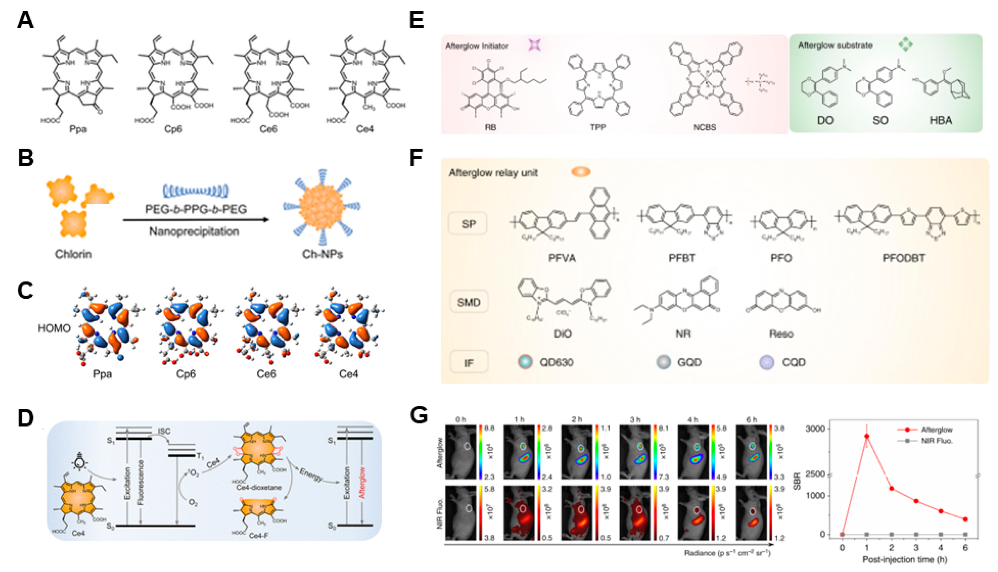
Surgical resection of tumor lesions demonstrates effective local control and therapeu-tic outcomes. However, accurately identifying the margins of tumor lesions during sur-gery remains challenging. Image-guided surgical resection can significantly address this issue [30,31,32,33]. Afterglow imaging agents possess the capability to emit photons gradually even after the cessation of light excitation, offering a distinct advantage in the realm of ultra-sensitive detection [34,35]. Recently, a variety of inorganic and organic photo-afterglow imaging probes have been employed for the monitoring and treatment of tumors. Particularly, organic afterglow probes with molecule-level specificity and minimal side effects are attracting increasing attention. Currently, to improve the signal-to-background ratio and sensitivity of imaging, many organic photo-afterglow probes have been developed. In 2022, Miao et al. [36] first reported a series of chlorin-based afterglow agents (Ppa, Cp6, Ce6, Ce4) and then they used an amphiphilic triblock copolymer PEG-b-PPG-b-PEG as a surface-capping agent to enhance the water solubility of the compound (Figure 1A,B). Among them, the NPs-Ce4 exhibited the strongest afterglow luminescence and an ultralong half-life (1.5 h), which was attributed to its low highest occupied molecular orbital (HOMO) levels (−5.08 ev) (Figure 1C). A mechanism study showed that NPs-Ce4 could produce 1O2 under light excitation to oxidize the vinylene bond (C=C) of NPs-Ce4 to form an active intermediate species followed by afterglow luminescence (Figure 1D). An in vivo imaging study indicated that the afterglow intensity was 26 times higher than that of the fluorescence signal after subcutaneous injection of NPs-Ce4. Furthermore, they injected NPs-Ce4 through the tail vein to detect the 4T1 tumors of different sizes. The result shows that the probe can identify tumors as small as 3 mm3, facilitating imaging-guided surgical resection of the tumor foci. In 2019, Pu et al. [37] selected RB (rose bengal octyl ester), TPP (meso-tetraphenylporphyrin), NCBS (silicon 2,3-naphthalocyanine bis(trihexylsilyloxide) as the afterglow initiators. DO (N, N dimethyl-4-(3-phenyl-5,6-dihydro-1,4-dioxin-2-yl) aniline), SO (N, N-dimethyl-4-(2-phenyl-5,6-dihydro-1,4-oxathiin-3-yl) aniline), and HBA (3-((1r,3r,5R,7S)-adamantan-2-ylidene (methoxy) methyl) phenol) were employed as the afterglow substrates. Semiconducting polymers (SPs), small molecule dyes (SMDs), and inorganic fluorophores (IFs) served as relay units for the afterglow process (Figure 1E,F). A series of afterglow luminescent nanoparticles (ALNPs) were synthesized via co-nanoprecipitation utilizing an amphiphilic copolymer, PEG-b-PPG-b-PEG. Among them, PFVA-N-DO was selected to detect tumors in mice. In vivo tumor imaging shows that at one hour post-injection, the signal-to-noise ratio of the afterglow signal at the tumor region (2922 ± 121) was 3 orders of magnitude higher than that of the fluorescence signal (~1) (Figure 1G). Moreover, the ALNPs were mainly cleared by the liver and had good biosafety.
Moreover, aggregation-induced emission (AIE) molecules have been widely used in bioimaging due to their bright luminescence and high photostability [38]. Benefiting from the excellent optical properties of the AIEgens, Ding et al. [39] synthesized an AIE probe with near-infrared afterglow luminescence properties (AGL AIE dots) (Figure 2A). The TPE-Ph-DCM moiety could produce 1O2 under light excitation to oxidize an enol ether precursor, forming an unstable intermediate with high energy and then emitting persistent luminescence. The luminescence could continue more than 10 days after the cessation of light and penetrate up to 7 mm of tissue. For in vivo imaging, the tumor-to-liver ratio of the afterglow signal was nearly 100-fold larger than that of the fluorescence signal, indicating excellent performance in image-guided cancer surgery with the AIE dots. Another example is from the work of Zhang et al. [40] who encapsulated photosensitizer 2-((5-(4(diphenylamino)phenyl)thiophen-2-yl)methylene)malononitrile (TTMN) and afterglow metricspoly[(9,9-di(2-ethylhexyl)-9H-fluorene-2,7-vinylene)-co-(1-methoxy-4-(2-ethylhexyloxy)-2,5phenylenevinylene)] (PFPV) with F127 to construct AIE afterglow imaging probe P-TNPs. They found that resonance energy transfer existed between TTMN and PFPV. After irradiation by a white LED lamp, the afterglow of PFPV-dioxetane intermediates could irradiate TTMN again to produce 1O2 (Figure 2B). This type of closed-loop probe can achieve effective imaging of subcutaneous tumors after injection through the tail vein. In order to improve the luminescence properties of AIE molecules, Liu et al. [41] balanced the twist and conjugation of molecules and reported a luminescent core (TPT), which possesses a higher molar extinction coefficient (ε) and fluorescent brightness than the conventional triphenylamine (TPA) and tetraphenylethene (TPE). Moreover, they introduced a strong electron acceptor into the TPT core to construct a D-A near-infrared AIE molecule (TPT-DCM) with a triazole luminescent core (Figure 2C), featuring a high molar extinction coefficient, high brightness, and efficient reactive oxygen species generation efficiency, which is attributed to the decreased energy gap (ΔEST). Furthermore, they encapsulated TPT-DCM and Schaap’s dioxetane precursor with DSPE-PEG 2000. The TPT-DCM can be light-irradiated to generate 1O2 to convert Schaap’s dioxetane precursor to Schaap’s dioxetane and then generate an afterglow with long time (up to 20 days), tumor-to-liver signal ratio (up to 187) and deep tissue penetration (1.6 cm). To enhance the effectiveness at the in vivo level, they synthesized a nanoparticle by encapsulating the TPT-DCM and AGL (enol ether precursor of Schapp’s dioxetane) with DSPE-PEG 2000; the nanoparticle was termed as TPT-DCM/AGL NPs. Notably, the tumor with a tiny size of <1 mm can be successfully detected by injecting nanoprobes into 4T1 tumor-bearing Balb/c mice through the tail vein. In 2024, Miao et al. [42] synthesized a single molecule (TPP-DO) by the covalent bonding of the afterglow substrate (DO) with the afterglow initiator (mesotetraphenyl porphyrin). To increase the hydrophilicity of TPP-DO, they co-assembled the TPP-DO with an amphiphilic triblock copolymer PEG-b-PPG-b-PEG to form TPP-DO NPs (Figure 2D). The covalent bond between the afterglow substrate and the energy transfer unit enhances intramolecular energy transfer efficiency, resulting in increased afterglow brightness. The mechanism study indicated that the TPP-DO NPs can be light-irradiated for TPP to generate 1O2 to convert the DO to its 1,2-dioxetane intermediate TPP-DO-dioxetane, which decomposed into TPP-DE, which can absorb the released energy to form excited TPP-DE*. The energy transfer excites the TPP moiety, producing bright NIR luminescence. An in vitro study showed that the TPP-DO NPs can penetrate a thickness of 6 cm with a SBR of 35. The in vivo imaging can be conducted even at TPP-DO NP concentrations as low as 0.1 nM. Furthermore, they prepared a GSH-activatable afterglow probe (Q-TPP-DO NPs) by co-precipitating the DSPE-PEG2000-ssBHQ3 and the TPP-DO (Figure 2E) for the detection of subcutaneous tumors as small as 0.048 mm3.
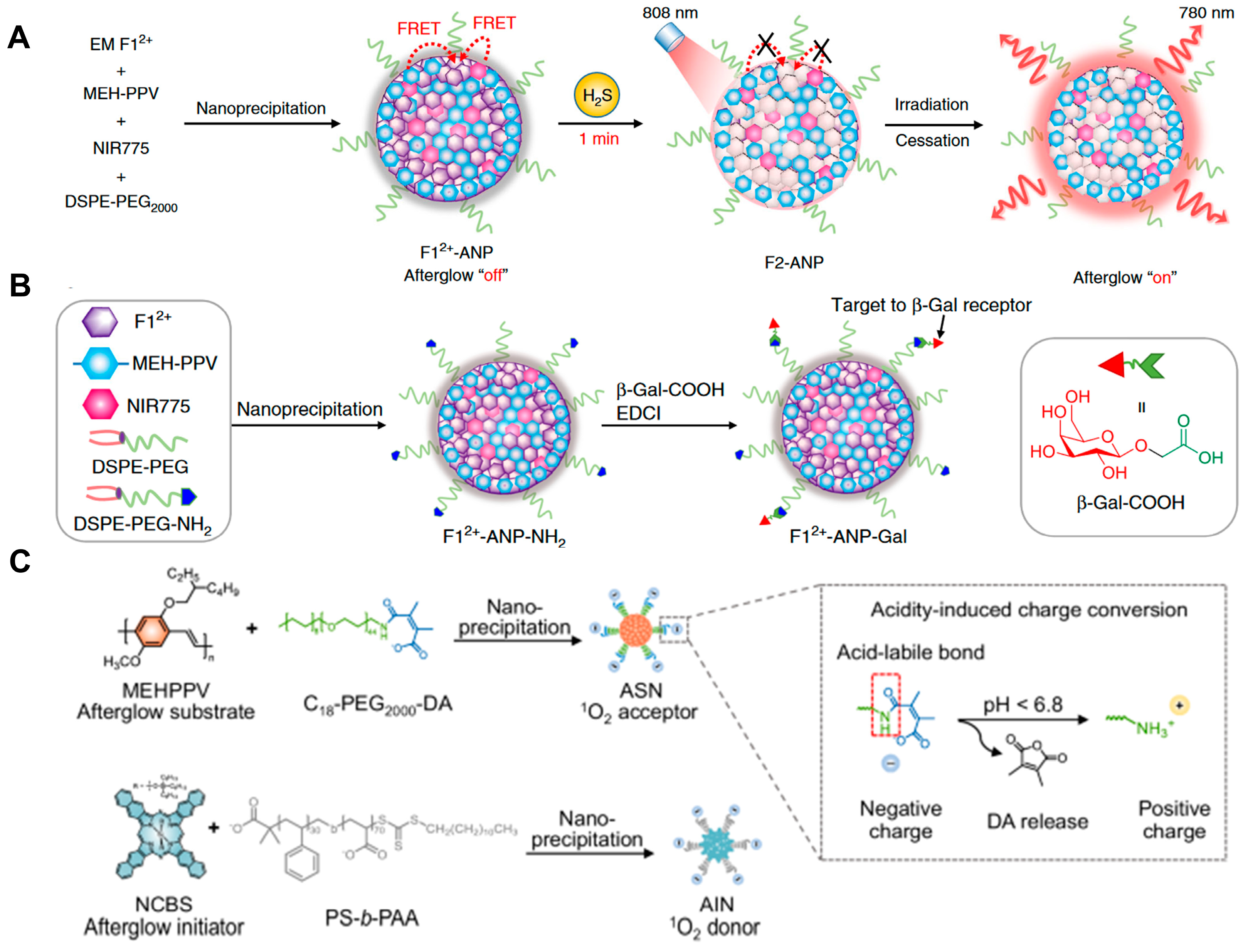
Aiming at achieving precise tumor imaging with high SBR of tumors, it is essential to enhance the in vivo targeting ability of imaging probes. Ye et al. [43] developed an H2S-activated afterglow probe (F12+-ANP) by integrating organic electrochromic material (EM F12+) into a NIR photosensitizer (silicon 2,3-naphthalocyanine bis(trihexylsilyloxide) and (poly[2-methoxy-5-(2-ethylhexyloxy)1,4-phenylenevinylene]) containing nanoparticles (Figure 3A). In the presence of H2S, the FRET (fluorescent resonance energy transfer) process was eliminated, leading to the recovery of 1O2 production in F2-ANP under 808 nm light irradiation. In addition, the F12+-ANP possessed positive reduction potential E red = +0.40 V, resulting in a faster reaction to H2S with a second-order reaction rate k2 = 3600 ± 130 M−1s−1. Under the 808 nm irradiation, the NIR775 produced 1O2 to oxidize the vinylene bond of MEH-PPV, accompanied by the emission of photons at 580 nm. This process was followed by energy transfer to NIR775, which ultimately activates the NIR afterglow luminescence at 780 nm. To enable precise detection of H2S in HCC HepG2 cells, the β-Gal was introduced to the surface of F12+-ANP to prepare F12+-ANP-Gal (Figure 3B). They further used the F12+-ANP-Gal probe for the imaging of subcutaneous and orthotopic HepG2 tumors (<3 mm in diameter) in mice. It is worth noting that F12+-ANP-Gal precisely delineated tumor margins in excised hepatic cancer specimens, which may be used for intraoperative guidance in liver cancer surgery. To break the limitation of energy transfer distance between the donor and the acceptor, Miao et al. [44] developed an organic PH-activatable upconversion afterglow luminescence cocktail of nanoparticles (ALCNs) consisting of an “energy” donor (AIN) and an “energy” acceptor (ASN) (Figure 3C). In the acidic microenvironment, the amide bond of ASN was broken and the surface charge of the nanoparticles changed from negative to positive, which reduced the transfer distance of singlet oxygen (1O2) in the aqueous solution, so that the afterglow substrate was oxidized to intermediate, and then produced afterglow. An in vitro afterglow imaging study proved that the intensity of the afterglow signal was proportional to the number of 4T1 cells and HepG2 cells. Furthermore, the ALCNs successfully achieved afterglow imaging of 4T1 subcutaneous tumors and orthotopic liver tumors. Notably, bio-ALCNs exhibited superior tumor-targeting capability and enhanced tumor-imaging performance compared to ALCNs.
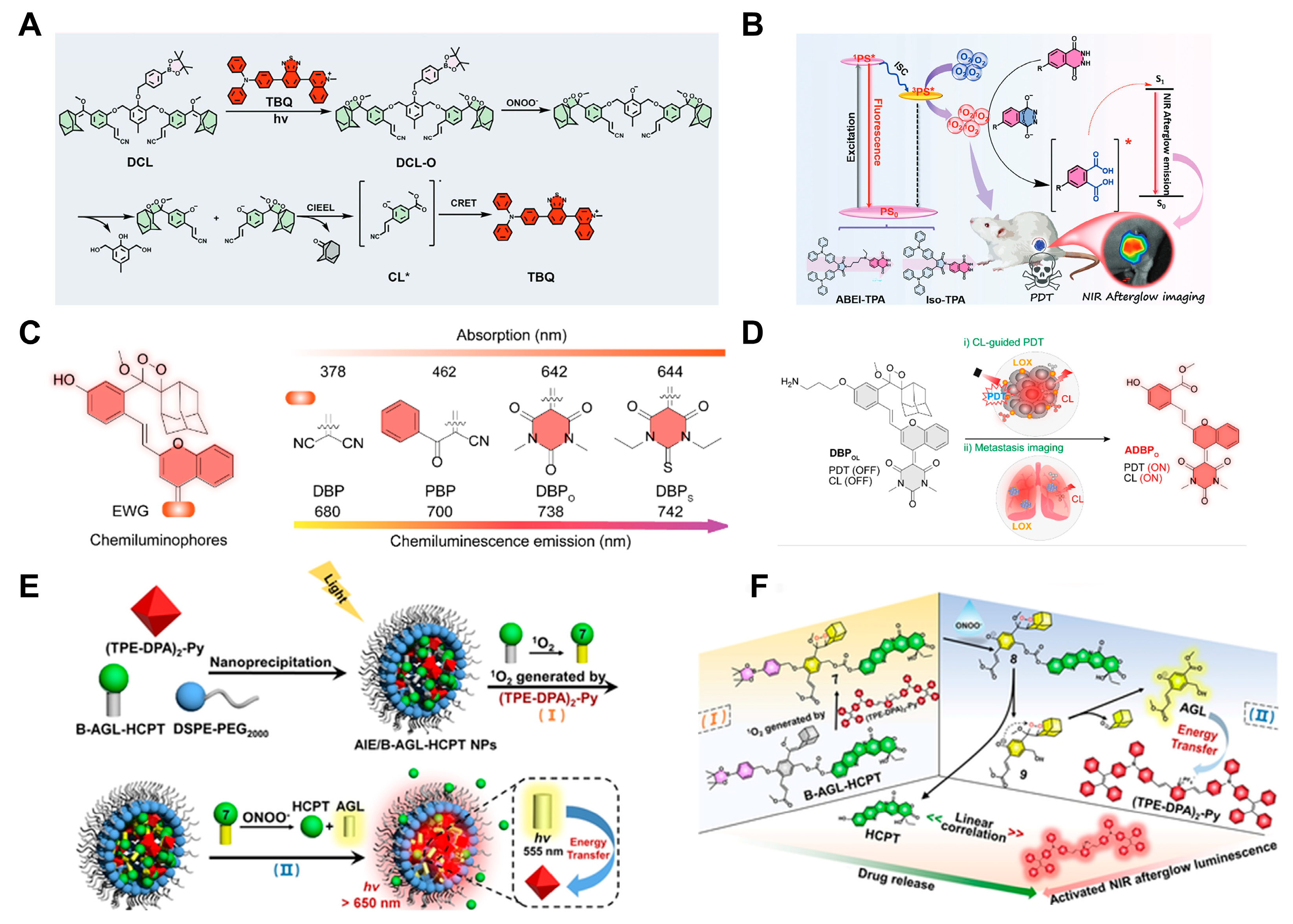
Due to the generation of ROS, afterglow luminescent probes can also be used for photodynamic therapy of tumors. As an exploration, Zeng successfully integrated the acceptor moiety TBQ, which possessed 1O2 generation capabilities, with the donor moiety DCL featuring an ONOO-responsive group, into a nanoparticle [45] (Figure 4A). This innovative construct enabled in vitro detection of ONOO- with a limit of detection (LOD) of 46.1 nM and achieved a penetration depth of 2 cm, thereby facilitating effective photodynamic therapy in vivo. Tang et al. [46] reported two novel photochemical afterglow luminescent nano-photosensitizers (ABEI-TPA and Iso-TPA) (Figure 4B). Two PSs with extended afterglow were created using TMAH and isoluminol derivatives through conjugate and non-conjugate links. While ABEI-TPA possessed higher photoluminescence quantum efficiency, Iso-TPA exhibited better effects of luminescent bioimaging such as high SNR and deep tissue penetration (32 mm), which were attributed to the higher ROS generation efficiency. Consequently, in vivo experiments indicated that two PS-based NPs possessed effective PDT therapeutic efficacy. Furthermore, activatable phototheranostic probes, characterized by specific “turn-on” signals, minimized tissue auto-fluorescence and side effects, have been used for phototheranostics of the tumor. In 2023, Pu et al. [47] synthesized a chemiluminophore (DBPO) by replacing the malononitrile group of dicyanomethylene-4H-benzopyran-phenoxyl-dioxetane (DBP) with A1,3-dimethyl barbituric acid (an electron-withdrawing group (EWGs) (Figure 4C,D). They further introduced the lysyl oxidase (LOX)-responsive group propylamine into DBPO to construct the LOX-activatable luminescent probe (DBPOL) with NIR afterglow and 1O2 production after laser irradiation. In the presence of lysyl oxidase (LOX), the CL signal and photodynamic activity could be turned on, enabling imaging-guided PDT therapy in vivo. As an exploration, Ding et al. [48] developed an afterglow ONOO-activatable theranostic nanoprobe AIE/B -AGL-HCPT NPs for inducing Immunogenic Cell Death (ICD), monitoring the process of ICD and the cold-to-hot tumor transformation. The AIE/B -AGL-HCPT NPs consisted of afterglow hydroxycamptothecin prodrug (B-AGL-HCPT) with the ONOO-activatable group and AIE photosensitizer ((TPE-DPA)2-Py). Under the irradiation of light, the AIE photosensitizer generated 1O2 to evoke ICD and oxidize the B-AGL-HCPT, forming a 1,2-dioxetane derivative, which had neither afterglow nor therapeutic effect except to retain the fluorescent signal (Figure 4E,F). With the development of ICD, the cold tumor can be converted to a hot tumor with increased ONOO levels. The ONOO can cleave the B-AGL-HCPT, thereby uncaging the afterglow unit, which can transfer the energy to (TPE-DPA)2-Py to emit NIR afterglow (>650 nm), while also releasing the HCPT to enhance the PDT-mediated ICD process. Furthermore, in vivo experiments demonstrated that AIE/B -AGL-HCPT NPs can effectively eliminate tumors and prevent tumor recurrence in the 4T1 tumor-bearing mice.

3. Sono-Afterglow Probes

Despite the elimination of auto-fluorescence of tissue, limited penetration (1~2 cm), light-scattering, and reabsorption in tissue still exist in photo-afterglow imaging. Conversely, ultrasound-triggered luminescence, which is characterized by light emission resulting from the energy-releasing process of piezoelectric inorganic materials, has great potential for application in biological imaging owing to its noninvasiveness, high spatiotemporal, deep penetrability (up to 10 cm), precise localization of lesions, and low cost [49,50,51,52].
To achieve ultrasound-activated luminescence imaging of tumor sites, Song et al. [53] reported an ultrasound-activated NIR chemiluminescence PNCL, which was prepared with sonosensitizer protoporphyrin IX (PpIX) and an enol ether precursor of Schaap’s dioxetane containing adicyanomethyl chromone (DCMC) acceptor scaffold (NCL) through click reaction. The PNCL could generate singlet oxygen under ultrasound irradiation (1 MHz). Oxidation of the enol ether of the NCL moiety by 1O2 resulted in the forming of active intermediates and then the producing of luminescence. The strongest afterglow luminescence peaked at 710 nm (Figure 5A), enabling US-activated afterglow imaging in deep tissue (∼2 cm). Furthermore, bright chemiluminescence was detected in vivo after tail vein injection of PNCL (Figure 5B). This strategy achieves ultrasound-induced luminescence imaging of the tumor. Aiming at mitigating the hypoxic microenvironment present in tumors and enhancing therapeutic outcomes for tumor treatment, Chen et al. [54] constructed a probe by combining Synechococcus elongatus PCC 7942 (PCC) with sono-afterglow Ce6 nanoparticles (NPs-Ce6). Under ultrasound stimulation, the afterglow emitted by NPs-Ce6 facilitates photodynamic conversion to generate oxygen, thereby alleviating the hypoxic microenvironment within the tumor. Concurrently, the singlet oxygen produced through ultrasound exposure can induce ferroptosis in tumor cells (Figure 5C).
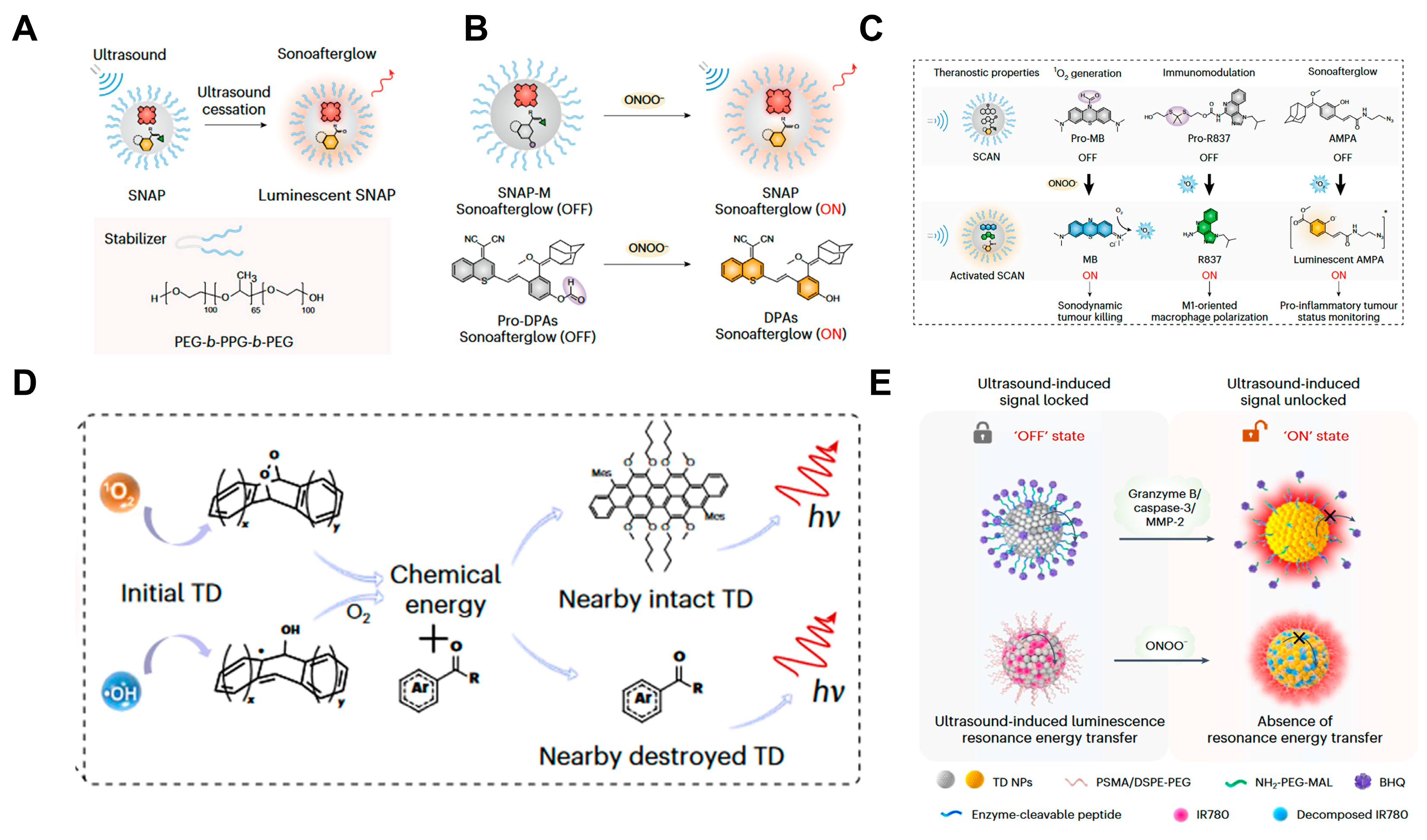
The immune system can maintain homeostasis in tumor initiation and development, and many activatable molecular sono-afterglow probes have been reported to evaluate immunotherapy and early diagnosis of tumors [55,56]. In 2022, Pu et al. [57] reported a NIR-I molecular sono-afterglow probe SNAP (peaked at 780 nm), which was designed by silicon 2,3-naphthalocyanine bis(trihexylsilyloxide) (NCBS) as a sonosensitizer moiety, and dicyanomethylene-4H-benzothiopyran-phenoxyl-adamantylidene (DPAs) as a sono-afterglow substrate (Figure 6A). When the ultrasound condition was 2.0 W cm−2 for 30 s, the mass ratio between NCBS and DPAs was 1:5, and the sono-afterglow signal of SNAP could be strong enough to penetrate tissue to a depth of 4 cm. To further evaluate the performance of biomarker-activated sono-afterglow imaging, they synthesized an activatable sono-afterglow probe SNAP-M, comprising a singlet oxygen initiator (NCBS) and a sono-afterglow substrate (DPAs), which is caged with ONOO responsive moiety (Pro-DPAs) (Figure 6B). The afterglow substrate could be activated by overproduced ONOO in the pro-inflammatory tumor microenvironment and subsequently reacted with 1O2, generated from NCBS under ultrasound irradiation, to produce long-lasting and deep tissue luminescence, which could be used to specifically and sensitively (LOD down to 0.3 μM) detect ONOO in deep tissues (4 cm). To further accelerate the in vivo use of sono-afterglow nanoparticles, SCAN, consisting of Pro-MB, Pro-R837, and DPAs, has been reported to achieve tumor immunotherapy. The MB could be activated in the ONOO overproduced tumor microenvironment and produced 1O2 under ultrasound condition (5 min, 2.0 W cm−2). Due to the 1O2 effect, R837 and DPAs were liberated from SCAN in situ for immunotherapy and accurate sono-afterglow imaging of subtle ONOO molecular changes in deep tumor tissue, respectively (Figure 6C). Moreover, Tan et al. [58] synthesized luminescence nanoparticle TD NPs, which enable early detection of tumors and monitoring cancer treatment. Due to the piezocatalytic effect of ultrasound, TD NPs could generate a large amount of ROS (1O2 and HO•) under ultrasonic excitation and then react with the TD to produce luminescence with a peak at 625~650 nm, which exhibited excellent tissue penetration (up to 2.2 cm) (Figure 6D). Whether in delayed or real-time imaging mode, the TD had excellent sono-afterglow performance. In the delayed imaging mode, its luminescence intensity was 2389 times higher than that of H2O, and in the real-time imaging mode, it was 1428 times higher. To investigate the luminescence ability in vivo, orthotopic GBM-bearing mice and pancreatic tumor-bearing mice were intraperitoneally injected with TD NPs, and then dynamically enhanced signals were observed. Moreover, the nanoparticle could produce strong sono-afterglow in metastatic tumors and lymph nodes. These results demonstrated the ability of in vivo imaging. Furthermore, based on the critical role of granzyme B in cell death through cytotoxic lymphocytes, the team further constructed a granzyme B-activated TD-Grz-BHQ probe for the precise immune response differentiation of different tumor types (Figure 6E).

4. Radio-Afterglow Probes

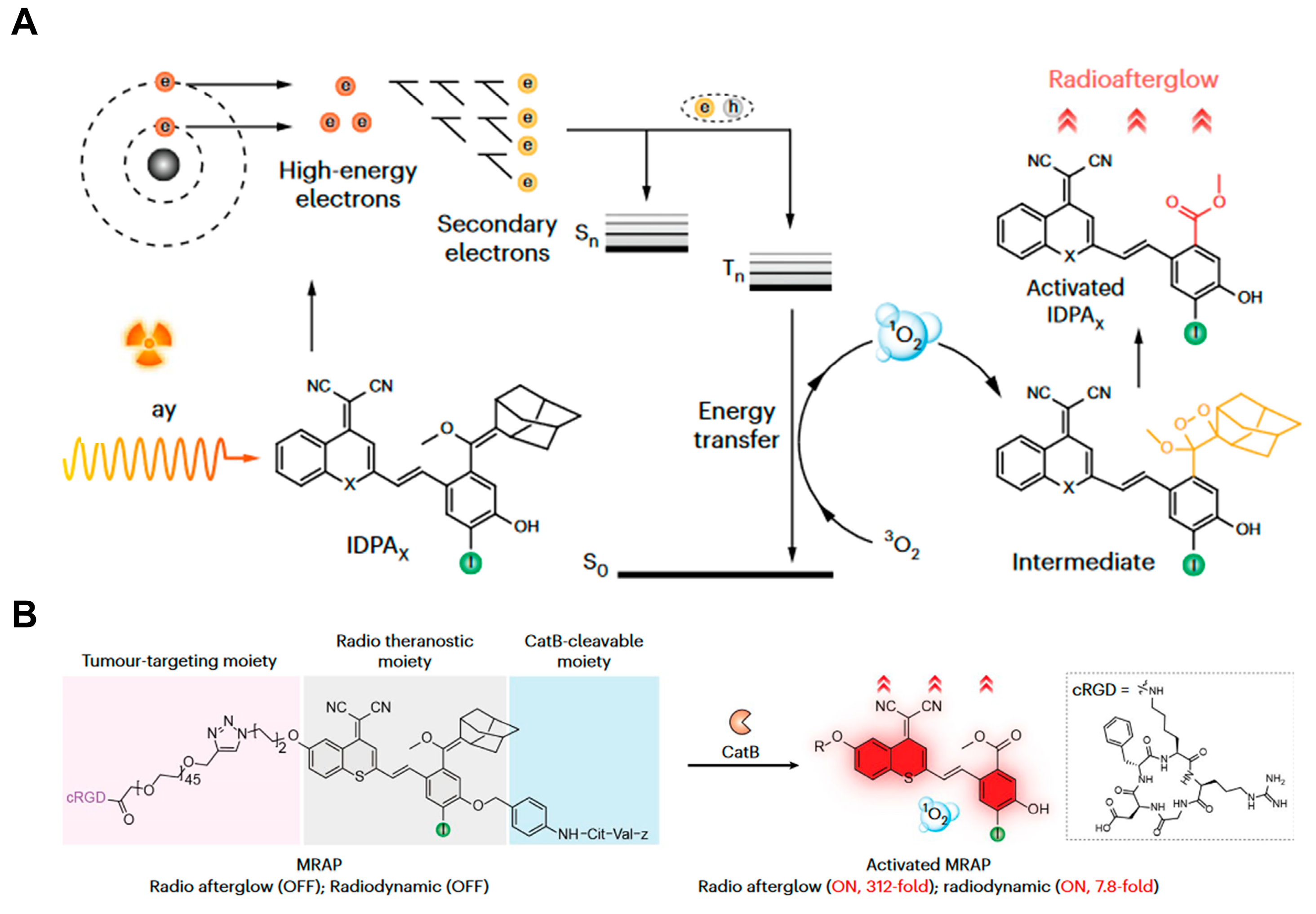
As invisible high-energy rays, X-rays have no penetration limitation, making X-ray-activated afterglow imaging useful for diagnostic imaging. Moreover, similar to photodynamics, X-ray can also enable radiodynamic therapy (RDT) of radio-afterglow probes by initiating the photodynamic process of optical agents [59,60,61,62,63,64,65,66]. However, it is still limited to a few inorganic nanophosphors [67]. Recently, Pu et al. [68] reported a series of organic radio afterglow probes with tunable afterglow emission (624 nm~792 nm) and 1O2 generation for cancer radiodynamic theranostics (Figure 7A,B). Mechanistic studies showed that the generation of 1O2 by IDPASu was the highest, which enabled stronger radio afterglow. The IDPASu exhibited deep tissue penetration (15 cm) in vitro and in vivo. To allow precise cancer diagnosis and therapy, the authors synthesized an organic radio afterglow dynamic probe (MRAP), comprising a tumor-targeting moiety (cyclic arginine-glycine-aspartate (cRGD)), a theranostic moiety and a CatB-cleavable peptide moiety (Cit-Val). In the CatB-upregulated microenvironment, the peptide moiety was specifically cleaved, and then the radio afterglow signal and radiodynamic of MRAP could be activated. It should be noted that the probe enables the detection of a diminutive tumor. This work provided a new avenue for precision theranostics.

5. Conclusions and Perspective

Afterglow imaging plays a crucial role in the field of biomedicine. At present, compared with inorganic afterglow probes, organic afterglow probes have advantages such as good biocompatibility, low toxicity, and easy adjustment of structure, which provide broad prospects for development in tumor imaging and treatment. The imaging effect of photo-afterglow probes is often affected by the unavoidable absorption of light by tissues. In contrast, due to the superior deep penetration capabilities of ultrasound and X-ray, both ultrasound-activated afterglow imaging and X-ray-activated afterglow imaging can effectively address this issue. Despite significant advancements, the design of promising organic afterglow imaging probes still faces many challenges.
Firstly, sono-afterglow probes and radio-afterglow probes can effectively enable deep tissue imaging. However, the development and reporting of organic sono-afterglow probes and radio-afterglow probes remain relatively underexplored, which highlights a significant opportunity for further investigation and innovation in this field [69,70].
Secondly, the reported probes are often composed of multiple components (stimuli-responsive units, singlet oxygen-generating units, luminescent units) encapsulated together. The construction process of these organic probes is relatively cumbersome, and the component ratio is not easily controlled precisely. Developing an activatable organic afterglow probe that integrates multiple units in one molecule is very meaningful.
Thirdly, the half-life of singlet oxygen which plays a crucial role in the process of afterglow imaging is very short (4 ms). This issue affects the efficiency of singlet oxygen transfer from the singlet oxygen generator to the afterglow substrate. The current solution is to prepare the imaging probe into a small nanoparticle. Exploring more methods to improve the efficiency of singlet oxygen transfer is urgent.
Fourthly, there are still very few organic luminescent probes with near infrared luminescence, especially in the second region of the near infrared. To improve the imaging resolution and depth, it is very necessary to develop the second region of NIR II afterglow probes.
Finally, the development of organic afterglow probes with near-infrared emission, long luminescence duration, bright afterglow intensity (more than 105 ps−1 cm−2 sr−1 measured by an IVIS imaging system) and multi-target detection is still in its initial stage. There is an urgent need to explore new organic afterglow substrates.
In summary, there are still many issues with probes used for precise imaging and treatment of tumors. We hope that this review will serve as a valuable resource, offering insightful and practical information about this specialized field. We aim to encourage and facilitate the development and eventual clinical application of afterglow imaging agents.

Author Contributions

Conceptualization, M.L. and L.T.; methodology, M.L.; software, M.L. and H.W.; validation, M.L. and H.W.; formal analysis, M.L. and L.T.; investigation, M.L. and L.T.; resources, M.L. and H.W; data curation, M.L. and L.T.; writing—original draft preparation, M.L. and L.T.; writing—review and editing, M.L. and L.T.; visualization, M.L.; supervision, Y.S. and J.L.; project administration, Y.S. and J.L.; funding acquisition, Y.S. and J.L. All authors have read and agreed to the published version of the manuscript.

Funding

This work was funded by the National Natural Science Foundation of China (22374055, 22022404, 22074050, 82172055), the National Natural Science Foundation of Hubei Province (22022CFA033), and the Fundamental Research Funds for the Central Universities (CCNU24JCPT001, CCNU24JCPT020). The authors have reported that they have no relationships relevant to the contents of this paper to disclose.

Institutional Review Board Statement

Not applicable.

Informed Consent Statement

Not applicable.

Data Availability Statement

No new data were created or analyzed in this study.

Conflicts of Interest

The authors declare no conflicts of interest.

References

  1. Winkler, R.; Ciria, M.; Ahmad, M.; Plank, H.; Marcuello, C. A Review of the Current State of Magnetic Force Microscopy to Unravel the Magnetic Properties of Nanomaterials Applied in Biological Systems and Future Directions for Quantum Technologies. Nanomaterials 2023, 13, 2585. [Google Scholar] [CrossRef] [PubMed]
  2. Luthra, R.; Chen, H.; Roy-Chowdhuri, S.; Singh, R.R. Next-Generation Sequencing in Clinical Molecular Diagnostics of Cancer: Advantages and Challenges. Cancers 2015, 7, 2023–2036. [Google Scholar] [CrossRef] [PubMed]
  3. Razgulin, A.; Ma, N.; Rao, J. Strategies for in vivo imaging of enzyme activity: An overview and recent advances. Chem. Soc. Rev. 2011, 40, 4186–4216. [Google Scholar] [CrossRef]
  4. Zhang, J.; Chai, X.; He, X.P.; Kim, H.J.; Yoon, J.; Tian, H. Fluorogenic Probes for Disease-Relevant Enzymes. Chem. Soc. Rev. 2019, 48, 683–722. [Google Scholar] [CrossRef]
  5. Peng, Y.; Wang, Y.; Zhou, C.; Mei, W.; Zeng, C. PI3K/Akt/mTOR Pathway and Its Role in Cancer Therapeutics: Are We Making Headway? Front. Oncol. 2022, 12, 819128. [Google Scholar] [CrossRef]
  6. Huang, J.; Li, J.; Lyu, Y.; Miao, Q.; Pu, K. Molecular Optical Imaging Probes for Early Diagnosis of Drug-Induced Acute Kidney Injury. Nat. Mater. 2019, 18, 1133–1143. [Google Scholar] [CrossRef]
  7. Wang, S.; Fan, Y.; Li, D.; Sun, C.; Lei, Z.; Lu, L.; Wang, T.; Zhang, F. Anti-Quenching NIR-II Molecular Fluorophores for in Vivo High-Contrast Imaging and pH Sensing. Nat. Commun. 2019, 10, 1058–1068. [Google Scholar] [CrossRef]
  8. Moore, C.; Chen, F.; Wang, J.; Jokerst, J.V. Listening for the Therapeutic Window: Advances in Drug Delivery Utilizing Photoacoustic Imaging. Adv. Drug Deliv. Rev. 2019, 144, 78–89. [Google Scholar] [CrossRef]
  9. Huang, Z.; Yu, P.; Tang, J. Characterization of Triple-Negative Breast Cancer MDA-MB-231 Cell Spheroid Model. OncoTargets Ther. 2020, 13, 5395–5405. [Google Scholar] [CrossRef]
  10. Shuhendler, A.J.; Pu, K.; Cui, L.; Uetrecht, J.P.; Rao, J. Real-time imaging of oxidative and nitrosative stress in the liver of live animals for drug-toxicity testing. Nat. Biotechnol. 2024, 32, 373–380. [Google Scholar] [CrossRef]
  11. Badr, C.E.; Tannous, B.A. Bioluminescence imaging: Progress and applications. Trends Biotechnol. 2011, 29, 624–633. [Google Scholar] [CrossRef] [PubMed]
  12. Suzuki, K.; Nagai, T. Recent progress in expanding the chemiluminescent toolbox for bioimaging. Curr. Opin. Biotechnol. 2017, 48, 135–141. [Google Scholar] [CrossRef] [PubMed]
  13. Jiang, Y.; Pu, K. Multimodal Biophotonics of Semiconducting Polymer Nanoparticles. Acc. Chem. Res. 2018, 51, 1840–1849. [Google Scholar] [CrossRef]
  14. Sun, S.K.; Wang, H.F.; Yan, X.P. Engineering Persistent Luminescence Nanoparticles for Biological Applications: From Biosensing/Bioimaging to Theranostics. Acc. Chem. Res. 2018, 51, 1131–1143. [Google Scholar] [CrossRef]
  15. Xu, Y.; Li, C.; An, J.; Ma, X.; Yang, J.; Luo, L.; Deng, Y.; Kim, J.S.; Sun, Y. Construction of a 980 nm laser-activated Pt(II) metallacycle nanosystem for efficient and safe photo-induced bacteria sterilization. Sci. China Chem. 2023, 66, 155–163. [Google Scholar] [CrossRef]
  16. Duan, Q.; Zhao, Z.; Zhang, Y.; Fu, L.; Yuan, Y.; Du, J.; Wang, J. Activatable fluorescent probes for real-time imaging-guided tumor therapy. Adv. Drug Deliv. Rev. 2023, 196, 114793. [Google Scholar] [CrossRef]
  17. Zeng, W.; Wu, L.; Ishigaki, Y.; Harimoto, T.; Hu, Y.; Sun, Y.; Wang, Y.; Suzuki, T.; Chen, H.; Ye, D. An Activatable Afterglow/MRI Bimodal Nanoprobe with Fast Response to H2S for In Vivo Imaging of Acute Hepatitis. Angew. Chem. Int. Ed. 2022, 134, e202111759. [Google Scholar] [CrossRef]
  18. Yang, J.; Yin, W.; Van, R.; Yin, K.; Wang, P.; Zheng, C.; Zhu, B.; Ran, K.; Zhang, C.; Kumar, M.; et al. Turn-on Chemiluminescence Probes and Dual-Amplification of Signal for Detection of Amyloid Beta Species in Vivo. Nat. Commun. 2020, 11, 4052. [Google Scholar] [CrossRef]
  19. Zhen, X.; Xie, C.; Pu, K. Temperature-Correlated Afterglow of a Semiconducting Polymer Nanococktail for Imaging-Guided Photothermal Therapy. Angew. Chem. Int. Ed. 2018, 57, 3938–3942. [Google Scholar] [CrossRef]
  20. Zhang, X.; Zhang, T.; Tuo, W.; Liu, Y.; Umar, T.; Chen, Y.; Wu, Z.; Zhou, Q.; Li, X.; Deng, G.; et al. Rising interest in the accurate and controllable anticancer strategy: Based on photon-evoked pyroptosis engineering perspective. Coord. Chem. Rev. 2024, 501, 215588. [Google Scholar] [CrossRef]
  21. Xu, Y.; Pang, Y.; Luo, L.; Sharma, A.; Yang, J.; Li, C.; Liu, S.; Zhan, J.; Sun, Y. De Novo Designed Ru(II) Metallacycle as a Microenvironment-Adaptive Sonosensitizer and Sonocatalyst for Multidrug-Resistant Biofilms Eradication. Angew. Chem. Int. Ed. 2024, 63, E202319966. [Google Scholar] [CrossRef] [PubMed]
  22. Zhang, Z.; Ye, H.; Cai, F.; Sun, Y. Recent advances on the construction of long-wavelength emissive supramolecular coordination complexes for photo-diagnosis and therapy. Dalton Trans. 2023, 52, 15193–15202. [Google Scholar] [CrossRef] [PubMed]
  23. Su, Y.; Phua, S.Z.F.; Li, Y.; Zhou, X.; Jana, D.; Liu, G.; Lim, W.Q.; Ong, W.K.; Yang, C.; Zhao, Y. Ultralong Room Temperature Phosphorescence from Amorphous Organic Materials toward Confidential Information Encryption and Decryption. Sci. Adv. 2018, 4, eaas9732. [Google Scholar] [CrossRef] [PubMed]
  24. Xie, C.; Zhen, X.; Miao, Q.; Lyu, Y.; Pu, K. Self-Assembled Semiconducting Polymer Nanoparticles for Ultrasensitive Near-Infrared Afterglow Imaging of Metastatic Tumors. Adv. Mater. 2018, 30, 1801331. [Google Scholar] [CrossRef]
  25. Li, C.; Tu, L.; Xu, Y.; Li, M.; Du, J.; Stang, P.J.; Sun, Y.; Sun, Y. A NIR-Light-Activated and Lysosomal-Targeted Pt(II) Metallacycle for Highly Potent Evoking of Immunogenic Cell Death that Potentiates Cancer Immunotherapy of Deep-Seated Tumors. Angew. Chem. Int. Ed. 2024, 63, e202406392. [Google Scholar] [CrossRef]
  26. Li, Y.; Gecevicius, M.; Qiu, J. Long Persistent Phosphors-from Fundamentals to Applications. Chem. Soc. Rev. 2016, 45, 2090–2136. [Google Scholar] [CrossRef]
  27. Chuang, Y.J.; Zhen, Z.; Zhang, F.; Liu, F.; Mishra, J.P.; Tang, W.; Chen, H.; Huang, X.; Wang, L.; Chen, X.; et al. Photostimulable Near-Infrared Persistent Luminescent Nanoprobes for Ultrasensitive and Longitudinal Deep-Tissue Bio-Imaging. Theranostics 2014, 4, 1112–1122. [Google Scholar] [CrossRef]
  28. Wu, S.; Yang, C.; Yan, X. A dual-functional persistently luminescent nanocomposite enables engineering of mesenchymal stem cells for homing and gene therapy of glioblastoma. Adv. Funct. Mater. 2017, 27, 1604992. [Google Scholar] [CrossRef]
  29. Zhou, H.; Zeng, X.; Li, A.; Zhou, W.; Tang, L.; Hu, W.; Fan, Q.; Meng, X.; Deng, H.; Duan, L.; et al. Upconversion NIR-II Fluorophores for Mitochondria-Targeted Cancer Imaging and Photothermal Therapy. Nat. Commun. 2020, 11, 6183. [Google Scholar] [CrossRef]
  30. Ren, J.; He, J.; Zhang, H.; Xia, Y.; Hu, Z.; Loughran, P.; Billiar, T.; Huang, H.; Tsung, A. Platelet TLR4-ERK5 Axis Facilitates NET-Mediated Capturing of Circulating Tumor Cells and Distant Metastasis after Surgical Stress. Cancer Res. 2021, 81, 2373–2385. [Google Scholar] [CrossRef]
  31. Ma, L.; Fei, B. Comprehensive review of surgical microscopes: Technology development and medical applications. J. Biomed. Opt. 2021, 26, 010901. [Google Scholar] [CrossRef] [PubMed]
  32. Bortot, B.; Mangogna, A.; Lorenzo, G.; Stabile, G.; Ricci, G.; Biffi, S. Image-guided cancer surgery: A narrative review on imaging modalities and emerging nanotechnology strategies. J. Nanobiotechnol. 2023, 21, 155. [Google Scholar] [CrossRef] [PubMed]
  33. Miao, Q.; Xie, C.; Zhen, X.; Lyu, Y.; Duan, H.; Liu, X.; Jokerst, J.V.; Pu, K. Molecular afterglow imaging with bright, biodegradable polymer nanoparticles. Nat. Biotechnol. 2017, 35, 1102–1110. [Google Scholar] [CrossRef] [PubMed]
  34. Pei, P.; Chen, Y.; Sun, C.; Fan, Y.; Yang, Y.; Liu, X.; Lu, L.; Zhao, M.; Zhang, H.; Zhao, D.; et al. X-ray-activated persistent luminescence nanomaterials for NIR-II imaging. Nat. Nanotechnol. 2021, 16, 1011–1018. [Google Scholar] [CrossRef]
  35. Hananya, N.; Shabat, D. A Glowing Trajectory between Bio- and Chemiluminescence: From Luciferin-Based Probes to Triggerable Dioxetanes. Angew. Chem. Int. Ed. 2017, 56, 16454–16463. [Google Scholar] [CrossRef]
  36. Wan, C.; Zhang, Y.; Li, Q.; Jiang, Y.; Zhou, H.; Liu, Y.; Miao, Q.; Gao, M. Near-Infrared Afterglow Luminescence of Chlorin Nanoparticles for Ultrasensitive In Vivo Imaging. J. Am. Chem. Soc. 2022, 144, 6719–6726. [Google Scholar]
  37. Jiang, Y.; Huang, J.; Zhen, X.; Li, J.; Xie, C.; Miao, Q.; Chen, J.; Chen, P.; Pu, K. A generic approach towards afterglow luminescent nanoparticles for ultrasensitive in vivo imaging. Nat. Commun. 2019, 10, 2064. [Google Scholar] [CrossRef]
  38. Niu, G.; Zhang, R.; Shi, X.; Park, H.; Xie, S.; Kwok, R.T.K.; Lam, J.W.Y.; Tang, B.Z. AIE Luminogens as Fluorescent Bioprobes. TrAC. Trends Anal. Chem. 2020, 123, 115769. [Google Scholar] [CrossRef]
  39. Ni, X.; Zhang, X.; Duan, X.; Zheng, H.L.; Xue, X.S.; Ding, D. Near-Infrared Afterglow Luminescent Aggregation-Induced Emission Dots with Ultrahigh Tumor-to-Liver Signal Ratio for Promoted Image-Guided Cancer Surgery. Nano Lett. 2019, 19, 318–330. [Google Scholar] [CrossRef]
  40. Xu, Y.; Yang, W.; Yao, D.; Bian, K.; Zeng, W.; Liu, K.; Wang, D.; Zhang, B. An Aggregation-Induced Emission Dye-Powered Afterglow Luminogen for Tumor Imaging. Chem. Sci. 2019, 11, 419–428. [Google Scholar] [CrossRef]
  41. Li, J.; Zhang, G.; Zhang, Y.; Tang, Y.; Ding, D.; Li, W.; Liu, Q. Building Highly Light-Harvesting Near-Infrared AIEgens Using Triazole-Based Luminescent Core for Improved Intravital Afterglow Imaging. Adv. Funct. Mater. 2023, 33, 2212380. [Google Scholar] [CrossRef]
  42. Yang, L.; Zhao, M.; Chen, W.; Zhu, J.; Xu, W.; Li, Q.; Pu, K.; Miao, Q. A Highly Bright Near-Infrared Afterglow Luminophore for Activatable Ultrasensitive In Vivo Imaging. Angew. Chem. Int. Ed. 2024, 63, e202313117. [Google Scholar] [CrossRef] [PubMed]
  43. Wu, L.; Ishigaki, Y.; Hu, Y.; Sugimoto, K.; Zeng, W.; Harimoto, T.; Sun, Y.; He, J.; Suzuki, T.; Jiang, X.; et al. H2S-Activatable near-Infrared Afterglow Luminescent Probes for Sensitive Molecular Imaging in Vivo. Nat. Commun. 2020, 11, 446. [Google Scholar] [CrossRef]
  44. Jiang, Y.; Zhao, M.; Miao, J.; Chen, W.; Zhang, Y.; Miao, M.; Yang, L.; Li, Q.; Miao, Q. Acidity-activatable upconversion afterglow luminescence cocktail nanoparticles for ultrasensitive in vivo imaging. Nat. Commun. 2024, 15, 2124. [Google Scholar] [CrossRef]
  45. Huang, S.; Bai, S.; Luo, T.; Zheng, F.; Fan, D.; Zhou, Y.; Dong, J.; Chen, F.; Zeng, W. Chemiluminescent Afterglow Material for Enhanced Tumor Diagnosis and Photodynamic Therapy. Adv. Funct. Mater. 2024. [Google Scholar] [CrossRef]
  46. Li, Y.; He, D.; Zheng, Q.; Tang, R.; Wan, Q.; Tang, B.; Wang, Z. Single-Component Photochemical Afterglow Near-Infrared Luminescent Nano-Photosensitizers: Bioimaging and Photodynamic Therapy. Adv. Healthc. Mater. 2024, 13, 2304392. [Google Scholar] [CrossRef]
  47. Huang, J.; Zhang, C.; Wang, X.; Wei, X.; Pu, K. Near-Infrared Photodynamic Chemiluminescent Probes for Cancer Therapy and Metastasis Detection. Angew. Chem. Int. Ed. 2023, 62, e202303982. [Google Scholar] [CrossRef]
  48. Gao, Z.; Jia, S.; Ou, H.; Hong, Y.; Shan, K.; Kong, X.; Wang, Z.; Feng, G.; Ding, D. An Activatable Near-Infrared Afterglow Theranostic Prodrug with Self-Sustainable Magnification Effect of Immunogenic Cell Death. Angew. Chem. Int. Ed. 2022, 61, e202209793. [Google Scholar] [CrossRef]
  49. Tong, Y.; Li, M.; Huang, H.; Long, S.; Sun, W.; Du, J.; Fan, J.; Wang, L.; Liu, B.; Peng, X. Urea-Bond Scission Induced by Therapeutic Ultrasound for Biofunctional Molecule Release. J. Am. Chem. Soc. 2022, 144, 16799–16807. [Google Scholar] [CrossRef]
  50. Dou, Y.; Wang, Y.; Tian, S.; Song, Q.; Deng, Y.; Zhang, Z.; Chen, P.; Sun, Y. Metal–organic framework (MOF)-based materials for pyroptosis-mediated cancer therapy. Chem. Commun. 2024, 60, 6476–6487. [Google Scholar] [CrossRef]
  51. Liu, T.; Zhang, N.; Wang, Z.; Wu, M.; Chen, Y.; Ma, M.; Chen, H.; Shi, J. Endogenous Catalytic Generation of O2 Bubbles for In Situ Ultrasound-Guided High Intensity Focused Ultrasound Ablation. ACS Nano 2017, 11, 9093–9102. [Google Scholar] [CrossRef] [PubMed]
  52. Zhang, Y.; Zhang, X.; Yang, H.; Yu, L.; Xu, Y.; Sharma, A.; Yin, P.; Li, X.; Kim, J.S.; Sun, Y. Advanced Biotechnology-Assisted Precise Sonodynamic Therapy. Chem. Soc. Rev. 2021, 50, 11227–11248. [Google Scholar] [CrossRef] [PubMed]
  53. Wu, R.; Yao, Z.; Chen, Z.; Ge, X.; Su, L.; Wang, S.; Wu, Y.; Song, J. Ultrasound-Activated NIR Chemiluminescence for Deep Tissue and Tumor Foci Imaging. Anal. Chem. 2023, 95, 11219–11226. [Google Scholar] [CrossRef]
  54. Yang, Z.; Shen, X.; Jin, J.; Jiang, X.; Pan, W.; Wu, C.; Yu, D.; Li, P.; Feng, W.; Chen, Y. Sonosynthetic Cyanobacteria Oxygenation for Self-Enhanced Tumor-Specific Treatment. Adv. Sci. 2024, 11, 2400251. [Google Scholar] [CrossRef]
  55. De Visser, K.E.; Eichten, A.; Coussens, L.M. Paradoxical Roles of the Immune System during Cancer Development. Nat. Rev. Cancer 2006, 6, 24–37. [Google Scholar] [CrossRef]
  56. Schmidt, R.E.; Grimbacher, B.; Witte, T. Autoimmunity and Primary Immunodeficiency: Two Sides of the Same Coin? Nat. Rev. Rheumatol. 2018, 14, 7–18. [Google Scholar] [CrossRef]
  57. Xu, C.; Huang, J.; Jiang, Y.; He, S.; Zhang, C.; Pu, K. Nanoparticles with Ultrasound-Induced Afterglow Luminescence for Tumour-Specific Theranostics. Nat. Biomed. Eng. 2022, 7, 298–312. [Google Scholar] [CrossRef]
  58. Wang, Y.; Yi, Z.; Guo, J.; Liao, S.; Li, Z.; Xu, S.; Yin, B.; Liu, Y.; Feng, Y.; Rong, Q.; et al. In Vivo Ultrasound-Induced Luminescence Molecular Imaging. Nat. Photonics 2024, 18, 334–343. [Google Scholar] [CrossRef]
  59. Xu, J.; Chen, Q.; Tian, K.; Liang, R.; Chen, T.; Gong, A.; Mathy, N.; Yu, T.; Chen, X. m6A methyltransferase METTL3 maintains colon cancer tumorigenicity by suppressing SOCS2 to promote cell proliferation. Oncol. Rep. 2020, 44, 973–986. [Google Scholar] [CrossRef]
  60. Zhang, Q.; Wang, N.; Ma, M.; Luo, Y.; Chen, H. Transferrin Receptor-Mediated Sequential Intercellular Nanoparticles Relay for Tumor Deep Penetration and Sonodynamic Therapy. Adv. Ther. 2019, 2, 1800152. [Google Scholar] [CrossRef]
  61. Chen, W.; Zhang, J. Using Nanoparticles to Enable Simultaneous Radiation and Photodynamic Therapies for Cancer Treatment. J. Nanosci. Nanotechnol. 2006, 6, 1159–1166. [Google Scholar] [CrossRef] [PubMed]
  62. Gao, W.; Liu, Y.; Zhang, H.; Wang, Z. Electrochemiluminescence Biosensor for Nucleolin Imaging in a Single Tumor Cell Combined with Synergetic Therapy of Tumor. ACS Sens. 2020, 5, 1216–1222. [Google Scholar] [CrossRef] [PubMed]
  63. Wang, X.; Sun, W.; Shi, H.; Ma, H.; Niu, G.; Li, Y.; Zhi, J.; Yao, X.; Song, Z.; Chen, L.; et al. Organic Phosphorescent Nanoscintillator for Low-Dose X-Ray-Induced Photodynamic Therapy. Nat. Commun. 2022, 13, 5091. [Google Scholar] [CrossRef] [PubMed]
  64. Chen, X.; Song, J.; Chen, X.; Yang, H. X-Ray-Activated Nanosystems for Theranostic Applications. Chem. Soc. Rev. 2019, 48, 3073–3101. [Google Scholar] [CrossRef]
  65. Chen, Z.; Wang, L.; Manoharan, D.; Lee, C.; Wu, L.; Huang, W.; Huang, E.; Su, C.; Sheu, H.; Yeh, C. Low Dose of X-Ray-Excited Long-Lasting Luminescent Concave Nanocubes in Highly Passive Targeting Deep-Seated Hepatic Tumors. Adv. Mater. 2019, 31, 1905087. [Google Scholar] [CrossRef]
  66. Shi, T.; Sun, W.; Qin, R.; Li, D.; Feng, Y.; Chen, L.; Liu, G.; Chen, X.; Chen, H. X-Ray-Induced Persistent Luminescence Promotes Ultrasensitive Imaging and Effective Inhibition of Orthotopic Hepatic Tumors. Adv. Funct. Mater. 2020, 30, 2001166. [Google Scholar] [CrossRef]
  67. Huang, K.; Le, N.; Wang, J.S.; Huang, L.; Zeng, L.; Xu, W.; Li, Z.; Li, Y.; Han, G. Designing Next Generation of Persistent Luminescence: Recent Advances in Uniform Persistent Luminescence Nanoparticles. Adv. Mater. 2022, 34, 2107962. [Google Scholar] [CrossRef]
  68. Huang, J.; Su, L.; Xu, C.; Ge, X.; Zhang, R.; Song, J.; Pu, K. Molecular Radio Afterglow Probes for Cancer Radiodynamic Theranostics. Nat. Mater. 2023, 22, 1421–1429. [Google Scholar] [CrossRef]
  69. Wilkinson, F.; Helman, W.P.; Ross, A.B. Rate Constants for the Decay and Reactions of the Lowest Electronically Excited Singlet State of Molecular Oxygen in Solution: An Expanded and Revised Compilation. J. Phys. Chem. Ref. Data 1995, 24, 663–677. [Google Scholar] [CrossRef]
  70. Lu, C.; Zhang, C.; Wang, P.; Zhao, Y.; Yang, Y.; Wang, Y.; Yuan, H.; Qu, S.; Zhang, X.; Song, G.; et al. Light-free Generation of Singlet Oxygen through Manganese-Thiophene Nanosystems for pH-Responsive Chemiluminescence Imaging and Tumor Therapy. Chem 2020, 6, 2314–2334. [Google Scholar] [CrossRef]
Scheme 1. Overview of organic afterglow probes for cancer diagnosis and treatments.
Targets 02 00019 sch001
Figure 1. (A) Chemical structures of chlorins. (B) Synthesis route of Ch-NPs. (C) The HOMO levels of chlorins. (D) Luminescence mechanism of NPs-Ce4 [36]. Reprinted (adapted) with permission from Ref. [36]. Copyright 2022 American Chemical Society. Chemical structures of (E) afterglow initiators, afterglow substrates, and (F) afterglow relay unit. (G) In vivo tumor afterglow imaging and fluorescence imaging [37]. Reprinted (adapted) with permission from Ref. [37]. Copyright 2019 Springer Nature.
Targets 02 00019 g001
Figure 2. (A) Synthetic route and luminescence mechanism of AGL AIE dot [39]. Reprinted (adapted) with permission from Ref. [39]. Copyright 2019 American Chemical Society. (B) Closed-loop luminescence mechanism of P-TNPs [40]. Reprinted (adapted) with permission from Ref. [40]. Copyright 2020 Royal Society of Chemistry. (C) The chemical structures of TPA, TPE, and TPT [41]. Reprinted (adapted) with permission from Ref. [41]. Copyright 2023 Wiley-VCH. The synthetic route of (D) TPP-DO NPs and (E) Q-TPP-DO NPs [42]. Reprinted (adapted) with permission from Ref. [42]. Copyright 2024 Wiley-VCH.
Targets 02 00019 g002
Figure 3. Synthesis route and response mechanism of (A) F12+-ANP and (B) F12+-ANP-Gal to hydrogen sulfide [43]. Reprinted (adapted) with permission from Ref. [43]. Copyright 2020 Springer Nature. (C) Synthetic route and PH response mechanism of ALCNs [44]. Reprinted (adapted) with permission from Ref. [44]. Copyright 2024 Springer Nature.
Targets 02 00019 g003
Figure 4. (A) Illustration of afterglow luminescence of DCL/TBQ activated by ONOO [45]. Reprinted (adapted) with permission from Ref. [45]. Copyright 2024 Wiley-VCH. (B) Schematic of in vivo luminescence imaging and PDT therapy of ABEI-TPA and ISO-TPA [46]. Reprinted (adapted) with permission from Ref. [46]. Copyright 2024 Wiley-VCH. (C) Molecular design of different chemiluminophores. (D) Illustration of LOX-activated afterglow imaging and PDT treatment [47]. Reprinted (adapted) with permission from Ref. [47]. Copyright 2023 Wiley-VCH. (E) Illustration of afterglow luminescence of AIE/B-AGL-HCPT NPs activated by ONOO. (F) The molecular structure conversion for the afterglow luminescence processes [48]. Reprinted (adapted) with permission from Ref. [48]. Copyright 2022 Wiley-VCH.
Targets 02 00019 g004
Figure 5. (A) Mechanism illustration of sono-afterglow of PNCL for in vivo imaging. (B) Sono-afterglow imaging of 4T1 tumor at different times [53]. Reprinted (adapted) with permission from Ref. [53]. Copyright 2023 American Chemical Society. (C) Schematic illustration of preparation of NPs-Ce6 and antitumor mechanisms [54]. Reprinted (adapted) with permission from Ref. [54]. Copyright 2024 Wiley-VCH.
Targets 02 00019 g005
Figure 6. Mechanism illustration of sono-afterglow imaging for (A) SNAP, (B) SNAP-M, (C) SCAN [57]. Reprinted (adapted) with permission from Ref. [57]. Copyright 2022 Springer Nature. Molecule mechanism of (D) TD NPs (E) TD-Grz-BHQ for sono-afterglow imaging [58]. Reprinted (adapted) with permission from Ref. [58]. Copyright 2024 Springer Nature.
Targets 02 00019 g006
Figure 7. (A) Molecule mechanism of IDPASu for radio-afterglow imaging. (B) Molecule mechanism of MRAP for radio-afterglow imaging and RDT [68]. Reprinted (adapted) with permission from Ref. [68]. Copyright 2023 Springer Nature.
Targets 02 00019 g007
Table 1. Key characteristics of organic afterglow probes used for cancer diagnosis and treatment.
Excitation Source Name of Afterglow ProbeAfterglow EmissionHalf-Life
Photo-afterglow ProbeNPs-Ce4680 nm1.5 h
PFVA-N-DO780 nm-
AGL-AIE550–850 nm48 min
TPT-DCM630 nm-
TPP-DO670 nm-
F12+-ANP780 nm6.6 min
DBPOL720 nm-
B-AGL-HCPT400–650 nm118.5 min
Sono-afterglow ProbePNCL710 nm-
NPs-Ce6680 nm-
NCBS/DPAs SNAP780 nm110 s
TD635–650 nm180 s
Radio-afterglow ProbeIDPAs624–792 nm9.2–196.8 min
Disclaimer/Publisher’s Note: The statements, opinions and data contained in all publications are solely those of the individual author(s) and contributor(s) and not of MDPI and/or the editor(s). MDPI and/or the editor(s) disclaim responsibility for any injury to people or property resulting from any ideas, methods, instructions or products referred to in the content.