Article

Identification of Biomarkers for Diagnosis and Prognosis of Head and Neck Cancer: Bioinformatics Approach

by
Alexandra Fernandes
1 and
Rui Vitorino
1,2,*
1
iBiMED, Department of Medical Sciences, University of Aveiro, 3810-193 Aveiro, Portugal
2
Cardiovascular R&D Centre-UnIC@RISE, Department of Surgery and Physiology, Faculty of Medicine of the University of Porto, 4200-319 Porto, Portugal
*
Author to whom correspondence should be addressed.
Targets 2024, 2(4), 470-480; https://doi.org/10.3390/targets2040026
Submission received: 15 October 2024 / Revised: 13 November 2024 / Accepted: 4 December 2024 / Published: 6 December 2024
(This article belongs to the Special Issue Multidisciplinary Approach to Oral Cavity Cancer: An Hard Enemy)

Abstract

:
Head and neck cancer (HNC) is the seventh most commonly diagnosed malignancy worldwide, and its incidence is expected to increase in coming years. Current diagnostic methods for HNC are often limited by suboptimal accuracy and speed, which can negatively impact therapeutic decision-making and patient outcomes. To address the shortcomings of conventional diagnostics, biomarker detection has attracted increasing clinical interest as a promising alternative. However, a major challenge is the identification of biomarkers with sufficient accuracy and sensitivity for HNC. The integration of bioinformatics tools with omics data analysis has proven to be a robust approach for biomarker discovery. In this study, we outline a bioinformatics protocol aimed at identifying differentially expressed genes (DEGs) in HNC and evaluating the diagnostic and prognostic relevance of specific genes, including FN1, LGALS3, MMP9, TIMP1, MMP2, and TIMP2, in this pathology. In addition, we performed an enrichment analysis for the genes of interest. The prognostic significance of the selected genes was evaluated in relation to patient survival. This study contributes to the growing body of knowledge by identifying potential biomarkers with diagnostic and prognostic utility in this malignancy.

1. Introduction

Head and neck cancer (HNC) comprises a group of biologically similar tumors that affect important anatomical regions, including the oral cavity, oropharynx, nasopharynx, hypopharynx, and larynx [1,2]. Globally, HNC is the seventh most commonly diagnosed cancer, with the incidence expected to increase by 30% by 2030 [3]. The incidence of HNC is significantly higher in men aged 50 to 70 years and varies depending on the anatomical site affected [2,4,5]. According to the 2022 GLOBOCAN report, most HNC cases occur in the oral cavity, larynx, and pharynx (International Agency for Research on Cancer, 2022).
Several risk factors are known to influence the occurrence of HNC, including tobacco and alcohol use, viral infections such as human papillomavirus (HPV) and Epstein–Barr virus (EBV), nutritional deficiencies (e.g., vitamin A and iron), a diet high in animal fats and low in fruits and vegetables, exposure to environmental toxins or pollutants, genetic predispositions (e.g., Li-Fraumeni syndrome or Fanconi anemia), and poor oral hygiene [4,5,6,7,8].
The five-year survival rate for HNC ranges between 50% and 65%, but can vary considerably depending on factors such as the tumor’s site of origin, the stage at diagnosis, patient age, and the presence of comorbidities [5,7]. Mortality rates are highest in Asia, Europe, and Africa, where HNC accounts for approximately 5% of all cancer-related deaths (International Agency for Research on Cancer, 2022; [8]).
A key challenge in the treatment of HNC is the lack of early diagnosis. Symptoms often do not appear until advanced stages, and current diagnostic methods are invasive and inaccurate [7,9]. In response to this challenge, biomarker detection has emerged as a promising area of research to improve HNC diagnosis and prognosis. Several researchers have been working in this direction, resulting in the identification of promising biomarkers. This study focuses on evaluating the diagnostic and prognostic potential of six genes: FN1, LGALS3, MMP9, TIMP1, MMP2, and TIMP2.
FN1 (fibronectin 1) is overexpressed in HNC and is associated with poor prognosis and reduced disease-free survival [10,11]. LGALS3 (Galectin-3) promotes tumor growth and aggressiveness by dysregulating the WNT pathway [12,13,14,15]. MMP9 (matrix metalloproteinase 9) is associated with cancer progression, metastasis, and reduced survival [16,17,18]. TIMP1 (Tissue Inhibitor of Metalloproteinases (1)) has a dual function: while its overexpression correlates with a poor prognosis, it also inhibits the degradation of the extracellular matrix and thus reduces tumor growth [16,17]. MMP2 (Matrix Metalloproteinase (2)) is associated with lymph node metastasis and poor survival [17], while TIMP2 (Tissue Inhibitor of Metalloproteinases 2) has shown tumor suppressive effects in HNC [17,19].
The integration of bioinformatics tools has become indispensable in cancer research, particularly through the application of proteomics and metabolomics to understand molecular changes involved in cancer development, progression, and metastasis. These approaches are invaluable for the identification of new biomarkers [20,21]. In HNC, bioinformatic studies have proven to be successful by better elucidating cancer progression and development. The aim of this study is to evaluate the diagnostic and prognostic utility of FN1, LGALS3, MMP9, TIMP1, MMP2, and TIMP2 with a bioinformatic strategy for HNC, and thus contribute to the development of more accurate and non-invasive diagnostic strategies for this malignancy.

2. Methods

To evaluate the diagnostic and prognostic potential of the FN1, LGALS3, MMP9, TIMP1, MMP,2 and TIMP2 genes in head and neck cancer (HNC), we first characterized the protein–protein interactions (PPIs) [22] between these six genes and their interactions with key regulatory factors using the Signor database in order to evaluate, within the datasets involved in this work, their interactions with each other, as well as with their positive and negative regulators. We then used the GEO and GEO2R [23] platforms provided by the National Center for Biotechnology Information (NCBI) to search and select datasets and to analyze the expression patterns of the candidate genes and their regulators in different datasets, respectively. For the selection of the datasets published on GEO, the following keyword model was applied: “Head and neck cancer”. Additionally, the following criteria were used: (1) datasets published between 2019 and 2024; (2) datasets that focused on Homo sapiens as the organism under study; (3) datasets with at least 10 samples (5 from control and 5 from HNC group); (4) datasets with detailed information about the samples under study; and (5) datasets with results integrating the identification of differentially expressed genes (DEGs) with statistical significance (p ≤ 0.05). GEO2R utilizes tools like R packages such as DeSeq2, GEOquery and limma to perform data analysis [23].
In addition, the combined diagnostic and prognostic value of these biomarkers was investigated using the Biomarker Exploration of Solid Tumors (BEST) platform [24]. This tool allowed us to assess these gene expression patterns in relation to various clinical factors, including the type of tissue (healthy vs. cancer), age, gender, and tumor stage. BEST also facilitated survival analysis and finding the prognostic value of genes of interest, which were evaluated through the parameters of disease-free survival (DFS) and disease-specific survival after treatment (DSS). Gene enrichment studies, including Gene Ontology, Kyoto Encyclopedia of Genes and Genomes (KEGG), and Hallmark analysis, and the evaluation of the potential of FN1, LGALS3, MMP9, TIMP1, MMP2, and TIMP2 as targets for immunotherapy in HNC, were performed as well.
Finally, we used the SecretomeP [25] platform to predict the secretion activity of these genes and evaluate their potential for detection by protein quantification methods in body fluids such as blood and plasma. This prediction sheds light on the feasibility of non-invasive diagnostic techniques for HNC.

3. Results

In exploring the potential of FN1, LGALS3, MMP9, TIMP1, MMP2, and TIMP2 as diagnostic and prognostic biomarkers for head and neck cancer (HNC), the identification of protein–protein interactions (PPIs) is critical, as it facilitates the understanding of the underlying mechanisms, signaling pathways, and biological processes involved in tumor progression and regulation. Using the Signor platform, we identified the PPI networks for these six proteins (Table 1). Remarkably, MMP2 and MMP9 had the highest number of interactions (n = 10), highlighting their extensive involvement in HNC-related molecular pathways, while TIMP1 had the fewest interactions (n = 2), suggesting a more limited but potentially significant regulatory role within the network. The analysis revealed that the proteins NME1, PZP, and A2M negatively impact the expression of MMP2, while the proteins MMP25, TWIST1, TP53, RUNX2, and NODAL promote its increase. For MMP9, the identified negative regulators were SPRY4, SPDEF, PZP, and A2M, whereas the positive regulators included ETS1, SNAI2, USP6, and Nfkb p65/p50 (Table 1). Additionally, it was observed that the proteins A2M and PZP negatively impact both metalloproteinases MMP2 and MMP9, while their expression downregulates A2M, PZP, and TGFB1, suggesting a shared regulatory relationship between the two MMPs. FN1′s negative regulators include FGG, Vincristine sulfate, and SERPINA5, while positive regulators include TWIST1, SNAIL/RELA/RAPP1, and DLK1 (Table 1). TWIST1 is a positive regulator shared by MMP2 and FN1. For LGALS3, only positive regulators were identified (Table 1). Two positive regulators and one negative were identified for TIMP2 and TIMP1, respectively (Table 1). Additionally, TIMP2, along with MMP2, showed to increase LRP2 expression, while TIMP1 is positively regulated by SPRY4, which also downregulates MMP9, supporting the hypothesis of an inverse relationship between the expression of MMP9 and TIMP1. These results provide valuable insights into the molecular context in which these biomarker candidates operate and provide a basis for further functional analysis and validation.
The GEO platform initially analyzed 78 datasets published between 2019 and 2024 that focus on Homo sapiens and contain at least ten samples. This selection criterion was used to ensure both the relevance and robustness of the results, as recommended by Conesa et al. [26]. After a manual review, only three datasets were selected for further analysis, as the majority of the remaining datasets did not comprehensively address gene expression profiles in HNC and the corresponding control samples (Figure 1). The selection of these datasets was based on their quality, size, and relevance to the study objectives. The robustness of the data provided by these datasets enables a more reliable and detailed analysis, helping us identify genetic expression patterns relevant for HNC diagnosis and prognosis.
The datasets selected for in-depth analysis were GSE130605, GSE138206, and GSE142083, all of which contained tissue samples from both control and HNC patients with oral and laryngeal carcinoma. In total, the differential gene expression of 127 control tissue samples and 117 tumor tissue samples was analyzed. Differentially expressed genes (DEGs) (p ≤ 0.01) were identified in all of these datasets, as shown in the volcano plots (Figure 1B), and the analysis revealed a positive correlation between the number of DEGs identified and the sample size in each dataset (Figure 1). These results emphasize the importance of sample size for robust identification of DEGs, which is critical for reliable conclusions in HNC biomarker studies.
The expression patterns of FN1, LGALS3, MMP9, TIMP1, MMP2, and TIMP2, and their regulatory elements, were analyzed within the selected datasets (Table 2). A higher number of samples was positively correlated with the statistical significance of expression profiles, suggesting that larger cohorts provide more robust and reliable insights into gene regulation. This finding suggests that studies with larger samples are likely to provide more accurate and meaningful results.
The analysis revealed different expression trends among the candidate genes. In particular, MMP9, MMP2, and FN1 showed increased expression levels in the HNC samples, while LGALS3 showed a decreased expression pattern in the HNC group (Table 2). These different expression profiles emphasize the potential of these genes as biomarkers for HNC and highlight their different roles in the pathophysiology of the disease.
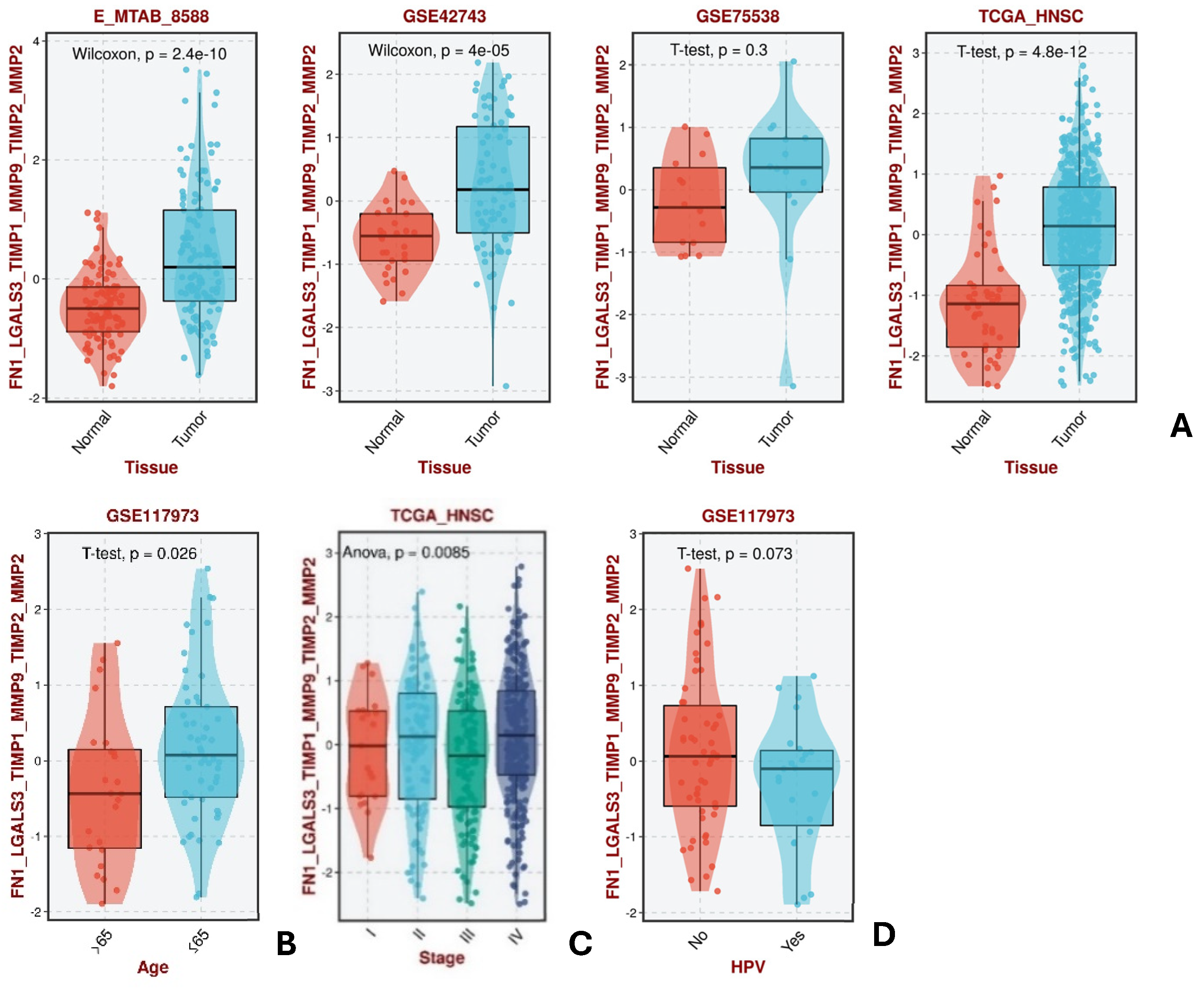
Combined analysis of the genes of interest by BEST revealed a significant increase (p ≤ 0.05) in their levels in tumor samples compared to control samples, and this behavior was observed in three of the four available datasets (E_MATB_8588; GSE42743; GSE75538; TCGA_HNSC), indicating the diagnostic biomarker potential of these genes (Figure 2A).
For several of the remaining parameters, only a single dataset was available for analysis. A statistically significant pattern of decreasing gene expression with increasing patient age was observed (Figure 2B). However, no clear pattern of expression was observed for the different cancer stages (Figure 2C). In addition, an upregulation of candidate genes was observed in HPV-negative tumors, although these results did not reach statistical significance (Figure 2D).
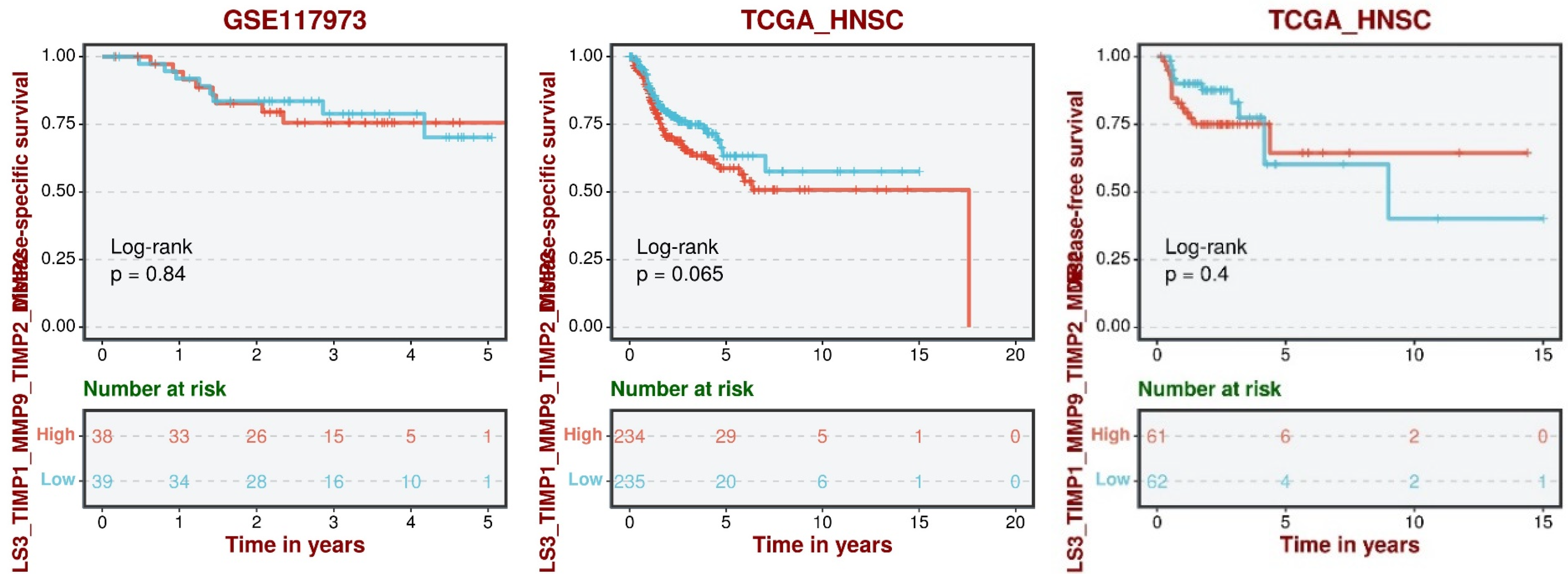
Next, we analyzed the survival data to evaluate the prognostic potential of the combined gene set (Figure 3). In terms of disease-specific survival (DSS), individuals with lower expression levels of the genes of interest were more likely to survive. A similar trend was observed for disease-free survival (DFS), where lower gene expression was associated with better outcomes. However, these results did not reach statistical significance on the BEST platform. Of note, the statistical significance of the results increased with larger sample size, emphasizing the importance of studying larger cohorts to more accurately determine the prognostic potential of these genes.
A combined gene enrichment analysis was performed using three different strategies on the BEST platform: Gene Ontology (GO) analysis, KEGG Pathway analysis, and Hallmark analysis. GO analysis identified the biological processes, cellular components, and molecular functions associated with the candidate genes and linked them to processes such as collagen fibril organization, extracellular matrix (ECM) organization and structure, angiogenesis, and metabolic processes. Other biological functions identified were actomyosin organization, focal adhesion, cell–substrate adhesion, and differentiation of various cell types, including endodermal cells, chondrocytes, and striated muscle cells.
KEGG pathway analysis also revealed that the candidate genes are involved in ECM receptor interactions, cell adhesion molecules, actin cytoskeleton regulation, and key signaling pathways such as WNT and TGF-β. These genes were also involved in complement and coagulation cascades, the transendothelial migration of leukocytes, and cardiac diseases such as dilated cardiomyopathy, cardiac hypertrophy, and arrhythmogenic right ventricular cardiomyopathy.
Hallmark analysis linked FN1, LGALS3, TIMP1, MMP9, TIMP2, and MMP2 to critical phenotypes such as epithelial–mesenchymal transition (EMT), the formation of cell junctions in muscle and epithelial cells, angiogenesis, and apoptosis. In addition, the genes were associated with several signaling pathways, including TGF-β, WNT, KRAS, TNF-α, NF-κB, NOTCH, and Hedgehog pathways. An examination of gene expression profiles associated with immunotherapeutic drugs revealed potential prognostic value for candidate genes in anti-PD-1/PD-L1 therapy, suggesting their relevance in the HNC immunotherapeutic landscape. However, no significant associations were found for other therapies, including anti-PD-1, anti-CTLA-4, CAR-T, or combined therapies such as anti-PD-1/CTLA-4 or anti-PD-L1.
In the SecretomeP analysis (Figure 4), LGALS3 was identified as secreted via a non-classical pathway, while FN1, MMP9, TIMP1, MMP2, and TIMP2 were predicted to be non-classically secreted. However, the presence of a signal peptide for these proteins indicates that they are likely to be secreted via the classical secretory pathway, suggesting potential utility for the detection of these proteins in body fluids for non-invasive diagnostic approaches.

4. Discussion

Analysis of expression data from GEO datasets showed that MMP9, MMP2, and FN1 exhibited increased expression patterns, while LGALS3 showed decreased expression in the HNC group. The matrix metalloproteinases (MMPs) showed a coordinated expression pattern, with both MMP9 and MMP2 showing increased levels, suggesting that they may be regulated by similar mechanisms. This pattern was also observed for FN1, whose expression appears to be influenced by its regulators. Although the relationship between MMPs and TIMPs (Tissue Inhibitors of Metalloproteinases) has not been fully elucidated, an inverse relationship was observed between MMP9 and TIMP1, which is consistent with previous findings [18]. However, further studies are needed to definitively establish the diagnostic potential of TIMP1 and TIMP2 in HNC, as the current data lack sufficient statistical power due to the limited sample size (less than 107 samples per dataset). Therefore, larger studies are recommended to more comprehensively investigate the expression of these genes in HNC. In the BEST analysis, upregulation of the combined genes was confirmed in HNC tissues, with decreasing expression observed with increasing age. This suggests that these genes could serve as diagnostic biomarkers, but it is important to define their expression levels in different age groups to enable more personalized diagnostic approaches. While these genes were effective in discriminating between HPV-positive and HPV-negative tumors, suggesting their role in the origin of HNC as HPV-negative, they did not facilitate the stratification of patients based on tumor stage, which may limit their utility in some clinical contexts. Nonetheless, the downregulation of these genes was associated with improved survival outcomes for both disease-specific survival (DSS) and disease-free survival (DFS), indicating their potential as prognostic biomarkers. Despite our results, further studies on the integration of clinical parameters should be conducted to improve the knowledge of the influence of these parameters on the expression pattern of the genes of interest. Functional enrichment analysis performed via the BEST platform linked FN1, LGALS3, TIMP1, MMP9, TIMP2, and MMP2 to biological processes and pathways involved in cancer development. These genes have been strongly associated with modulating the tumor microenvironment, providing nutrients for tumor growth, and promoting tumor invasion and metastasis, particularly through their role in collagen and extracellular matrix (ECM) organization, cell adhesion, and angiogenesis [2,27,28,29,30]. In addition, these genes have been associated with important signaling pathways involved in HNC progression, including WNT and TGF-β signaling [2,31,32]. Although indirect associations with HNC have been identified, further research is needed to determine the exact role of these genes in the development of this malignancy, including experimental validation of these biomarkers.
Regarding secretory activity, all genes except LGALS3 were identified as secreted via the classical pathway. These results are consistent with previous studies showing that LGALS3 is secreted via a non-classical mechanism that allows LGALS3 to interact with ECM components in the extracellular space and regulate tumor progression [33,34,35,36], although it is not yet fully understood. For FN1, MMP9, TIMP1, MMP2, and TIMP2, previous studies have confirmed the presence of a signaling peptide at the amino terminus, suggesting that these proteins are secreted via the classical secretory pathway involving the endoplasmic reticulum and Golgi apparatus before being released into the extracellular space [37,38,39,40,41,42]. These results suggest that the quantification of these proteins in body fluids such as blood or plasma can be achieved through the combined use of non-invasive sample collection techniques and proteomic detection methods such as mass spectrometry, ELISA, or Western blotting [37,38,39,40,41,42,43]. Nevertheless, experimental validation is required to confirm this hypothesis. The identification of non-invasive biomarkers offers a less invasive and often faster approach to early diagnosis than the available HNC diagnostic methods such as computed tomography or biopsy. This may allow earlier treatment and open a new avenue for the research and discovery of therapeutic targets, which may impact patient outcomes and survival rates [37,38,39,40,41,42,44].

5. Limitations

This work made significant progress in identifying diagnostic and prognostic biomarkers for HNC through bioinformatics tools. However, it is important to recognize certain limitations noticed during this process. One major limitation was the heterogeneity of the data sources, which manifested in variations in data formats and different methodologies used across studies. This lack of uniformity posed challenges in data integration, analysis, and interpretation, potentially affecting the accuracy of the results. Additionally, the absence of metadata, such as sample characteristics or collection and extraction protocols, in most of the datasets, further limited the analysis. A shortage of datasets specifically studying the genetic expression of HNC and potential publication bias may have led to underrepresentation of less-studied genes or negative results, which could have impacted the findings’ accuracy. Furthermore, the frequency of updates on the platforms used, as well as limitations in statistical analysis functions, were noted. While the platforms were sufficient for the scope of this study, more advanced tools could be necessary for other types of research. Finally, the lack of user manuals on some platforms hindered the selection of appropriate protocols, especially for tools with less intuitive interfaces. The experimental validation of biomarkers, including FN1, LGALS3, MMP9, TIMP1, MMP2, and TIMP2, was also planned using dried blood samples (DBSs), but was not feasible until the end of this work due to external improper handling and misidentification of sample types, which led to contamination of HNC and control samples.

6. Future Perspectives

The results of this study open up new avenues in the investigation of biomarkers for head and neck cancer (HNC), particularly in the field of bioinformatics. Future research building on the results of this work should include larger and more diverse cohorts to validate the identified biomarkers in different populations. Experimental validation is essential to confirm the diagnostic and prognostic potential of the genes studied. Furthermore, the combination of these biomarkers with other diagnostic tools and therapeutic strategies could lead to more personalized and effective diagnostic and treatment approaches for HNC. In addition, further investigation of the potential for patient stratification using these biomarkers could improve clinical outcomes by enabling tailored therapeutic interventions.
The investigation of these and other biomarkers should incorporate the integration of multi-omics data, including genomics, transcriptomics, proteomics, and metabolomics, to achieve a more holistic understanding of tumor biology and its interaction with the tumor microenvironment. Such multi-omics integration provides a more comprehensive approach for identifying therapeutic targets and deeper insight into the molecular mechanisms that determine tumor development, progression, and resistance to therapy. This approach could also help to uncover the metabolic pathways responsible for therapy resistance, enabling the development of more precise, targeted treatments for HNC.

7. Conclusions

This study represents a significant advance in the identification of diagnostic and prognostic biomarkers for head and neck cancer (HNC) and highlights several genes that have the potential to improve diagnostic precision and therapeutic outcomes. Through comprehensive proteomic analyses, including gene expression profiling, survival analyses, and therapeutic response assessment, this study identified biomarkers that may be of diagnostic and prognostic significance. In particular, the upregulation of FN1, MMP9, and MMP2 in HNC, together with the downregulation of LGALS3, suggests that these genes may serve as reliable diagnostic biomarkers for this malignancy. In addition, the observed association between the downregulation of FN1, LGALS3, MMP9, TIMP1, MMP2, and TIMP2 and improved survival in HNC patients suggests that these genes may also have therapeutic significance. Targeting the overexpression of these genes could represent a novel therapeutic strategy to improve patient survival. However, despite these promising results, further studies are needed to validate these biomarkers in larger and more diverse cohorts. In addition, experimental studies are essential to confirm these results and translate them into clinically applicable diagnostic and therapeutic approaches.

Author Contributions

Conceptualization: R.V. and A.F.; Writing—original draft preparation: R.V. and A.F.; Formal analysis and investigation: R.V. and A.F.; Writing—review and editing: R.V. and A.F. All authors have read and agreed to the published version of the manuscript.

Funding

This work was supported the scope of the national funds through FCT-Portuguese Foundation for Science and Technology, under iBiMED (UIDB/04501/2020 and POCI-01-0145-FEDER-007628), and by Cardiovascular R&D Center-UnIC (UIDB/00051/2020 and UIDP/00051/2020).

Institutional Review Board Statement

Not applicable.

Informed Consent Statement

Not applicable.

Data Availability Statement

The original contributions presented in the study are included in the article, further inquiries can be directed to the corresponding author.

Conflicts of Interest

The authors declare no conflicts of interest.

References

  1. Yiu, C.Y.; Liu, C.C.; Wu, J.Y.; Tsai, W.W.; Liu, P.H.; Cheng, W.J.; Chen, J.Y.; Hung, K.C. Efficacy of the Geriatric Nutritional Risk Index for Predicting Overall Survival in Patients with Head and Neck Cancer: A Meta-Analysis. Nutrients 2023, 15, 4348. [Google Scholar] [CrossRef] [PubMed]
  2. Vakili, S.; Barzegar Behrooz, A.; Wichelo, R.; Fernades, A.; Emwas, A.-H.; Jaremko, M.; Markowski, J.; Łos, M.J.; Ghavami, S.; Vitorino, R. Progress in Precision Medicine for Head and Neck Cancer. Preprints 2024, 16, 3716. [Google Scholar] [CrossRef] [PubMed]
  3. Marur, S.; Forastiere, A.A. Head and neck cancer: Changing epidemiology, diagnosis, and treatment. Mayo Clin. Proc. 2008, 83, 489–501. [Google Scholar] [CrossRef] [PubMed]
  4. Barros, O.; D'Agostino, V.G.; Santos, L.; Ferreira, R.; Vitorino, R. Multi-omics approach reveals promising salivary protein markers for head and neck squamous cell carcinoma prognosis. Oral. Oncol. Rep. 2023, 7, 100084. [Google Scholar] [CrossRef]
  5. Rettig, E.M.; D'Souza, G. Epidemiology of head and neck cancer. Surg. Oncol. Clin. N. Am. 2015, 24, 379–396. [Google Scholar] [CrossRef]
  6. Galbiatti, A.L.; Padovani-Junior, J.A.; Maníglia, J.V.; Rodrigues, C.D.; Pavarino, É.C.; Goloni-Bertollo, E.M. Head and neck cancer: Causes, prevention and treatment. Braz. J. Otorhinolaryngol. 2013, 79, 239–247. [Google Scholar] [CrossRef]
  7. Gormley, M.; Creaney, G.; Schache, A.; Ingarfield, K.; Conway, D.I. Reviewing the epidemiology of head and neck cancer: Definitions, trends and risk factors. Br. Dent. J. 2022, 233, 780–786. [Google Scholar] [CrossRef]
  8. Souza de Oliveira, N.; Rech, N. Biological aspects of Head and Neck Cancer. Interdiscip. J. Appl. Sci. 2023, 7. [Google Scholar] [CrossRef]
  9. Kassirian, S.; Dzioba, A.; Hamel, S.; Patel, K.; Sahovaler, A.; Palma, D.A.; Read, N.; Venkatesan, V.; Nichols, A.C.; Yoo, J.; et al. Delay in diagnosis of patients with head-and-neck cancer in Canada: Impact of patient and provider delay. Curr. Oncol. 2020, 27, e467–e477. [Google Scholar] [CrossRef]
  10. Huang, Q.; Hsueh, C.Y.; Shen, Y.J.; Guo, Y.; Huang, J.M.; Zhang, Y.F.; Li, J.Y.; Gong, H.L.; Zhou, L. Small extracellular vesicle-packaged TGFβ1 promotes the reprogramming of normal fibroblasts into cancer-associated fibroblasts by regulating fibronectin in head and neck squamous cell carcinoma. Cancer Lett. 2021, 517, 1–13. [Google Scholar] [CrossRef]
  11. Sheng, S.; Guo, B.; Wang, Z.; Zhang, Z.; Zhou, J.; Huo, Z. Aberrant Methylation and Immune Microenvironment Are Associated With Overexpressed Fibronectin 1: A Diagnostic and Prognostic Target in Head and Neck Squamous Cell Carcinoma. Front. Mol. Biosci. 2021, 8, 753563. [Google Scholar] [CrossRef] [PubMed]
  12. Funasaka, T.; Raz, A.; Nangia-Makker, P. Galectin-3 in angiogenesis and metastasis. Glycobiology 2014, 24, 886–891. [Google Scholar] [CrossRef] [PubMed]
  13. Hara, A.; Niwa, M.; Noguchi, K.; Kanayama, T.; Niwa, A.; Matsuo, M.; Hatano, Y.; Tomita, H. Galectin-3 as a Next-Generation Biomarker for Detecting Early Stage of Various Diseases. Biomolecules 2020, 10, 389. [Google Scholar] [CrossRef]
  14. Tan, Y.; Zheng, Y.; Xu, D.; Sun, Z.; Yang, H.; Yin, Q. Galectin-3: A key player in microglia-mediated neuroinflammation and Alzheimer’s disease. Cell Biosci. 2021, 11, 78. [Google Scholar] [CrossRef] [PubMed]
  15. Tokmak, S.; Arık, D.; Pınarbaşlı, Ö.; Gürbüz, M.K.; Açıkalın, M.F. Evaluation and Prognostic Significance of Galectin-3 Expression in Oral Squamous Cell Carcinoma. Ear Nose Throat J. 2021, 100, 578s–583s. [Google Scholar] [CrossRef]
  16. Cai, M.; Zheng, Z.; Bai, Z.; Ouyang, K.; Wu, Q.; Xu, S.; Huang, L.; Jiang, Y.; Wang, L.; Gao, J.; et al. Overexpression of angiogenic factors and matrix metalloproteinases in the saliva of oral squamous cell carcinoma patients: Potential non-invasive diagnostic and therapeutic biomarkers. BMC Cancer 2022, 22, 530. [Google Scholar] [CrossRef]
  17. Fornieles, G.; Núñez, M.I.; Expósito, J. Matrix Metalloproteinases and Their Inhibitors as Potential Prognostic Biomarkers in Head and Neck Cancer after Radiotherapy. Int. J. Mol. Sci. 2023, 25, 527. [Google Scholar] [CrossRef]
  18. Pietruszewska, W.; Bojanowska-Poźniak, K.; Kobos, J. Matrix metalloproteinases MMP1, MMP2, MMP9 and their tissue inhibitors TIMP1, TIMP2, TIMP3 in head and neck cancer: An immunohistochemical study. Otolaryngol. Pol. 2016, 70, 32–43. [Google Scholar] [CrossRef]
  19. Chen, F.; Zheng, A.; Li, F.; Wen, S.; Chen, S.; Tao, Z. Screening and identification of potential target genes in head and neck cancer using bioinformatics analysis. Oncol. Lett. 2019, 18, 2955–2966. [Google Scholar] [CrossRef]
  20. Tayanloo-Beik, A.; Sarvari, M.; Payab, M.; Gilany, K.; Alavi-Moghadam, S.; Gholami, M.; Goodarzi, P.; Larijani, B.; Arjmand, B. OMICS insights into cancer histology; Metabolomics and proteomics approach. Clin. Biochem. 2020, 84, 13–20. [Google Scholar] [CrossRef]
  21. Zhao, H.; Shi, C.; Han, W.; Luo, G.; Huang, Y.; Fu, Y.; Lu, W.; Hu, Q.; Shang, Z.; Yang, X. Advanced progress of spatial metabolomics in head and neck cancer research. Neoplasia 2024, 47, 100958. [Google Scholar] [CrossRef] [PubMed]
  22. Szklarczyk, D.; Kirsch, R.; Koutrouli, M.; Nastou, K.; Mehryary, F.; Hachilif, R.; Gable, A.L.; Fang, T.; Doncheva, N.T.; Pyysalo, S.; et al. The STRING database in 2023: Protein-protein association networks and functional enrichment analyses for any sequenced genome of interest. Nucleic Acids Res. 2023, 51, D638–D646. [Google Scholar] [CrossRef] [PubMed]
  23. Clough, E.; Barrett, T.; Wilhite, S.E.; Ledoux, P.; Evangelista, C.; Kim, I.F.; Tomashevsky, M.; Marshall, K.A.; Phillippy, K.H.; Sherman, P.M.; et al. NCBI GEO: Archive for gene expression and epigenomics data sets: 23-year update. Nucleic Acids Res. 2023, 52, D138–D144. [Google Scholar] [CrossRef] [PubMed]
  24. Liu, Z.; Liu, L.; Weng, S.; Hui, X.; Xing, Z.; Ren, Y.; Ge, X.; Wang, L.; Guo, C.; Li, L.; et al. BEST: A web application for comprehensive biomarker exploration on large-scale data in solid tumors. J. Big Data 2022, 10, 165. [Google Scholar] [CrossRef]
  25. Nielsen, H.; Petsalaki, E.I.; Zhao, L.; Stühler, K. Predicting eukaryotic protein secretion without signals. Biochim. Biophys. Acta (BBA) Proteins Proteom. 2019, 1867, 140174. [Google Scholar] [CrossRef]
  26. Conesa, A.; Madrigal, P.; Tarazona, S.; Gomez-Cabrero, D.; Cervera, A.; McPherson, A.; Szcześniak, M.W.; Gaffney, D.J.; Elo, L.L.; Zhang, X.; et al. A survey of best practices for RNA-seq data analysis. Genome Biol. 2016, 17, 13. [Google Scholar] [CrossRef]
  27. Eke, I.; Cordes, N. Focal adhesion signaling and therapy resistance in cancer. Semin. Cancer Biol. 2015, 31, 65–75. [Google Scholar] [CrossRef]
  28. Hyytiäinen, A.; Wahbi, W.; Väyrynen, O.; Saarilahti, K.; Karihtala, P.; Salo, T.; Al-Samadi, A. Angiogenesis Inhibitors for Head and Neck Squamous Cell Carcinoma Treatment: Is There Still Hope? Front. Oncol. 2021, 11, 683570. [Google Scholar] [CrossRef]
  29. Kretschmer, M.; Rüdiger, D.; Zahler, S. Mechanical Aspects of Angiogenesis. Cancers 2021, 13, 4987. [Google Scholar] [CrossRef]
  30. Pereira, A.L.; Veras, S.S.; Silveira, E.J.; Seabra, F.R.; Pinto, L.P.; Souza, L.B.; Freitas, R.A. The role of matrix extracellular proteins and metalloproteinases in head and neck carcinomas: An updated review. Braz. J. Otorhinolaryngol. 2005, 71, 81–86. [Google Scholar] [CrossRef]
  31. Kang, J.J.; Ko, A.; Kil, S.H.; Mallen-St Clair, J.; Shin, D.S.; Wang, M.B.; Srivatsan, E.S. EGFR pathway targeting drugs in head and neck cancer in the era of immunotherapy. Biochim. Biophys. Acta Rev. Cancer 2023, 1878, 188827. [Google Scholar] [CrossRef] [PubMed]
  32. Xie, J.; Huang, L.; Lu, Y.G.; Zheng, D.L. Roles of the Wnt Signaling Pathway in Head and Neck Squamous Cell Carcinoma. Front. Mol. Biosci. 2020, 7, 590912. [Google Scholar] [CrossRef] [PubMed]
  33. Fortuna-Costa, A.; Gomes, A.M.; Kozlowski, E.O.; Stelling, M.P.; Pavão, M.S. Extracellular galectin-3 in tumor progression and metastasis. Front. Oncol. 2014, 4, 138. [Google Scholar] [CrossRef] [PubMed]
  34. Gao, Z.; Liu, Z.; Wang, R.; Zheng, Y.; Li, H.; Yang, L. Galectin-3 Is a Potential Mediator for Atherosclerosis. J. Immunol. Res. 2020, 2020, 5284728. [Google Scholar] [CrossRef] [PubMed]
  35. Guo, Y.; Shen, R.; Yu, L.; Zheng, X.; Cui, R.; Song, Y.; Wang, D. Roles of galectin-3 in the tumor microenvironment and tumor metabolism (Review). Oncol. Rep. 2020, 44, 1799–1809. [Google Scholar] [CrossRef]
  36. Kim, S.J.; Chun, K.H. Non-classical role of Galectin-3 in cancer progression: Translocation to nucleus by carbohydrate-recognition independent manner. BMB Rep. 2020, 53, 173–180. [Google Scholar] [CrossRef]
  37. Adhikari, N.; Halder, A.K.; Mallick, S.; Saha, A.; Saha, K.D.; Jha, T. Robust design of some selective matrix metalloproteinase-2 inhibitors over matrix metalloproteinase-9 through in silico/fragment-based lead identification and de novo lead modification: Syntheses and biological assays. Bioorg Med. Chem. 2016, 24, 4291–4309. [Google Scholar] [CrossRef]
  38. An, J.; Wang, C.; Jian, S.; Gang, Y.; Wen, C.; Hu, B. Construction of wound repair model and function of recombinant TIMP from Hyriopsis cumingii. Fish. Shellfish. Immunol. 2021, 119, 533–541. [Google Scholar] [CrossRef]
  39. Feser, R.; Opperman, R.; Nault, B.; Maiti, S.; Chen, V.; Majumder, M. Breast Cancer Cell Secretome Analysis to Decipher miRNA Tumor Biology and Discover Potential. Biomarkers. Res. Sq. 2022. [Google Scholar] [CrossRef]
  40. Li, T.; Li, X.; Feng, Y.; Dong, G.; Wang, Y.; Yang, J. The Role of Matrix Metalloproteinase-9 in Atherosclerotic Plaque Instability. Mediat. Inflamm. 2020, 2020, 3872367. [Google Scholar] [CrossRef]
  41. Peeney, D.; Liu, Y.; Lazaroff, C.; Gurung, S.; Stetler-Stevenson, W.G. Unravelling the distinct biological functions and potential therapeutic applications of TIMP2 in cancer. Carcinogenesis 2022, 43, 405–418. [Google Scholar] [CrossRef] [PubMed]
  42. Zakiyanov, O.; Kalousová, M.; Zima, T.; Tesař, V. Matrix metalloproteinases and tissue inhibitors of matrix metalloproteinases in kidney disease. Adv. Clin. Chem. 2021, 105, 141–212. [Google Scholar] [CrossRef] [PubMed]
  43. Das, S.; Dey, M.K.; Devireddy, R.; Gartia, M.R. Biomarkers in Cancer Detection, Diagnosis, and Prognosis. Sensors 2023, 24, 37. [Google Scholar] [CrossRef]
  44. Zakari, S.; Niels, N.K.; Olagunju, G.V.; Nnaji, P.C.; Ogunniyi, O.; Tebamifor, M.; Israel, E.N.; Atawodi, S.E.; Ogunlana, O.O. Emerging biomarkers for non-invasive diagnosis and treatment of cancer: A systematic review. Front. Oncol. 2024, 14, 1405267. [Google Scholar] [CrossRef] [PubMed]
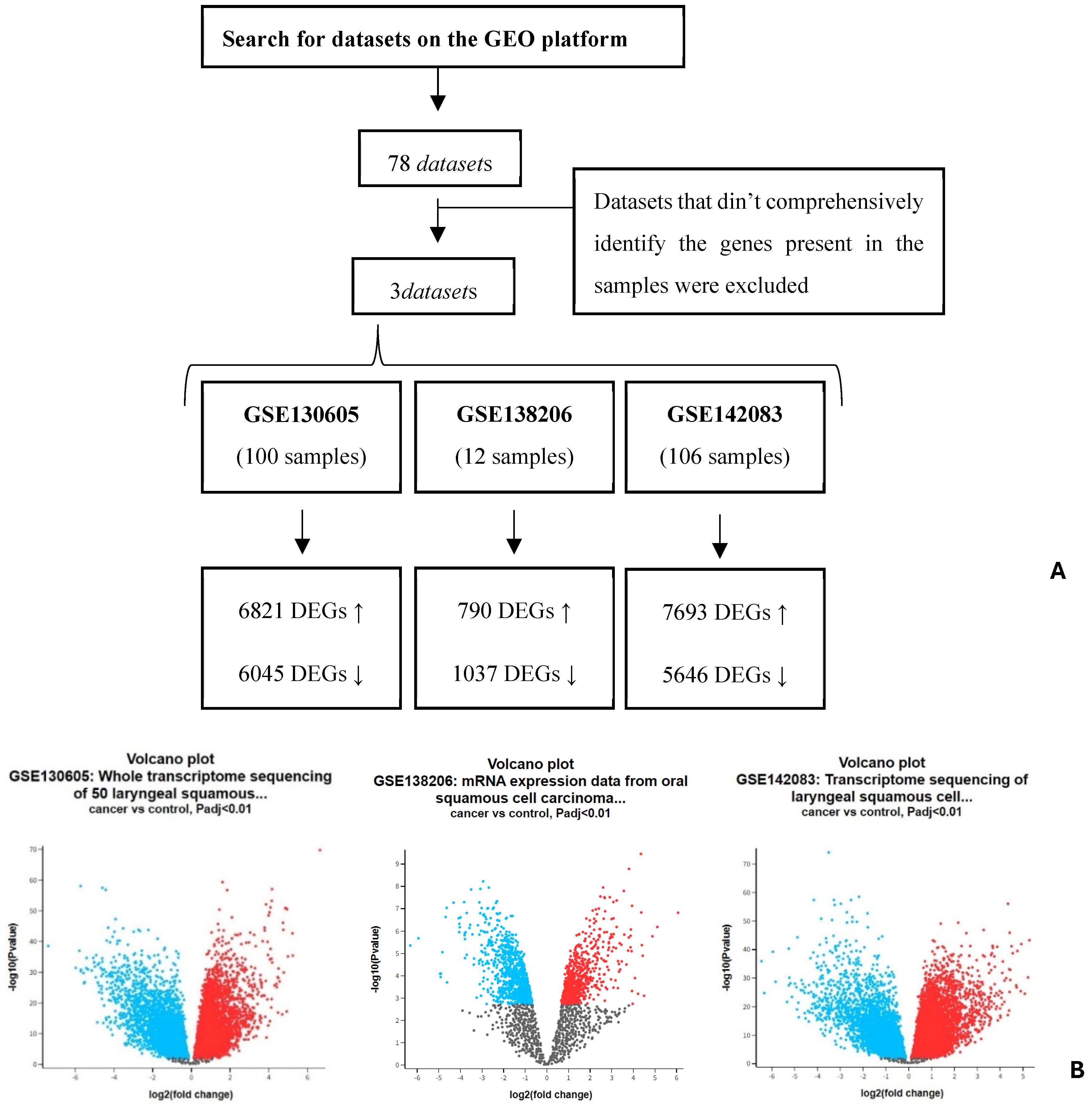
Figure 1. (A) Results from the search in GEO and GEO2R platforms. (B) Volcano plots of DEGs from the datasets identified on GEO.As shown in the volcano plot (B), differentially expressed genes with high statistical significance are highlighted, with the top DEGs marked for head and neck cancer.
Targets 02 00026 g001
Figure 2. Comparative analysis of combined gene expression data from the BEST platform between (A) normal and tumor tissues; and according to (B) the individual’s age; (C) tumor development stage; and (D) HPV infection status.
Targets 02 00026 g002
Figure 3. Analysis of disease-free survival (DFS) and disease-specific survival (DSS) patterns associated with the combined genes.
Targets 02 00026 g003
Figure 4. Identification of the secretion activity of FN1 (P02751), LGALS3 (P17931), MMP9 (P14780), TIMP1 (P01033), MMP2 (P08253), and TIMP2 (P16035).
Targets 02 00026 g004
Table 1. Negative and positive regulators of FN1, LGALS3, MMP9, TIMP1, MMP2, and TIMP2 identified through the analysis of protein interactions performed by the Signor 3.0.
Protein of InterestNegative RegulatorsPositive Regulators
FN1FGG; SERPINA5; Viocristine sulfateTWIST1; SNAIL/RELA/RAPP1; DLK1
LGALS3NDCSNK1A1; ABL2; GSK3B
MMP9SPRY4; SPDEF; PZP; A2METS1; SANI2; USP6; Nfkb-p65/p50
TIMP1NDSPRY4
MMP2NME1; PZP; A2MMMP25; TWIST1; TP53; RUNX2; NODAL
TIMP2DNMT3A; PTTG1ND
Table 2. Main results obtained from the analysis of the expression of the genes FN1, LGALS3, MMP9, TIMP1, MMP2, and TIMP2 in the selected datasets from the GEO platform.
DatasetMain Conclusion on Gene Expression Pattern
GSE130605Only MMP9 and MMP2 were identified with statistical significance, and they exhibited a similar increased expression profile. Their positive regulators were found to be upregulated and negative ones downregulated.
GSE138206Only MMP9 and TIMP1 were identified with statistical significance, and the results obtained support the inverse expression relationship between these two proteins. For MMP9, only positive regulators were found to be upregulated.
GSE142083For FN1, LGALS3, MMP2, and MMP9, a statistically significant difference in expression levels was identified between the control group and HNC. All showed increased expression in HNC except for LGALS3, which was downregulated. The pattern expression of FN1, MMP2, and MMP9 seemed to show to a lack of negative regulators, while LGALS3 did not seem to be affected by its regulators.
Disclaimer/Publisher’s Note: The statements, opinions and data contained in all publications are solely those of the individual author(s) and contributor(s) and not of MDPI and/or the editor(s). MDPI and/or the editor(s) disclaim responsibility for any injury to people or property resulting from any ideas, methods, instructions or products referred to in the content.