Review

Trilobite Eyes and Their Evolution

by
Brigitte Schoenemann
Department of Zoology (Neurobiology/Comparative Animal Physiology), Biology Centre, University of Cologne, Zülpicherstrasse 47b, 50673 Cologne, Germany
Arthropoda 2025, 3(1), 3; https://doi.org/10.3390/arthropoda3010003
Submission received: 23 August 2024 / Revised: 3 January 2025 / Accepted: 6 February 2025 / Published: 14 February 2025
(This article belongs to the Special Issue Trilobites and Their Kin: Evolution, Diversity, and Fossil Insights)

Abstract

:
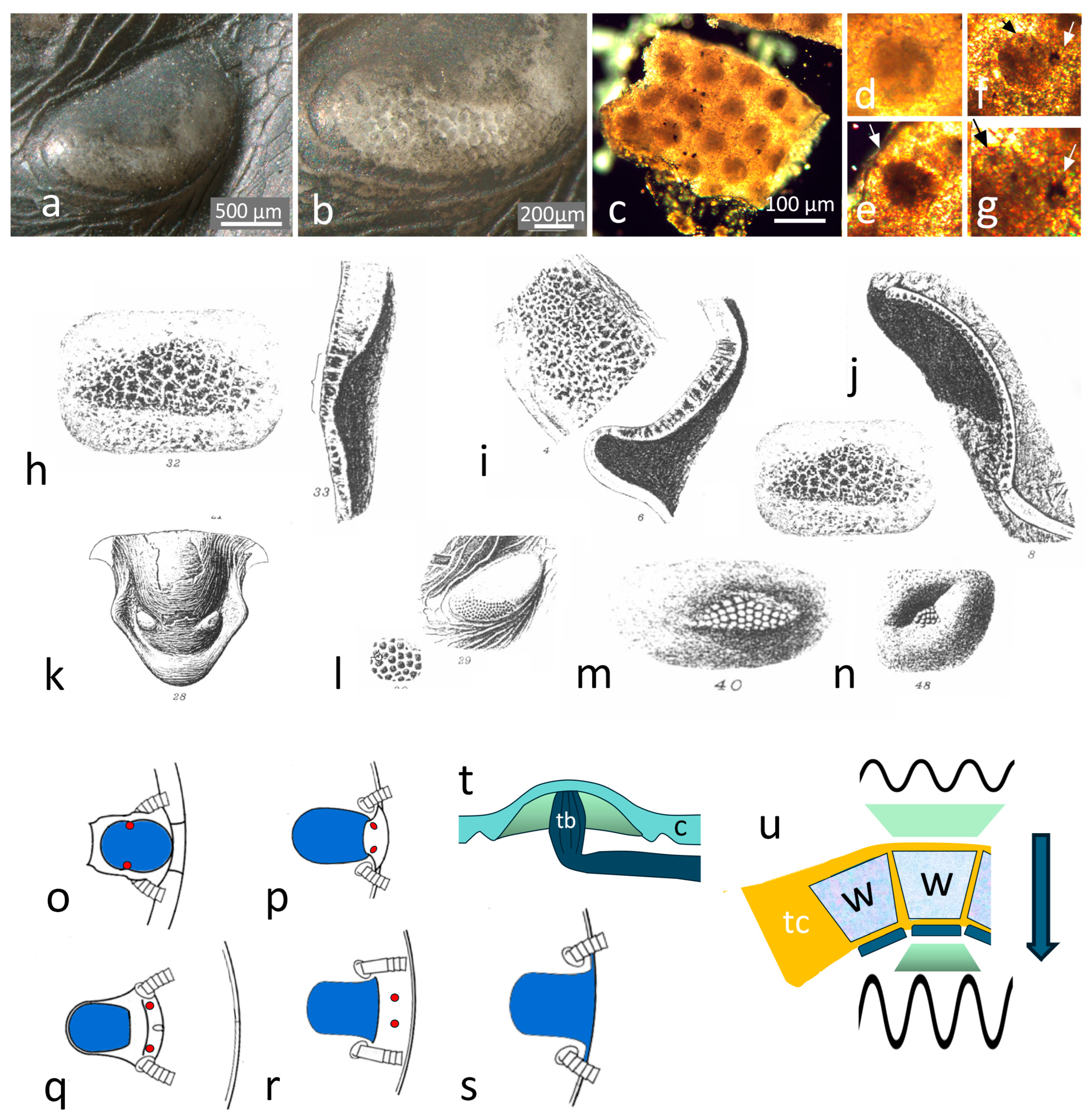
Trilobites, as typical euarthropods, possess compound eyes. In 1901, Lindström was the first to describe them in detail; on the one hand, we reconsider his descriptions of the different modes of trilobite eyes; on the other hand, we expand this by compiling the observations that have been possible in recent years. There are two, perhaps three kinds of trilobite compound eyes. The first are the primordial holochroal eyes, which are actually apposition compound eyes, similar to those of many modern diurnal crustaceans and insects. The abathochroal eyes, often referred to as the second form, are probably a subtype of the holochroal eyes. Consequently, the second is the schizochroal eye of phacopid trilobites, which are hyper-compound eyes composed of numerous small compound eyes below each of the big lenses, which appear from outside as one big lateral eye each. Thirdly, one may call the maculae light-sensitive organs, but this is still uncertain. Comparing what are probably the oldest trilobite eyes described so far with other forms, it is possible to conclude that the sensory apparatus is much older than the fossil record of trilobite eyes and probably developed in Precambrian times. The refractive apparatus, however, was developed later and separately within the systematic groups. This explains why, for example, the mandibulates have a lens and a crystalline cone. Still, the chelicerate xiphosurans, such as horseshoe crabs or eurypterids, possess a lens cylinder with an index gradient but no crystalline cone. Furthermore, this can explain why the calcite character of trilobites is unique in the arthropod kingdom. An important discovery is the probably epidermal, lens-building cells encompassing a prospective lens of Schmidtiellus reetae Bergström 1973 from the early Lower Cambrian of Estonia. We reconsider the morphology of hypostome maculae and interpret them as a potential phylogenetic relict and a potential predecessor of all arthropod ommatidial compound eyes. It will be of great relevance for future research to understand the evolution of compound eyes and vision because we witness the emergence of the first lenses in the trilobite, if not the arthropod kingdom.

Graphical Abstract

1. Introduction

Trilobites, from the very beginning of their appearance in the fossil record, c 522 million years ago, close to the very beginning of the Cambrian explosion of life, were equipped with elaborate compound eyes. During their existence over more than 272 million years, they developed multiple forms and principles of visual systems well-adapted to the most different ecological habitats and environmental conditions, as is well-documented and compiled in several overview articles so far [1,2,3]. Since those reviews, important new insights have developed regarding the structure and function of these ancient systems. These insights, in turn, make it possible to develop an idea of how the formation of compound eyes of trilobites could have occurred during evolution. Even though trilobites represent a separate group of arthropods, in other groups, this process could have taken place differently, both independently and in parallel. We can follow a common evolutionary pathway for the establishment of a proper compound eye. We shall see what are the limits of this ambitious undertaking, because essential elements of the system evolved before they could be preserved in the fossil record and thus escape observation. So, we will become aware that the concept of the fine structure of the sensory apparatus/system of a trilobite compound eye is older than the fossil record of arthropods because it appears completely formed before the dawn of the Cambrian period. Therefore, it was a part of the developmental toolkit available to the common ancestors of trilobites and other arthropods. This concept, as it is found in a representative of the earliest trilobites, in principle, is still present and typical for diurnal insects and crustaceans living today, and it is the basis for derived systems, such as superposition eyes [4,5]. The course of development of the optical apparatus was novel in the trilobite line and different from other arthropods. It was also independent of other arthropod lines, which sheds light on the possibility of determining whether only trilobites in the arthropod kingdom had lenses made of calcite [6].
As in modern arthropods, we know of derived sensory systems in trilobites. As such, in this article, we shall follow the development of different types of compound eyes in trilobites from their very beginning to more advanced sensory systems. There is a non-compound-eye system in trilobites also, as is typical for all euarthropods, the ocellar median eyes, which is evolutionarily older than compound eyes and not homologous to them. These so-called ’Median Eyes’ were recently described and discussed in more detail elsewhere [7]. Finally, there are the enigmatic maculae on the ventral side of hypostomes of some forms of trilobites. We discuss the possible sensory functions and come to suggest that they may represent mechanoreceptive fields.

2. Trilobite Eyes and Lindström’s First Descriptions

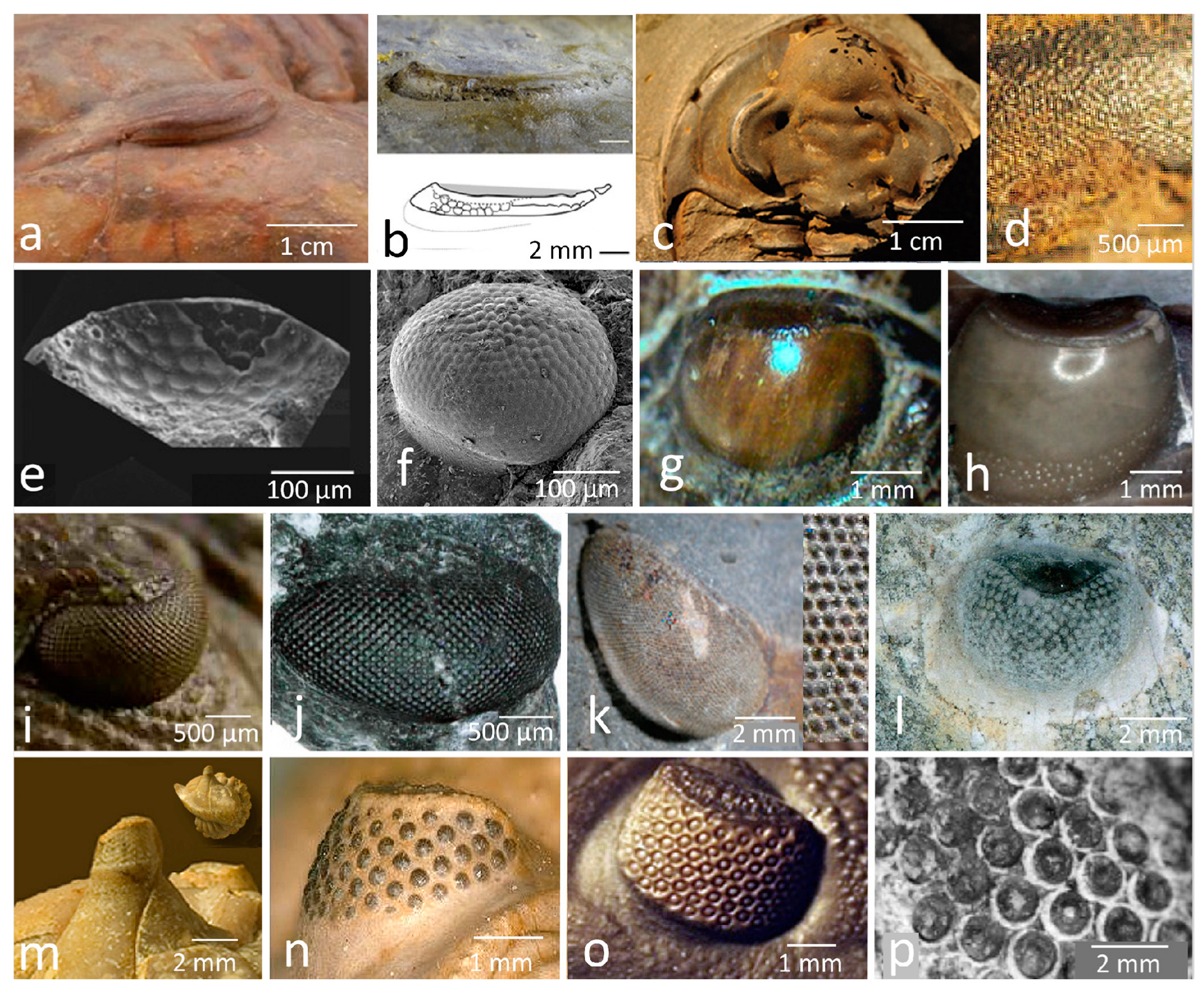
Trilobite eyes received significant attention from the pioneers of vision research (e.g., [8,9,10]), but Gustav Lindström [11] provided the first analytical and comprehensive description in 1901. Lindström described two principal types of trilobite compound eyes, the holochroal (Figure 1a–h, Figure 2a–l, Figure 3 and Figure 4a–d,g,h,j–t) and the aggregate eyes (schizochroal eyes sensu Clarke 1889 [12] (Figure 1i,j). The holochroal eyes, he subdivided into two subgroups. In his first group of holochroal eyes, corneal lenses are covered by a pellucid, smooth, and glossy integument, which is quite similar to that of many modern crustacea. From outside, it is difficult to discern any lenses, which, in a vertical section in these trilobites, appear as columnar prisms comparable in shape to basalt ([11], p.28). In a transverse section, they appear as regular hexahedrons (e.g., Figure 1d–f, Figure 3c and Figure 4b,q–u). These lenses show on the exterior a plane surface, and the interior surface is convex (Figure 1d–f and Figure 4b,q–t). Lindström gave examples of trilobites being equipped with this type of eye, such as representatives of the genera Nileus, Proetus (Gerastos), Bumastus (Scutellum), and Asaphus. The latter was also investigated and described in greater detail by Clarkson in 1973 [13]. He found that these thick, prismatic lenses are always accompanied by a thick cuticle of the trilobites and probably an adaptation against stronger currents. For Nileus, Lindström noted that the exterior integument above the lenses was so exceedingly thick [14] that he doubted if the eyes of this species were able to function as visual organs ([11], p.28).
The second subgroup of holochroal eyes is equipped with globular or more flattened, biconvex lenses, ordinated beside each other continuously or separated by a faint dividing line. The horizontal section shows a round or hexahedral shape (Figure 1a,b). Often, the lenses form an extremely thin stratum in contrast with the surrounding cuticle of the cephalon. As examples of this concept of holochroal eyes, Lindström notes Cyrtometopus, Ctenopyge, or Sphaerophthalmus, all from the Upper Cambrian period, and some of the Phacopida, mainly the Silurian [11].
One of the oldest trilobites Lindström mentions is Peltura (Upper Cambrian). The outer visual surface is smooth, and on its interior side stands out a low relief of semi-globular facets as regular incrassations of the cornea (Figure 1a), but they do not form free lenses. Intriguingly, he reports one small (<1 mm in total), probably young specimen of Acerocare, with one eye showing slightly convex lenses and the other eye resembling that of Peltura ([11], p. 30).
The second type Lindström describes is eyes represented just in the family Phacopida (just in the subfamily Phacopina); he called them aggregate eyes [11] (=schizochroal eyes sensu Clarke [12]) (Figure 1i,j, Figure 2n–q and Figure 4e,f,i). These eyes have almost globular, big lenses, each of them covered by its own cornea, and they are equipped with a capsule below the lens. As he assumed, the capsule was floored by a small retina. In schizochroal eyes, the lenses are widely separated from each other by cuticular material. Because the lenses decay heterogeneously, Lindström suggests that the interior of these lenses might have been of different compositions ([11], p. 30). Today, much more is known about these schizochroal eyes, and we shall come back to them later.
A third form of trilobite eyes, the so-called abathochroal eyes, was first described in 1975 by Jell [16,17] (Figure 2e). This type is restricted to the small trilobites (Pagetia) of the suborder Eodiscina, Order Agnostida.
Most recently, Agnostida were considered to be closely related to polymerid trilobites and classified as a sister group [18]; earlier researchers even discussed a relation to crustaceans [19] (but, see [20,21]). Generally, the abathochroal eyes today are interpreted as a form of holochroal eyes, which matches Lindström’s original ideas.
The recently discovered compound eyes, which are very similar to those of trilobites, of the artiopod Pygmaclypeatus daziensis Zhang et al., 2000 [22] push down the origin of trilobite’s compound eyes to the Artiopoda.
In the following, we shall see what the structural background of this diversity may be and that Lindström’s subdivision [11] should be slightly modified.

2.1. The Holochroal Eyes

In the Cambrian period, the eyes of early trilobites look similar through all systematic groups and do not appreciably diversify before the Furongian period, the last series of the Cambrian period. It is during the transition to the Ordovician period that trilobites left their benthic realm and invaded the pelagic and mesopelagic realms [23,24,25], and even planktonic forms evolved [26]. With this change of habitats and lifestyles, the requirements for visual orientation became more diverse, and so became the eye shapes of primarily holochroal and later also schizochroal eyes, as an expression of their functional structures.
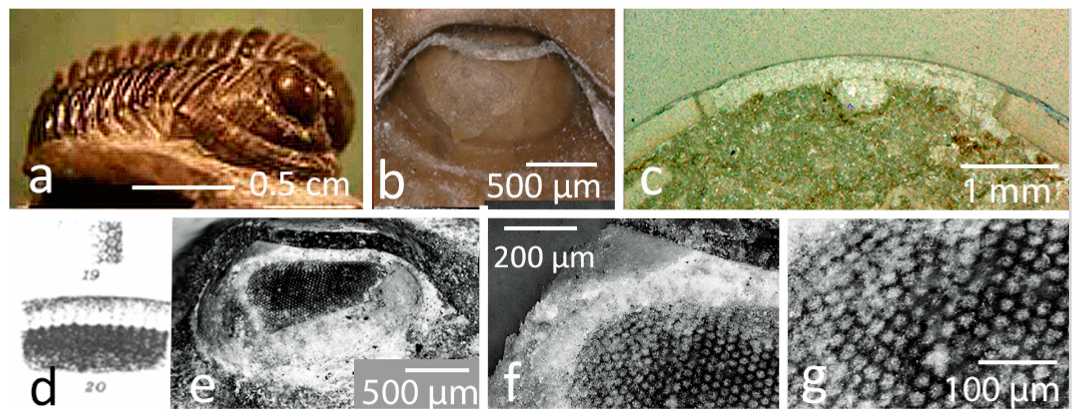
During the Early Cambrian period, the eyes of trilobites were slit-formed, with a narrow, smooth, vertical visual surface that was not structured in any way by facets (Figure 2a,b). Horizontally, the field of view was often enormously wide (about 130° for each eye). However, the palpebral lobe shaded the upward field of vision so that no upward vision was possible (Figure 2a–c).
Just as interesting as the uniform external appearance is the internal one. Insights regarding the interiors of the eyes of one of the oldest trilobites found so far, Schmidtiellus reetae Bergström, 1973 [(Olenelloidea), Saviranna Section, Lükati Fm., Dominopolian Stage, Estonia, Global lower Stage 2, corresponding to the Atdabanian of Siberia, Lower Early Cambrian period] give us important facts for understanding the early evolution of compound eyes in trilobites [27].

2.2. The Structure of Holochroal Compound Eyes of Trilobites

To more easily understand what S. reetae reveals, it might be useful to consider the most common type of arthropod compound eye, the so-called apposition eye (Figure 5h) briefly. It is most common among diurnal insects and crustaceans living today but also in primordial chelicerates, such as the xiphosuran Limulus, or, just preserved in fossils, eurypterids. Adapted to different ecological constraints, there developed many modifications, especially under poorer lighting conditions (e.g., superposition eyes), and the light-collecting dioptric apparatus shows great diversity, especially among the different systematic groups of arthropods. With moderate modifications, the principle of the sensory apparatus, however, is common to all of them.
Compound eyes are plesiomorphic to all euarthropods [28]. They normally consist of numerous units, including, sometimes, up to 30,000 individual visual units (as in dragonflies, such as Aeschna [5] of more or less identical structure, the so-called ommatidia. Each ommatidium consists of two components—the sensory and the dioptric apparatus. The sensory part typically consists of eight, or, in some crustaceans [29] and xiphosurans [30], eleven or more elongated receptor cells. They are grouped around a central light-conducting rod, the rhabdom. It is part of the sensory cells, containing the visual pigments which, through the energy of the incident light, change their steric configuration and thereby generate a small electrical signal that passes through the optical nerves to the central nervous system to be processed. The dioptric apparatus consists of a cuticular lens and, below a cellular clear system, the crystalline cone. The crystalline cone allows space to bring the light focused by the lens onto the rhabdom. In terrestrial systems, the refractive power of the lens is high enough to ensure good focusing. In aquatic systems, the difference in the refractive indices between water and the cuticular material is not high enough, and the crystalline cone, through different, often sophisticated mechanisms, such as being formed to index gradient lenses, overtakes the task of efficient focusing. Because all contrasts, colors, and other details in the field of view of any ommatidium are focused on one point, often the tip of the rhabdom (the focal apposition eye) or a bit deeper (the afocal apposition eye), all of this information is summed up in one signal, and over the whole compound eye arises a mosaic-like vision (Figure 5h). The accuracy depends on the number of facets, the opening angle of the rhabdom, and other factors, while the light sensitivity depends mainly on the sensitivity of the receptor cells and the aperture of the rhabdom and the lenses. Finally, to avoid cross-talk, the ommatidia are separated from each other by screening pigment cells.
While the geometry, with some functional modifications, of the sensory apparatus is quite uniform in the arthropod realm, the dioptric apparatus varies widely among the systematic groups and habitats.
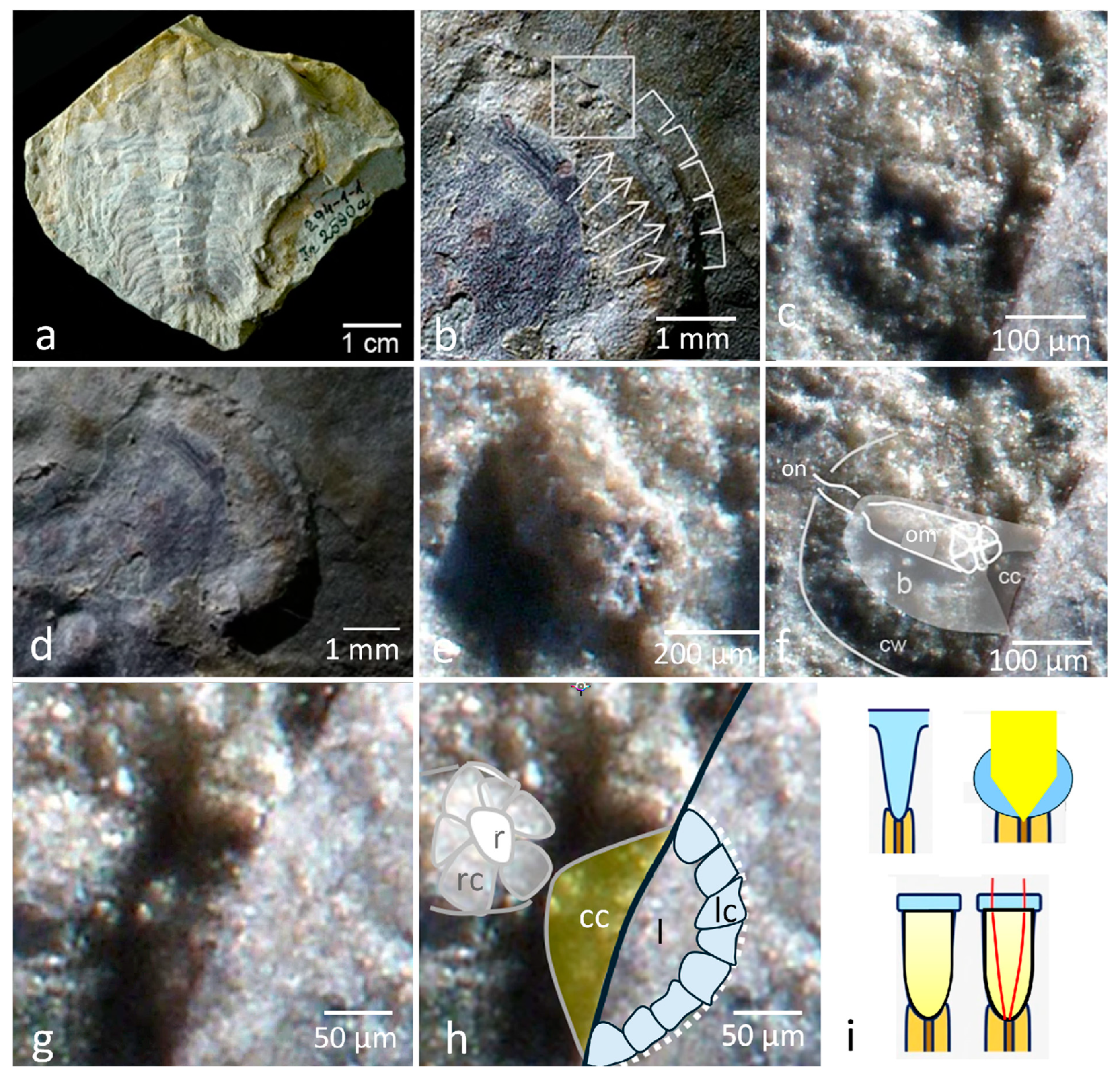
Myriapods, for example, possess modified systems derived from apposition compound eyes’ aggregated systems merged to ocelli, with bigger lenses on top of individual units; only the Scutigeromorpha secondarily developed ommatidial structures again [31]. Among the Chelicerata, just the extinct eurypterids and the Xiphosura (genus Limulus, [10,32]), as mentioned, possessed the typical ommatidial compound eyes; all other chelicerates with eyes developed derived retinae, built from the former sensory elements of their former ommatidial eye (for an overview, see [4,33]). The latter chelicerates possess no crystalline cone, and the xiphosurans developed a cone-like, cuticular lens cylinder with an index gradient instead (Figure 5h and Figure 6i).
In 1859, when Darwin doubtfully pondered the difficulties of his theory of the origin of species, he developed a sequence that illustrates how the human eye, and eyes in general, could have evolved. His hypothesis started with a simple, light-sensitive nerve epithelium whose sensory cells are embedded in pigment tissue, and it culminates in highly complex compound and vertebrate eyes. He found that these types of eyes were so complex that this could lead to difficulties in his theory and that everything could have arisen solely through the classical evolutionary factors of mutation and selection ([34] chapter 6, p. 190ff).
Similar series were established by Paulus [35] to understand the phylogeny of myriapods, insects, and crustaceans in terms of eye structures, as well as by Bitsch and Bitsch [36] considering the evolution of arthropods’ eye structure in general. More recently, Dan-Eric Nilsson, e.g., [37,38], has argued for the homology of all eye types on a common genetic basis (opsin families, similarities of membrane sticking, transduction cascades, and developmental genes). According to Nilsson, the complexity of different eye types (compound eyes, camera lens eyes) evolved later and sometimes several times separately according to different visual requirements due to changing environments during the Earth’s history, e.g., [37,38].
Important insights into the evolution of trilobite eyes, if not arthropod eyes as a whole, are provided below.

The Eye of Schmidtiellus Reetae Bergström 1973

Schmidtiellus reetae Bergström 1973 (Figure 6), from the Early Cambrian period, is one of the oldest trilobites in the fossil record [26,27] at more than half a billion years old. The outer shape of the eye overall is already trilobitic, as is typical for all Early Cambrian trilobites. It is reniform, with the anterior lateral side slightly longer than the posterior part. The upper side is covered by a palpebral lobe (eyelid), and the visual surface is smooth and slit-formed (Figure 2b and Figure 6a,d). Very recently, it has been shown that this typical shape is very probably a heritage of older predecessors [23]. Fortuitously, one of the eyes was slightly abraded, revealing the interior and thus the functional structure of the compound eye of the fossilized trilobite (Figure 6b–h). Phosphatization (as observed in S. reetae) is a mode of fossilization that preserves very fine detail, and it is a specific form of preservation revealing the finest structures, sometimes just a few micrometers in size [39,40]. The outer shape of the eye of S. reetae is flat and slit-formed, as is typical for the compound eyes of trilobites of the Cambrian period, forming a field of view of about 132° for each eye [27] and with no upward vision. It comprises a few horizontal rows of visual units (Figure 2b), roughly 100 of them. These lie as elongated columns far apart from each other embedded in a kind of cellular basket (Figure 6b–f), and consist, as is typical of euarthropods, of about eight (relicts of) receptor cells, grouped around a central rod (Figure 6c,e,f), presumably the former rhabdom. The proximal part of this typically formed ommatidium ends up with a thin section, which is probably the (relicts of) the optical nerve. So far, we have the characteristics of the typical sensory apparatus of an apposition compound eye. Not as typical are the cellular ‘baskets’ (Figure 6c) and the dioptric apparatus, because, more or less, the lenses still seem to be missing here. Just some very fine circular lines may indicate the arising development of a lens inside of the thin cuticle (Figure 6f).
As is typical for Early Cambrian trilobites, clear patterns of facets are not visible on the visual surface of the eye. There is a crystalline cone (Figure 6f–h), which would indicate, phylogenetically, a relation to the Tetraconata/Pancrustacea (insects + crustaceans) rather than to the Chelicerata, which do not possess crystalline cones. A new discovery in this old material [27] is about 16 probably epidermal lens-building cells (just about 8 of them can be seen here). They encompass a round structure of about 200 µm in diameter, which is not thicker than the still very thin cuticle (the dark line in Figure 6h). This, very surely, is the predecessor of a proper prospective lens, possibly one of the oldest that we have so far in trilobites. It may be pointed out that these cells closely resemble those that can be observed on the hypostomal maculae surrounding the round structures (Figure 7n,o).
There is, however, a perhaps slightly younger trilobite from the same location, Holmia kjerulfi (Linnarson, 1871) (Figure 2c,d), which shows compound eyes completely equipped with hundreds, if not thousands, of small lenses with a remarkably high resolution so early in trilobite history. Later trilobites typically have well-developed lenses of different shapes. They still, however, keep the sensory part, as described here, as shown in the Silurian Aulacopleura koninckii (Barrande 1846) [41]. One may conclude that the evolution of lenses started during the Early Cambrian period and diversified, especially during the Furongian period (see Ctenopyge, Sphaerophthalmus, etc., Figure 2f and Figure 4h), at the threshold of the Ordovician period, as trilobites started to invade different ecological niches with different environmental constraints.
Altogether, the first traces of lenses in S. reetae and the later diversification point to the important conclusion that the dioptric apparatus in trilobites developed independently from other arthropods (structures that allow for focusing (mandibulates: lens and crystalline cone; chelicerates (eurypterids, xiphosures): lens cylinders, soft-bodied arthrtopodean predecessors: primitive (initially individual?) spherical blobs of mucus functioning as lenses (=> crystalline cones, cuticular lenses developing later)) comparable to the so-called ocelli of soft-bodied invertebrates living today are conceivable here). The well-described fantastic compound eyes of the Australian stem arthropods (Cambrian Series 2), which are just slightly younger than S. reetae, are equipped with a grid in which thousands of lenses must have been located [42,43,44]. Many of these eyes are anomalocarid eyes outside of the crown group arthropods. The refracting and focusing systems of the other arthropods may also have evolved inside of the systematic groups independently and may be the reason why the xiphosurans have, in principle, the same sensory system as the others but possess lens cylinders as a dioptric apparatus, which is different from the tetracontane lens–crystalline cone systems.
This, in turn, may take the wind out of the puzzle as to why and how trilobites were the only arthropod form to develop calcite lenses, as they developed them independently of all other groups of euarthropods. So, in total, one may conclude that the sensory part of an ommatidium is pleiomorphic for all euarthropods, and the dioptric apparatus is apomorphic for each systematic group.

2.3. The Process of Evolving Proper, Focusing Lenses

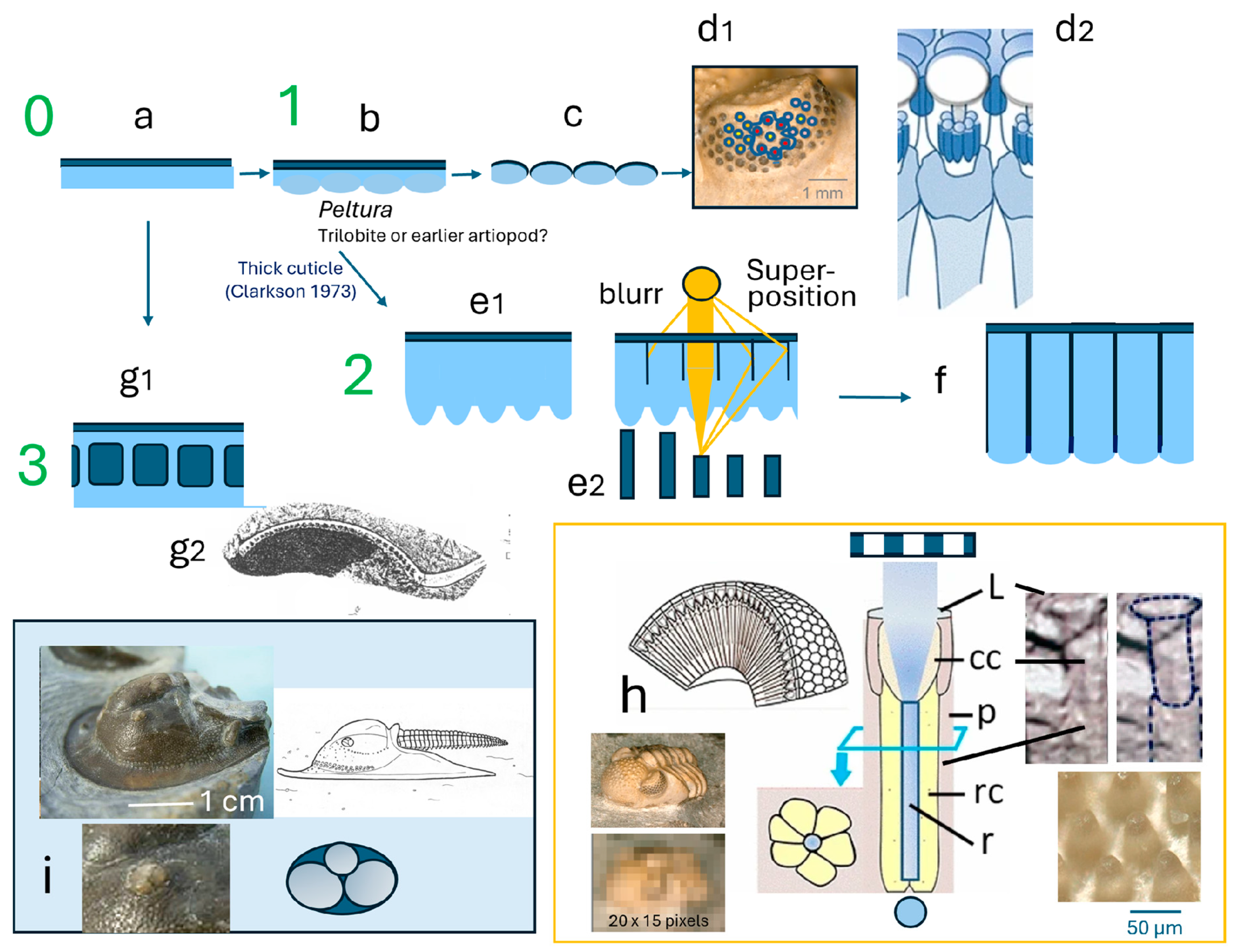
As we have seen in one of the oldest trilobites, Schmidtiellus reetae Bergström, 1973 (Series 3/Atdabanian, Early Lower Cambrian), the arrays of sensory units are just covered by a thin, continuous, smooth cuticular cornea, as is seemingly typical for most Cambrian trilobites. S. reetae, in its geological section, is the first complete trilobite to appear in the fossil record. In the strata below, the lowest fossil-bearing strata in this area, are only trace fossils, and, above this area, there are layers with only fragments of trilobites, which are probably also Schmidtiellus species, until the first complete trilobite, S. reetae, appears. Consequently, one may assume that the exoskeleton of S. reetae, in principle, was still very thin, and, later, when the cuticle became thicker, it perhaps remained thinner above the light-sensitive units of the eye, where the cuticle surely was at least translucent. This principle may have remained in some later trilobites, such as Gerastos and other proetids. This ‘window through the shell’ (Figure 3b), with its smooth surface, still has no function physically because it has no relevant focusing properties. Each sensory unit of an ommatidium, consisting of its receptor cells ensembled around the central rhabdom, perceives a section of its environment to which it is directed, forming a mosaic-like image overall (Figure 5h). This changes with Lindström’s following observation.
There is an intriguing figure in Lindstrom ([11], Pl. III, 40): a vertical section through the eye of the mid- to late-Cambrian Peltura scarabaeoides Wahlenberg 1821, Olenidae (Alum Shale Fm, southern Scandinavia). Lindström describes the superior surface as quite smooth and evenly rounded; on the “interior side stand out, somewhat distantiated, in low relief, semi-globular facets, quite as regular incrassation of the cornea, thus not forming free lenses …” ([11], p. 30) (Figure 1a). Because the lower side of the thin cuticle, however, now develops slightly bulging, hemispherical domes, these act as semi-convex lenses that concentrate/focus the light onto the rhabdom. This makes better use of the available light and allows the eyes to work even under weaker light conditions.
In principle, this Peltura system seems to have been retained in later trilobites, such as the asaphids of the Ordovician period. Here, the lenses consist of basalt-like calcite columns formed from the cuticle and arranged hexagonally, showing also a hemispherical half-lens proximally (Figure 1d–f, Figure 4b,q–t and Figure 5e1,f). The ‘new feature’ here is a fine separation of the individual lens elements, which are probably too fine to consist of cuticular material. One can probably assume that this separation consisted of organic material, something like a thin membrane or a fluid with a lower refractive index than calcite. As soon as it is thicker than a critical size, then at least some of the obliquely incident light rays would also be focused on the rhabdom due to total reflection (Figure 5e2). This would widen the field of view of each unit, but, by overlapping with its neighbors, the image would be less sharp for the individual unit; however, the sensitivity/exploitation of light would be increased.
Proetids, with a similar eye system ([11], Pl. VI, 20), persisted as the longest-surviving trilobites. They survived several mass extinctions until they finally became extinct at the Permian–Triassic boundary during the most disastrous ecological catastrophe ever [45,46,47]. Below the columnar lenses formed by the smooth cuticle spanning the visual surface, the relicts of the sensory arrays can be seen at some distance from each other (Figure 3). Because some of these arrays show relicts of the receptor cells and/or the central rhabdoms, the white material does not represent stuck cones, such as broken lens cylinders comparable to those of xiphosurans, as one might assume according to the appearance of the whole system. Two primordial characteristics may have been retained in this system—the cuticular ‘window’ and the distantiated receptor units.
In most holochroal eyes, the sensory apparatus is probably identical, more or less, and similar to an apposition compound eye [27,41]. Considering only the formation/evolution of proper lenses in trilobites, one may establish a theoretical series of steps (Figure 5a–f), showing how a smooth, transparent cuticle could have developed into functioning, i.e., light-focusing lenses over time without any reference to phylogenetic relationships. This sequence is a model, and the processes may have occurred independently and at different times in the different systematic groups, so the different types of lenses trilobites possess may represent different stages of achievements.
Comparisons with modern crustacean eyes suggest another remarkable conclusion—that the columnar lenses may have been an adaptation to deeper water, which is different from most crustaceans with ‘normal’ apposition compound eyes, which are adapted to well-lit conditions. Mid-water, long-bodied decapod crustaceans, such as shrimps, lobster crayfish, etc., possess a strict geometry of their compound eyes and developed the so-called reflecting superposition eyes. Preconditions are, among others, reflecting cylinders with a squared floor area, a defined geometry, and a spherical shape of the whole eye [48,49]. This means that not only the light rays that emanate directly from an object are focused on the corresponding rhabdom and can be captured there but also light rays that enter obliquely and hit neighboring facets because they are reflected from there appropriately. Normally, the rhabdoms in reflecting superposition eyes lie deeper in the eye than in apposition eyes to give space for the reflected rays to reach the relevant rhabdom. Because light is exploited much better, these eyes enable their owners to live in deeper areas of the ocean or under poorer light conditions. Perhaps trilobites with columnar lenses, separated by organic material and an almost spherical shape (e.g., Figure 4t, Asaphus. raniceps Dalman 1827), developed comparable mechanisms.
It is also possible that trilobite lenses evolved in an opposite way by starting the development of lenses by bulging the outermost thin layer of the cuticle. The outermost thin layer of the cuticle bulges into numerous small contiguous domes, which are semi-convex lenses (Figure 4j–l). This seems to be the case, for example, in Nileus platys (Figure 4j–p). This cuticle is so thin that in exuviae, the underlying matrix shows through (Figure 4j,m). It seems to be the case that these lenses are not covered by their own thin and pellucid membrane, as with the other types of lenses. Below this lens-forming layer, there is a second. In the first specimen of Figure 4l, there is a homogenous, darker one. The second specimen of N. platys (Figure 4m–p) tells us a bit more about this stratum. The thin outer lentiferous layer here is lost, and so we have a direct view of this in the fossil’s darker second layer below. This is characterized by dark, translucent spots (Figure 4n–p), which reinforce the idea of windows through a thick cuticle. A vertical fracture shows that the translucent dark spot is indeed a small cone (Figure 4o,p), which is triangular in its vertical section. Elongated elements surround it, and all of these structures are positioned above a wider cylinder underneath (Figure 4o,p). It is easy to see that here again we have relics of a typical ommatidium of an apposition/holochroal eye. The element forming the translucent spot must be interpreted as the relict of the crystalline cone, typically built by four cone cells. Consequently, the encompassing elements represent relicts of the primary pigment cells, and the underlying tube represents the relict of the sensory unit, surrounded by relicts of secondary pigment cells. Much clearer than in the examples before [27,41,50,51], here, the crystalline cone in trilobites becomes evident, indicating an affinity of trilobites to the Mandibulata.
This interpretation (thin lenses and the ‘dark cubes’ within the cuticle -> crystalline cones) would be in accordance with the hypothesis developed by Schoenemann and Clarkson [50] that eye systems of trilobites with thin lenses develop a proper crystalline cone, which probably supports refractive tasks, while for systems with thick biconvex lenses, such as in phacopid trilobites or Aulacopleura koninkii (Barrande, 1846), the refractive power of the big lenses, especially due to the high refractive index of calcite, is efficient enough to focus the light properly, and so here crystalline cones are missing or reduced [41].
Therefore, if we look at the eye of Nileus, we find a compound eye with lenses made of a fine cuticle, a long crystalline cone, which possibly takes over the transmission and focusing of light, as the lens is too weak for proper refraction; this is reminiscent of an eye typical of many crustaceans (Figure 6i), which is also represented in the fossil of the predatory Dollocaris ingens (Van Straelen, 1923) from the Jurassic period [52].
A quick survey of other trilobite eyes helps reveal the evolutionary pattern. There have been described crystalline cones for trilobites of different systematic groups (Schmidtiellus reetae Bergström 1973, Redlichiida [27]; Aulacopleura koninckii (Barrande, 1846), Proetida [41]; Archegonus (Waribole) warsteiniensis Richter and Richter 1926, Proetida [50,51], and now Nileus armadillo Dalman 1827, Asaphida). While the lenses of these trilobites are very different or even missing (S. reetae), they all possess crystalline cones. Thus, it becomes evident that possessing a crystalline-cone-like structure is independent of the systematic group, suggesting it is primitive for trilobites (see S. reetae Berström, 1973 [27]). It is a feature typical of trilobites probably from the very beginning (see Schmidtiellus reetae Bergström, 1973 [27]). Crystalline cones are sustained just in systems with thin lenses; they became modified, were often reduced Aulacopleura koninckii (Barrande, 1846), Proetida [41], or even became lost if the lenses became thicker phacopids with schizochroal eyes. Thick lenses have a high refractive power, even more so if they are of calcite.
So far, we have several types of holochroal eyes—those with a pure, smooth visual surface, as is typical for the Early Cambrian trilobites, and those equipped with lenses, including semi- (Nileus) or biconvex (e.g., Sphaerophthalmus, Ctenopyge, Telephina, phacopids) and columnar (e.g., Asaphus, Proteus, Gerastos); there exist many transitions.

2.4. Schizochroal Eyes

For the eyes of phacopid trilobites (subfamily Phacopina), finally, it has been shown that the big, separated lenses and their sensory equipment developed through fusion of several neighboring ommatidia (Figure 1i,j, Figure 2n–q, Figure 4e,f and Figure 5d1,2). The small sensory systems gathered underneath the big lens, each of them equipped with a tiny lens of its own, share the light-gathering properties of the all-covering big lens in common [53]. Therefore, below each of the big lenses, a small compound eye of its own is located. So, in total, the lateral eye of a phacopid trilobite (suborder Phacopina) represents a hyper-compound eye. Furthermore, there is a central elaborate system of still unknown functions. In summary, here, we have a very derived, advanced, younger system existing in this elaborated form probably since the Devonian period. The function of this system is still unknown; conceivable functions are sharing functions between the elements, such as vision of different colors, perception of polarized light, pooling of signals to higher sensitivity, etc.
The big lenses show a sophisticated internal differentiation (Figure 4f), and their function under certain conditions could include the correction of spherical and chromatic aberrations, which could well avoid a blurred image [54]. Bifocality and the ability to perceive polarized light have been widely discussed [54,55,56,57]. The phacopid eye may remind us of the tiny eyes of the parasitic strepsiterian insect Xenos peckii Kirby, 1813 (males have a size of c 4mm) because of the relatively large lenses, which stay apart from each other. The dioptric apparatus forms a cone on the inside of the eye tightly enclosed by a small kind of retina [58,59]. The retina consists, founded in its phylogenetic context, of cells that, during evolutionary history, turned from former ommatidia to retinal cells. This highly specialized structure is probably a consequence of the miniaturization of the eye, especially when working under reduced light levels. Because the light receptors need a certain amount of photons to work, very small eyes often reach this limit. Like here, other mechanisms are being developed to capture enough photons. Earlier trilobite researchers (e.g., [60,61]) assumed that the capsule beneath the phacopid lens contained a small retina, which is why the similarity to X. peckii was assumed. This idea of the phacopid retina, however, had to be revised, and today we know that the eyes of X. peckii are structurally very different from the hyper-compound eyes of the phacopid trilobites.

The Third Type of Eye?

One of Lindström’s great concerns in his famous 1901 article [11] is the hypostomal maculae, paired small areas oriented ventrally on either side of the hypostomes (Figure 7k). Interestingly, these seem to occur only among species with natant (free-floating, not fixed) hypostomes [61], which are thought to belong to trilobites with generalized particle feeding habits [14], such as representants of the orders Asaphida, Harpetida, and Ptychopariida, post-Devonian proetids, and two primitive phacopids (Pharostomia and Bavarilla) [62]. This suggests a function of the maculae as mechano- or chemosensory organs to recognize their food.
The function of the maculae, however, is still not conclusively clarified. While Lindström [11] argues that they are reduced ventral compound eyes that have lost their lenses, as did, for example, Spencer, Hanström, and Raymond [63,64,65], Hupé, Størmer, Whittington, and Evitt [66,67,68,69,70] interpret these structures as muscle attachments. Muscle attachments are often manifested as small ditches or dark depressions, especially on the glabella, and they look very similar to many maculae. Vertical thin sections show prismatic hollow spaces within the cuticle (Figure 7h–jmany of Lindström’s [11] specimens (e.g., Figure 7l–n), clearly shows a surface that appears to be composed of many delicate translucent or even transparent spherical elements (Figure 7a,b), probably thus functionally forming proper lenses. Consequently, we distinguish a second type of maculae, here called ‘smooth type’, with a structureless surface. All authors notice that the cuticle of the maculae is much thinner than the rest of the trilobite’s cuticle) sometimes filled with a spongy material and probably fluid, which surely had no lens function (focusing light) but would allow light to come through a thick cuticle easier than at other places, and it could have been a primordial ‘attempt’ to establish light-permeable windows through an opaque cuticle.
Very striking are the horizontal thin sections through the maculae with smooth, structureless surfaces (Figure 7c,d,h–j). They show an almost regular arrangement of irregular patches of material, which are more light-dense than the rest. The cellular arrangement around the patches (Figure 7c,d), not known so far, indicates that here we are probably already below the cuticle. For a normal arthropod compound eye, one would have reached the sensory level, and one would expect at least indications of an ommatidial structure, namely retinular cells arranged around a central axis, the rhabdom, surrounded by pigment cells containing melanin, which are often very stable over the course of millions of years (e.g., [71,72]).
At first glance, here we find, as Lindström did [11] (Figure 7h–j), relicts of an unstructured tissue, which would be expected to represent the remains of a small retina composed of receptor cells and perhaps isolated by pigment cells, which would make possible a better resolution of the input to individual sensory cells. Retinal compound eyes in arthropods, however, are very unusual. All retinal compound eyes in modern arthropods are secondary, as in myriapods, chelicerates, and the specialized eyes of insects (e.g., larval eyes of insects (stemmata) or the eyes of Xenos peckii Kirby, 1813 (Strepsiteria)).
A point that speaks against the retinal character of these maculae’s patches is the globular structures that obviously make up the patches (Figure 7f,g). They consist of a dark center, encompassed by smaller petal-shaped (relicts of) cells, arranged in a star shape around this center (Figure 7f,g). The two examples that can be observed here are vaguely reminiscent of the two states of pulsating vacuoles, one closely surrounded by cells and the other one larger in its dark center and surrounded by more elongated structures. Pulsatory vacuoles, however, are organelles of unicellular organisms (Ciliatae) with osmoregulating function. Maybe one of the organs is simply larger than the other.
This gives the structure the appearance of a small (sub-)organ of unknown function. They show a certain similarity to ocelli (Latin: small eyes), small, sometimes tiny light-receptive organs represented in many invertebrates, such as Cnidarians, worms, mollusks, lobopodians, and onychophorans, and the median eyes of euarthropods, just some to name a few. Usually, these consist of a cup of epidermal cells containing light-receptive and pigment cells, isolating the sensory cells to achieve a more differentiated signal. These pigment cells could be the reason for the darker appearance of the small globes. More advanced ocelli can contain more or less differentiated lenses. Very speculatively, this would result in the assumption that the maculae were compound eyes indeed not yet containing ommatidial structures but ocelli, perhaps predecessors of the latter. This would be unique so far.
Another question is why a trilobite might have had eyes on the ventral side of the body. It would not be a unique feature for trilobites, as ventral light-sensitive organs also occur in arthropods, including chelicerates (e.g., larval Limulus, [73,74,75,76]) and myriapods [65], and perhaps this ventral ‘eye’ is even a primordial organ. It generally could have been a larval organ, as in Limulus [76], and/or a relict organ of use for specialized back swimming larval stages. What these light-sensory organs were used for remains a matter of speculation. It has been suggested that they were of help when the trilobites swam upside–down or to detect day and night [77]. Well-equipped with compound eyes dorsally, these trilobites, however, probably would not have needed a further visual organ; it may have been, however, a relict organ.
Fully developed trilobites with natant hypostomes often live close to the ground, feeding on fine material, so the hypostome is hardly ever exposed to light. Alternatives to discuss are that maculae are chemo- or mechanoreceptors.
Different from Seilacher and Gishlick’s assumption ([78], p. 370), the maculae described here do not possess any pores. Pores of chemoreceptors below or within a cuticle are needed so that the substance that should be perceived can reach the receptors. [The pores described by Seilacher and Gishlick [78] in plate 22.5, for comparison, are elements of pustules in the outer cuticle of the glabella (not maculae), and it may be that these pores were chemosensory entrances, but, more probably, alternatively, it may be suggested that they had a secretory function: a slimy trilobite.] Our maculae here do not show any pores; we may therefore exclude a chemosensory function here.
Their idea of a stridulation organ, producing sounds by using the cephalic legs scratching along the maculae [78], similar to the scroll bars of cricket wings or at the legs of locusts, seems improbable, because in contrast to the insect’s scroll bars, the surfaces of the maculae’s domes are by far too thin and delicate.
What remains is the mechanoreceptor function. Mechanoreceptors, for example, can sense shear forces, expansion, and compression forces, but, above all, pressure is often caused by vibrations of matter of various frequencies. In arthropods, normally, perception occurs through hairy receptors, which are often used for proprioception, such as, for example, measuring relative positions of body parts (e.g., legs to thorax), but it also plays a great role in any kind of ‘hearing’. The perception of vibrations in the air or in the substrate was suggested by Seilacher and Gishlick [78]. While the argumentation of these authors is raised mainly by the position of the maculae on the ventral side of the hypostome, it may be worthwhile to test this hypothesis against a physical background.
At first glance, the maculae may resemble a field of campaniform sensillae (Figure 7t), a class of mechanosensory organs of insects measuring local stress and strain within the cuticle. Similar to our organs on the hypostomes, these consist of a flexible dome of thin cuticle, innervated by a single, bipolar neuron, and they often occur in groups of sensory fields. Campaniform sensillae are located all over the insect body, especially where stresses are high, and act as proprioceptors in the cuticle, detecting mechanical load as resistance to muscle contraction [79]. Such receptors could be worthwhile for detecting the vibrations of small living organisms moving on the ground. Arguments against this hypothesis are that campaniform sensillae are restricted to insects, so they are a phylogenetically ‘new’ development, and that the tissue below the cupulae of the maculae does not match the internal structure of the campaniform sensillae (Figure 7c–g,t).
Mechanoreception through a relatively thick cuticle is difficult because vibrations can hardly be transmitted, but the thin cuticle in the maculae may facilitate mechanoreception and, thus, in principle, hearing. We have in the smooth form of maculae (without lenses) an outer thin and an inner thin cuticular membrane (Figure 7h–j) between the spongy hollow spaces mentioned and an assumed sensory membrane directly underneath. Because of the incompressibility of water, vibrations like sound but also vibrations caused by movements in water are transferred four times faster than in the air and three times less absorbed and are thus much more distinguishable than in the air. If the spongy hollow spaces between the inner and outer cuticular membranes in the smooth-surfaced maculae were filled with fluid also, the vibration could have been transferred effectively through the thicker cuticle toward the underlying receptive sensory field. If the ‘lenses’ of the lens-bearing maculae are filled with fluid also, the shape of the outer domes of the cuticle may provide directional characteristics. Due to the curvature of the macula surface, the upper thin cuticular membrane is always wider than the lower (Figure 7h–j,u); consequently, the pressure of the vibration onto the outer surface is transduced to the smaller area of the lower, which resulted, ideally, in an amplification of the signal, which is similar to the conditions of the round and oval window in the human inner ear. However, we must admit that the difference between both surfaces of the hollow spaces is very small in reality.
Perhaps, however, the holey structure of the cuticle above the patches made the cuticle weaker and able to swing in resonance with mechanic vibrations. This is more likely, as one may assume that the calcification here was decreased, as we know of differentiation of calcite content within other sensory organs of trilobites (lenses of phacopids, [6,54,80,81]).
It well may be that the small globular sub-organs within the patches reacted to pressure as a result; ideally, we may have in the form of small organs within the patches of the maculae (Figure 7e,f) an effective mechanoreceptive field perceiving vibrations of sound or vibrations in the ground and water, perhaps caused by social partners or predators, thus forming in total a ‘compound ear’ in the widest sense. It may be a very plausible assumption that this mechanoreceptive field (the macula) was testing the consistency of the ground while searching for small food particles. In later trilobites with fixed hypostomes, this function may have been overtaken by the cephalic legs.
The idea that the globular elements work as proprioceptors and statoliths to inform the trilobite about the position of the mobile hypostome seems to be rather improbable because the statoliths are not particles but cellularly formed structures and star-shaped small organs that probably are not free but fixed in their context. We probably will never know. The darker color of the interior area may be caused by pigments, such as melanin; it is stable over many millions of years (e.g., [71,72]), which leads to a different idea.
Finally, weighing up the arguments, one may assume to be confronted here with a highly fascinating primordial ‘window’ through the thick cuticle, equipped with a flat assemblage of small ocelli underneath the cuticle to perceive light, overall establishing a unique, initially non-ommatidial compound eye, a relict and perhaps a predecessor of all arthropod ommatidial compound eyes. A second option seems to be as probable—that the maculae formed mechanoreceptors composed of a field of subunits consisting themselves of small mechanoreceptive organs. On the one hand, they may have perceived vibrations, such as sound or other vibrations, within the water, or they may have been of use when testing the consistency of the ground while searching for food particles. The compilation of the maculae of homologous organs of various trilobitomorphs (Figure 7o–s), as performed by Cotton [82,83], may inspire further ideas.

3. Discussion

It would be desirable to be able to establish a relationship between the high diversity of different eye concepts and phylogenetic relations. From their very beginning, all trilobites seem to have holochroal (appositional) compound eyes—if they have any. Blindness in trilobites is secondary, and if eyes once had been lost, they never became reestablished [2]. The abathochroal eyes of the small systematic group of the eodiscid trilobites [16] nowadays generally are understood to be holochroal eyes, and the occurrence of schizochroal eyes is restricted just to a subfamily of the order Phacopida, the Phacopina. According to Nilsson [37,38], in recent arthropods, the constraints of the environment may be stronger than phylogenetic contexts in the formation of eye types, and the great diversity of environmental conditions and demands may generate a multiplicity of concepts. The problem may be demonstrated as follows. Representatives of the genus Asaphus are cited by Lindström [11] and Clarkson [13] as examples of trilobites with columnar lenses. Within the asaphids, however, the length of the lenses can vary considerably (Figure 1d–f and Figure 4q–t), but, still, they always seem to possess more or less columnar lenses. The columnar character depends on the thickness of the cuticle, and this, in turn, depends on the flow conditions in the surrounding area [13]. Another example we find in Gerastos cuvieri (Steininger, 1831) (Figure 3c). Gerastos belongs to the order Proetida. If we accept these lenses as columnar, it is shown that their expression can exist independently from asaphids and thus the phylogenetic context (convergence). So, we find columnar lenses within a systematic group, albeit with greater variability, but also without. Whether there are other trilobites aside from the Asaphida and Proetida with columnar lenses is not known so far. Generally, not much more is known about the relationship between eye designs and phylogenetic context, and it would be worthwhile to investigate.
As we have seen, within the group of trilobites, we find a wide variety to develop light-refractive systems, such as semi- or biconvex lenses, sometimes with a columnar shape, probably starting from a smooth cuticular visual surface. Because the sensory apparatus, consisting of several receptor cells grouped around the central light perceiving structure, the so-called rhabdom, already exists in the oldest form of the trilobite compound eye, it is rather probable that this principle is a heritage from earlier ancestors and was developed far before the development of hard shells and thus before what the fossil record at the moment can tell us. Trilobites share this pattern of an apposition compound eye with many other arthropods. It is the plesiomorph ground pattern of an arthropod compound eye [28], today still present especially among diurnal insects and crustaceans, and it has been described also for extinct forms, like the system of xiphosurans, such as eurypterids [32]. It is very similar to that of today’s living xiphosuran Limulus [30].
In an example of Asaphus kowalewskii (Lessnikova, 1949) and some other species, it can be nicely observed how the lenses finally could have been formed sidewards from the cuticle during evolution (Figure 4f), very probably adapting to the constraints of changing environments, as in recent arthropods. Firstly, from a homogeneous cuticle, they are separated as vertical blocks. In other species, these compartments separate from each other, and in a third, the small ‘blocks’ are equipped with slight bulging on both sides. So, finally, they form a proper biconvex lens, focusing the light precisely onto the central rhabdom.
This goes, however, hand in hand with the fact that light falling at a critical angle is directed out of the system or into the neighboring unit, causing ambiguous information. The visual units (ommatidia) must from now on stay apart from each other or be shielded by light-absorbing pigments to isolate the visual units optically. This results in sharp images for perception. While in S. reetae the visual units are still widely separated from each other, in more densely packed compound eyes, the ommatidia are separated by pigment cells. That these pigments exist in younger and more advanced trilobite eye systems has been previously demonstrated (Aulacopleura koninkii (Barrande 1846), Silurian) [41], and it is demonstrated here for Nileus.
In the strict geometrically arranged eye of some asaphids, even mechanisms of reflecting superposition may have become realized, whereby reflection inside of the columns of neighboring ommatidia may be supported with additional light supply (Figure 5e2). The strictly round cross-section of the eye of Asaphus (Figure 4t) suggests that superposition may have been possible. It would be worthwhile to investigate the geometry here.
It is noteworthy that different stages of the systems occur in different but more or less contemporary trilobites. For example, Holmia kjerulfi Linnarson, 1871, a holmiid trilobite like Schmidtiellus only slightly younger (~4000 years) than Schmidtiellus at the same location, already has densely packed biconvex lenses, while S. rental is equipped with a very initial system [27].
Because we can nicely follow here the development of different modes of dioptric apparatuses inside of the trilobite group, it becomes obvious that the development of lenses occurs later than that of the sensory part of the compound eye and that it happened individually for trilobites compared to other groups of arthropods. This may explain why xiphosurans with their lens cylinders went their own evolutionary way, as did the other mandibulates with their multitude of different lens systems, as these were developed after the sensory system and individually within each group. This is also important for the discussion of whether it is possible that trilobites alone possessed a (mainly) calcite lens [6] while all other arthropods have a chitinous one. If the lenses of compound eyes develop from the cuticle separately within each systematic group, this has consequences for the understanding of the evolution of lenses for trilobites with their high content of calcite in their cuticle (calcite and calcium phosphate minerals in a lattice of chitin [84], as the lenses also became mainly calcitic. That the calcite character of trilobite lenses is not primary was recently assumed by Lindgren and colleagues [85]. They found an Eocene insect that surely in vivo had a cuticular lens but, as a fossil, had calcite-replaced lenses in a calcitic rock. By analogy, they doubted the original calcite character of the trilobite lenses. There is, however, strong evidence that trilobite lenses were of calcite in vivo, because under most different modes of fossilization, these calcite lenses in the fossils are always retained. The calcite disappears last when the calcific cuticle is altered, such as, for example, during silicification, which can only happen if the lenses were calcite originally and in vivo [86]. There is even mineralogical evidence that the result of alteration through diagenesis over time, which results in the lenses we find today, is possible if it started in calcite lenses in vivo ([80,81,87,88]). As mentioned, calcite lenses brought the advantage of a high refractive index and a high refracting power in water. Different from crustaceans, for example, where the crystalline cone instead of the weak cuticular lenses often overtakes the refraction by building index gradient systems, in trilobites, the crystalline cone is often reduced or even missing if the lenses are big enough. In combination with very thin lenses, as in Archegonus wahrsteiniensis Richter and Richter, 1926, or Nileus armadillo Dalman 1827, a crystalline cone seems to be formed out [50], perhaps providing an index gradient also, and it thus resembles an eye, as is typical of many crustaceans. The existence of crystalline cones brings the trilobites systematically assigned to the mandibulates.
Furthermore, the birefringence of the calcite lenses forms a differentiated light intensity pattern depending on the direction of the incident polarized sunlight. It enables, at least for phacopid trilobites with their sophisticated sensory system, the perception of information regarding the sun’s position, even in deeper water, where the sun’s position cannot be located, and it makes a compass orientation at least possible [57]. In holochroal eyes, both signals are combined on the rhabdom, and this information is probably not available to be used for the trilobite. The often-asked question of why, if it is so advantageous, these calcific lenses are not more common in aquatic visual systems can now be answered: it is only in trilobites that there is a cuticle with such an exceptionally high calcite content, and because the lenses here evolved independently from other euarthropods, they are unique.
The important question of whether the compound eye was created once or multiple times must now, therefore, be answered in a different manner. The sensory part of the ommatidium of an apposition eye probably arose once because it is plesiomorph for all euarthropods, and it probably existed in Precambrian times. Its differentiation, for example, into superposition eyes, came later (superposition eyes have existed possibly since the Silurian period [89] and individually inside of the different systematic groups. Each system then developed a specific dioptric device, separately and within the systematic groups, and it still adapts today to changing environmental constraints.
Thus, there is no need to try to derive the trilobite dioptric apparatus from the crustaceans and insects or chelicerates, or the other way round, as the development of the dioptric apparatus from the cuticle occurs later than the formation of the sensory part of the ommatidium and, at least for trilobites, individually in this systematic group, following the individual constraints. Consequently, it may well be that the lenses of trilobites are simply different from those of all other arthropods, calcitic, even if the sensory part of the ommatidium is shared.
Finally, we interpret the maculae to be a light-sensitive organ consisting of a compound system consisting of patches containing several ocelli each. These ‘eyes’ may be predecessors of ommatidial compound eye systems and relict organs without an obvious function. A second way to interpret the internal structure of maculae is to interpret them as a mechanoreceptive field. Each of the units of a macula consists of patches of small sub-organs, each encompassed by a ring of cells. If these globular tiny organs reacted to pressure, the patches containing them may form mechanoreceptive fields that may have functioned as ‘compound ears’ or may have tested the ground for consistency to find organic particles to feed and differentiate them from harder ground material. Perhaps the latter is the more probable hypothesis, with the one of the third, the relict eye, however, not to be excluded.

Funding

This research received no external funding.

Institutional Review Board Statement

The study did not require ethical approval.

Data Availability Statement

All data are included in the text.

Acknowledgments

This manuscript is dedicated to Euan Clarkson, my mentor and friend, who died in August 2024, and to whom I am grateful for the many discussions and support, also to the two reviewers for their helpful comments, to T. Hegna for his valuable discussions and suggestions as his support in Figure 1, and to P. Freitag (specimens of Asaphus kowalewskii Lawrow, 1856), H. Prescher (ⴕ) (specimens of phacopid trilobites), A. Rückert (specimen in Figure 3), and H. Schöning (specimen in Figure 2m. for the trilobite materials. I am also thankful to O. Dülfer, Steinmann Institute at the University of Bonn, for preparing the thin sections and the Biodiversity Heritage website and the Project Gutenberg online library, which make Lindström’s work available online.

Conflicts of Interest

The author declares no conflicts of interest.

References

  1. Clarkson, E.N.K.; Levi-Setti, R.; Horváth, G. The eyes of trilobites: The oldest preserved visual system. Arthropod Struct. Dev. 2006, 35, 247–259. [Google Scholar] [CrossRef] [PubMed]
  2. Schoenemann, B. Evolution of eye reduction and loss in trilobites and some related fossil arthropods. Emerg. Sci. J. 2018, 2, 272–286. [Google Scholar] [CrossRef]
  3. Schoenemann, B. An overview on trilobite eyes and their functioning. Arthropod Struct. Dev. 2021, 61, 101032. [Google Scholar] [CrossRef] [PubMed]
  4. Land, M.F. Optics and Vision in Invertebrates. In Handbook of Sensory Physiology; Autrum, H., Ed.; Springer: Berlin/Heidelberg, Germany; New York, NY, USA, 1981; pp. 471–592. [Google Scholar]
  5. Land, M.F.; Nilsson, D.E. Animal Eyes; OUP Oxford: Oxford, UK, 2012. [Google Scholar]
  6. Towe, K.M. Trilobite eyes: Calcified lenses in vivo. Science 1973, 179, 1007–1009. [Google Scholar] [CrossRef]
  7. Schoenemann, B.; Clarkson, E.N.K. The median eyes of trilobites. Sci. Rep. 2023, 13, 3917. [Google Scholar] [CrossRef] [PubMed]
  8. Grenacher, H. Untersuchungen über das Arthropoden-Auge; Stiller: Berlin, Germany, 1877; Volume 15. [Google Scholar]
  9. Grenacher, H. Untersuchungen über das Sehorgan der Arthropoden, Insbesondere der Spinnen, Insecten und Crustaceen; Vandenhoeck & Ruprecht: Göttingen, Germany, 1879. [Google Scholar]
  10. Exner, S. Die Physiologie der Facettirten Augen von Krebsen und Insecten: Eine Studie; Franz Deuticke: Leipzig, Germany, 1891. [Google Scholar]
  11. Lindström, G. Researches on the visual organs of the trilobites. Kongliga Sven. Vetenskapsakademiens Handl. 1901, 34, 1–87. [Google Scholar]
  12. Clarke, J.M. The structure and development of the visual area in the trilobite, Phacops rana, Green. J. Morphol. 1898, 2, 253–270. [Google Scholar] [CrossRef]
  13. Clarkson, E.N.K. The eyes of Asaphus raniceps Dalman (Trilobita). Palaeontology 1973, 16, 425–444. [Google Scholar]
  14. Fortey, R.A.; Wilmot, N.V. Trilobite cuticle thickness in relation to palaeoenvironment. Paläontol. Z. 1991, 65, 141–151. [Google Scholar] [CrossRef]
  15. Schöning, H. Ein Fund von Scopelochasmops wrangeli (Schmidt, 1881) aus einem mittelordovizischen Geschiebe. Geschiebekd 1997, 13, 75–81. [Google Scholar]
  16. Jell, P.A. The abathochroal eye of Pagetia, a new type of trilobite eye. Foss. Strat. 1975, 4, 33–43. [Google Scholar]
  17. Zhang, X.-g.; Clarkson, E.N. Phosphatized eodiscoid trilobites from the Cambrian of China. Palaeontogr. Abt. A 2012, 297, 1–121. [Google Scholar] [CrossRef]
  18. Moysiuk, J.; Caron, J.B. Burgess Shale fossils shade light on the agnostid problem. Proc. R. Soc. B 2019, 286, 20182314. [Google Scholar] [CrossRef] [PubMed]
  19. Müller, K.J.; Walossek, D. Morphology, ontogeny and life habit of Agnostus pisiformis from the Upper Cambrian of Sweden. Foss. Strat. 1987, 19, 1–124. [Google Scholar]
  20. Cotton, T.J.; Fortey, R.A. Comparative morphology and relationships of the Agnostina. In Crustacea and Arthropod Relationships(19); Koenemann, S., Jenner, R., Eds.; CRC Press: Boca Raton, FL, USA, 2005; pp. 95–136. [Google Scholar]
  21. Fortey, R. Trilobite systematics: The last 75 years, J. Paleontol. 2001, 75, 1141–1151. [Google Scholar] [CrossRef]
  22. Schmidt, M.; Schoenemann, B.; Hou, X.-G.; Melzer, R.R.; Liu, Y. A unique 1 Lower Cambrian arthropod with two different compound eye systems. Commun. Biol. 2025; in press. [Google Scholar]
  23. McCormick, T.; Fortey, R.A. Independent testing of a paleobiological hypothesis: The optical design of two Ordovician pelagic trilobites reveals their relative paleo bathymetry. Paleobiology 1998, 24, 235–253. [Google Scholar] [CrossRef]
  24. Schoenemann, B.; Clarkson, E.N.K. Eyes and vision in the coeval Furongian trilobites Sphaerophthalmus alatus (Boeck, 1938) and Ctenopyge (Mesoctenopyge) tumida Westergård, 1922, from Bornholm, Denmark. Palaeontology 2015, 58, 133–140. [Google Scholar] [CrossRef]
  25. Tanaka, G.; Schoenemann, B.; El Hariri, K.; Ono, T.; Clarkson, E.; Maeda, H. Vision in a Middle Ordovician trilobite eye. Palaeogeogr. Palaeoclimatol. Palaeoecol. 2015, 433, 129–139. [Google Scholar] [CrossRef]
  26. Schoenemann, B.; Clarkson, E.N.K.; Ahlberg, P.; Alvarez, M.E. A tiny eye indicating a planktonic trilobite. Palaeontology 2010, 53, 695–701. [Google Scholar] [CrossRef]
  27. Schoenemann, B.; Pärnaste, H.; Clarkson, E.N. Structure and function of a compound eye, more than half a billion years old. Proc. Natl. Acad. Sci. USA 2017, 114, 13489–13494. [Google Scholar] [CrossRef] [PubMed]
  28. Ax, P. Multicellular Animals: The Phylogenetic System of the Metazoa; Springer Science & Business Media: Berlin/Heidelberg, Germany, 2000. [Google Scholar]
  29. Hallberg, E.; Nilsson, H.L.; Elofsson, R. Classification of amphipod compound eyes—The fine structure of the ommatidial units (Crustacea, Amphipoda). Zoomorphologie 1980, 94, 279–306. [Google Scholar] [CrossRef]
  30. Fahrenbach, W.H. The morphology of the eyes of Limulus: II. Ommatidia of the compound eye. Z. Zellforsch. Mikrosk. Anat. 1968, 93, 451–483. [Google Scholar] [CrossRef] [PubMed]
  31. Paulus, H.F. Eye structure and the monophyly of the arthropod eye. In Arthropod Phylogeny; Gupta, A.P., Ed.; Van Nostrand Reinhold & Co.: New York, NY, USA, 1979; pp. 299–383. [Google Scholar]
  32. Schoenemann, B.; Poschmann, M.; Clarkson, E.N.K. Insights into the 400 million-year-old eyes of giant sea scorpions (Eurypterida) suggest the structure of Palaeozoic compound eyes. Sci. Rep. 2019, 9, 17797. [Google Scholar] [CrossRef]
  33. Land, M.F. Structure of the retinae of the principal eyes of jumping spiders (Salticidae: Dendryphantinae) in relation to visual optics. J. Exp. Biol. 1958, 51, 443–470. [Google Scholar] [CrossRef] [PubMed]
  34. Darwin, C. On the Origin of Species by Means of Natural Selection, or the Preservation of Races in the Struggle of Life, 1st ed.; John Murray: London, UK, 1859. [Google Scholar]
  35. Paulus, H.F. Phylogeny of the Myriapoda–Crustacea–Insecta: A new attempt using photoreceptor structure. J. Zool. Syst. Evol. Res. 2000, 38, 189–208. [Google Scholar] [CrossRef]
  36. Bitsch, C.; Bitsch, J. Evolution of eye structure and arthropod phylogeny. Crustac. Issues 2005, 16, 185–214. [Google Scholar]
  37. Nilsson, D.-E. The evolution of eyes and visually guided behavior. Philos. Trans. R. Soc. Lond. B Biol. Sci. 2009, 364, 2833–2847. [Google Scholar] [CrossRef] [PubMed]
  38. Nilsson, D.-E. Eye evolution and its functional basis. Vis. Neurosci. 2013, 30, 5–20. [Google Scholar] [CrossRef] [PubMed]
  39. Walossek, D. The upper Cambrian Rehbachiella and the phylogeny of Branchiopoda and Crustacea. Foss. Strat. 1993, 32, 1–102. [Google Scholar]
  40. Maas, A.; Braun, A.; Dong, X.P.; Donoghue, P.C.; Müller, K.J.; Olempska, E.; Repetski, J.E.; Siveter, D.J.; Stein, M.; Waloszek, D. The ‘Orsten’—More than a Cambrian Konservat-Lagerstätte yielding exceptional preservation. Palaeoworld 2006, 15, 266–282. [Google Scholar] [CrossRef]
  41. Schoenemann, B.; Clarkson, E.N.K. Insights into a 429-million-year-old compound eye. Sci. Rep. 2020, 10, 12029. [Google Scholar] [CrossRef]
  42. Lee, M.S.; Jago, J.B.; García-Bellido, D.C.; Edgecombe, G.D.; Gehling, J.G.; Paterson, J.R. Modern optics in exceptionally preserved eyes of Early Cambrian arthropods from Australia. Nature 2011, 474, 631–634. [Google Scholar] [CrossRef]
  43. Paterson, J.R.; García-Bellido, D.C.; Lee, M.S.; Brock, G.A.; Jago, J.B.; Edgecombe, G.D. Acute vision in the giant Cambrian predator Anomalocaris and the origin of compound eyes. Nature 2011, 480, 237–240. [Google Scholar] [CrossRef] [PubMed]
  44. Paterson, J.R.; Edgecombe, G.D.; García-Bellido, D.C. Disparate compound eyes of Cambrian radiodonts reveal their developmental growth mode and diverse visual ecology. Sci. Adv. 2020, 6, eabc6721. [Google Scholar] [CrossRef] [PubMed]
  45. Clarkson, E.N.K. Invertebrate Palaeontology and Evolution; John Wiley & Sons: Hoboken, NJ, USA, 1998. [Google Scholar]
  46. McGhee, G.R., Jr.; Clapham, M.E.; Sheehan, P.M.; Bottjer, D.J.; Droser, M.L. A new ecological-severity ranking of major Phanerozoic biodiversity crises. Palaeogeogr. Palaeoclimatol. Palaeoecol. 2013, 370, 260–270. [Google Scholar] [CrossRef]
  47. Algeo, T.J.; Shen, J. Theory and classification of mass extinction causation. Natl. Sci. Rev. 2024, 11, nwad237. [Google Scholar] [CrossRef] [PubMed]
  48. Vogt, K. The optical system of the crayfish eye. J. Comp. Physiol. 1980, 135, 1–19. [Google Scholar] [CrossRef]
  49. Vogt, K. Optics of the crayfish eye. Z. Naturforschung 1975, 30, 691. [Google Scholar] [CrossRef]
  50. Schoenemann, B.; Clarkson, E.N. Points of view in understanding trilobite eyes. Nat. Commun. 2021, 12, 2081. [Google Scholar] [CrossRef]
  51. Scholtz, G.; Staude, A.; Dunlop, J.A. Trilobite compound eyes with crystalline cones and rhabdoms show mandibulate affinities. Nat. Commun. 2019, 10, 2503. [Google Scholar] [CrossRef] [PubMed]
  52. Vannier, J.; Schoenemann, B.; Gillot, T.; Charbonnier, S.; Clarkson, E. Exceptional preservation of eye structure in arthropod visual predators from the Middle Jurassic. Nat. Commun. 2016, 7, 10320. [Google Scholar] [CrossRef] [PubMed]
  53. Schoenemann, B.; Clarkson, E.N.K.; Bartels, C.; Südkamp, W.; Rössner, G.E.; Ryck, U. A 390 million-year-old hyper-compound eye in Devonian phacopid trilobites. Sci. Rep. 2021, 11, 19505. [Google Scholar] [CrossRef] [PubMed]
  54. Clarkson, E.N.K.; Levi-Setti, R. Trilobite eyes and the optics of Des Cartes and Huygens. Nature 1975, 254, 663–667. [Google Scholar] [CrossRef] [PubMed]
  55. Gál, J.; Horváth, G.; Clarkson, E.N.; Haiman, O. Image formation by bifocal lenses in a trilobite eye? Vis. Res. 2000, 40, 843–853. [Google Scholar] [CrossRef]
  56. Egri, Á.; Horváth, G. Possible optical functions of the central core in lenses of trilobite eyes: Spherically corrected monofocality or bifocality. J. Opt. Soc. Am. A 2012, 29, 1965–1976. [Google Scholar] [CrossRef]
  57. Schoenemann, B.; Hoekstra, H.J.; Horváth, G.; Clarkson, E.N.K. Vision of Trilobites and Polarized Light. In Polarization Vision and Environmental Polarized Light; Springer Nature: Cham, Switzerland, 2024; pp. 347–403. [Google Scholar]
  58. Buschbeck, E.K.; Ehmer, B.; Hoy, R.R. The unusual visual system of the Strepsiptera: External eye and neuropils. J. Comp. Physiol. A 2003, 189, 617–630. [Google Scholar] [CrossRef] [PubMed]
  59. Buschbeck, E.; Ehmer, B.; Hoy, R. Chunk versus point sampling: Visual imaging in a small insect. Science 1999, 286, 1178–1180. [Google Scholar] [CrossRef]
  60. Campbell, K.S.W. The functional anatomy of phacopid trilobites: Musculature and eyes. J. Proc. R. Soc. New South Wales 1975, 108, 168–188. [Google Scholar] [CrossRef]
  61. Clarkson, E.N.K. The eye: Morphology, function and evolution. In Treatise on Invertebrate Palaeontology; Moore, R.C., Ed.; Part O- Arthropoda (Trilobitomorpha); Geological Society of America and University of Kansas Press: Lawrence, KS, USA, 1997; pp. 14–132. [Google Scholar]
  62. Fortey, R.A. Ontogeny, hypostome attachment and trilobite classification. Palaeontology 1990, 33, 529–576. [Google Scholar]
  63. Spencer, W.K. The Hypostomic Eyes of Trilobites. Geol. Mag. 1903, 10, 489–492. [Google Scholar] [CrossRef]
  64. Hanström, B. Untersuchungen über die Relative Größe der Gehirnzentren Verschiedener Arthropoden unter Berücksichtigung der Lebensweise; Zeitschrift für Mikr.-Anatom. Forschung Bd. 7 Heft 2/3; Akademische verkassgesellschaft: Cambridge, MA, USA, 1926. [Google Scholar]
  65. Raymond, P.E. The appendages, anatomy, and relationships of the trilobites. Mem. Conn. Acad. Arts Sci. 1920, 7, 1–169. [Google Scholar]
  66. Hupé, P. Sur les affinités des trilobites. Bull. de la Société Géologique de Fr. 1951, 6, 469–486. [Google Scholar] [CrossRef]
  67. Størmer, L. Studies on trilobite morphology, part I, the thoracic appendages and their phylogenetic significance. Nor. Geol. Tidsskr. 1939, 19, 143–273. [Google Scholar]
  68. Størmer, L. Studies on trilobite morphology, part II, the larval development, the segmentation and the sutures, and their bearing on trilobite classification. Nor. Geol. Tidsskr. 1942, 21, 49–163. [Google Scholar]
  69. Størmer, L. Studies on trilobite morphology, part III, the ventral cephalic structures with remarks on the zoological position of trilobites. Nor. Geol. Tidsskr. 1951, 29, 108–158. [Google Scholar]
  70. Whittington, H.B.; Evitt, W.R. Silicified Middle Ordovician Trilobites; Geological Society of America: Boulder, CO, USA, 1953; Volume 59, pp. 1–137. [Google Scholar]
  71. Vinther, J.; Briggs, D.E.; Prum, R.O.; Saranathan, V. The colour of fossil feathers. Biol. Lett. 2008, 4, 522–525. [Google Scholar] [CrossRef] [PubMed]
  72. Lindgren, J.; Uvdal, P.; Sjövall, P.; Nilsson, D.E.; Engdahl, A.; Schultz, B.P.; Thiel, V. Molecular preservation of the pigment melanin in fossil melanosomes. Nat. Commun. 2012, 3, 824. [Google Scholar] [CrossRef] [PubMed]
  73. Clark, A.W.; Millecchia, R.; Mauro, A. The ventral photoreceptor cells of Limulus: I. The microanatomy. J. Gen. Physiol. 1969, 54, 289. [Google Scholar] [CrossRef] [PubMed]
  74. Herzog, E.D.; Barlow, R.B. The Limulus-eye view of the world. Vis. Neurosci. 1992, 9, 571–580. [Google Scholar] [CrossRef]
  75. Johansson, G. Beiträge zur Kenntnis der Morphologie und Entwicklung des Gehirns von Limulus polyphemus. Acta Zool. 1933, 14, 1–100. [Google Scholar] [CrossRef]
  76. Fahrenbach, W.H. The visual system of the horseshoe crab Limulus polyphemus. Int. Rev. Cytol. 1975, 41, 285–349. [Google Scholar]
  77. Bruton, D.L.; Haas, W. The puzzling eye of Phacops. Spec. Pap. Palaeontol. 2003, 70, 349–362. [Google Scholar]
  78. Seilacher, A.; Gishlick, A.D. Sensory Organs II: Trilobite Eyes. In Morphodynamics; Seilacher, A., Gishlick, A.D., Eds.; Taylor & Francis: Oxfordshire, UK, 2015; pp. 370–379. [Google Scholar]
  79. Zill, S.N.; Chaudhry, S.; Büschges, A.; Schmitz, J. Directional specificity and encoding of muscle forces and loads by stick insect tibial campaniform sensilla, including receptors with round cuticular caps. Arthropod Struct. Dev. 2013, 42, 455–467. [Google Scholar] [CrossRef] [PubMed]
  80. Lee, M.R.; Torney, C.; Owen, A.W. Magnesium-rich intralensar structures in schizochroal trilobite eyes. Palaeontology 2007, 50, 1031–1037. [Google Scholar] [CrossRef]
  81. Lee, M.R.; Torney, C.; Owen, A.W. Biomineralization in the Palaeozoic oceans: Evidence for simultaneous crystallization of high and low magnesium calcite by phacopine trilobites. Chem. Geol. 2012, 314, 33–44. [Google Scholar] [CrossRef]
  82. Cotton, T.J. The Phylogeny and Morphological Evolution of Cambrian Trilobites and Their Relatives. Doctoral Dissertation, University of Bristol, Bristol, UK, 2002. Available online: https://research-information.bris.ac.uk/ws/portalfiles/portal/34498254/393951.pdf (accessed on 5 February 2025).
  83. Cotton, T.J.; Braddy, S.J. The phylogeny of arachnomorph arthropods and the origin of the Chelicerata. Trans. R. Soc. Edinb. Earth Sci. 2004, 94, 169–193. [Google Scholar] [CrossRef]
  84. Teigler, D.J.; Towe, K.M. Microstructure and composition of the trilobite exoskeleton. Foss. Strat. 1975, 4, 137–149. [Google Scholar]
  85. Lindgren, J.; Nilsson, D.E.; Sjövall, P.; Jarenmark, M.; Ito, S.; Wakamatsu, K.; Kear, B.P.; Schultz, B.P.; Sylvestersen, R.L.; Madsen, H.; et al. Fossil insect eyes shed light on trilobite optics and the arthropod pigment screen. Nature 2019, 573, 122–125. [Google Scholar] [CrossRef] [PubMed]
  86. Klug, C.; Schulz, H.; De Baets, K. Red Devonian trilobites with green eyes from Morocco and the silicification of the trilobite exoskeleton. Acta Palaeontl. Polon. 2009, 54, 117–123. [Google Scholar] [CrossRef]
  87. Torney, C.; Lee, M.R.; Owen, A.W. An Electron Backscatter Diffraction Study of Geesops: A Broader View of Trilobite Vision? Cuadernos del Museo Geominero, n°9 Advances in Trilobite Research; Instituto Geológico y Minéro de España: Madird, Spain, 2008; pp. 389–394. [Google Scholar]
  88. Torney, C.; Lee, M.R.; Owen, A.W. Microstructure and growth of the lenses of schizochroal trilobite eyes. Palaeontology 2014, 57, 783–799. [Google Scholar] [CrossRef]
  89. Gaten, E. Optics and phylogeny: Is there an insight? The evolution of superposition eyes in the Decapoda (Crustacea). Contrib. Zool. 1998, 67, 223–235. [Google Scholar] [CrossRef]
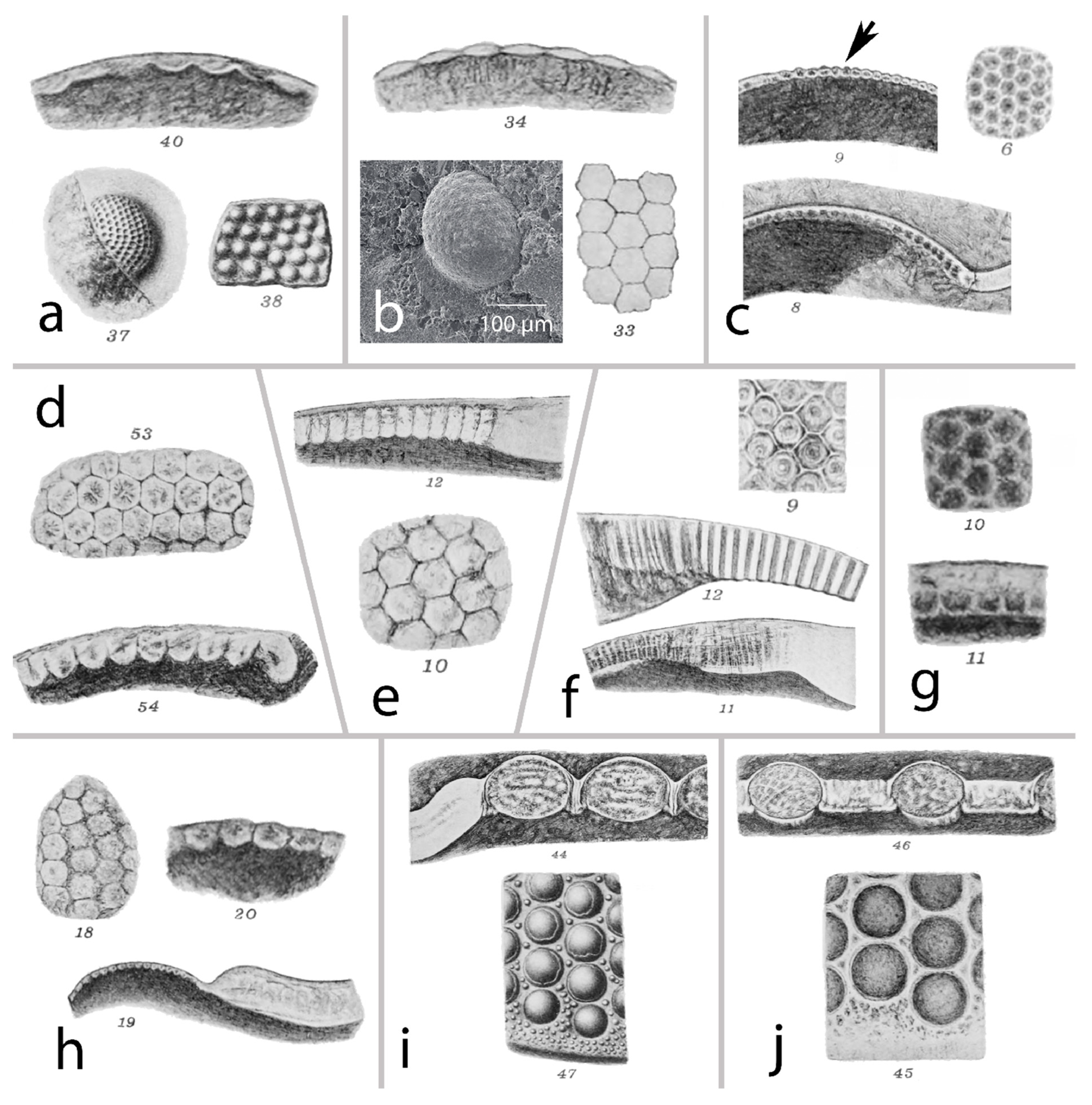
Figure 1. Selected examples of Lindström’s illustrations in 1901 [11]. Current generic nomenclature is used, with Lindström’s original generic assignments in brackets. It may be by chance, but note the increasing shaping into biconvex or columnar lenses with decreasing age. (ag) Holochroal eyes. (ij) Schizochroal eyes. (a) Peltura scaraboides (Wahlenberg,1821), Upper Cambrian period, Pl. III 37,38,40. (b) Sphaerophthalmus [Sphaerophthalmus] alatus (Boeck, 1838), Upper Cambrian period, Pl. III, 33,34. (c) Eobronteus [Bronteus] laticauda (Wahlenberg, 1821), Ordovician period, Pl. II 6,8,9. Arrow in (c): note the ’cubes’ formed as lenses -> lens function possible. (d) Archaegonus [Dysplanus] centrotus Dalman, 1937, Lower Ordovician period, Pl. III 53,54. (e) Nileus [Nileus] armadillo Dalman, 1827, Middle Ordovician period, Pl. V 10,12. (f) Asaphus [Asaphus] fallax Angelin, 1854, Middle Ordovician period, Pl. I 11,12,18. (g) Encrinurus laevis (Angelin 1851), Phacopida-no schizochroal eye, Middle Ordovician period, Pl. IV 10,11. (h) Cyrtometopus [Cyrtometopus] clavifrons (Dalman, 1827), Middle Ordovician period, Pl. III, 18–20. (Transitional form between holochroal and schizochroal eyes?). (i) Dalmanites [Dalmanites] imbriculatus (Angelin, 1851), Phacopida-schizochroal eyes, Silurian period, Pl. III 44,47. (j) Dalmanites [Dalmanites] obtusus (Lindström, 1885), Phacopida-schizochroal eyes, Silurian period, Pl. III, 45,46.
Arthropoda 03 00003 g001
Figure 2. Holochroal with different visual surfaces and schizochroal eyes. (a,b) Unspecified specimen; typical Early Cambrian compound eye with smooth visual surface and no discernible facets. (c,d) Eye of Holmia kjerulfi (Linnarson, 1871), Early Cambrian period, with thousands of facets. (e) Abathochroal eye of Pagetia sp., Middle Cambrian period, China. (f) Sphaerophthalmus alatus (Boeck, 1938), Upper Cambrian period, Sweden. (g) Gerastos cuvieri (Bronn, 1835) Middle Devonian period, Germany. (h) Aulacopleura koninckii (Barrande, 1846), Middle Silurian period, Czech Republic. (i) Scutellum sp. Middle Devonian, Germany. (j) Telephina bicuspis (Angelin, 1854), Middle Ordovician period, Norway. (k) Pricyclopyge bindosa (Barrande, 1872), Czech Geological Survey, CGS XB 139, Middle Ordovician, Czech Republic. (l) Aulacoplaura koninckii (Barrande, 1846), Czech Republic. (m) Scopelochasmops wrangeli (Schmidt, 1881), Phacopina, probably schizochroal eye, Middle Ordovician period, Geschiebe (glacially transported boulder) (comp. [15]). (n) Geesops schlotheimi (Bronn 1825), Middle Devonian period, Germany. (o) Eldredgeops crassituberculata (Stumm, 1953), Middle Devonian period, USA. (p) Unspec. Phacopid triloite, Lower Middle Devonian period, Germany. Note that the lower part of the lens (intralensar bowl) is well-preserved.
Arthropoda 03 00003 g002
Figure 3. The eye of Gerastos (picture is smaller (less wide) than the others), just 1 column wide. (a) Gerastos cuvieri (Steininger, 1831), Ahrdorf Fm., Eifelian, Middle Devonian period, Gees, Eifel, Germany. (b) Compound eye of G. cuvieri with a smooth surface. (c) Thin section of the eye. Note that the visual surface integrated into the cuticle is a specialized part of the cuticle. (d) Lindstöm’s plate VI, 20, Proetus [Proetus] concinnus Dalman, 1827. (e) Broken eye of G. cuvieri. Note the thick visual surface and the relics of the visual units below (white dots). (f) Magnification of a part of (e). (g) Relicts of the sensory units are separated quite far from each other.
Arthropoda 03 00003 g003
Figure 4. Different modes of lenses in trilobite eyes and their substructures. (ad) Sequence of building lenses. (a) Slit-formed holochroal eye of Cambropallas telesto (Geyer, 1993), Middle Cambrian period, Morocco. (b) Columnar lenses of Asaphus expansus Wahlenberg 1821, Lower Ordovician period, Sweden. (c) ‘Block-lenses’ Paralejurus sp., Silurian-Middle Devonian period, Morocco. (d) Biconvex lenses Telephina bicuspis (Angelin, 1854), Middle Ordovician period, Norway. (e) Unspec. phacopid trilobite, Morocco, schizochroal eye. (f) Dalmanitina socialis (Barrande, 1846), probably Ordovician, unknown provenance, showing the internal differentiation of a phacopid lens. (g) Paralejurus sp., Silurian–Middle Devonian period, Morocco, nicely showing the lens being part of the cuticle. (h) Ctenopyge sp., Upper Cambrian period, Sweden. (i) Chotecops ferdinandi Kayser 1880, Lower Devonian period, German. Note the separated lenses of the schizochroal eye. (j) Nileus platys Schrank 1972, Ordovician period, Sweden. (k) Visual surface of (j) with lenses. (l) Side view of (k); note the thin, cuticle-forming lenses. Insert: note the thin cuticle and the darker (former sensory) area underneath. (m) Nileus platys Schrank 1972, Ordovician period, Sweden. (n) Visual surface of (m). (o) Fraction and lateral view of (n); arrow indicates ommatidium. (p) Magnification of the ommatidium in (o); cc, crystalline cone; pp, primary pigment cells; sp, secondary pigment cells; r, rhabdom. (q) Thin section of the stalked eye of Asaphus kowalewskii Lawrow, 1856, Middle Ordovician period, Russia. Insert: stalked eye and visual surface. (r) Columnar lenses of (q) built from the cuticle at the right side. (s) Columnar lenses with domed inner surface forming semi-convex lenses. From (q). (t) Thin section through an eye of A. raniceps Dalman 1827. Note the almost perfect spherical shape, with the potential to enable a superposition.
Arthropoda 03 00003 g004
Figure 5. Stages of lens development through evolution and interior of an ommatidium. Green numbers indicate different ways/sequences to develop a trilobite lens. (a) Smooth visual surface of an Early Cambrian compound eye. (b) Forming bulged inner surface (Pagetia) and thus semi-convex lenses. (c) Biconvex lenses (Telephina). (d) Schizochchroal eyes, combined with 7 neighboring ommatidia (insert left). (e1) Columnar lenses through elongation or a lens forming inside of a thick cuticle (Clarkson, 1973). (e2) Columnar lenses with separations and their possible functioning. Left: obliquely entering rays are absorbed. Right: total reflection if the refractive index of the interface is lower than that of calcite, resulting in a support to capture light through neighboring units. (f) Elongated columnar lenses (semi-convex). (g) System of a cuticle with more translucent areas (‘cubes’). (h) The system of an ommatidium (apposition eye). A compound eye (top left) is normally composed of numerous identical units, the ommatidia. cc, crystalline cone; L, lens; r, rhabdom; rc, receptor cell; p, screening pigment cell. As a result of perception, the striped pattern is perceived as a summed-up blue signa (dot below). Bottom left: mosaic-like vision of a compound eye. Bottom right: crystalline cone in a system with a thin lens Peltura scarabaeoides Wahlenberg 1821, Late Cambrian period, and a lens cylinder of a xiphosuran, Limulus sp. I) Coalesced, reduced compound eye of Harpes macrocephalus (Goldfuss, 1839), Lower Middle Devonian period, Germany.
Arthropoda 03 00003 g005
Figure 6. The eye of Schmidtiellus reetae Bergström, 1973. (a) Schmidtiellus reetae Bergström, 1973, Series 3/Atdabanian, Early Lower Cambrian, Estonia. (b) Arrangement of ommatidia, indicated by arrows and square brackets; rectangle -> (c). (c) Single ommatidium in the cellular ‘basket’. (d) Right eye of (a), which already has the typical shape of a Cambrian trilobite compound eye. (e) Ommatidium, with relicts of receptor cells grouped around the central rhabdom. (f) Explanative depiction. b, ‘basket’; cc, crystalline cone; cw, cellular wall of the ‘basket’; om, ommatidium on the optic nerve. (g) Situation of the crystalline cone. (h) Interpretation of (g): cc, crystalline cone; r, rhabdom; rc, receptor cells; l, ‘lens’; lc, epidermal lens-building cells. (i) Different forms of lens systems: top left lens: cylinder eurypterids, horseshoe crabs; top right: ray path through thick lenses; bottom right: thin lens and long crystalline cone with index gradient; bottom right: ray path; note the bent rays.
Arthropoda 03 00003 g006
Figure 7. Maculae. (a,b) Maculae of unspec. (asaphid?) trilobite. (c) Horizontal thin section of a macula (unspec. trilobite) under polarized light. (dg) Individual ‘patches’ of (c) under polarized light. (e) Arrows indicate the encompassing (relicts) of cells. (f) Patch with discernible small organs indicated by arrows. (g) Magnification of (f). Arrows indicate small organs encompassed by even smaller cells, forming a flower-like structure. (hn) Examples by Lindström (1901) [11]. (h) Illaenus [Illaenus] esmarki (Schlotheim, 1823), Pl. IV 32, 33 32 in transmitted light. (i,j) Megitaspis [Megalaspis] attenuata (Wahlenberg, 1821), Pl. V, 4, 6. (k) Example of a typical hypostome with maculae, Scutellum [Bronteus] polyactin Ang., Pl. II 22. (l) Eobronteus [Bronteus] sp., macula. Pl. II 29, 30. (m) Illaenus [Illaenus] roemeri (Volborth, 1864), Pl. IV 40. (n) Lichas [Lichas] sp., Pl. IV, 48. (os) Positions of possible homologies of maculae in examples in the phylogenetic tree, showing perhaps transitional modes from a natant to a fixed hypostome and the consequences for the maculae (getting lost, finally, in (s). (o) Ceraurinella typa Cooper, 1953, Cheiruridae, Trilobita. (p) Kuamaia lata (Hou, 1987), Artiopoda, Trilobitomorpha. (q) Naraoia (Misszhouia) longicaudata (Zhang & Hou, 1985), Naraoiidae, Nectaspida. (r) Cindarella eucalla Chen et al., 1996, Xandarellida, Trilobitomorpha. (s) Emeraldella brocki Walcott 1912, Vicissicaudata, Artiopoda. Drawings (os) changed after Cotton 2002. (t) Exemplary schematic drawing of a campaniform sensillum (mechanoreceptor), restricted to insects. (u) Model of the ‘compound ear’ (macula). Mechanical signal arriving at the window within the cuticle, emerging at the proximal side, amplified due to the smaller surface at the inner part. tb, tubular body of the sensory dendrite; tc, thick cuticle; w, ‘window’; cubic hollow, space within the cuticle. Compare (hj); arrow in (u) shows the direction of the signal.
Arthropoda 03 00003 g007
Disclaimer/Publisher’s Note: The statements, opinions and data contained in all publications are solely those of the individual author(s) and contributor(s) and not of MDPI and/or the editor(s). MDPI and/or the editor(s) disclaim responsibility for any injury to people or property resulting from any ideas, methods, instructions or products referred to in the content.