Article

Potential Involvement of Protein Phosphatase PPP2CA on Protein Synthesis and Cell Cycle During SARS-CoV-2 Infection: A Meta-Analysis Investigation

by
Luca P. Otvos
1,†,
Giulia I. M. Garrito
1,†,
Gabriel E. Jara
2,
Paulo S. Lopes-de-Oliveira
2,3 and
Luciana E. S. F. Machado
1,*
1
Departamento de Genética e Biologia Evolutiva, Instituto de Biociências, Universidade de São Paulo, São Paulo 05508-090, SP, Brazil
2
Brazilian Biosciences National Laboratory (LNBio), Brazilian Center for Research in Energy and Materials (CNPEM), Campinas 13083-100, SP, Brazil
3
Graduate Program in Pharmaceutical Sciences, Faculty of Pharmaceutical Sciences, University of Campinas, Campinas 13083-970, SP, Brazil
*
Author to whom correspondence should be addressed.
These authors contributed equally to this work.
Kinases Phosphatases 2025, 3(1), 4; https://doi.org/10.3390/kinasesphosphatases3010004
Submission received: 12 December 2024 / Revised: 10 February 2025 / Accepted: 11 February 2025 / Published: 18 February 2025

Abstract

:
Coronavirus disease 2019 is a multi-systemic syndrome that caused a pandemic. Proteomic studies have shown changes in protein expression and interaction involved in signaling pathways related to SARS-CoV-2 infections. Protein phosphatases play a crucial role in regulating cell signaling. In this study, we assessed the potential involvement of protein phosphatases and their associated signaling pathways during SARS-CoV-2 infection by conducting a meta-analysis of proteome databases from COVID-19 patients. We identified both direct and indirect interactions between human protein phosphatases and viral proteins, as well as the expression levels and phosphorylation status of intermediate proteins. Our analyses revealed that PPP2CA and PTEN are key phosphatases involved in cell cycle and apoptosis regulation during SARS-CoV-2 infection. We also highlighted the direct involvement of PPP2CA in the cell division throughout its interaction with CDC20 protein (cell division cycle protein 20 homolog). This evidence strongly suggests that both proteins play critical roles during SARS-CoV-2 infection and represent potential targets for COVID-19 treatment.

1. Introduction

Coronavirus disease 2019 (COVID-19) is caused by the severe acute respiratory syndrome coronavirus 2 (SARS-CoV-2), which reached pandemic levels and posed a significant global health threat. The COVID-19 pandemic recorded 676,609,955 global cases and 6,881,955 total deaths over four years (2020–2023), averaging to 144,116,214 cases and 1000 deaths per day, according to the Coronavirus Resource Center of Johns Hopkins University (JHU) website https://coronavirus.jhu.edu/map.html (accessed on 10 March 2023) [1].
The high transmission rate and global lethality of COVID-19 has driven the research efforts towards the understanding of disease mechanisms to allow drugs and vaccine development to prevent and treat the disease. This global effort has resulted in approximately 200,000 scientific papers on “COVID-19” and “SARS-CoV-2” in just under two years.
The SARS-CoV-2 virus belongs to the genus betacoronavirus (β-COVs) and consists of a single-stranded positive-sense RNA molecule surrounded by an envelope. The genome of SARS-CoV-2 features a 5′ capped mRNA, a poly-A tail at 3′ end, a 5′ and 3′ untranslated region (UTR), and codes for accessory proteins. It also encodes four structural proteins, including spike (S), membrane (M), glycoprotein envelope (E) and nucleocapsid (N) proteins, as well as 11 open reading frames (ORFs). These include orf1a and orf1b, which contain the coding sequences for sixteen non-structural proteins (nsp1–16), along with orf3a, orf3b, orf6, orf7a, orf7b, orf9b, orf9c, and orf10 [2].
The structural proteins of SARS-CoV-2 are important for viral assembling and budding: the M protein, which is composed of three transmembrane domains and defines the shape of the viral envelope; the E protein, a transmembrane protein with ion-channel activity; the S protein, a trimeric glycoprotein that interacts with host cell receptors to facilitate viral entry; and the N protein, which wraps the RNA genome and forms a ribonucleoprotein complex. On the other hand, the non-structural proteins play crucial roles in viral transcription and/or replication (nsp7, nsp8, nsp9, nsp10, nsp13, nsp14, nsp15, nsp16, nsp11, and nsp12, which is a RNA-dependent RNA polymerase), blocking the host innate immune response (nsp1, nsp3), cleaving the viral polyprotein (nsp5), acting as scaffold protein (nsp4, nsp6), or with an unknown function (nsp2) [2,3]. Finally, the accessory proteins are important for facilitating viral replication, virus release, and modulating the host immune response.
Although the COVID-19 was initially described as a respiratory disease, it is better understood as a multisystem syndrome that affects different organs and tissues in infected patients [4]. A multi-level analysis of transcripts, proteins, and post-translational modifications in cultured cells, human biopsies, and bodily fluids revealed a strong correlation between the inflammatory response, T-cell activity, and the severity of COVID-19 disease. Pro-inflammatory cytokines such as IL-6 and TNFα, along with chemokines like CXCL10 and CXCL12, and the transcription factor NFκB signaling pathway, all increase with disease severity [5,6,7,8,9,10,11,12,13,14]. In contrast, T-cell activation is observed in mild cases but appears to be impaired in severe disease [15,16].
Studies have commonly shown that protein kinases are activated during cell signaling in response to SARS-CoV-2 infection, suggesting that protein phosphorylation plays a crucial role in facilitating viral infection [9,14,17]. Protein phosphatases and kinases are essential regulators of cell signaling in both physiological and pathological functions, including cell-cycle control, cell survival, apoptosis, cancer, diabetes, cardiovascular diseases, and more. In these processes, kinases phosphorylate and phosphatases dephosphorylate their substrates, thereby regulating the level of protein phosphorylation [18,19] and controlling the duration (initiation, sustainment, and termination) and amplitude of signaling [20,21,22].
Protein phosphatases are classified into two families based on their substrate specificities: protein serine/threonine phosphatases (PSPs) and protein tyrosine phosphatases (PTPs). The PSPs family is further divided into three classes: classical protein serine/threonine phosphatases (PPP), metal-dependent protein phosphatases (PPM) and aspartate-based protein phosphatases (FCP/SCP). The PTP family is also divided into three classes: classical protein tyrosine phosphatases, dual-specificity phosphatases (DUSPs), which act on tyrosine, serine, and threonine, and aspartate- or histidine-based phosphatases. Additionally, classical PTPs can be further classified into two groups: cytoplasmic PTPs (cPTPs) and receptor PTPs (rPTPs).
For example, certain protein phosphatases, including serine/threonine phosphatases PPP1 and PPP2A, as well as the protein tyrosine phosphatases SHP2 and SHP1, are known to be regulated during viral infections. These interactions promote viral replication by regulating transcription elongation, viral transcription, and RNA packaging [23,24,25].
For instance, the influenza A virus activates SHP2, which regulates the epidermal growth factor receptor (EGFR) pathway, leading to ERK kinase activation. This, in turn, promotes viral replication, cell-cycle progression, cell survival, and proliferation. On the other hand, SHP2 inhibition blocks the EGFR/ERK pathway, enhancing the antiviral innate immune response by increasing interferon and interleukin production. These findings suggest SHP2 as a promising target for controlling influenza A virus infection [26]. In contrast, while SHP2 expression remains normal, SHP1 is downregulated during hepatitis C virus (HCV) infection. The viral protein C induces methylation of the SHP1 promoter, which deregulates phosphotyrosine signaling and alters the T-cell receptor (TCR) signaling pathway. Treatment with DNA methyltransferase inhibitor blocks DNA methylation, restoring SHP1 expression [27]. Additionally, HCV NS3 protein interacts with and promotes the ubiquitination of PPM1A, leading to its degradation via the proteasoma. This degradation enhances the cell migration and invasiveness of hepatocellular carcinoma through the activation of the transforming growth factor (TGF) signaling pathway. Treatment with the proteasomal inhibitor MG132 abolishes the NS3-mediated degradation of PPM1A [28].
PP1 has been shown to dephosphorylate the core protein of the hepatitis B virus (HBV) nucleocapsid, facilitating viral RNA packaging. The inhibition of PP1 by tautomycin keeps the core protein phosphorylated, reducing RNA packing into the nucleocapsid [29]. Furthermore, PPP2CA is crucial for influenza A virus replication and to maintaining the viability of infected cells, as silencing PPP2CA decreases viral titers and increases cell death [30]. Interestingly, the regulatory B56 subunit of PPP2CA is overexpressed in chronic hepatitis B patients, increasing PPP2CA activity. This elevation promotes cell-cycle arrest through the p53 pathway, resulting in apoptosis and liver injury in HBV infected cells [31]. These findings highlight that PPP1 and PPP2CA are not only central to viral replication but also represent potential therapeutic targets for controlling hepatitis B virus and influenza A infections.
Since viruses are entirely dependent on host-cell machinery and manipulate many protein phosphatases to promote viral replication and infection, we hypothesized that the SARS-CoV-2 virus could similarly manipulate the expression and activity of specific protein phosphatases, particularly those involved in cell-cycle arrest, cell proliferation, survival and apoptosis regulation. To test this hypothesis, we investigated protein phosphatase levels, protein phosphatase–protein interactions, and the phosphorylation status of proteins interacting with phosphatases, using data from various databases, as summarized in Figure 1 and Table S1.

2. Results and Discussion

2.1. Expression Levels of Protein Phosphatases During SARS-CoV-2 Infection

To investigate the role of host protein phosphatases during SARS-CoV-2 infection, we analyzed their expression levels across various datasets. These studies were conducted using cell models or samples from confirmed SARS-CoV-2 infected patients (including urine, blood cells, plasma, pulmonary tissues) [5,6,7,8,10,12,14,17,32,33,34,35,36,37,38,39,40,41,42]. Interestingly, we observed a consistent expression profile of protein phosphatases within their sub-families between infected and control samples. This pattern was independent of the analyzed model—whether cell models, patient tissues or fluids—and showed higher expression for PTPs and a lower expression of PPPs.
In detail, for the cell models, most of the differences in expression levels between infected and control cells were observed in the liver tumor cell (Huh7) [33], which showed higher expression levels of cPTPs (numbers 15, 20, 24, and 27 in Figure 2A), particularly rPTPs (numbers 14, 16–19, 26, 29, 31, and 32 in Figure 2A) and lower expression levels for PPMs (numbers 1, 2, 5, 7, and 8 in Figure 2A). In contrast, other cell lines such as the kidney-derived HEK293, lung-derived A459, and colorectal adenocarcinoma Caco-2 cells exhibited higher expression levels only in PPP1CB (number 28 in Figure 2A) and PPP4C (number 22 in Figure 2A) for HEK293 cells [37], and PTPN2 (number 25 in Figure 2A) for A459 cells [14]. Lower expression levels were observed only in PTPN13 (number 10 in Figure 2A) and PTPRF (number 9 in Figure 2A) for A459 cells [14], and in PTPRF for Caco-2 cells [17] (Figure 2A, Table S2).
In patient samples (fluids/cells), the protein phosphatases with the highest expression levels were PTPs, including numbers 10, 13, 15, 16, and 18 for PBMC cells [41]; numbers 17 and 20 for lung tissue [5,40]; and PTPN1 (number 19) for the renal cortex [35]. Conversely, the protein phosphatases with lower expression levels included PPM1B (number 4), PPM1K (number 5), and PPM1M (number 3) for PBMC cells [41]; PGAM1 (number 8), PPP2CA (number 6), PTPN6 (number 2), and PTPRC (number 1) for lung tissue [5]; and PGAM1 (number 7) for spleen [35] (Figure 2B, Table S2).
In the patient fluids and secretions, the protein phosphatase with highest expression levels were cPTPs (numbers 29, 30, 41, 43, 44, 46, and 50 in Figure 2C), rPTPs (numbers 23–28, 30, 31, 33–35, 37, 47, and 53 in Figure 2C) and DUSPs (numbers 22, 32, 36, 38–40, 42, 45, and 52 in Figure 2C) in bronchoalveolar lavage fluid [41], as well as PTPN18 (number 51 in Figure 2C) in a nasopharynx swab [38]. Conversely, the protein phosphatases with lower expression levels, including PPPs (number 7, 14–16 in Figure 2C); PTPs (numbers 2, 3, 6, 8–13, 17, and 18 in Figure 2C) in bronchoalveolar lavage fluid [41]; PTPRF (number 21 in Figure 2C) and PTPRJ (number 20 in Figure 2C) in plasma [36,39]; and PTPRN (number 1 in Figure 2C) in urine [7] (Figure 2C, Table S2).
In summary, we observed that the Huh7 cell model and bronchoalveolar lavage fluid exhibited high expression levels of the protein tyrosine phosphatase family (DUSPs, non-receptor PTPs and receptor PTPs), particularly PTPN12 and PTEN. In contrast, the canonical protein serine/threonine phosphatases, such as PPP1CA, PPP1CC, PPP2CA, and metal-dependent phosphatases, were expressed at lower levels in these samples, as well as in lung tissue.

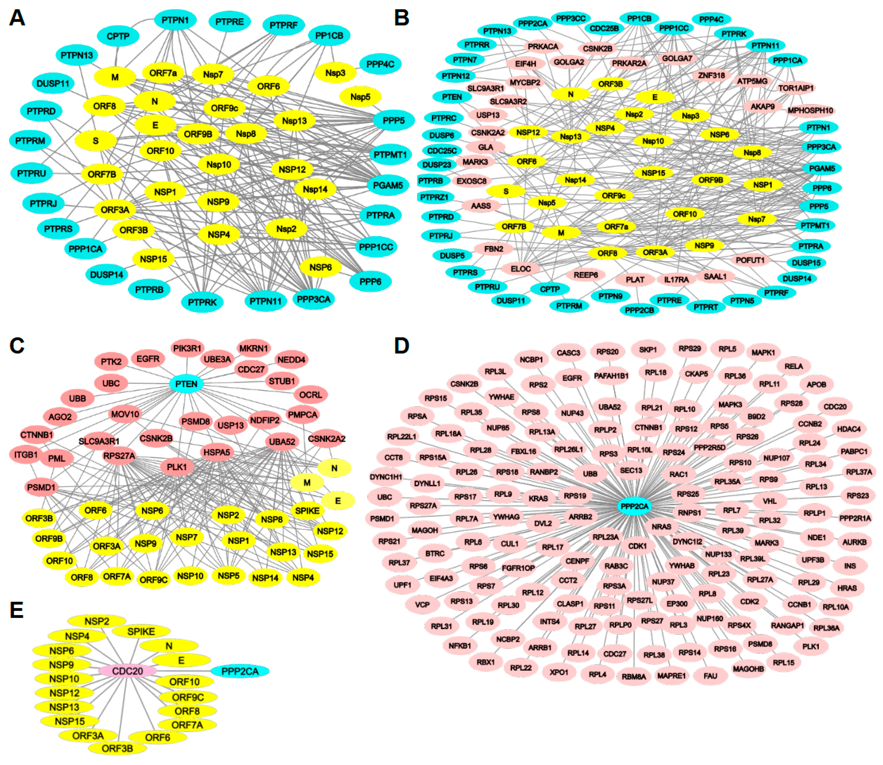
2.2. Protein Phosphatases Interact Directly with Viral Proteins

Given the importance of protein phosphatases to host cells, we analyzed the interaction network of these proteins with viral proteins, as well as their interactions with other proteins that connect protein phosphatases and viral proteins (hereafter referred to as intermediate proteins) during SARS-CoV-2 infection. To achieve this, we searched the scientific literature for protein interaction data during infection and identified protein phosphatases that directly interact with the virus or with other intermediate proteins that interact with viral proteins [7,14,37,43,44,45,46,47,48].
To identify host protein targets for SARS-CoV-2 proteins, the expression of 26 viral proteins (as explained in the Introduction section) in HEK-293T cells revealed 332 high-confidence human–protein interactions with SARS-CoV-2 proteins, as determined by affinity purification and mass spectrometry [43]. From this database, we found thirteen protein phosphatases that directly interact with viral proteins: six protein serine/threonine phosphatases (PPP1CA, PPP1CB, PPP1CC, PPP2CA, PPP3CA, PPP6C and PGAM5) and seven protein tyrosine phosphatases (PTP), including two cPTP (PTPN1 and PTPN11), two rPTP (PTPRK and PTPRF), and three DUSPs (DUSP11, DUSP14, and DUSP23/PTPMT1). Most of the identified PPPs can bind to several viral proteins, while PTPs appear to be more specific, binding to fewer viral proteins (Figure 3A, Table S3).
Using A459 lung carcinoma cells expressing individual SARS-CoV-2 proteins and analyzed through affinity purification and mass spectrometry, we identified the interaction of seven protein tyrosine phosphatases, namely PTPMT1, PTPN11, PTPRA, PTPRF, PTPRJ, PTPRM, and PTPRS, with the viral proteins ORF3 and/or ORF7B [14] (Figure 3A, Table S3). We then mined a curated dataset of host and viral proteins interaction obtained from 151 publications. This dataset was compiled by curators from the International Molecular Exchange (IMEx) Consortium and included 4400 interactions for the proteins described above. From these interactions, only PPP1CB was found to interact with SARS-CoV-2 proteins (nsp8, nsp10, nsp13, and orf9b) [48] (Figure 3A, Table S3). Additionally, Li et al. (2021) overexpressed plasmids encoding each SARS-CoV-2 gene with N-terminal FLAG epitope in HEK293 cells and identified 286 cellular protein interactions using affinity purification and mass spectrometry. From this dataset, we found that PTPRD interacts with the viral protein orf3a of SARS-CoV-2 [6]. In another study, we identified two PTPs (PTPN1 and PTPN13) and two PSPs (PPP1CC, PPP3CA) interacting with some viral proteins. This study transfected 17 SARS-CoV-2-encoded viral proteins with an HA epitope into 293T cells, labeled them with biotin, and isolated them using streptavidin; we then analyzed the results by mass spectrometry and immunoblotting, identifying 2422 host proteins that interact with viral proteins [47] (Figure 3A, Table S3). Moreover, the expression of viral proteins and subsequent immunoprecipitation and mass spectrometry in human bronchial epithelial cells (16HBEo-) revealed that 189 host proteins interact with viral proteins. From these data, we identified eleven protein phosphatases: two PSPs (PPP5, PPP6), eight PTPs (PTPRA, PTPRB, PTPRD, PTPRE, PTPRF, PTPRU PTPMT1, CPTP), and the histidine-based protein phosphatase phosphoglucomutase 5 (PGAM5) interacting with viral proteins [46] (Figure 3A, Table S3).
The above-mentioned protein phosphatases interacting with viral proteins were grouped in five functional clusters: (a) serine/threonine phosphatases, (b) protein tyrosine phosphatase, (c) transmembrane receptor protein phosphatase, (d) DUSPs, and (e) focal adhesion assembly. Furthermore, the proteins’ networks identified in these studies were mapped into important cellular pathways affected by each viral proteins in HEK293 cells [6]: (i) ATP biosynthesis and metabolic processes correlated with the M viral protein; (ii) mRNA transport with the N viral protein; (iii) melanoma differentiation-associated protein 5 and retinoic acid-inducible gene I RNA sensing signaling with the Nsp1 viral protein; (iv) nucleotide-excision repair with the Nsp4 viral protein; (v) protein methylation and alkylation with the Nsp5 viral protein; (vi) translation initiation with the Nsp9 viral protein; (vii) cellular amino-acid metabolic process and neutrophil chemotaxis with the Nsp10 viral protein; (viii) reactive oxygen species’ metabolic process with the Nsp14 vrial protein; (ix) Golgi to plasma membrane transport with the Nsp16 viral protein; (x) endoplasmic reticulum stress and IFN or IL-6 signaling pathways with the Orf3a; (xi) mRNA transport and NF-κB pathways with the Orf6; or in A549 cells [14]: (xii) mitochondrial dysregulation by the ORF9b; (xiii) innate immunity by the ORF7b; (xiv) stress response components and DNA damage response mediators by the ORF7a; and (xv) cholesterol metabolism by the NSP6 viral protein.

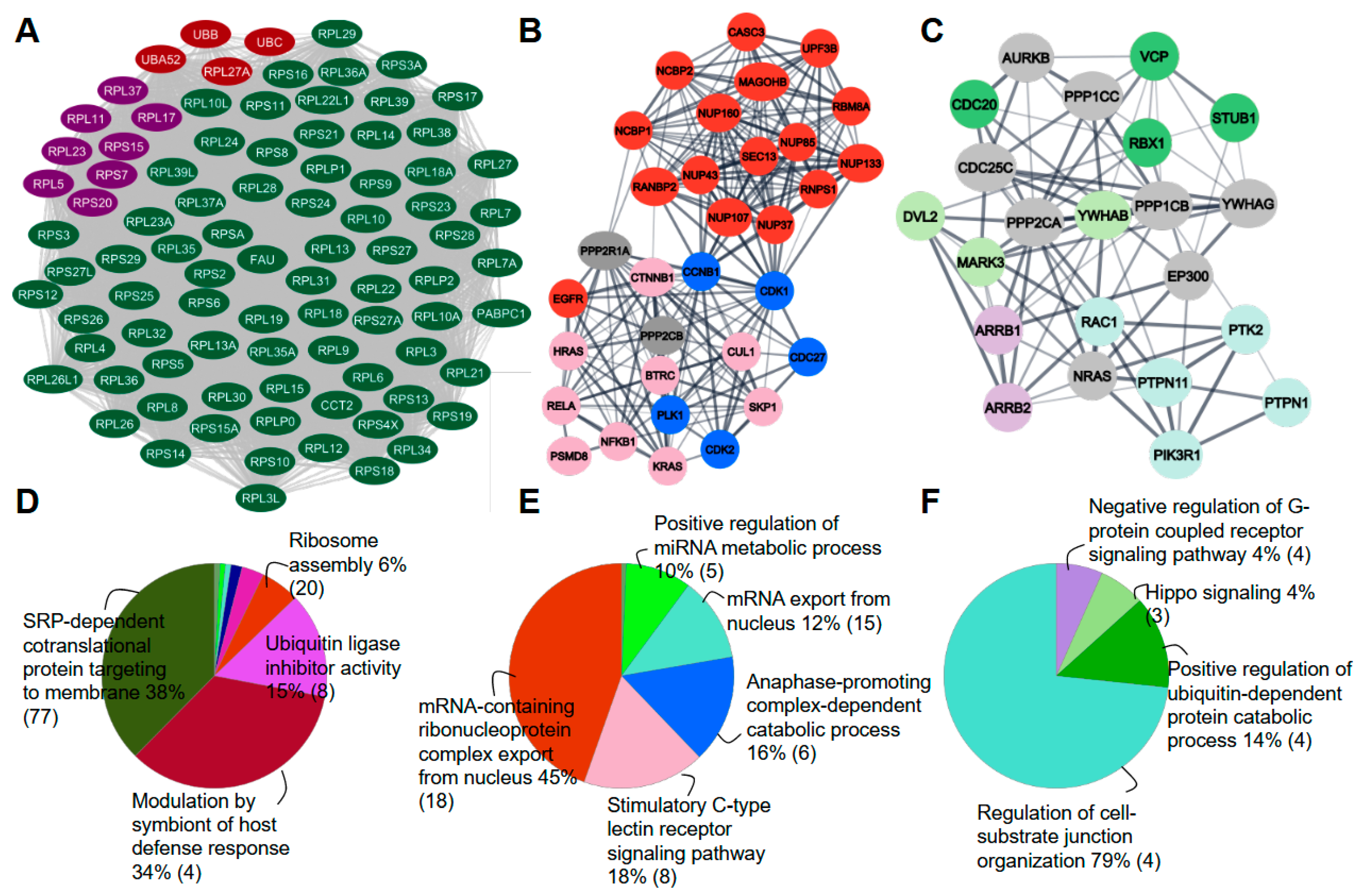
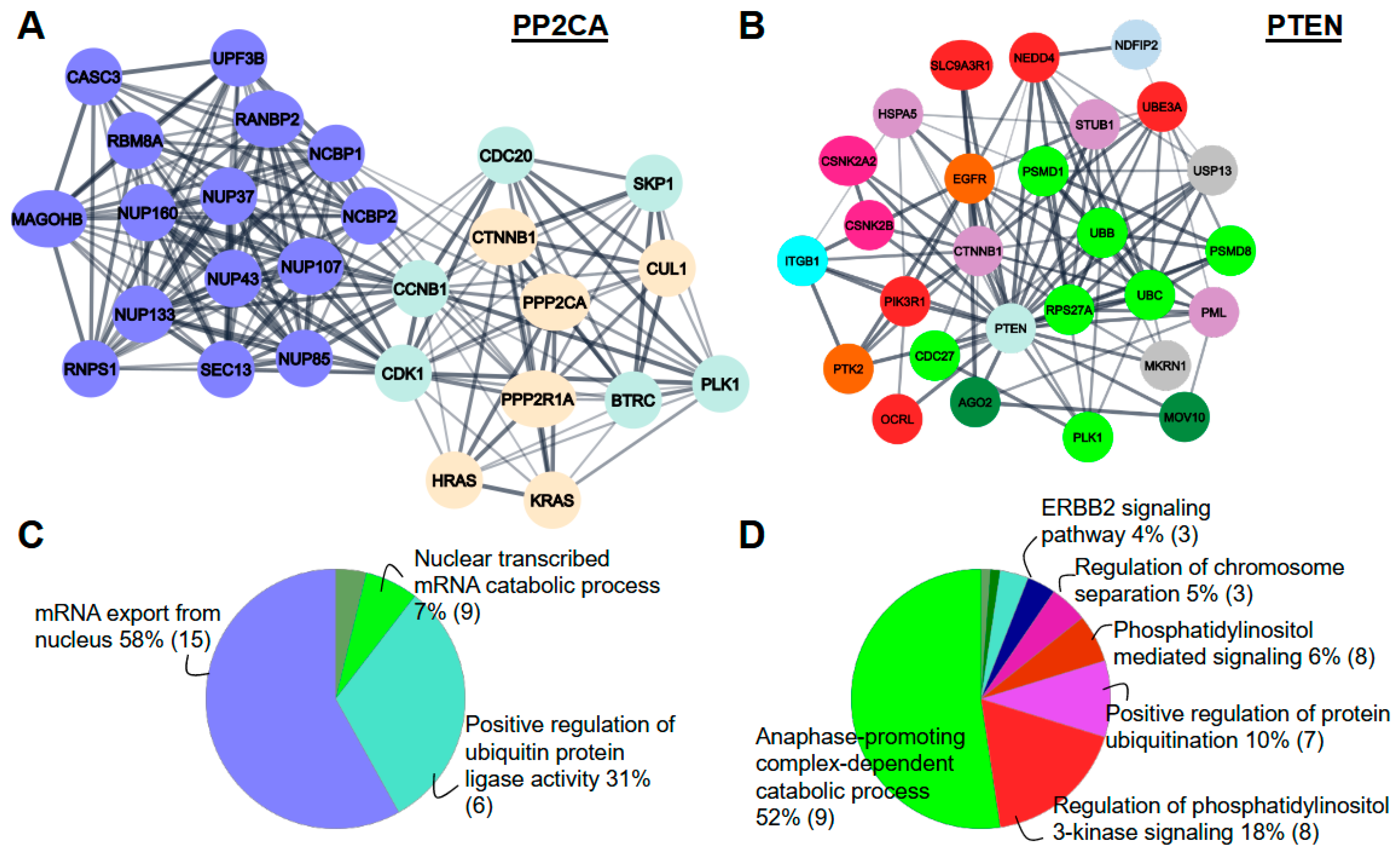
2.3. Protein Phosphatases Interact with Intermediate Proteins

Although some studies show the direct interaction between protein phosphatases and viral proteins, other studies present protein–protein interaction (PPI) information describing intermediate proteins that made this binding. From that, we investigate the interactions between protein phosphatases and intermediate proteins that interact directly with viral proteins. These interactions generate a network between many proteins that are important in a specific cell-signaling pathways during SARS-CoV-2 infection once protein–protein interactions (PPIs) drive the cellular signal transduction mechanisms.
A protein–protein interaction prediction (HiPPIP) was implemented to assemble the interactome of host and viral proteins, generating a set of novel PPIs through a string-extended PPI network, which should be important during SARS-CoV-2 infection and replication [44]. These data were extracted from 332 human proteins that interacted with the SARS-CoV-2 proteins as analyzed by Gordon et al., 2020. They identified 4408 host proteins that were likely involved in 6076 interactions with viral proteins [44].
Analyzing the interactome list, we identified twenty-seven protein phosphatases and twenty-six intermediate proteins that interact with viral proteins. Of the protein phosphatases identified, six are PSPs (PPP1CB, PPP1CC, PPP2CA, PPP2CB, PPP3CC) and twenty-one PTPs. Of these, eight cytosolic PTPs (PTPN1, PTPN5, PTPTN7, PTPN9, PTPN11, PTPN12, PTPN13, CPTP), six receptor PTPs (PTPRA, PTPRC, PTPRK, PTPRR, PTPRT, PTPRZ1) and seven DUSPs (DUSP5, DUSP6, DUSP15, DUSP23, PTEN, CDC25B and CDC25C) (Figure 3B, Table S3). As a result, twenty-one new protein phosphatases interacting with intermediate proteins were identified in the interactome of SARS-CoV-2 infection.
Nadeau et al. (2020) [45] analyzed the viral-host PPI network obtained from Gordon et al., 2020 [43] and performed an in-depth computational analysis of the interactome of SARS-CoV-2 using the GoNet tool. This tool evaluated the clustering of GO terms in PPI networks to identify biological processes and protein complexes, which involve three layers of interaction: (i) viral-host PPI network, SARS-CoV-2 proteins interacting with human proteins; (ii) string-augmented PPI network, interactions between human proteins in the viral-host PPI network; and (iii) string-extended PPI network, proteins that bind with human interactors of SARS-CoV-2 proteins [45]. Analyzing these datasets, the protein serine/threonine phosphatase 2A catalytic subunit alpha (PPP2CA) was identified to bind with 166 human interactors of SARS-CoV-2 proteins and PTEN interacting with 28 human interactors of SARS-CoV-2 proteins (Figure 3C,D, Table S3). Interestingly, many of these intermediate proteins have also been identified to interact with the human protein phosphatases listed in the Biogrid database. Additionally, a recent study transfected 28 SARS-CoV-2 proteins, each with a specific N-terminal-tag, into Saccharomyces cerevisiae for a high-throughput yeast two-hybrid (Y2H) screen, and also transfected them into Caco-2 cells for tandem mass-tag affinity purification followed by mass spectrometry (TMT-AP-MS) to analyze the human protein–protein interactome. They reported 299 and 472 human-SARS-CoV-2 PPIs via Y2H screen and TMT-AP-MS methodologies, respectively [49]. From these, we searched for the intermediate proteins identified in our meta-analysis and found 33 intermediates proteins that interact with viral proteins (Table S3).
Each intermediate protein in the PPP2CA network presented in Figure 3D shows many interactions with viral proteins, with CDC20 as an example (Figure 3E). This protein is involved in the cell division regulation. Interesting, many studies have shown that PPP2CA seems to interact and dephosphorylate CDC20, activating the anaphase-promoting complex (APC/C) in mitosis [50,51,52,53]. Then, the interaction of SARS-CoV-2 virus proteins with the CDC20 could be a way to manipulate the PPP2CA into the cell, resulting in apoptosis and tissue injury as shown in the HBV infected cells [31].
Consequently, to understand the role of CDC20 and other intermediate proteins’ interactions with these protein phosphatases, we analyzed the biological functions using MCODE and ClueGo plugins in the Cytoscape software. The MCODE plugin identified seven function clusters, being three of them predominant (Figure 4). It is important to highlight that these software create a network based on nodes of interaction, with the protein with the highest interaction observed as the central node, providing a higher percentage related to its function [54]. In the first cluster, most proteins were shown to be involved in four signaling pathways: (i) SRP-dependent cotranslational protein targeting to the membrane; (ii) modulation by the symbiont of host defense response; (iii) ubiquitin ligase inhibitor activity; and (iv) ribosome assembly (Figure 4A,D). Three of these pathways were related to protein synthesis, while the other is related to the immunity defense against the virus.
In the second cluster, most proteins were shown to be involved in five signaling pathways: (i) mRNA-containing ribonucleoprotein complex export from nucleus; (ii) stimulatory C-type lectin receptor signaling pathway; (iii) anaphase-promoting complex-dependent catabolic process; (iv) mRNA export from nucleus; and (v) positive regulation of the miRNA metabolic process (Figure 4B,E). While two of these functions are related to protein synthesis, one refers to cell mitosis.
In the third cluster, most proteins were shown to be involved in other four signaling pathways: (i) regulation of cell–substrate junction organization; (ii) positive regulation of the ubiquitin-dependent protein catabolic process; (iii) Hippo signaling; (iv) negative regulation of the G-protein coupled-receptor signaling pathway (Figure 4C,F). In this cluster, we also observed functions related to cell organization and protein degradation. In summary, those proteins work in cell organization, protein degradation, and synthesis. It is known that the effect of SARS-CoV-2 infection promotes inflammatory response and cell damage. We suggest that the protein phosphatases in partnership with intermediate proteins might be involved in the critical cellular processes to prevent cell damage caused by the SARS-CoV-2 protein virus.
Taken together, our results suggest that protein serine/threonine phosphatase 2A (PPP2CA) and phosphatase and tensin homolog (PTEN) play important biological roles during SARS-CoV-2 infection, based on their interactions with various intermediate proteins (Figure 3C,D, Table S3). We then proceeded with MCODE and ClueGo analysis using only the PPP2CA or PTEN networks (Figure 5). For PPP2CA, we identified two main clusters: the first is consistent across all proteins (Figure 4A,D), while the second is associated with mRNA export from the nucleus and the positive regulation of ubiquitin protein ligase activity (Figure 5A,C). For PTEN, we observed a principal cluster of interactions involved in the anaphase-promoting complex-dependent catabolic process, the positive regulation of protein ubiquitination and phosphatidylinositol regulation (Figure 5B,D). Based on these findings, we propose a potential role for PPP2CA in protein turnover, and for PTEN in mitosis and the phosphatidylinositol signaling pathway.
The export of mRNAs is a crucial process for encoding antiviral factors in the body and is essential for producing proteins that counteract viral replication and help control infections [55]. Therefore, the enrichment of proteins involved in these function may reflect the body’s urgent need to combat the virus.
SARS-CoV-2 infection is known to trigger multiple processes of cell apoptosis as part of the inflammatory response [56]. One of the key functions of PTEN is to regulate mitosis, along with proteins involved in the anaphase-promoting complex-dependent catabolic process [57]. On the other hand, dephosphorylated PTEN associated with chromatin is removed by APC/C during mitotic exit [58]. The anaphase-promoting complex is a protein complex that mediate cycling degradation during anaphase, driving the exit from mitosis and enabling cell growth. This complex also promotes the degradation of proteins that hold sister chromatids together, leading to their separation during anaphase and their migration to opposite poles of the cell [59]. The presence of a large proportion of proteins involved in the anaphase-promoting complex-dependent catabolic process during SARS-CoV-2 infection suggests that cell turnover is activated as a mechanism to facilitate viral propagation.
PTEN is also involved in the regulation of the phosphatidylinositol 3-kinase signaling pathway through its lipid phosphatase activity. Among the key players, we can highlight NEDD4 (Figure 5B), which is known to be a major E3 ubiquitin ligase for PTEN, promoting its ubiquitination and degradation, thereby leading to the activation of PI3K activation [60]. We observed that growth factor receptor (GFR) signaling and PI3K was activated in Caco-2 and Huh7 cells infected with SARS-CoV-2, respectively [10,17]. This activation leads to Akt phosphorylation and subsequent mTOR activation in Huh7 cells during SARS-CoV-2 infection [10], which regulates apoptosis, cell survival, host transcription, and translation. Notably, viral replication decreases when GFR signaling is inhibited by pictilisib (a PI3K inhibitor) and omipalisib (a PI3K and mTOR inhibitor) [17].
Therefore, understanding these cellular pathways is crucial for gaining insight into the mechanisms of COVID-19 infection and identifying potential targets for the development of therapeutic agents.

2.4. Phosphorylation Level of Intermediate Proteins

In this study, we investigated the role of protein phosphatases during viral infection. Then, we hypothesized that these proteins might dephosphorylate the viral proteins and/or the intermediate proteins involved in signaling pathway described. To assess this, we analyzed the phosphorylation levels of these proteins using phosphoproteome data from SARS-CoV-2-infected cells [9,14,38]. Since protein kinases phosphorylate and protein phosphatases dephosphorylate their substrates, we infer that intermediate proteins with higher phosphorylation levels in SARS-CoV-2-infected samples compared to controls may have either increased kinase activity or decreased phosphatase activity. Conversely, proteins with lower phosphorylation levels in the infected samples suggest increased phosphatase activity and decreased kinase activity.
To determine whether protein kinases are involved during SARS-CoV-2 infection, three studies analyzed the levels of host protein phosphorylation and kinase activities in Vero E6 cells, A549 cells infected with the virus, and nasopharynx swab samples from infected patients. These studies identified 3036, 4643, and over 8500 human proteins/phosphorylation sites phosphorylated upon infection, which are involved in various cellular functions such as the cell cycle, apoptosis, and DNA replication [9,14,38]. They also identified kinases that were either activated or downregulated during infection and demonstrated that kinase inhibition reduced viral replication [9]. Interestingly, both studies identified phosphorylation sites in the SARS-CoV-2 proteins M, N, S, nsp3, nsp9, nsp14, and orf9b [9,14].
Analyzing the phosphorylation levels of intermediate proteins identified in our meta analysis, we observed lower phosphorylation levels (indicating protein phosphatase activity) in FGFRTOP_S160 (1), RPS17_S113 (2), CDC20_T70 (3), ZNF318_S2091 (4), CENPF_S3054 (6), RANBP2_S2510 (7), and others in the model cells (Figure 6A, Table S4). The intermediate proteins with higher phosphorylation levels (indicating protein kinase activity) were MAPK1_Y187 and T185 (17, 18), RBM8A_S56 (19), MYCBP2_S3931, S3932 (20), and RPS20_T9 (21), among others (Figure 6A, Table S4). Interestingly, in the patient samples, we also observed lower phosphorylation levels for RPS17_S113 (8), RANBP2_S2900 (1), and members of the RPL and RPS families (3, 4, 5), while higher phosphorylation levels were observed for ZNF318 (12, 15, 17), though at different residues, as well as for PML proteins (14, 16) (Figure 6B, Table S4). The RPS and RPL proteins are involved in the SRP-dependent cotranslational protein targeting to the membrane and ubiquitin ligase inhibitor activity, as described above (Figure 4A,D), and they predominantly interact with PPP2CA in the network (Figure 3D). Numerous studies have demonstrated the regulation of RPS proteins’ phosphorylation/dephosphorylation. Acidic ribosomal proteins (P1, P2 and P0) and RPS3 can be dephosphorylated and regulated by PPP2CA, playing an important role in protein synthesis [61,62]. RPS6 is regulated by protein phosphatase 1 and 2B (PPP1, PPP2B) after mitogenic stimulation [63,64,65]; and RPL5 interact with PPP1 [66].
Furthermore, a recent analysis searching for PPP2CA substrates used dTAG proteolysis-targeting chimeras to selectively degrade dTAG-PPP2CA in HEK293 cells. In this study, they identified 7589 proteins, with 5829 phosphorylated and 3353 showing significant increases in abundance. Only 11 proteins showed a significant decrease in abundance in dTAG-13-treated cells compared to DMSO-treated controls. These proteins are involved in cell cycle, RNA transport, ubiquitin-mediated proteolysis, and the spliceosome [67]. We identified 141 intermediate proteins that bind to both PPP2CA and SARS-CoV-2 virus in our meta-analysis, which were also observed in that study (Table S5). Of these, 59 proteins showed a significant increase in phosphorylation abundance in dTAG-13-treated cells compared to DMSO-treated controls (Table S5), with particular emphasis on the RPS, RPL, and CDC20 proteins.
The intermediate proteins with lower phosphorylation levels appear to be dephosphorylated primarily by PPP2CA (Figure S1), as we observed interaction between them (Figure 3D). In contrast, the intermediate proteins with higher phosphorylation levels are likely phosphorylated by protein kinases, with minimal involvement from the protein phosphatases PPP2CA, PTEN, PPP1CB, PPP1CC, and PTPN12 (Figure S1).
SARS-CoV-2 is known to induce changes in host cells that can lead to apoptosis, prompting the organism to generate new cells and activate the defense system to combat the virus [56]. CDC20 plays a crucial role in the mitotic checkpoint, regulating cell division through ubiquitination. The Ube2S (E2) protein extends the ubiquitin chain on CDC20, which is required for APC/C substrate degradation in a dependent manner of phosphorylation [53,68,69]. If protein phosphatases interact with the virus and other intermediate proteins during infection, they may affect CDC20’s activity, potentially influencing new cell formation, as CDC20 is central to the regulation of cell division. It is well known that the protein phosphatase PPP2CA is essential for the viral life cycle due to its critical role in controlling the cell cycle and apoptosis [70]. Additionally, PPP2CA is important for activating the anaphase-promoting complex (APC/C), suggesting that CDC20 dephosphorylation by PPP2CA is necessary for proper APC/C activation [50,51,52,53]. In fact, the depletion of PPP2CA reduced the interaction of Ube2S with CDC20 and APC/C, thereby interfering with its activation. Thus, the interaction of PPP2CA with APC/C requires the presence of CDC20, Ube2S, and kinetochore protein Knl1, which is important for the proper binding of the PPP2CA-B56-CDC20-Ube-2S-APC/C complex at the kinetochore [53]. During infection, if the phosphorylation level of CDC20 is dysregulated, it can be inferred that APC/C may not activate correctly, leading to cell-cycle arrest and/or cell apoptosis. Furthermore, SARS-CoV-2 infection stimulates the p38/MAPK and Casein kinase 2 (CK2) signaling pathways while inhibiting mitotic kinases, resulting in cell-cycle arrest [9].
These studies highlight the balance between protein kinases and phosphatases during cell infection and viral replication. Therefore, further research is needed to clarify the direct roles of protein kinases and phosphatases in regulating SARS-CoV-2 infection and replication in human cells.

2.5. PPP2CA Recognizes a Conserved Motif in Their Substrates

Protein phosphatases interact with their substrates to facilitate dephosphorylation. A conserved motif in the substrates of PPP2CA-B56 has been identified, characterized by a hydrophobic amino acid at position +1 and +4 and a negatively charged amino acid at position +6, with variable amino acids in between. This motif is specifically recognized by the regulatory B56 subunit of PPP2CA, which determines the substrates for PPP2CA activity. The most characteristic motif is LxxIxE [71,72,73].
While searching for the motif variant L-x-x-[ILVMAPF]-x-[ED] in potential substrates identified in our meta analysis of PPP2CA-B56, we found 48 proteins containing these motifs (Table S6). Among these, CDC20 (T70, T106), RPS17 (S113), and FGR1OP (S160) emerged as significant proteins with notably low phosphorylation levels, suggesting an interaction between PPP2CA and these substrates during SARS-CoV-2 infection. To further investigate this interaction, we used an AlphaFold 3 server to predict the complex structure of PPP2CA-B56 and CDC20 with phosphorylation at T70 (Figure 7A), which aligns with the reduced phosphorylation levels shown in Figure 6 and Figure S1. The B56 subunit is crucial for recognizing CDC20 and regulating the anaphase-promoting complex (APC/C) during cell division [50,51,52].
We observed that the phosphorylated T70 residue in CDC20 was predicted by the AlphaFold3 server to be located within the active site of PPP2CA, and its stability was confirmed during the Molecular Dynamics Simulations (Figure S2). This interaction involves active site residues in PPP2CA (D57, H59, N117, H167, and H241) (Figure 7A,B and Figure S3). Additionally, CDC20 contains three regions featuring the L-x-x-[ILVMAPF]-x-[ED] motif that potentially interact with B56: residues 198 to 203, 315 to 320, and 393 to 397. In three independent 1 μs Molecular Dynamics Simulations, the two last regions are near B56 throughout the entire simulation (Figure 7B). The residues R322 and C388 of CDC20 have the highest interaction persistence. These residues are near to the L-x-x-[ILVMAPF]-x-[ED] motif (Figures S4 and S5) These findings support the hypothesis that PPP2CA-B56 forms a stable association with CDC20, facilitating its dephosphorylation.
Furthermore, docking analyses for the other two complexes, PPP2CA-B56-RPS17 and PPP2CA-B56-FGR1OP, were performed using AlphaFold 3. These substrate proteins were also identified with lower phosphorylation, as shown in Figure 6 and Figure S1, at S113 and S160, respectively. The complex models indicate that the residue S113 in RPS17 or S160 in FGR1OP are situated in the active site of PPP2CA, with the predicted motifs LxxxIxE for RPS17 and LxxLxD for FGR1OP located near B56 (Figure S6).

3. Conclusions

During SARS-CoV-2 infections, interactions between host and viral proteins are essential for initiating viral replication. In this study, we demonstrated changes in protein phosphatase levels, with the protein tyrosine phosphatase family showing higher expression and the protein serine/threonine phosphatase family showing lower expression during SARS-CoV-2 infection. We identified interactions between protein phosphatases and SARS-CoV-2 proteins. By expanding our database searches, we also found numerous intermediate proteins participating in the interaction network. This suggests an important relationship between these proteins, as each one contributes to a specific signaling pathway. These interactions may also affect the immune response in various ways and potentially contribute to the different symptoms observed.
We identified two phosphatases with notable interactions: PPP2CA and PTEN. Among their functions, the most prominent were mRNA exporting to the nucleus and the anaphase-promoting complex-dependent catabolic process. This led us to infer that, while the virus infects our cells and triggers inflammatory and apoptotic processes, cell turnover is activated in an attempt to prevent excessive cells loss and tissue damage.
We also identified the intermediate proteins CDC20, RPS17, and FGR1OP, which exhibited significant decreases in their phosphorylation levels, indicating they are among the most affected by the infection. Interestingly, RPS proteins have been shown to be regulated by protein serine/threonine phosphatases, and CDC20 demonstrated a stable interaction with the protein phosphatase PPP2CA. This regulation and interaction are necessary for protein synthesis and the anaphase-promoting process, respectively. Given the established role of PPP2CA in being targeted by different viruses, our data suggest that SARS-CoV-2 may also modulate this protein.
Therefore, this work highlights PPP2CA as a promising target for COVID-19 treatment, to impair its interaction with CDC20, RPS17, and FGR1OP. Additionally, previous computational studies have identified potential inhibitors for PTEN in the context of SARS-CoV-2 infection [74]. Future experiments—such as protein–protein interaction via tag-affinity methods, pull-down assays, fluorescence, isothermal calorimetry (ITC), and protein–inhibitor interaction through molecular design, synthesis, and ITC—should be designed to further investigate the findings presented in our paper.

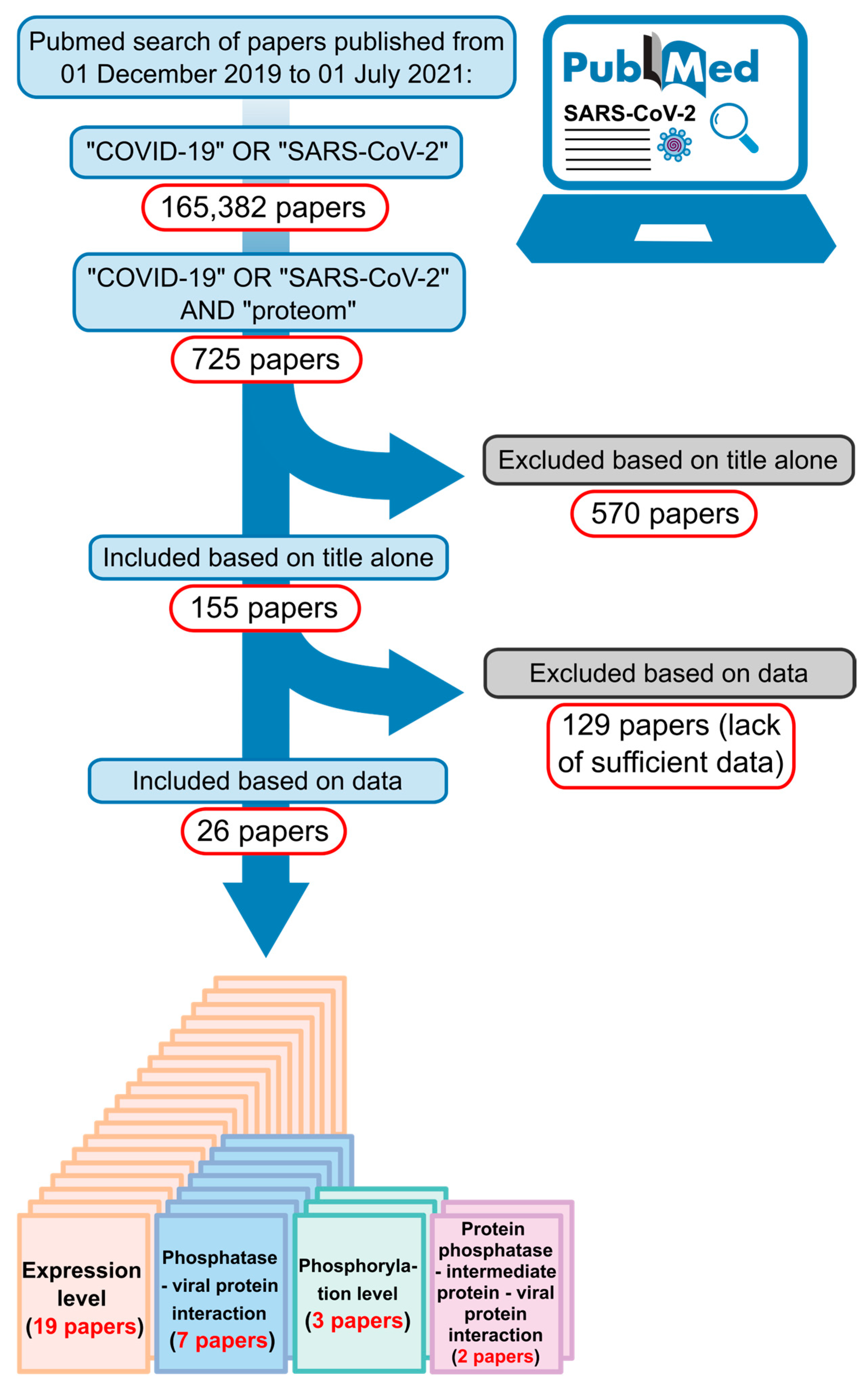
4. Material and Methods

4.1. In Silico Analysis

Here, we present an in silico meta-analysis that utilizes data from the reanalysis of 26 scientific papers and their corresponding databases [5,6,7,8,9,10,12,14,17,32,33,34,35,36,37,38,39,40,41,42,43,44,45,46,47,48] (Figure 1, Table S1). Specifically, we selected studies that included proteomic analyses conducted on various model cells or on blood cells, plasma, urine, and lung tissues from SARS-CoV-2-infected patients. The search for the expression and interaction information regarding protein phosphatases and the phosphorylation levels of intermediate proteins (Tables S2–S5) was conducted using both supplementary files provided by the authors and from the PRIDE and ProteomeXchange databases [75]. Information regarding the source data (PRIDE identifier, website links for supplementary tables, and others dataset) is summarized in Table S1.

4.2. Protein–Protein Interaction Network

The generation of interaction networks between host and viral proteins was performed using Cyoscape software with data extracted from publicly available databases (Table S3) [76]. The functions of proteins interacting with protein phosphatases and viral proteins were analyzed using Uniprot program [77]. We provide a preliminary dataset containing all proteins predicted to interact with protein phosphatases and viral proteins. This dataset was used as input in Cytoscape, employing the MCODE and ClueGo plugins [54,78] to identify the signaling pathways in which they are involved.

4.3. Statistical Analysis

The statistical analysis and graphs depicting the expression and the phosphorylation levels of protein phosphatases and intermediate proteins were generated using GraphPad Prism 9. We utilized the fold change data and log10 p-value values for each extracted sample. These data refer to the ratio of samples from control groups compared to those from infected groups, according to the equation FC = log2 (infected/control), where FC ≥ 1.0 indicates a higher level of protein phosphorylation compared to the control, and FC ≤ 1.0 indicates a lower level of protein phosphorylation compared to the control (Tables S2 and S4). Data were considered statistically different when p < 0.05 (log10 p value > 1.3).

4.4. Docking

The complex models were generated on the Alphafold 3 Server [79] using the website https://alphafoldserver.com/ and analyzed using Pymol software 2.5.2. The PPP2CA sequence was loaded with the addition of the metal Mn2+, while the sequence of CDC20, RPS17, and FGRO1P were loaded with phosphate added to the residues T70, S113, and S160, respectively. The motif L-x-x-[ILVMAPF]-x-[ED] and the proteins with higher or lower degrees of phosphorylation were used as input in ScanProsite [80] to identify the motifs in these proteins.

4.5. Molecular Dynamics

First, the structure was prepared for Molecular Dynamics (MD). From the five models generated by AlphaFold 3, the highest-confidence model was selected for further refinement [79]. To optimize the simulation box size, the first 60 residues of CDC20 (pIDDT < 50 and largely unfolded) were removed, as they did not contribute to the study. Structural analysis with MolProbity [81] addressed steric clashes and added hydrogen atoms. The model was then relaxed with 1000 steps of steepest descent in implicit solvent, using: GB-Neck2 model [82], 1000 Å non-bonded cut-off, and a surface tension of 0.007 kcal/mol/Å2. Based on the relaxed coordinates, protonation states were optimized using the H++ server [83]. The final protonated structure served as the starting point for MD simulations.
For the MD setup, the complex was solvated in a truncated octahedral water box with a 15 Å minimum boundary distance. Na+ and Cl- ions were added to achieve neutrality and a salt concentration of 0.15 M. The Amber ff14SB force-field was used for protein and phosphorylated threonine residues [84], the TIP3P model for water molecules [85], and Joung–Cheatham parameters for monovalent ions [86]. Based on studies of other manganese enzymes [87,88], the Mn2+ ions in the PPP2CA active site were described using the 12-6-4 LJ-type non-bonded model [89]. Tleap from AmberTools23 was used to generate topology and coordinates [90]. The equilibration started by optimizing side chains, solvent, and ions with 2500 steps each of steepest descent and conjugate gradient, while restraining the backbone with a 100 kcal/mol/Å2 force constant, followed by an unrestrained minimization. The system was heated from 10 K to 300 K over 0.5 ns by NVT simulations with backbone restraints (10 kcal/mol/Å2), and then equilibrated at 300 K for 1.5 ns. Five subsequent NPT simulations (0.5 ns each) gradually reduced backbone restraints from 10 to 2 kcal/mol/Å2. The final equilibration step was a 10 ns unrestrained NPT simulation. After equilibration, three replicas of 1000 ns production MD simulation were run under NPT conditions, with trajectory frames saved every 2 ns. For all MD simulations, temperature control was achieved using a stochastic Berendsen thermostat with a coupling time of 1 ps [91], while pressure was maintained at 1 bar with a Monte Carlo barostat and a relaxation time of 1 ps [92]. A 4 fs time step with Hydrogen Mass Repartitioning and the SETTLE algorithm for water was applied [93]. All the simulations were performed using PMEMD.cuda in AMBER22 [94,95].
Finally, the root-mean-square deviation (RMSD) of the whole system was calculated using a trajectory aligned to the B56 backbone coordinates of the starting point. RMSD analysis was performed with Bio3D [96]. The results indicated that the complex reached structural equilibration after 400 ns; therefore, the final 600 ns of each production run were used for contact map analysis. Contact maps were generated using CPPTRAJ from AmberTools23 and normalized to 1 (contact with 100% persistent) using a custom R script [97]. Visualizations were created using ggplot2 in Rstudio [98,99].

Supplementary Materials

The following supporting information can be downloaded at: https://www.mdpi.com/article/10.3390/kinasesphosphatases3010004/s1, Figure S1: Phosphorylation degree of the intermediate proteins network. Figure S2: RMSD of the complex backbone over simulation time for the three replicas. Figure S3: Contact map between PPP2CA and CDC20. Figure S4: Contact map between B56 and CDC20. Figure S5: Contact map between B56 and CDC20. Figure S6: Docking of the phosphorylated complexs PPP2CA-B56-RPS17 and PPP2CA-B56-FGFRO1P. Table S1: Overview of the database used in this paper. Table S2: Expression degree of proteins phosphatase. Table S3: Protein-protein interaction–PPI. Table S4: Phosphorylation levels of interaction proteins. Table S5: Proteins identified as a PPP2CA substrates treated with dTAG-13 for 24 h. Data extracted from Brewer et al., 2024 [67]. Table S6: PPP2CA-B56 motif L-x-x-[ILVMAPF]-x-[ED].

Author Contributions

G.I.M.G., L.P.O. and L.E.S.F.M. contributed to data analysis and to figure drawing and editing. G.E.J. and P.S.L.-d.-O. contributed to Molecular Dynamics Simulation, analysis and figure drawing. G.I.M.G. and L.E.S.F.M. conceptualized the study, wrote and prepared the original draft. All authors have read and agreed to the published version of the manuscript.

Funding

This work was supported by grant 2019/02605-3 to L.E.S.F.M., the fellowship 2021/10474-6 to L.P.O. and 2020/10168-0 to L.E.S.F.M. from São Paulo Research Foundation (FAPESP) and PIBIC 2021/2022 to G.I.M.G. from National council for Scientific and Technological Development (CNPq).

Data Availability Statement

All the data are referenced in this paper and provided as a Supplementary Tables S1–S6.

Acknowledgments

We would like to thank Marcus Oliveira and Francisco Prosdocimi for the critical reading of this manuscript. We also thank the Brazilian Biosciences National Laboratory (LNBio) at the Brazilian Center for Research in Energy and Materials (CNPEM) for access to the High-Performance Computing Cluster and scientific infrastructure.

Conflicts of Interest

The authors declare no conflicts of interest.

References

  1. COVID-19 Map. In Johns Hopkins Coronavirus Resource Center. Available online: https://coronavirus.jhu.edu/map.html (accessed on 11 December 2024).
  2. Naqvi, A.A.T.; Fatima, K.; Mohammad, T.; Fatima, U.; Singh, I.K.; Singh, A.; Atif, S.M.; Hariprasad, G.; Hasan, G.M.; Hassan, M.I. Insights into SARS-CoV-2 genome, structure, evolution, pathogenesis and therapies: Structural genomics approach. Biochim. Biophys. Acta Mol. Basis Dis. 2020, 1866, 165878. [Google Scholar] [CrossRef] [PubMed]
  3. Malik, Y.A. Properties of Coronavirus and SARS-CoV-2. Malays. J. Pathol. 2020, 42, 3–11. [Google Scholar] [PubMed]
  4. Harrison, A.G.; Lin, T.; Wang, P. Mechanisms of SARS-CoV-2 Transmission and Pathogenesis. Trends Immunol. 2020, 41, 1100–1115. [Google Scholar] [CrossRef] [PubMed]
  5. Leng, L.; Cao, R.; Ma, J.; Mou, D.; Zhu, Y.; Li, W.; Lv, L.; Gao, D.; Zhang, S.; Gong, F.; et al. Pathological features of COVID-19-associated lung injury: A preliminary proteomics report based on clinical samples. Signal Transduct. Target. Ther. 2020, 5, 240. [Google Scholar] [CrossRef] [PubMed]
  6. Li, J.; Guo, M.; Tian, X.; Wang, X.; Yang, X.; Wu, P.; Liu, C.; Xiao, Z.; Qu, Y.; Yin, Y.; et al. Virus-Host Interactome and Proteomic Survey Reveal Potential Virulence Factors Influencing SARS-CoV-2 Pathogenesis. Med 2021, 2, 99–112.e7. [Google Scholar] [CrossRef] [PubMed]
  7. Li, Y.; Wang, Y.; Liu, H.; Sun, W.; Ding, B.; Zhao, Y.; Chen, P.; Zhu, L.; Li, Z.; Li, N.; et al. Urine proteome of COVID-19 patients. Urine 2020, 2, 1–8. [Google Scholar] [CrossRef] [PubMed]
  8. Bojkova, D.; Klann, K.; Koch, B.; Widera, M.; Krause, D.; Ciesek, S.; Cinatl, J.; Münch, C. Proteomics of SARS-CoV-2-infected host cells reveals therapy targets. Nature 2020, 583, 469–472. [Google Scholar] [CrossRef]
  9. Bouhaddou, M.; Memon, D.; Meyer, B.; White, K.M.; Rezelj, V.V.; Correa Marrero, M.; Polacco, B.J.; Melnyk, J.E.; Ulferts, S.; Kaake, R.M.; et al. The Global Phosphorylation Landscape of SARS-CoV-2 Infection. Cell 2020, 182, 685–712.e19. [Google Scholar] [CrossRef]
  10. Appelberg, S.; Gupta, S.; Svensson Akusjärvi, S.; Ambikan, A.T.; Mikaeloff, F.; Saccon, E.; Végvári, Á.; Benfeitas, R.; Sperk, M.; Ståhlberg, M.; et al. Dysregulation in Akt/mTOR/HIF-1 signaling identified by proteo-transcriptomics of SARS-CoV-2 infected cells. Emerg. Microbes Infect. 2020, 9, 1748–1760. [Google Scholar] [CrossRef] [PubMed]
  11. Zeng, H.-L.; Chen, D.; Yan, J.; Yang, Q.; Han, Q.-Q.; Li, S.-S.; Cheng, L. Proteomic characteristics of bronchoalveolar lavage fluid in critical COVID-19 patients. FEBS J. 2020, 288, 5190–5200. [Google Scholar] [CrossRef]
  12. Gisby, J.; Clarke, C.L.; Medjeral-Thomas, N.; Malik, T.H.; Papadaki, A.; Mortimer, P.M.; Buang, N.B.; Lewis, S.; Pereira, M.; Toulza, F.; et al. Longitudinal proteomic profiling of dialysis patients with COVID-19 reveals markers of severity and predictors of death. Elife 2021, 10, e64827. [Google Scholar] [CrossRef] [PubMed]
  13. Filbin, M.R.; Mehta, A.; Schneider, A.M.; Kays, K.R.; Guess, J.R.; Gentili, M.; Fenyves, B.G.; Charland, N.C.; Gonye, A.L.; Gushterova, I.; et al. Longitudinal proteomic analysis of severe COVID-19 reveals survival-associated signatures, tissue-specific cell death, and cell-cell interactions. CR Med. 2021, 2, 100287. [Google Scholar] [CrossRef] [PubMed]
  14. Stukalov, A.; Girault, V.; Grass, V.; Karayel, O.; Bergant, V.; Urban, C.; Haas, D.A.; Huang, Y.; Oubraham, L.; Wang, A.; et al. Multilevel proteomics reveals host perturbations by SARS-CoV-2 and SARS-CoV. Nature 2021, 594, 246–252. [Google Scholar] [CrossRef] [PubMed]
  15. Chen, Z.; John Wherry, E. T cell responses in patients with COVID-19. Nat. Rev. Immunol. 2020, 20, 529–536. [Google Scholar] [CrossRef] [PubMed]
  16. Rümke, L.W.; Smit, W.L.; Bossink, A.; Limonard, G.J.M.; Muilwijk, D.; Haas, L.E.M.; Reusken, C.; van der Wal, S.; Thio, B.J.; van Os, Y.M.G.; et al. Impaired SARS-CoV-2 specific T-cell response in patients with severe COVID-19. Front. Immunol. 2023, 14, 1046639. [Google Scholar] [CrossRef] [PubMed]
  17. Klann, K.; Bojkova, D.; Tascher, G.; Ciesek, S.; Münch, C.; Cinatl, J. Growth Factor Receptor Signaling Inhibition Prevents SARS-CoV-2 Replication. Mol. Cell 2020, 80, 164–174.e4. [Google Scholar] [CrossRef]
  18. Novak, B.; Kapuy, O.; Domingo-Sananes, M.R.; Tyson, J.J. Regulated protein kinases and phosphatases in cell cycle decisions. Curr. Opin. Cell Biol. 2010, 22, 801–808. [Google Scholar] [CrossRef]
  19. Smoly, I.; Shemesh, N.; Ziv-Ukelson, M.; Ben-Zvi, A.; Yeger-Lotem, E. An Asymmetrically Balanced Organization of Kinases versus Phosphatases across Eukaryotes Determines Their Distinct Impacts. PLoS Comput. Biol. 2017, 13, e1005221. [Google Scholar] [CrossRef] [PubMed]
  20. Fischer, E.H. Cell signaling by protein tyrosine phosphorylation. Adv. Enzyme Regul. 1999, 39, 359–369. [Google Scholar] [CrossRef]
  21. Hornberg, J.J.; Bruggeman, F.J.; Binder, B.; Geest, C.R.; de Vaate, A.J.M.B.; Lankelma, J.; Heinrich, R.; Westerhoff, H.V. Principles behind the multifarious control of signal transduction. ERK phosphorylation and kinase/phosphatase control. FEBS J. 2005, 272, 244–258. [Google Scholar] [CrossRef] [PubMed]
  22. Hunter, T. Protein kinases and phosphatases: The yin and yang of protein phosphorylation and signaling. Cell 1995, 80, 225–236. [Google Scholar] [CrossRef]
  23. Corda, P.O.; Bollen, M.; Ribeiro, D.; Fardilha, M. Emerging roles of the Protein Phosphatase 1 (PP1) in the context of viral infections. Cell Commun. Signal 2024, 22, 65. [Google Scholar] [CrossRef]
  24. Sontag, E. Protein phosphatase 2A: The Trojan Horse of cellular signaling. Cell Signal 2001, 13, 7–16. [Google Scholar] [CrossRef] [PubMed]
  25. Ouellet, M.; Barbeau, B.; Tremblay, M.J. Protein tyrosyl phosphatases in T cell activation: Implication for human immunodeficiency virus transcriptional activity. Prog. Nucleic Acid Res. Mol. Biol. 2003, 73, 69–105. [Google Scholar] [CrossRef] [PubMed]
  26. Wang, Q.; Pan, W.; Wang, S.; Pan, C.; Ning, H.; Huang, S.; Chiu, S.-H.; Chen, J.-L. Protein Tyrosine Phosphatase SHP2 Suppresses Host Innate Immunity against Influenza A Virus by Regulating EGFR-Mediated Signaling. J. Virol. 2021, 95, e02001-20. [Google Scholar] [CrossRef] [PubMed]
  27. Devi, P.; Ota, S.; Punga, T.; Bergqvist, A. Hepatitis C Virus Core Protein Down-Regulates Expression of Src-Homology 2 Domain Containing Protein Tyrosine Phosphatase by Modulating Promoter DNA Methylation. Viruses 2021, 13, 2514. [Google Scholar] [CrossRef] [PubMed]
  28. Zhou, Y.; Zhao, Y.; Gao, Y.; Hu, W.; Qu, Y.; Lou, N.; Zhu, Y.; Zhang, X.; Yang, H. Hepatitis C virus NS3 protein enhances hepatocellular carcinoma cell invasion by promoting PPM1A ubiquitination and degradation. J. Exp. Clin. Cancer Res. 2017, 36, 42. [Google Scholar] [CrossRef]
  29. Hu, Z.; Ban, H.; Zheng, H.; Liu, M.; Chang, J.; Guo, J.-T. Protein phosphatase 1 catalyzes HBV core protein dephosphorylation and is co-packaged with viral pregenomic RNA into nucleocapsids. PLoS Pathog. 2020, 16, e1008669. [Google Scholar] [CrossRef] [PubMed]
  30. Gerlt, V.; Mayr, J.; Del Sarto, J.; Ludwig, S.; Boergeling, Y. Cellular Protein Phosphatase 2A Regulates Cell Survival Mechanisms in Influenza A Virus Infection. Int. J. Mol. Sci. 2021, 22, 11164. [Google Scholar] [CrossRef]
  31. He, C.; Qiu, Y.; Han, P.; Chen, Y.; Zhang, L.; Yuan, Q.; Zhang, T.; Cheng, T.; Yuan, L.; Huang, C.; et al. ER stress regulating protein phosphatase 2A-B56γ, targeted by hepatitis B virus X protein, induces cell cycle arrest and apoptosis of hepatocytes. Cell Death Dis. 2018, 9, 762. [Google Scholar] [CrossRef]
  32. D’Alessandro, A.; Thomas, T.; Dzieciatkowska, M.; Hill, R.C.; Francis, R.O.; Hudson, K.E.; Zimring, J.C.; Hod, E.A.; Spitalnik, S.L.; Hansen, K.C. Serum Proteomics in COVID-19 Patients: Altered Coagulation and Complement Status as a Function of IL-6 Level. J. Proteome Res. 2020, 19, 4417–4427. [Google Scholar] [CrossRef] [PubMed]
  33. Schmidt, N.; Lareau, C.A.; Keshishian, H.; Ganskih, S.; Schneider, C.; Hennig, T.; Melanson, R.; Werner, S.; Wei, Y.; Zimmer, M.; et al. The SARS-CoV-2 RNA-protein interactome in infected human cells. Nat. Microbiol. 2021, 6, 339–353. [Google Scholar] [CrossRef] [PubMed]
  34. Roh, J.D.; Kitchen, R.R.; Guseh, J.S.; McNeill, J.N.; Aid, M.; Martinot, A.J.; Yu, A.; Platt, C.; Rhee, J.; Weber, B.; et al. Plasma Proteomics of COVID-19-Associated Cardiovascular Complications: Implications for Pathophysiology and Therapeutics. JACC Basic. Transl. Sci. 2022, 7, 425–441. [Google Scholar] [CrossRef]
  35. Nie, X.; Qian, L.; Sun, R.; Huang, B.; Dong, X.; Xiao, Q.; Zhang, Q.; Lu, T.; Yue, L.; Chen, S.; et al. Multi-organ proteomic landscape of COVID-19 autopsies. Cell 2021, 184, 775–791.e14. [Google Scholar] [CrossRef] [PubMed]
  36. Wang, H.; Wu, X.; Zhang, X.; Hou, X.; Liang, T.; Wang, D.; Teng, F.; Dai, J.; Duan, H.; Guo, S.; et al. SARS-CoV-2 Proteome Microarray for Mapping COVID-19 Antibody Interactions at Amino Acid Resolution. ACS Central Sci. 2020, 6, 2238–2249. [Google Scholar] [CrossRef] [PubMed]
  37. Almasy, K.M.; Davies, J.P.; Plate, L. Comparative Host Interactomes of the SARS-CoV-2 Nonstructural Protein 3 and Human Coronavirus Homologs. Mol. Cell. Proteom. 2021, 20, 100120. [Google Scholar] [CrossRef]
  38. Vanderboom, P.M.; Mun, D.-G.; Madugundu, A.K.; Mangalaparthi, K.K.; Saraswat, M.; Garapati, K.; Chakraborty, R.; Ebihara, H.; Sun, J.; Pandey, A. Proteomic Signature of Host Response to SARS-CoV-2 Infection in the Nasopharynx. Mol. Cell. Proteom. 2021, 20, 100134. [Google Scholar] [CrossRef]
  39. Xu, B.; Lei, Y.; Ren, X.; Yin, F.; Wu, W.; Sun, Y.; Wang, X.; Sun, Q.; Yang, X.; Wang, X.; et al. SOD1 is a Possible Predictor of COVID-19 Progression as Revealed by Plasma Proteomics. ACS Omega 2021, 6, 16826–16836. [Google Scholar] [CrossRef] [PubMed]
  40. Wu, M.; Chen, Y.; Xia, H.; Wang, C.; Tan, C.Y.; Cai, X.; Liu, Y.; Ji, F.; Xiong, P.; Liu, R.; et al. Transcriptional and proteomic insights into the host response in fatal COVID-19 cases. Proc. Natl. Acad. Sci. USA 2020, 117, 28336–28343. [Google Scholar] [CrossRef]
  41. Tan, Y.; Zhang, W.; Zhu, Z.; Qiao, N.; Ling, Y.; Guo, M.; Yin, T.; Fang, H.; Xu, X.; Lu, G.; et al. Integrating longitudinal clinical laboratory tests with targeted proteomic and transcriptomic analyses reveal the landscape of host responses in COVID-19. Cell Discov. 2021, 7, 42. [Google Scholar] [CrossRef] [PubMed]
  42. Li, N.; Zhao, L.; Zhan, X. Quantitative proteomics reveals a broad-spectrum antiviral property of ivermectin, benefiting for COVID-19 treatment. J. Cell. Physiol. 2021, 236, 2959–2975. [Google Scholar] [CrossRef] [PubMed]
  43. Gordon, D.E.; Jang, G.M.; Bouhaddou, M.; Xu, J.; Obernier, K.; White, K.M.; O’Meara, M.J.; Rezelj, V.V.; Guo, J.Z.; Swaney, D.L.; et al. A SARS-CoV-2 protein interaction map reveals targets for drug repurposing. Nature 2020, 583, 459–468. [Google Scholar] [CrossRef] [PubMed]
  44. Karunakaran, K.B.; Balakrishnan, N.; Ganapathiraju, M.K. Interactome of SARS-CoV-2/nCoV19 modulated host proteins with computationally predicted PPIs. Res. Sq. 2020. [Google Scholar] [CrossRef]
  45. Nadeau, R.; Shahryari Fard, S.; Scheer, A.; Hashimoto-Roth, E.; Nygard, D.; Abramchuk, I.; Chung, Y.-E.; Bennett, S.A.L.; Lavallée-Adam, M. Computational Identification of Human Biological Processes and Protein Sequence Motifs Putatively Targeted by SARS-CoV-2 Proteins Using Protein-Protein Interaction Networks. J. Proteome Res. 2020, 19, 4553–4566. [Google Scholar] [CrossRef] [PubMed]
  46. Bamberger, C.; Pankow, S.; Martínez-Bartolomé, S.; Diedrich, J.K.; Park, R.S.K.; Yates, J.R. Analysis of the Tropism of SARS-CoV-2 Based on the Host Interactome of the Spike Protein. J. Proteome Res. 2023, 22, 3742–3753. [Google Scholar] [CrossRef]
  47. Meyers, J.M.; Ramanathan, M.; Shanderson, R.L.; Beck, A.; Donohue, L.; Ferguson, I.; Guo, M.G.; Rao, D.S.; Miao, W.; Reynolds, D.; et al. The proximal proteome of 17 SARS-CoV-2 proteins links to disrupted antiviral signaling and host translation. PLoS Pathog. 2021, 17, e1009412. [Google Scholar] [CrossRef] [PubMed]
  48. Perfetto, L.; Pastrello, C.; Del-Toro, N.; Duesbury, M.; Iannuccelli, M.; Kotlyar, M.; Licata, L.; Meldal, B.; Panneerselvam, K.; Panni, S.; et al. The IMEx coronavirus interactome: An evolving map of Coronaviridae-host molecular interactions. Database 2020, 2020, baaa096. [Google Scholar] [CrossRef]
  49. Zhou, Y.; Liu, Y.; Gupta, S.; Paramo, M.I.; Hou, Y.; Mao, C.; Luo, Y.; Judd, J.; Wierbowski, S.; Bertolotti, M.; et al. A comprehensive SARS-CoV-2-human protein-protein interactome reveals COVID-19 pathobiology and potential host therapeutic targets. Nat. Biotechnol. 2023, 41, 128–139. [Google Scholar] [CrossRef] [PubMed]
  50. Lee, S.J.; Rodriguez-Bravo, V.; Kim, H.; Datta, S.; Foley, E.A. The PP2AB56 phosphatase promotes the association of Cdc20 with APC/C in mitosis. J. Cell Sci. 2017, 130, 1760–1771. [Google Scholar] [CrossRef] [PubMed]
  51. Fujimitsu, K.; Yamano, H. PP2A-B56 binds to Apc1 and promotes Cdc20 association with the APC/C ubiquitin ligase in mitosis. EMBO Rep. 2020, 21, e48503. [Google Scholar] [CrossRef]
  52. Labit, H.; Fujimitsu, K.; Bayin, N.S.; Takaki, T.; Gannon, J.; Yamano, H. Dephosphorylation of Cdc20 is required for its C-box-dependent activation of the APC/C. EMBO J. 2012, 31, 3351–3362. [Google Scholar] [CrossRef]
  53. Craney, A.; Kelly, A.; Jia, L.; Fedrigo, I.; Yu, H.; Rape, M. Control of APC/C-dependent ubiquitin chain elongation by reversible phosphorylation. Proc. Natl. Acad. Sci. USA 2016, 113, 1540–1545. [Google Scholar] [CrossRef] [PubMed]
  54. Bindea, G.; Mlecnik, B.; Hackl, H.; Charoentong, P.; Tosolini, M.; Kirilovsky, A.; Fridman, W.-H.; Pagès, F.; Trajanoski, Z.; Galon, J. ClueGO: A Cytoscape plug-in to decipher functionally grouped gene ontology and pathway annotation networks. Bioinformatics 2009, 25, 1091–1093. [Google Scholar] [CrossRef]
  55. Kuss, S.K.; Mata, M.A.; Zhang, L.; Fontoura, B.M.A. Nuclear Imprisonment: Viral Strategies to Arrest Host mRNA Nuclear Export. Viruses 2013, 5, 1824–1849. [Google Scholar] [CrossRef]
  56. Melenotte, C.; Silvin, A.; Goubet, A.-G.; Lahmar, I.; Dubuisson, A.; Zumla, A.; Raoult, D.; Merad, M.; Gachot, B.; Hénon, C.; et al. Immune responses during COVID-19 infection. Oncoimmunology 2020, 9, 1807836. [Google Scholar] [CrossRef] [PubMed]
  57. Song, M.S.; Carracedo, A.; Salmena, L.; Song, S.J.; Egia, A.; Malumbres, M.; Pandolfi, P.P. Nuclear PTEN regulates the APC-CDH1 tumor-suppressive complex in a phosphatase-independent manner. Cell 2011, 144, 187–199. [Google Scholar] [CrossRef] [PubMed]
  58. Choi, B.H.; Pagano, M.; Huang, C.; Dai, W. Cdh1, a substrate-recruiting component of anaphase-promoting complex/cyclosome (APC/C) ubiquitin E3 ligase, specifically interacts with phosphatase and tensin homolog (PTEN) and promotes its removal from chromatin. J. Biol. Chem. 2014, 289, 17951–17959. [Google Scholar] [CrossRef] [PubMed]
  59. Pesin, J.A.; Orr-Weaver, T.L. Regulation of APC/C Activators in Mitosis and Meiosis. Annu. Rev. Cell Dev. Biol. 2008, 24, 475–499. [Google Scholar] [CrossRef]
  60. Shao, C.; Li, Z.; Ahmad, N.; Liu, X. Regulation of PTEN degradation and NEDD4-1 E3 ligase activity by Numb. Cell Cycle 2017, 16, 957–967. [Google Scholar] [CrossRef]
  61. Pilecki, M.; Grzyb, A.; Zień, P.; Sekuła, O.; Szyszka, R. Yeast protein phosphatase active with acidic ribosomal proteins. J. Basic Microbiol. 2000, 40, 251–260. [Google Scholar] [CrossRef] [PubMed]
  62. Kim, T.-S.; Kim, H.D.; Shin, H.-S.; Kim, J. Phosphorylation status of nuclear ribosomal protein S3 is reciprocally regulated by protein kinase Cδ and protein phosphatase 2A. J. Biol. Chem. 2009, 284, 21201–21208. [Google Scholar] [CrossRef]
  63. Andres, J.L.; Johansen, J.W.; Maller, J.L. Identification of protein phosphatases 1 and 2B as ribosomal protein S6 phosphatases in vitro and in vivo. J. Biol. Chem. 1987, 262, 14389–14393. [Google Scholar] [CrossRef] [PubMed]
  64. Andres, J.L.; Maller, J.L. Purification and characterization of a novel protein phosphatase highly specific for ribosomal protein S6. J. Biol. Chem. 1989, 264, 151–156. [Google Scholar] [CrossRef]
  65. Hutchinson, J.A.; Shanware, N.P.; Chang, H.; Tibbetts, R.S. Regulation of ribosomal protein S6 phosphorylation by casein kinase 1 and protein phosphatase 1. J. Biol. Chem. 2011, 286, 8688–8696. [Google Scholar] [CrossRef] [PubMed]
  66. Hirano, K.; Ito, M.; Hartshorne, D.J. Interaction of the ribosomal protein, L5, with protein phosphatase type 1. J. Biol. Chem. 1995, 270, 19786–19790. [Google Scholar] [CrossRef] [PubMed]
  67. Brewer, A.; Sathe, G.; Pflug, B.E.; Clarke, R.G.; Macartney, T.J.; Sapkota, G.P. Mapping the substrate landscape of protein phosphatase 2A catalytic subunit PPP2CA. iScience 2024, 27, 109302. [Google Scholar] [CrossRef]
  68. Yu, H. Regulation of APC-Cdc20 by the spindle checkpoint. Curr. Opin. Cell Biol. 2002, 14, 706–714. [Google Scholar] [CrossRef]
  69. Qiao, R.; Weissmann, F.; Yamaguchi, M.; Brown, N.G.; VanderLinden, R.; Imre, R.; Jarvis, M.A.; Brunner, M.R.; Davidson, I.F.; Litos, G.; et al. Mechanism of APC/CCDC20 activation by mitotic phosphorylation. Proc. Natl. Acad. Sci. USA 2016, 113, E2570–E2578. [Google Scholar] [CrossRef]
  70. Barski, M.S.; Minnell, J.J.; Maertens, G.N. PP2A Phosphatase as an Emerging Viral Host Factor. Front. Cell Infect. Microbiol. 2021, 11, 725615. [Google Scholar] [CrossRef]
  71. Wang, X.; Bajaj, R.; Bollen, M.; Peti, W.; Page, R. Expanding the PP2A Interactome by Defining a B56-Specific SLiM. Structure 2016, 24, 2174–2181. [Google Scholar] [CrossRef]
  72. Hertz, E.P.T.; Kruse, T.; Davey, N.E.; López-Méndez, B.; Sigurðsson, J.O.; Montoya, G.; Olsen, J.V.; Nilsson, J. A Conserved Motif Provides Binding Specificity to the PP2A-B56 Phosphatase. Mol. Cell 2016, 63, 686–695. [Google Scholar] [CrossRef] [PubMed]
  73. Wang, X.; Garvanska, D.H.; Nasa, I.; Ueki, Y.; Zhang, G.; Kettenbach, A.N.; Peti, W.; Nilsson, J.; Page, R. A dynamic charge-charge interaction modulates PP2A:B56 substrate recruitment. Elife 2020, 9, e55966. [Google Scholar] [CrossRef] [PubMed]
  74. Fu, Y.; Fang, Y.; Gong, S.; Xue, T.; Wang, P.; She, L.; Huang, J. Deep learning-based network pharmacology for exploring the mechanism of licorice for the treatment of COVID-19. Sci. Rep. 2023, 13, 5844. [Google Scholar] [CrossRef]
  75. Deutsch, E.W.; Bandeira, N.; Sharma, V.; Perez-Riverol, Y.; Carver, J.J.; Kundu, D.J.; García-Seisdedos, D.; Jarnuczak, A.F.; Hewapathirana, S.; Pullman, B.S.; et al. The ProteomeXchange consortium in 2020: Enabling “big data” approaches in proteomics. Nucleic Acids Res. 2020, 48, D1145–D1152. [Google Scholar] [CrossRef]
  76. Shannon, P.; Markiel, A.; Ozier, O.; Baliga, N.S.; Wang, J.T.; Ramage, D.; Amin, N.; Schwikowski, B.; Ideker, T. Cytoscape: A software environment for integrated models of biomolecular interaction networks. Genome Res. 2003, 13, 2498–2504. [Google Scholar] [CrossRef] [PubMed]
  77. UniProt Consortium. UniProt: The universal protein knowledgebase in 2021. Nucleic Acids Res. 2021, 49, D480–D489. [Google Scholar] [CrossRef] [PubMed]
  78. Bindea, G.; Galon, J.; Mlecnik, B. CluePedia Cytoscape plugin: Pathway insights using integrated experimental and in silico data. Bioinformatics 2013, 29, 661–663. [Google Scholar] [CrossRef] [PubMed]
  79. Abramson, J.; Adler, J.; Dunger, J.; Evans, R.; Green, T.; Pritzel, A.; Ronneberger, O.; Willmore, L.; Ballard, A.J.; Bambrick, J.; et al. Accurate structure prediction of biomolecular interactions with AlphaFold 3. Nature 2024, 630, 493–500. [Google Scholar] [CrossRef]
  80. de Castro, E.; Sigrist, C.J.A.; Gattiker, A.; Bulliard, V.; Langendijk-Genevaux, P.S.; Gasteiger, E.; Bairoch, A.; Hulo, N. ScanProsite: Detection of PROSITE signature matches and ProRule-associated functional and structural residues in proteins. Nucleic Acids Res. 2006, 34, W362–W365. [Google Scholar] [CrossRef] [PubMed]
  81. Williams, C.J.; Headd, J.J.; Moriarty, N.W.; Prisant, M.G.; Videau, L.L.; Deis, L.N.; Verma, V.; Keedy, D.A.; Hintze, B.J.; Chen, V.B.; et al. MolProbity: More and better reference data for improved all-atom structure validation. Protein Sci. 2018, 27, 293–315. [Google Scholar] [CrossRef]
  82. Nguyen, H.; Roe, D.R.; Simmerling, C. Improved Generalized Born Solvent Model Parameters for Protein Simulations. J. Chem. Theory Comput. 2013, 9, 2020–2034. [Google Scholar] [CrossRef]
  83. Anandakrishnan, R.; Aguilar, B.; Onufriev, A.V. H++ 3.0: Automating pK prediction and the preparation of biomolecular structures for atomistic molecular modeling and simulations. Nucleic Acids Res. 2012, 40, W537–W541. [Google Scholar] [CrossRef]
  84. Maier, J.A.; Martinez, C.; Kasavajhala, K.; Wickstrom, L.; Hauser, K.E.; Simmerling, C. ff14SB: Improving the Accuracy of Protein Side Chain and Backbone Parameters from ff99SB. J Chem. Theory Comput. 2015, 11, 3696–3713. [Google Scholar] [CrossRef] [PubMed]
  85. Jorgensen, W.L.; Chandrasekhar, J.; Madura, J.D.; Impey, R.W.; Klein, M.L. Comparison of simple potential functions for simulating liquid water. J. Chem. Phys. 1983, 79, 926–935. [Google Scholar] [CrossRef]
  86. Joung, I.S.; Cheatham, T.E. Molecular dynamics simulations of the dynamic and energetic properties of alkali and halide ions using water-model-specific ion parameters. J. Phys. Chem. B 2009, 113, 13279–13290. [Google Scholar] [CrossRef] [PubMed]
  87. Josino, L.P.C.; Alves, C.N.; Lima, A.H. A molecular model to study FosA enzyme inhibition. J. Mol. Graph. Model. 2021, 107, 107978. [Google Scholar] [CrossRef] [PubMed]
  88. Bonetta, R.; Hunter, G.J.; Trinh, C.H.; Borowski, T.; Fenech, A.G.; Kulp, M.; Tabares, L.C.; Un, S.; Hunter, T. Substitution of histidine 30 by asparagine in manganese superoxide dismutase alters biophysical properties and supports proliferation in a K562 leukemia cell line. Eur. Biophys. J. 2021, 50, 571–585. [Google Scholar] [CrossRef] [PubMed]
  89. Li, P.; Merz, K.M. Taking into Account the Ion-induced Dipole Interaction in the Nonbonded Model of Ions. J. Chem. Theory Comput. 2014, 10, 289–297. [Google Scholar] [CrossRef] [PubMed]
  90. Case, D.A.; Aktulga, H.M.; Belfon, K.; Cerutti, D.S.; Cisneros, G.A.; Cruzeiro, V.W.D.; Forouzesh, N.; Giese, T.J.; Götz, A.W.; Gohlke, H.; et al. AmberTools. J. Chem. Inf. Model. 2023, 63, 6183. [Google Scholar] [CrossRef]
  91. Bussi, G.; Donadio, D.; Parrinello, M. Canonical sampling through velocity rescaling. J. Chem. Phys. 2007, 126, 014101. [Google Scholar] [CrossRef] [PubMed]
  92. Åqvist, J.; Wennerström, P.; Nervall, M.; Bjelic, S.; Brandsdal, B.O. Molecular dynamics simulations of water and biomolecules with a Monte Carlo constant pressure algorithm. Chem. Phys. Lett. 2004, 384, 288–294. [Google Scholar] [CrossRef]
  93. Hopkins, C.W.; Le Grand, S.; Walker, R.C.; Roitberg, A.E. Long-Time-Step Molecular Dynamics through Hydrogen Mass Repartitioning. J. Chem. Theory Comput. 2015, 11, 1864–1874. [Google Scholar] [CrossRef] [PubMed]
  94. Miyamoto, S.; Kollman, P.A. Settle: An analytical version of the SHAKE and RATTLE algorithm for rigid water models. J. Comput. Chem. 1992, 13, 952–962. [Google Scholar] [CrossRef]
  95. Salomon-Ferrer, R.; Götz, A.W.; Poole, D.; Le Grand, S.; Walker, R.C. Routine Microsecond Molecular Dynamics Simulations with AMBER on GPUs. 2. Explicit Solvent Particle Mesh Ewald. J. Chem. Theory Comput. 2013, 9, 3878–3888. [Google Scholar] [CrossRef] [PubMed]
  96. Grant, B.J.; Rodrigues, A.P.C.; ElSawy, K.M.; McCammon, J.A.; Caves, L.S.D. Bio3d: An R package for the comparative analysis of protein structures. Bioinformatics 2006, 22, 2695–2696. [Google Scholar] [CrossRef]
  97. Roe, D.R.; Cheatham, T.E. PTRAJ and CPPTRAJ: Software for Processing and Analysis of Molecular Dynamics Trajectory Data. J. Chem. Theory Comput. 2013, 9, 3084–3095. [Google Scholar] [CrossRef]
  98. Wickham, H. ggplot2: Elegant Graphics for Data Analysis; Springer: New York, NY, USA, 2016; Available online: https://ggplot2.tidyverse.org/authors.html#citation (accessed on 31 October 2024).
  99. RStudio Team. RStudio: Integrated Development for R. In Posit Support. 2020. Available online: https://support.posit.co/hc/en-us/articles/206212048-Citing-RStudio (accessed on 31 October 2024).
Figure 1. Flowchart of paper selection based on defined inclusion and exclusion criteria. Scientific papers were searched in Pubmed using the terms “COVID-19” OR “SARS-CoV-2”, resulting in 165,382 papers. The search was refined by adding AND “proteom”, yielding 725 papers published between 1 December 2019 and 1 July 2021. Based on content of the abstracts, any papers that did not mention protein expression, phosphorylation, or protein–protein interaction analysis were excluded. This led to the removal of 570 papers, leaving 155 papers for analysis. Due to a lack of sufficient data, such as fold change and statistical analysis, an additional 129 papers were excluded. Ultimately, 26 papers were analyzed: 19 papers focused on the expression levels of protein phosphatases, 7 papers examined protein phosphatase–viral protein interactions, 3 papers assessed the phosphorylation levels of intermediate proteins, and 2 papers investigated interactions involving protein phosphatases, intermediate proteins, and viral proteins. The image was created in the Inkscape software 1.4, an open-source software that can be used for both personal or professional work.
Kinasesphosphatases 03 00004 g001
Figure 2. Volcano plot displaying the expression levels of protein phosphatases. FC refers to the fold change in expression levels, representing the ratio between SARS-CoV-2 infected cells and non-infected (control) cells in (A) the cell model, (B) patient tissues and cells, and (C) patient fluids and secretions. FC ≥ 1.0 indicates a higher level of protein expression compared to the control (red). FC ≤ 1.0 indicates a lower level of protein expression compared to the control (blue). The fold change is calculated as FC = log2 (infected/control). The graph was plotted using GraphPadPrism 9.0. Number identifications are provided in sheet 2 of Table S2 with the references cited in the main text.
Kinasesphosphatases 03 00004 g002
Figure 3. Protein–protein interaction network. (A) Interaction network between protein phosphatases (cyan) and SARS-CoV-2 viral proteins (yellow). (B) Interaction network between protein phosphatases (cyan) and intermediate proteins (magenta), which also interact with SARS-CoV-2 viral proteins (yellow). (C) Interaction network of PTEN (cyan) with intermediate proteins (magenta) that interact with SARS-CoV-2 viral proteins (yellow). (D) Interaction network of PPP2CA (cyan) with intermediate proteins (magenta) that interact with SARS-CoV-2 viral proteins. (E) Example of an intermediate protein (magenta) interacting with SARS-CoV-2 viral proteins (yellow) and PPP2CA (cyan).
Kinasesphosphatases 03 00004 g003
Figure 4. Functional clusters of intermediate proteins. (AC) PPI network of intermediate proteins that interact with protein phosphatases and viral proteins, generated in MCODE, organized into three clusters according to functional groups. (DF) Specific functional groups generated in ClueGo. The numbers in parentheses indicate the number of genes associated with each function (term). The percentages represent the most significant genes, with the term that leads the network interaction highlighted within the functional groups. (A,D) Cluster 1; (B,E) Cluster 2; (C,F) Cluster 3.
Kinasesphosphatases 03 00004 g004
Figure 5. Functional clusters of PPP2CA and PTEN protein. (A) PPI network of intermediate proteins that interact with protein the phosphatase PPP2CA and viral proteins, generated in MCODE and organized into three cluster according to their functional groups, with cluster 2 presented in the figure. (B) PPI network of intermediate proteins that interact with the phosphatase PTEN, generated in Cytoscape. (C,D) Functional groups generated in ClueGo. The numbers in parentheses indicate the number of genes associated with each function (term). The percentages represent the most significant genes, the leading term in the network interaction highlighted within the functional groups.
Kinasesphosphatases 03 00004 g005
Figure 6. Phosphorylation degree of intermediate proteins. The volcano plot illustrates the phosphorylation levels of intermediate proteins in (A) cell models and (B) patient samples. FC ≥ 1.0 indicates a higher level of protein phosphorylation compared to the control (red). FC ≤ 1.0 indicates a lower level of protein phosphorylation compared to the control (blue). The fold change is calculated as FC = log2 (infected/control). The graph was plotted using GraphPadPrism 9.0, and the number identifications are provided in sheet 2 of Table S4 with the references cited in the main text.
Kinasesphosphatases 03 00004 g006
Figure 7. Docking and Molecular Dynamics Simulation of the PPP2CA-B56-CDC20 complex. (A) On the left, the complex is depicted in a cartoon representation generated by AlphaFold 3 server. On the right, the CDC20 motif (magenta) is positioned near the B56 subunit, with the T70 phosphorylated residue of CDC20 located in the active site of PPP2CA. (B) On the left, the AlphaFold 3 server model (cartoon) overlaid with the complex configurations sampled by the Molecular Dynamics Simulation (transparent cartoon), both displayed in the new cartoon. On the right, the AlphaFold 3 server model highlights the three motifs L-x-x-[ILVMAPF]-x-[ED] on CDC20 (magenta), also shown in the new cartoon. PPP2CA is colored wheat, B56 is green, and CDC20 is gray, with the motifs L-x-x-[ILVMAPF]-x-[ED] in magenta.
Kinasesphosphatases 03 00004 g007
Disclaimer/Publisher’s Note: The statements, opinions and data contained in all publications are solely those of the individual author(s) and contributor(s) and not of MDPI and/or the editor(s). MDPI and/or the editor(s) disclaim responsibility for any injury to people or property resulting from any ideas, methods, instructions or products referred to in the content.

