Review

Periarticular Calcifications: Clinical Features and Treatment Options

by
Romain Dalla-Torre
1,
Benoit Le Goff
1,2 and
Christelle Darrieutort-Laffite
1,2,*
1
Service de Rhumatologie, CHU Nantes, 44000 Nantes, France
2
Nantes Université, Oniris, CHU Nantes, INSERM, Regenerative Medicine and Skeleton, RMeS, UMR 1229, 44000 Nantes, France
*
Author to whom correspondence should be addressed.
Gout Urate Cryst. Depos. Dis. 2024, 2(3), 266-274; https://doi.org/10.3390/gucdd2030020
Submission received: 24 June 2024 / Revised: 25 August 2024 / Accepted: 29 August 2024 / Published: 2 September 2024

Abstract

:
Periarticular calcifications are a common condition for rheumatologists. They are characterized by deposition of carbonated apatite in tendons or connective tissues around joints. It most commonly affects patients between 30 and 60, and the main location is the shoulder (rotator cuff tendons), followed by the hip. Although the disease is frequent, factors associated with the appearance of the deposits or their spontaneous resorption remain unclear. In this review, we will summarize the available data about mechanisms underlying the constitution of the deposits and their resorption and describe the various affected sites and the associated symptoms. In the last part, we will discuss current treatment options.

1. Introduction

Periarticular calcifications are characterized by apatite deposits in extra-articular structures such as tendons, bursa, ligaments or other connective tissue structures. The disease mainly affects people between 30 and 60, with a higher prevalence in women. Although the shoulder is the main involved joint, other locations are common around the hips, hands and feet or cervical spine. Depending on the stage of the disease, patients present either chronic pain, affecting their daily and professional activities, or acute and intense pain with sudden onset and rapid recovery in the case of deposit resorption.
Rotator cuff calcific tendonitis is the most widely studied location. Among risk factors, calcific tendonitis is associated with endocrine pathologies such as hypothyroidism or diabetes [1] but there is no link with manual labor or dominant side [2]. A lower level of urinary phytate was observed in patients with rotator cuff calcifications compared to healthy controls [3]. The main sources of phytate in the diet are cereals and vegetables. Phytate is considered as an inhibitor of apatite crystallization and, in animals, soft tissue calcification was reduced by a phytin-enriched diet [4]. These data suggested a tissue deficit in this crystallization inhibitor in patients and a potential role of food in the disease. Recently, an association between tyrosine kinase inhibitors intake and calcific tendonitis was reported [5]. However, there are no clear mechanisms identified to support a causal relationship between the treatment and tendon calcifications so far.
In this review, we will first discuss the data available on the mechanisms involved in the constitution of apatite deposits and those involved in the inflammatory resorption phase. We will describe the most common locations and related symptoms. In the last part, we will discuss therapeutic options.

2. Mechanisms Leading to the Deposits and Pro-Inflammatory Effects of Apatite Crystals

The analysis of calcifications extracted from the rotator cuff by infrared spectroscopy showed carbonated apatite associated with proteins. These proteins belong to biological processes such as endochondral ossification, vesicles and exocytosis, cartilage development, apoptosis or phosphate metabolism [6]. Mechanisms underlying the constitution of the deposits have been identified 30 years ago by Uhthoff [7]. The hypothesis was based on histological observations. A four-phase process was proposed: it starts with a pre-calcific stage, followed by a crystal formation stage, then by a resorption phase and ends with a repair phase. The mineralization process starts with a fibrocartilaginous metaplasia and a crystal deposition that looks to be actively mediated by chondrocyte-like cells [7]. This fibrocartilaginous metaplasia is not similar to enthesis fibrocartilage since it does not contain collagen II [6,7]. These chondrocyte-like cells observed in the fibrocartilaginous area might be derived from tendon stem progenitor cells (TSPC) or from tenocytes. Indeed, cells extracted from human rotator cuff tendons were able to differentiate in mineralizing cells, displaying a hypertrophic chondrocytes phenotype (increased expression of SOX9, MMP13, COLL10 and TNAP (Tissue Non-Specific Alkaline Phosphatase)) [6]. However, factors involved in the onset of the metaplasia remain unknown. Recently, Hu et al. established that a downregulation of PPP1R3A (Protein Phosphatase 1 regulatory subunit 3A) was associated with a dysregulation of intracellular calcium levels in calcific tendonitis, suggesting a potential causal role for this regulator of membrane channels and receptors [8]. In addition, mechanical stimuli may play a role in the development of rotator cuff calcifications through friction or shearing, since a large majority of deposits are located close to the junction between the supraspinatus and infraspinatus tendons [9]. This mechanical stress might have an effect on the phenotype of tenocytes or TSPC located in this area.
The natural course of calcific deposits ends via an acute inflammatory phase. Macrophages and multinucleated cells have been observed around broken-up deposits. These phagocytic cells might be responsible for the initiation of the resorption [10]. Then, the inflammation induced is mainly mediated by IL-1β through the NLRP3 inflammasome as reported in other crystal-associated disorders [11,12]. However, to date, mechanisms triggering resorption remain to be identified.

3. Clinical Presentations

Patients with calcific tendonitis suffer either acute and intense pain with a rapid onset in resorption cases or chronic pain, generally more moderate, in case of impingement. In acute cases, the main differential diagnosis is septic arthritis. In most cases, the diagnosis is based on X-ray, with blurred and fragmented calcifications contrasting with the dense and homogeneous aspect of calcifications when asymptomatic or responsible for chronic pain. To classify the deposits with X-rays, the two most widely used classifications were provided by Molé [13] and Gärtner [14]. Molé’s type C and Gartner’s type III refer to deposits in the course of resorption. However, it is important to note that inter-reader reliability is slight to fair for these classifications: Kappa values at 0.18 to 0.25 and 0.33 to 0.47 for Molé and Gärtner, respectively [15,16,17]. With ultrasound, four types of deposits have been described: arc-shaped (an echogenic arc with clear shadowing), fragmented or punctate (at least two separated echogenic spots or plaques with or without shadowing), or nodular (an echogenic nodule without shadowing). In some cases, computed tomography (CT) is necessary to rule out differential diagnosis or to explore deep locations such as the spine.

3.1. Upper Limb Involvement

Rotator cuff calcifications are the most frequent and the most widely studied. Calcific tendonitis is considered the cause of chronic shoulder pain in 10 to 40% of cases. In most cases, deposits are observed in the supra-spinatus tendon. Asymptomatic deposits are found in 3 to 10 % of patients [18]. Symptoms occur when the size is over 15 mm on X-rays [18]. In ultrasound, larger calcifications (>14 mm) are associated with pain. In addition, a power Doppler signal and a widening of the sub-acromial bursa were also associated with symptomatic calcifications [19]. During resorption, the intra-osseous migration of the calcification has sometimes been observed leading to humeral greater tuberosity osteolysis [20,21]. Migration of calcium deposits into the deltoid muscle is also possible [22]. More uncommon locations have been described around the shoulder, such as the long head of the biceps or the insertion of the pectoralis major. In the latter location, cortical erosion adjacent to the calcification may suggest a tumoral process [23].
Tendon insertions around the elbow may be affected by apatite deposits, especially wrist extensors or wrist flexors or triceps brachii tendon [24]. In Figure 1, we present the case of a 20-year-old woman with acute pain and limited mobility of the elbow. X-rays showed a blurred calcific deposits within the wrist extensors, close to the lateral epicondyle (Figure 1A).
The most common location around the wrist is the insertion of the flexor carpi ulnaris tendon, but cases involving the flexor digitorum, extensor digitorum or abductor pollicis brevis have also been reported [25]. Periarticular apatite deposits may also be observed around the metacarpophalangeal (MCP) or interphalangeal (IP) joints. In this 45-year-old woman with acute dorsal wrist pain (Figure 1B), the deposit was in the wrist joint capsule. One month later, the deposit has completely disappeared.
Finally, in older patients, another rare apatite-induced acute shoulder pain is possible. Milwaukee shoulder refers to a destructive arthropathy associated with advanced rotator cuff tears and deposition of basic calcium phosphate crystals including apatite crystals. It affects patients between 50 and 90 years. Radiographic findings show advanced articular surface destruction with intra-articular bodies [26].

3.2. Lower Limb Involvement

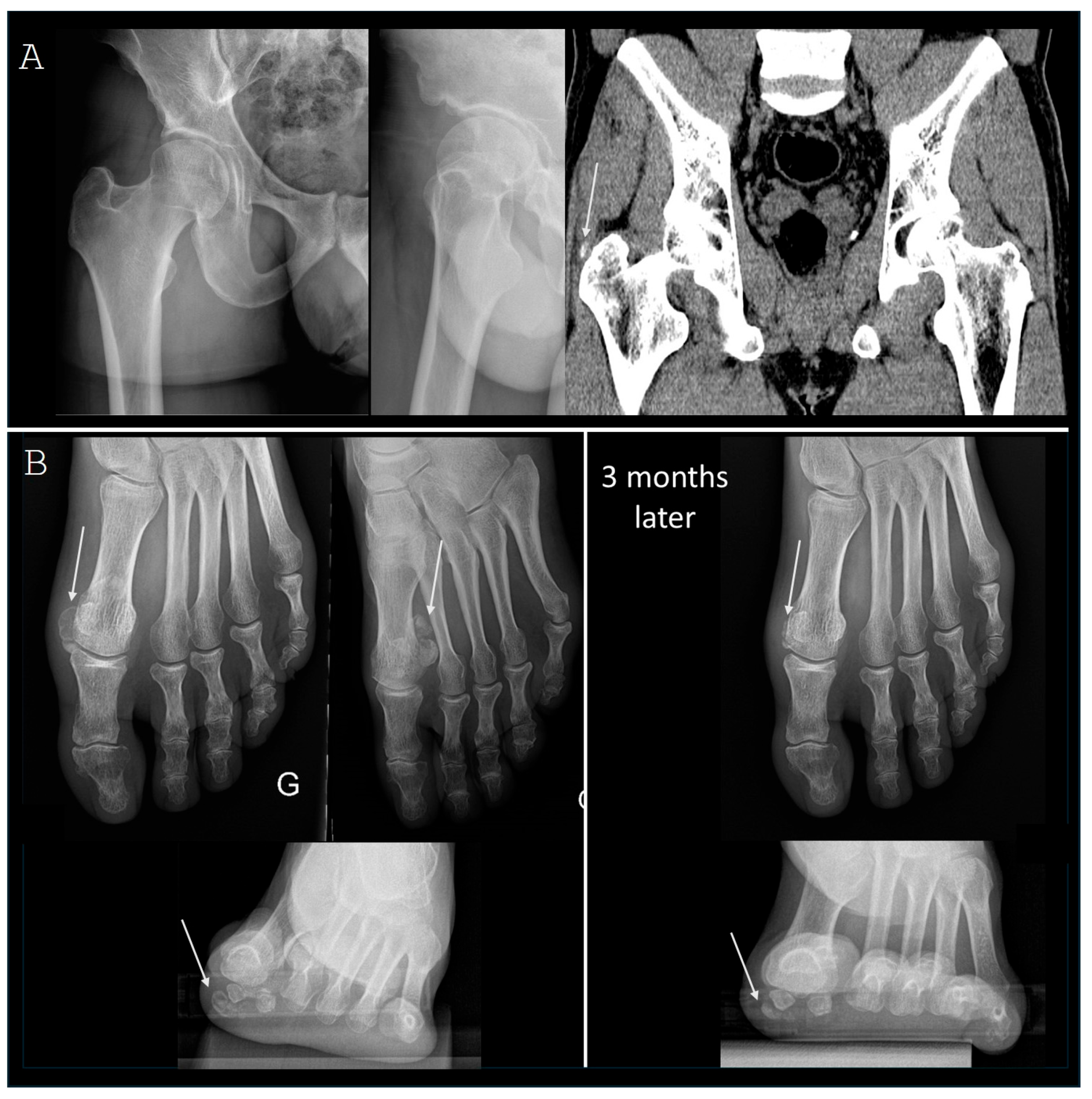
Around the hip, deposits are mainly found in the gluteus medius tendon (Figure 2A), followed by the reflected head of the rectus femoris muscle, which inserts at the superolateral aspect of the acetabulum. In Figure 2A, we report the case of a 33-year-old man suffering from severe hip pain. The examination of the hip was very painful and limited. A CT scan was finally necessary to identify the deposits located in the gluteus medius tendon. X-rays and ultrasound were considered normal. Other sites are more rarely encountered: direct head of the rectus femoris, iliopsoas, piriformis, gemellus superior and inferior or in the joint capsule [27,28]. When deposits resorption occurs within the reflected rectus femoris tendon, a coxofemoral effusion is possible, mimicking septic hip arthritis.
More rarely, calcification resorption occurs around knees, with a small number of cases reported in the literature involving the medial and lateral collateral ligaments, popliteal tendon, quadricipital tendon and biceps femoris [29].
Around the feet, periarticular deposits are common on the hallux, especially on the medial part of the first metatarsophalangeal (MTP) joint or near the lateral sesamoid bone (insertion of the flexor pollicis brevis). Calcifications may also be found at the insertion of the tibialis posterior on the navicular, in the fibularis longus or around other MTP joints or IP joints. In Figure 2B, we report the case of a 60-year-old runner consulting for pain under the first MTP for 1 month. X-rays showed a voluminous blurred and heterogeneous calcification under the medial sesamoid bone. In this case, the pain had finally improved after 3 months. Calcium deposit had decreased at this stage but was still present (Figure 2B).

3.3. Spinal Locations

The superior oblique portion of the longus colli muscle is the most commonly reported location. The muscle arises from the anterior tubercles of the transverse processes of the third, fourth and fifth cervical vertebrae and, ascending obliquely with a medial inclination, is inserted via a narrow tendon into the tubercle on the anterior arch of the atlas. On X-rays, the calcific deposit is visible in front of C1-C2. Patients suffer from neck pain and stiffness associated with odynophagia or dysphagia due to its location behind the pharynx. The differential diagnosis is retropharyngeal abscess and CT is usually required [30,31]. Cervical interspinous involvement has also been described [32]. Outside the cervical spine, apatite resorption around the coccyx is possible. In Figure 3, the X-ray and CT scan show a pericoccygeal deposit: the 25-year-old woman was suffering from acute pain in the coccyx area with nocturnal pain for two days.

3.4. Diffuse and Family Diseases

Forms with diffuse deposits around joints have been described and are responsible for recurrent painful flares [33]. Diffuse calcific periarthritis have been described in familial cases and may have reveal an underlying disease such as hypophosphatasia [34]. More recently, Cudrici et al. characterized diffuse periarticular calcifications in patients with a deficiency of CD73 [35]. Arterial calcification due to a deficiency of CD73 (ACDC) is a hereditary autosomal recessive ectopic mineralization syndrome caused by loss-of-function mutations in the ecto-50-nucleotidase gene. CD73 deficiency is thought to promote tissue calcification through a reduction in extracellular adenosine and an increase in tissue non-specific alkaline phosphatase activity. In this disease, joint involvement combines periarticular calcifications, especially around hands, intervertebral disk calcifications and mixed erosive-degenerative joint changes.

4. Treatments

In the case of resorption, nonsteroidal anti-inflammatory drugs are usually quickly effective. If they cannot be used due to contraindications (ischaemic heart disease, stomach or duodenal ulcers,...), oral steroids or local steroid injection are effective alternatives. Little data are available on IL-1 blockers to treat acute flares of calcific tendonitis: anakinra was able to quickly decrease apatite calcification-induced joint pain [36,37]. However, this treatment remains of limited use given the efficacy of other anti-inflammatory treatments.
In the case of chronic pain, as it is a self-resolving disease, conservative treatments are preferred as first-line treatments. The vast majority of studies concern rotator cuff calcifications. Treatments that have been evaluated include steroid injections, extracorporeal shockwave therapy, imaging-guided percutaneous lavage (UGPL) and surgery.
Steroid infiltration is generally given after failure of physiotherapy and symptomatic medication. In a group of 102 patients who received subacromial injection, 37% did not require further treatment during the 2-year follow-up period [38]. When steroid injection is ineffective, or only temporarily effective, extracorporeal shockwave therapy or UGPL may be considered.
High-energy extracorporeal shockwave therapy is more effective than placebo, both in terms of pain relief and functional recovery [39]. UGPL is also effective in reducing pain and calcification size. One- and two-needle techniques has been described. After local anesthesia, the lavage is performed with saline, and steroid injection is usually performed at the end of the procedure. A randomized controlled study demonstrated the benefits of this steroid injection after UGPL on pain and function and showed no negative impact on the deposit resorption [40]. Obtaining a communication between the calcification and the subacromial bursa was associated with disappearance of calcification at 3 and 12 months after UGPL [41]. After UGPL, patient follow-up is needed, as pain may flare up between 6 weeks and 3 months after the lavage. In addition, if pain persists with little changes on X-ray, a second lavage should be performed. Indeed, deposits changes on X-ray at 3 months are predictive of the clinical outcome at 12 months [42]. Finally, ultrasound features could be predictive of UGPL outcomes. Indeed, calcifications without shadowing seem to have a better prognosis [43], whereas dense calcifications with shadowing were more often associated with the need for a second lavage (15% of patients required a second procedure) [44]. To help remove hard rotator cuff deposits, sodium thiosulfate injection during UGPL was evaluated. However, adding sodium thiosulfate did not give better results than standard UGPL [45]. Although mainly performed to treat shoulder calcifications, UGPL also demonstrated good efficacy in extra-shoulder locations [46].
Steroid injection and UGPL efficacy showed rapid and similar effects in the first few weeks, but improvement over the long term (1 year) is only observed after UGPL. When the follow-up was extended to 5 years, 65% of patients who initially had a steroid injection finally required UGPL [47]. Studies comparing UGPL and high-energy extracorporeal shockwave therapy showed the efficacy of both techniques, but the superiority of UGPL in improving pain and reducing the calcification size [48].
Regarding surgical management, three options were studied: evacuation of the calcification alone, acromioplasty or both combined. Recovery and return to normal activities appear to be faster when acromioplasty was not performed [49]. To date, there is no strong evidence that surgery is superior to UGPL to improve symptoms so it looks reasonable to try UGPL first [50,51].
Finally, when patients suffer from mild to moderate symptoms, a wait-and-see strategy, i.e., not using invasive treatments aiming to remove the deposits (UGPL or arthroscopy), may be discussed since the condition ultimately resolves spontaneously. No difference in clinical outcomes were observed at 5 and 10 years in patients who underwent UGPL compared with those who did not [52]. The blurred appearance on X-rays and absence of shadowing on ultrasound were associated with favorable outcomes in patients managed with conservative therapy (physiotherapy and NSAIDs) [53].

5. Conclusions

Periarticular calcifications are a frequent condition. Despite their frequency, the mechanisms triggering tendon mineralization and the circumstances leading to resorption remain unknown. Regarding treatments, different options are available to reduce pain and improve function. As it is a self-resolving disorder, conservative treatments are preferred, and surgery should be only offered in a limited number of cases.

Author Contributions

All authors have read and agreed to the published version of the manuscript.

Funding

This research received no external funding.

Conflicts of Interest

The authors declare no conflicts of interest.

References

  1. Su, Y.C.; Chung, C.H.; Ke, M.J.; Chen, L.C.; Chien, W.C.; Wu, Y.T. Increased risk of shoulder calcific tendinopathy in diabetes mellitus: A nationwide, population-based, matched cohort study. Int. J. Clin. Pract. 2021, 75, e14549. [Google Scholar] [CrossRef] [PubMed]
  2. Harvie, P.; Pollard, T.C.; Carr, A.J. Calcific tendinitis: Natural history and association with endocrine disorders. J. Shoulder Elbow Surg. 2007, 16, 169–173. [Google Scholar] [CrossRef]
  3. Grases, F.; Muntaner-Gimbernat, L.; Vilchez-Mira, M.; Costa-Bauzá, A.; Tur, F.; Prieto, R.M.; Torrens-Mas, M.; Vega, F.G. Characterization of deposits in patients with calcific tendinopathy of the supraspinatus. Role of phytate and osteopontin. J. Orthop. Res. 2015, 33, 475–482. [Google Scholar] [CrossRef] [PubMed]
  4. Grases, F.; Prieto, R.M.; Sanchis, P.; Saus, C.; De Francisco, T. Role of phytate and osteopontin in the mechanism of soft tissue calcification. J. Nephrol. 2008, 21, 768–775. [Google Scholar]
  5. Canon, A.; Roy, L.; Chevalier, X.; Giraudier, S.; Eymard, F. Calcific tendinopathy: An unexpected side effect of tyrosine kinase inhibitor? Leuk. Lymphoma 2022, 63, 3175–3180. [Google Scholar] [CrossRef] [PubMed]
  6. Darrieutort-Laffite, C.; Arnolfo, P.; Garraud, T.; Adrait, A.; Couté, Y.; Louarn, G.; Trichet, V.; Layrolle, P.; Le Goff, B.; Blanchard, F. Rotator Cuff Tenocytes Differentiate into Hypertrophic Chondrocyte-like Cells to Produce Calcium Deposits in an Alkaline Phosphatase-Dependent Manner. J. Clin. Med. 2019, 8, 1544. [Google Scholar] [CrossRef]
  7. Uhthoff, H.K.; Loebr, J.W. Calcific tendinopathy of the rotator cuff: Pathogenesis, diagnosis, and management. J. Am. Acad. Orthop. Surg. 1997, 5, 183–191. [Google Scholar] [CrossRef]
  8. Hu, C.; Ma, L.; Gao, S.; Yang, M.Y.; Mu, M.D.; Chang, L.; Huang, P.; Ye, X.; Wang, W.; Tao, X.; et al. PPP1R3A inhibits osteogenesis and negatively regulates intracellular calcium levels in calcific tendinopathy. iScience 2023, 26, 107784. [Google Scholar] [CrossRef] [PubMed]
  9. Sougué, C.; Darrieutort-Laffite, C.; Maugars, Y.; Le Goff, B. Location of calcifications of rotator cuff on ultrasound in 74 patients: Near the junction between the supraspinatus and infraspinatus tendons in 96% of the cases. Jt. Bone Spine 2021, 88, 105–107. [Google Scholar] [CrossRef]
  10. Archer, R.S.; Bayley, J.I.; Archer, C.W.; Ali, S.Y. Cell and matrix changes associated with pathological calcification of the human rotator cuff tendons. J. Anat. 1993, 182, 1–11. [Google Scholar]
  11. Herman, J.; Le Goff, B.; De Lima, J.; Brion, R.; Chevalier, C.; Blanchard, F.; Darrieutort-Laffite, C. Pro-inflammatory effects of human apatite crystals extracted from patients suffering from calcific tendinopathy. Arthritis Res. Ther. 2021, 23, 131. [Google Scholar] [CrossRef] [PubMed]
  12. Jin, C.; Frayssinet, P.; Pelker, R.; Cwirka, D.; Hu, B.; Vignery, A.; Eisenbarth, S.C.; Flavell, R.A. NLRP3 inflammasome plays a critical role in the pathogenesis of hydroxyapatite-associated arthropathy. Proc. Natl. Acad. Sci. USA 2011, 108, 14867–14872. [Google Scholar] [CrossRef] [PubMed]
  13. Molé, D.; Kempf, J.F.; Gleyze, P.; Rio, B.; Bonnomet, F.; Walch, G. Results of endoscopic treatment of non-broken tendinopathies of the rotator cuff. 2. Calcifications of the rotator cuff. Rev. Chir. Orthop. Reparatrice Appar. Mot. 1993, 79, 532–541. [Google Scholar] [PubMed]
  14. Gärtner, J.; Heyer, A. Calcific tendinitis of the shoulder. Orthopade 1995, 24, 284–302. [Google Scholar]
  15. Maier, M.; Schmidt-Ramsin, J.; Glaser, C.; Kunz, A.; Küchenhoff, H.; Tischer, T. Intra-and interobserver reliability of classification scores in calcific tendinitis using plain radiographs and CT scans. Acta Orthop. Belg. 2008, 74, 590–595. [Google Scholar] [PubMed]
  16. De Witte, P.B.; Van Adrichem, R.A.; Selten, J.W.; Nagels, J.; Reijnierse, M.; Nelissen, R.G.H.H. Radiological and clinical predictors of long-term outcome in rotator cuff calcific tendinitis. Eur. Radiol. 2016, 26, 3401–3411. [Google Scholar] [CrossRef] [PubMed]
  17. Louwerens, J.K.; Claessen, F.M.; Sierevelt, I.N.; Eygendaal, D.; Van Noort, A.; Van Den Bekerom, M. Radiographic assessment of calcifying tendinitis of the rotator cuff: An inter- and intraobserver study. Acta Orthop. Belg. 2020, 86, 525–531. [Google Scholar]
  18. Louwerens, J.K.; Sierevelt, I.N.; van Hove, R.P.; van den Bekerom, M.P.; van Noort, A. Prevalence of calcific deposits within the rotator cuff tendons in adults with and without subacromial pain syndrome: Clinical and radiologic analysis of 1219 patients. J. Shoulder Elbow Surg. 2015, 24, 1588–1593. [Google Scholar] [CrossRef]
  19. Le Goff, B.; Berthelot, J.M.; Guillot, P.; Glémarec, J.; Maugars, Y. Assessment of calcific tendonitis of rotator cuff by ultrasonography: Comparison between symptomatic and asymptomatic shoulders. Jt. Bone Spine 2010, 77, 258–263. [Google Scholar] [CrossRef]
  20. Paruthikunnan, S.M.; Boily, M.; Martin, M.H.; Assaf, A.; Jaffer, R. Intra-osseous migration in calcific rotator cuff tendinopathy- a novel depiction of temporal evolution on multimodality imaging. BJR Case Rep. 2022, 8, 20210156. [Google Scholar] [CrossRef]
  21. Zampa, V.; Aringhieri, G.; Rossi, P.; Capanna, R.; Caramella, D. Humeral greater tuberosity osteolysis as a complication of intraosseous calcification migration: Natural history depicted by imaging. Acta Biomed. 2021, 92, e2021052. [Google Scholar]
  22. Iovane, A.; Terrasi, M.; Iovane, E.M.; Mantia, C.; Messina, G.; Mantia, F. Intramuscular migration of calcium deposits into the deltoid muscle: Two cases of a rare complication of rotator cuff calcific tendinopathy. J. Ultrasound 2023, 26, 929–933. [Google Scholar] [CrossRef] [PubMed]
  23. Cahir, J.; Saifuddin, A. Calcific tendonitis of pectoralis major: CT and MRI findings. Skeletal Radiol. 2005, 34, 234–238. [Google Scholar] [CrossRef] [PubMed]
  24. Vaishya, R.; Siddalingeshwara, G.I.; Vaishya, A. Calcific Tendonitis of the Elbow in an Adult—A Case Report and Review of the Literature. J. Orthop. Case Rep. 2020, 10, 57–60. [Google Scholar]
  25. Kim, J.K.; Park, E.S. Acute calcium deposits in the hand and wrist; comparison of acute calcium peritendinitis and acute calcium periarthritis. J. Hand Surg. (Eur. Vol.) 2014, 39, 436–439. [Google Scholar] [CrossRef]
  26. Nadarajah, C.V.; Weichert, I. Milwaukee shoulder syndrome. Case Rep. Rheumatol. 2014, 2014, 458708. [Google Scholar] [CrossRef]
  27. Park, S.M.; Baek, J.H.; Ko, Y.B.; Lee, H.J.; Park, K.J.; Ha, Y.C. Management of acute calcific tendinitis around the hip joint. Am. J. Sports Med. 2014, 42, 2659–2665. [Google Scholar] [CrossRef]
  28. Yang, W.B.; Xu, Q.K.; Liu, X.H.; Bakhshi, P.; Wang, H.; Shao, Z.W.; Meng, C.Q.; Huang, W. Arthroscopic Treatment of Calcific Tendinitis of Gemellus Superior and Gemellus Inferior: A Case Report and Literature Review. Orthop. Surg. 2022, 14, 621–627. [Google Scholar] [CrossRef]
  29. Watura, K.; Greenish, D.; Williams, M.; Webb, J. Acute calcific periarthiritis of the knee presenting with calcification within the lateral collateral ligament. BMJ Case Rep. 2015, 2015, bcr2014209041. [Google Scholar] [CrossRef]
  30. Turner, V.L.; Martinez, C.; Rocha, J.; Valenzuela, A. Acute calcific tendinitis of the longus colli: A case report. Radiol. Case Rep. 2024, 19, 2650–2653. [Google Scholar] [CrossRef] [PubMed]
  31. Reitz, I.; Allen, C.; Rappaport, D.E. An Unusual Cause of Fever, Neck Pain, and Neck Stiffness: Acute Calcific Tendinitis of the Longus Colli Muscle. J. Emerg. Med. 2023, 65, e307–e309. [Google Scholar] [CrossRef] [PubMed]
  32. Urrutia, J.; Contreras, O. Calcium hydroxyapatite crystal deposition with intraosseous penetration involving the posterior aspect of the cervical spine: A previously unreported cause of neck pain. Eur. Spine J. 2017, 26 (Suppl. S1), 53–57. [Google Scholar] [CrossRef] [PubMed]
  33. Amor, B.; Cherot, A.; Delbarre, F. Hydroxyapatite rheumatism (multiple tendon calcification disease). I.-Clinical study. Rev. Rhum. Mal. Osteoartic. 1977, 44, 301–308. [Google Scholar] [PubMed]
  34. Guanabens, N.; Mumm, S.; Moller, I.; González-Roca, E.; Peris, P.; Demertzis, J.L.; Whyte, M. Calcific periarthritis as the only clinical manifestation of hypophosphatasia in middle-aged sisters. J. Bone Miner. Res. 2014, 29, 929–934. [Google Scholar] [CrossRef]
  35. Cudrici, C.D.; Newman, K.A.; Ferrante, E.A.; Huffstutler, R.; Carney, K.; Betancourt, B.; Miettinen, M.; Siegel, R.; Katz, J.D.; Nesti, L.J.; et al. Multifocal calcific periarthritis with distinctive clinical and radiological features in patients with CD73 deficiency. Rheumatology 2021, 61, 163–173. [Google Scholar] [CrossRef] [PubMed]
  36. Zufferey, P.; So, A. A pilot study of IL-1 inhibition in acute calcific periarthritis of the shoulder. Ann. Rheum. Dis. 2013, 72, 465–467. [Google Scholar] [CrossRef] [PubMed]
  37. Zufferey, P.; Valcov, R.; Thomas, M.; Dumusc, A.; Forien, M.; So, A.; Ottaviani, S. Efficacy of anakinra in acute hydroxyapatite calcification-induced joint pain: A retrospective study of 23 cases. Jt. Bone Spine 2019, 86, 83–88. [Google Scholar] [CrossRef]
  38. Maugars, Y.; Varin, S.; Gouin, F.; Huguet, D.; Rodet, D.; Nizard, J.; N’Guyen, J.M.; Guillot, P.; Glémarec, J.; Passutti, N.; et al. Treatment of shoulder calcifications of the cuff: A controlled study. Jt. Bone Spine 2009, 76, 369–377. [Google Scholar] [CrossRef] [PubMed]
  39. Bannuru, R.R.; Flavin, N.E.; Vaysbrot, E.; Harvey, W.; McAlindon, T. High-energy extracorporeal shock-wave therapy for treating chronic calcific tendinitis of the shoulder: A systematic review. Ann. Intern. Med. 2014, 160, 542–549. [Google Scholar] [CrossRef]
  40. Darrieutort-Laffite, C.; Varin, S.; Coiffier, G.; Albert, J.D.; Planche, L.; Maugars, Y.; Cormier, G.; Le Goff, B. Are corticosteroid injections needed after needling and lavage of calcific tendinitis? Randomised, double-blind, non-inferiority trial. Ann. Rheum. Dis. 2019, 78, 837–843. [Google Scholar] [CrossRef]
  41. Dumoulin, N.; Cormier, G.; Varin, S.; Coiffier, G.; Albert, J.D.; Le Goff, B.; Darrieutort-Laffite, C. Factors associated with clinical improvement and the disappearance of calcifications after ultrasound-guided percutaneous lavage of rotator cuff calcific tendinopathy: A post hoc analysis of a randomized controlled trial. Am. J. Sports Med. 2021, 49, 883–891. [Google Scholar] [CrossRef]
  42. Dalla-Torre, R.; Fouasson-Chailloux, A.; Le Goff, B.; Darrieutort-Laffite, C. Development of a new radiographic score for the follow-up of calcific tendinopathy of the rotator cuff. Clin. Exp. Rheumatol. 2024, 42, 619–625. [Google Scholar] [CrossRef] [PubMed]
  43. Farin, P.U.; Räsänen, H.; Jaroma, H.; Harju, A. Rotator cuff calcifications: Treatment with ultrasound-guided percutaneous needle aspiration and lavage. Skeletal Radiol. 1996, 25, 551–554. [Google Scholar] [CrossRef]
  44. De Conti, G.; Marchioro, U.; Dorigo, A.; Boscolo, N.; Vio, S.; Trevisan, M.; Meneghini, A.; Baldo, V.; Angelini, F. Percutaneous ultrasound-guided treatment of shoulder tendon calcifications: Clinical and radiological follow-up at 6 months. J. Ultrasound 2010, 13, 188–198. [Google Scholar] [CrossRef] [PubMed]
  45. Darrieurtort-Laffite, C.; Bertrand-Vasseur, A.; Garraud, T.; Planche, L.; Le Goff, B. Tolerance and effect of sodium thiosulfate in calcific tendinitis of the rotator cuff. Clin. Rheumatol. 2020, 39, 561–569. [Google Scholar] [CrossRef] [PubMed]
  46. Albano, D.; Viglino, U.; Messina, C.; Fusco, S.; Gitto, S.; Lacelli, F.; Sconfienza, L.M. US-guided percutaneous irrigation of extra-shoulder calcific tendinitis. Br. J. Radiol. 2024, 97, 267–273. [Google Scholar] [CrossRef] [PubMed]
  47. de Witte, P.B.; Kolk, A.; Overes, F.; Nelissen, R.G.H.H.; Reijnierse, M. Rotator Cuff Calcific Tendinitis: Ultrasound-Guided Needling and Lavage Versus Subacromial Corticosteroids: Five-Year Outcomes of a Randomized Controlled Trial. Am. J. Sports Med. 2017, 45, 3305–3314. [Google Scholar] [CrossRef] [PubMed]
  48. Del Castillo-González, F.; Ramos-Alvarez, J.J.; Rodríguez-Fabián, G.; González-Pérez, J.; Jiménez-Herranz, E.; Varela, E. Extracorporeal shockwaves versus ultrasound-guided percutaneous lavage for the treatment of rotator cuff calcific tendinopathy: A randomized controlled trial. Eur. J. Phys. Rehabil. Med. 2016, 52, 145–151. [Google Scholar] [PubMed]
  49. Marder, R.A.; Heiden, E.A.; Kim, S. Calcific tendonitis of the shoulder: Is subacromial decompression in combination with removal of the calcific deposit beneficial? J. Shoulder Elbow Surg. 2011, 20, 955–960. [Google Scholar] [CrossRef] [PubMed]
  50. Chen, F.; Deng, Z.; Liu, Y.; Chen, R.; Chen, K.; Xu, J. Arthroscopic Surgery Versus Nonoperative Treatment for Calcific Tendinitis of the Shoulder: A Retrospective Cohort Study. Am. J. Sports Med. 2024, 52, 461–473. [Google Scholar] [CrossRef]
  51. Angileri, H.S.; Gohal, C.; Comeau-Gauthier, M.; Owen, M.M.; Shanmugaraj, A.; Terry, M.A.; Tjong, V.K.; Khan, M. Chronic calcific tendonitis of the rotator cuff: A systematic review and meta-analysis of randomized controlled trials comparing operative and nonoperative interventions. J. Shoulder Elbow Surg. 2023, 32, 1746–1760. [Google Scholar] [CrossRef] [PubMed]
  52. Serafini, G.; Sconfienza, L.M.; Lacelli, F.; Silvestri, E.; Aliprandi, A.; Sardanelli, F. Rotator cuff calcific tendonitis: Short-term and 10-year outcomes after two-needle us-guided percutaneous treatment—Nonrandomized controlled trial. Radiology 2009, 252, 157–164. [Google Scholar] [CrossRef] [PubMed]
  53. Ogon, P.; Suedkamp, N.P.; Jaeger, M.; Izadpanah, K.; Koestler, W.; Maier, D. Prognostic factors in nonoperative therapy for chronic symptomatic calcific tendinitis of the shoulder. Arthritis Rheum. 2009, 60, 2978–2984. [Google Scholar] [CrossRef] [PubMed]
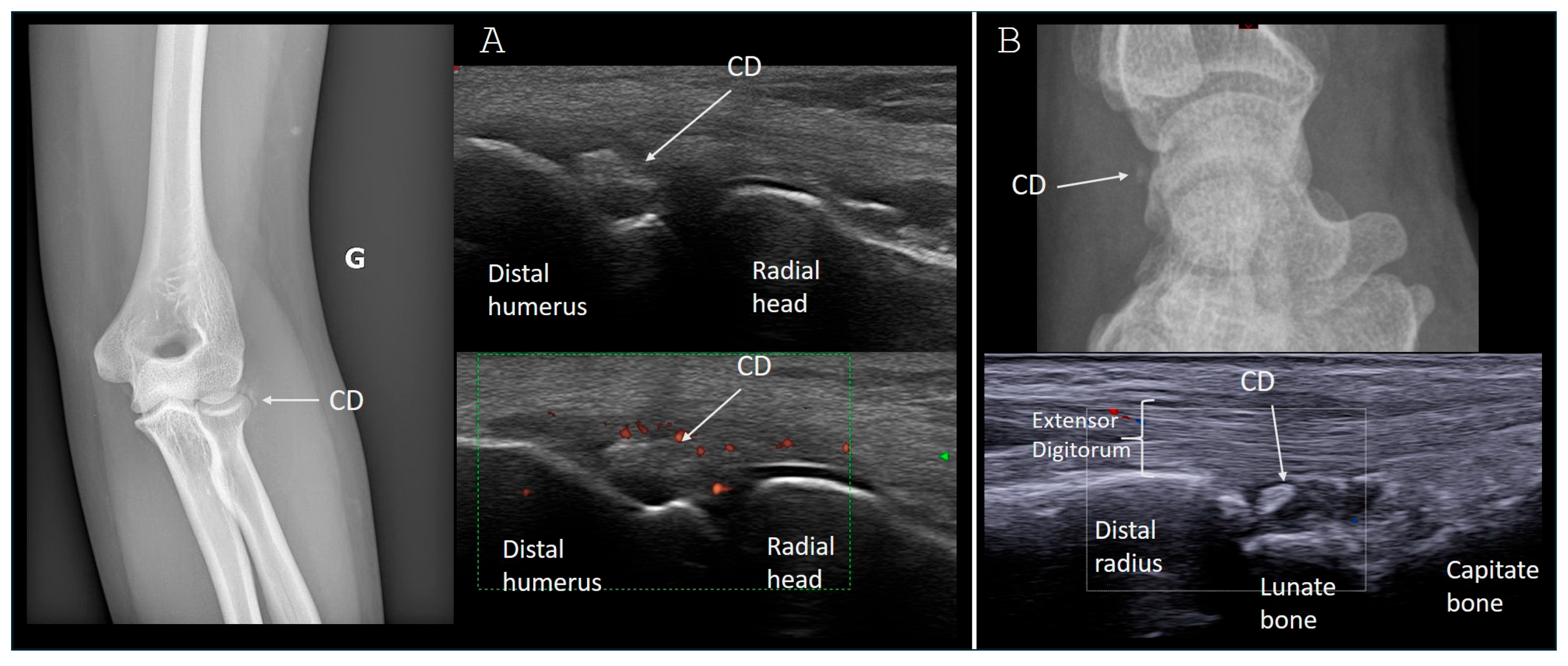
Figure 1. Upper limb examples of calcific deposits resorption on X-rays and ultrasound. (A) Calcification resorption in the wrist extensors close to the lateral epicondyle in a 20-year-old woman with acute and intense elbow pain. (B) Calcification resorption in the wrist capsule in a 45-year-old woman with acute dorsal wrist pain. CD = calcific deposit.
Gucdd 02 00020 g001
Figure 2. Lower limb examples of calcific deposits resorption on X-rays and CT. (A) Calcification resorption in the gluteus medius tendon in a 33-year-old man. The diagnosis required a CT scan as the calcific deposit was not dense enough to be visible on X-rays. (B) Calcification resorption around the medial sesamoid bone in a 60-year-old woman with acute hallux pain. The white arrows indicate the apatite deposits.
Gucdd 02 00020 g002
Figure 3. Apatite pericoccygeal resorption. A CT scan (right image) was necessary to confirm the presence of a calcification in resorption process. The patient suffered from acute pain in the coccyx area for 2 days with persistent and nocturnal symptoms. The white arrow indicates the apatite deposit.
Gucdd 02 00020 g003
Disclaimer/Publisher’s Note: The statements, opinions and data contained in all publications are solely those of the individual author(s) and contributor(s) and not of MDPI and/or the editor(s). MDPI and/or the editor(s) disclaim responsibility for any injury to people or property resulting from any ideas, methods, instructions or products referred to in the content.