Article

A Pilot Study to Investigate the Antimicrobial Activity of Pulsed UVA and UVC

Medical Technologies Innovation Facility (MTIF), Clifton Lane, Nottingham Trent University, Nottingham NG11 8NS, UK
*
Author to whom correspondence should be addressed.
Aerobiology 2023, 1(2), 82-97; https://doi.org/10.3390/aerobiology1020007
Submission received: 13 September 2023 / Revised: 3 November 2023 / Accepted: 7 November 2023 / Published: 13 November 2023

Abstract

:
UV irradiation has shown potential in reducing bacterial and viral loadings. This is a pilot study aimed at investigating the antimicrobial effect of a novel pulsed UVA and UVC technology on bacteria and human coronavirus 229E. The selection of these microorganisms is based on their relevance and significance in real-world scenarios. This study consists of independent experiments for the assessment of antibacterial and antiviral activities by using a lawn plate approach, measuring levels of adenine triphosphate (ATP) in three bacterial strains, Escherichia coli, Staphylococcus epidermidis and Bacillus subtilis, and performing Median Tissue Culture Infectious Dose (TCID50) of HCoV-229E on MRC-5 human lung fibroblast cell line. The results demonstrated the ability of UVA and UVC irradiation to reduce levels of adenine triphosphate (ATP) over a 12 h exposure period in all three bacterial strains, comparative to dark and artificial/natural light conditions using non-pulsing experiments. In addition to this, there was a reduction in colonies exposed to UVA and UVC pulsing experiments for E. coli K12 and S. epidermis compared to bacteria stored in artificial/natural and dark conditions. Furthermore, using dose-dependent modelling, it was demonstrated that the cross-contamination risk was reduced by 50% using E. coli as a typical model. Regarding the antiviral assay, the results showed that TCID50 of HCoV-229E was reduced after the first cycle of UV engagement. No cytopathic effect (CPE) was detected after three cycles using Protocol 1. The findings showed that UVA and UVC were effective under the conditions outlined in this paper for a reduction in the number of bacteria with additional applications to viruses.

1. Introduction

The feasibility of using ultraviolet (UV) light in an antimicrobial setting to reduce microbial load has been extensively explored and includes applications such as the recycling of water for food purposes [1], disinfection of surfaces in hospital rooms [2], wastewater treatment [3], reduction of pathogenic and spoilage bacteria in foods [4], and prevention of biofilm formation in urine and on urethral catheters [5]. UV irradiation has been considered for use on different surfaces, including water, air, and solids. Air is of particular interest due to the concerns of bioaerosols spreading disease, reactions, and allergies. However, microorganisms can survive on numerous surfaces and in liquids for extended periods of time [2,3]. Routes of human exposure have been thoroughly researched and include the skin, eyes, nose, and mouth [6]. Various measures have been implemented to minimise human exposure to microorganisms, encompassing a range of strategies such as elimination, substitution, engineering controls, administrative controls, safe work practices, and the use of preventative protective equipment [6]. Hence, due to microorganisms such as viruses and bacteria causing some of the aforementioned healthcare concerns, it is vital to explore methods in which bacterial and viral loadings can be reduced, and exposure to UV light has shown promise [6].
The UV–visible (UV-vis) spectrum can be further divided into ultraviolet A (UVA; 315–400 nm), ultraviolet B (UVB; 280–315 nm), ultraviolet C (UVC; 200–280 nm), and ultraviolet D (UVD; 100–200 nm). UVC is deemed one of the most effective [7] and has advantages for bacterial inactivation due to its ability to destroy the DNA and outer cells of pathogenic microorganisms. UVC light treatment at 260 nm to 265 nm has been proposed as a low-cost and maintenance approach to decontamination [4]. However, there are other wavelengths of UV that have been considered, including UVD, that have been observed to remove endotoxins [7]. It was reported elsewhere that UVA, in combination with blue light, has been used to prevent the formation of biofilms on medical devices made of silicone [5].
Indeed, there are a variety of different wavelengths in the UV range alone, which could be considered to destroy and/or reduce microorganisms in a system. Moreover, applications in the food industry have been explored by McLeod et al., whereby microbial load reduction has been monitored on chicken fillet surfaces. UVC effectively reduced bacterial loadings [4]. Herein, McLeod et al. demonstrated the use of pulsed (from 5 to 300 s) UV light to decontaminate the surface of food products; however, the investigation considers pulsing with a combination of infrared, visible, and UV light (200–1100 nm). Pulsed UV has been previously suggested as an energy- and cost-saving alternative to constant irradiation. Successful experiments have demonstrated the applicability of pulsed UVC on E. coli [8]. UVA and UVC used separately have provided variable results [9]. In an experiment using 20 min UVA pre-radiation, higher rates of activation were determined in E. coli ATCC 11229, 15597, and 700891 comparatively to solely UVA and UVC investigations [9]. Moreover, the impact of UV light irradiation is not limited to bacteria. For instance, viruses have also been irradiated with UV light [10]. Therefore, there is a need for further investigation of the effect of different types of UV exposure on microorganisms. The aim of the current research is to assess the effect of UVA and UVC lamps on human coronavirus HCoV-229E and on Gram-positive (S. epidermidis and B. subtilis) and Gram-negative (E. coli) bacteria. The selection of these microorganisms is based on their relevance and significance in real-world scenarios. HCoV-229E, a respiratory virus, is chosen due to its role in causing respiratory tract infections, contributing to the burden of respiratory diseases globally. S. epidermidis is selected as it can cause opportunistic infections, particularly in healthcare settings, and is associated with biofilm formation on medical devices, highlighting the need for effective prevention and management strategies. B. subtilis, though generally non-pathogenic, is included as a model organism with potential applications in sterilisation and decontamination studies, particularly due to its spore-forming ability. Finally, E. coli is chosen due to its versatility and relevance in both clinical and environmental contexts, including its association with various infections, foodborne illnesses, and antimicrobial resistance. To decrease risks associated with the experiments, the UV lamp was placed in a sealed lightbox [11]. The findings of this study have a significant potential impact, particularly in the context of preventive measures for facilities. Optimising the use of pulsed UVA and UVC irradiation techniques can lead to more efficient and cost-effective decontamination practices. This can have broad implications in various industries, healthcare settings, and public facilities where controlling microbial contamination is crucial for preventing infections and ensuring a safe environment.
This is an evaluation of an invention by Helios Shield Limited [12,13]. The technology consisted of two UVA and UVC lights that pulse at different powers. The schematic images of the setup are shown in Figure 1 and Figure 2. The methodology and results of the antibacterial assessment were reported in the patent elsewhere [12]. The results of the TCID50 (Tissue Culture Infectious Dose) assay provided in this paper were also mentioned in the second patent [13].

2. Materials and Methods

2.1. Safety Consideration

Safe use of the lamp was considered prior to commencing experiments. To decrease risks associated with the experiments, the UV lamp was placed in a sealed lightbox [11]. Eyes were covered with a UV protective shield, and a lab coat was always worn to cover the skin during the experiments. Experiments involving HCoV-229E were carried out in a level 2 safety hood. Labelling of the UV lightbox was in place.

2.2. Materials

For the antibacterial assay, Oxoid Ltd. (Basingstoke, Hampshire, UK) was the supplier of maximum recovery diluent, nutrient agar, Tryptone soya broth, and Violet-Red Bile Glucose agar. Single vent Petri dishes and UltraSnap Adenosine triphosphate surface tests were obtained from Scientific Lab Supplies Ltd. (Nottingham, UK) and Hygiena International Ltd. (Watford, UK), respectively [12]. Blades Biological Ltd. (East Sussex, UK) was used as the supplier for Escherichia coli K12 (E. coli K12), Bacillus subtilis (B. subtilis), and Staphylococcus epidermidis (S. epidermidis). To protect the operator from UVA and UVC exposure, the equipment was placed into a Syngene Bioimaging lightbox (Figure 1). ATP readings were performed using a Hygiena luminometer. Eclipse 17 was the autoclave used. A Genlab incubator was used for incubation of microorganisms at 37 °C.
For the antiviral assessment, American Type Culture Collection (ATCC) was the supplier of MRC-5 human fibroblast cells (ATCC® CCL-171™), HCoV-229E (ATCC VR-740), and Eagle’s Minimum Essential Medium ((EMEM) ATCC® 30-2003™) [13]. EMEM included L-glutamine and glucose. Merck Life Science UK Limited was a supplier of surface cell culture flasks (CellBIND, 25 cm2) and trypsin-EDTA sterile-filtered solution (0.25%). Moreover, dimethyl sulfoxide (DMSO) was purchased from ChemCruz. Dulbecco’s phosphate-buffered saline (DPBS) was obtained from Merck Life Science UK Limited. The UVA and UVC light equipment was provided by Helios Shield Limited and tested for inactivation of HCoV-229E and bacteria.

2.3. Antibacterial Assessment of UVA and UVC Exposure

This study was focused on assessing antibacterial activity of UVA and UVC lamps.

2.3.1. Microorganisms and Culture Methods

For the antibacterial essay, B. subtilis, S. epidermidis, and E. coli K12 were used [12]. The preparation of Violet-Red Bile agar and nutrient agar was performed according to the manufacturer’s protocol prior to sterilisation at 121 °C and 110.4 kPa for an hour-long period. These agar preparations were placed into single-vent Petri dishes, after which they were allowed to dry and set. Both agar preparations were then stored at 4 °C until use. Tryptone soya broth was used to grow B. subtilis and S. epidermidis. Both organisms underwent an incubation at 37 °C for 12 h. The streaking method on Violet-Red Bile Glucose was used to validate E. coli K12. Nutrient agar was employed to validate B. subtilis and S. epidermidis. A dilution of 1:10,000 of E. coli K12, B. subtilis, and S. epidermidis in previously autoclaved maximal recovery diluent was performed. The organisms were then counted using a lawn plate approach. Subsequently, under sterile conditions, 10 µL of E. coli K12 were pipetted onto various locations across the three nutrient agar plates. Afterwards, the plates were stored for different lengths of time under dark, natural light, and UV light conditions. Detailed information regarding the exposure of the plates to UV light can be found in Protocol I and Protocol II (Section 2.3.2 and Section 2.3.3). The plates were incubated at 37 °C for 24 h following the exposure to UV light. The resulting number of colonies was quantified. ATP measurements were performed when bacteria were too numerous to count (TNTC). Protocols for the antibacterial assessment are named Protocols I and II.

2.3.2. Protocol I

Different power levels (42 mW, 65 mW, and 117 mW) of UVA and UVC were continually applied simultaneously to the agar plates during a period of 720 min. All microorganisms in the investigation, E. coli K12, B. subtilis, and S. epidermidis, underwent the same treatment and were subsequently grown for 24 h before data acquisition.

2.3.3. Protocol II

Power levels of 42 mW and 62 mW were used to pulse UVA and UVC light, respectively. The bacteria (E. coli and S. epidermidis) underwent 10 to 13 cycles of between 270 and 399 min, respectively, of, firstly, engaging UVC for 3 min and then disengaging, followed by 30 min of UVA engagement and subsequent disengagement [12]. The E. coli K12 and S. epidermidis strains were then cultured for 24 h, and data were collected.

2.3.4. ATP Measurement

Where bacterial colonies were TNTC or the agar plates showed no visible colonies, ATP measurements were performed. The UltraSnap swabs, which are stored at 21 °C, were equalised to room temperature. The Petri dish surface area on which the bacteria had been pipetted was thoroughly swabbed. The swab was placed back into the tube prior to the addition of liquid-stable reagent included with the UltraSnap swab. The liquid-stable reagent optimises sample recovery and facilitates bioluminescence, providing more reliable results with superior sensitivity (0.001 fmol) [12]. ATP levels within the UltraSnap tube were detected in 30 s with the Hygiena luminometer employing a novel solid-state photodiode, which allows for the quantification and detection of low levels of light. The light emitted is directly proportionate to the ATP levels of the sample. A reading below 10 relative luminescence units (RLU) is considered clean. Readings within the range of 11–29 RLU are interpreted as a warning. Lastly, readings that exceed 30 RLU are characteristic of a dirty surface. The detection of ATP levels to determine the cleanliness of surfaces is a widely used method across healthcare and food manufacturing settings.
The equipment setup, which varied for the experiments on bacteria and coronavirus, is schematically represented in Figure 1 and Figure 2, respectively.
A distance of 32 cm between the lamp and the Petri dish was established across all experiments unless stated otherwise. The UVA and UVC light exposures were controlled through the use of dials, which allowed for 16 different settings. The enclosing box was maintained at a room temperature fluctuating between 18 and 25 °C. To ensure full exposure of the Petri dish to the light, the wire of the lamp was secured around the clamp. The experimental design was adapted from Bolton and Linden [11].

2.4. Antiviral Assessment of UVA and UVC Exposure

The next set of experiments was focused on antiviral assessment of UVA and UVC using different protocols (see below) [13]. TCID50 was performed after 1, 3, 5, 7, 9, and 11 cycles of UV exposure in order to investigate any decrease in viral infectivity. MRC-5 human lung cells and HCoV-229E were used for the assay [13]. The MRC-5 cells underwent four processing stages to prepare them for experimentation: maintenance, reviving from frozen, passaging of cells, and infecting with HCoV-229E. Prior to use, fresh EMEM was modified by adding FCS and penicillin–streptomycin antibiotics to a final concentration of 10% and 1%, respectively. Thawed MRC-5 cells were diluted to 1 in 6 with prepared EMEM and subsequently centrifuged (1200 rpm for 5 min). After centrifugation, cell pellets were mixed with EMEM and incubated at 37 °C for up to 72 h.

2.4.1. Infecting with HCoV-229E

MRC-5 were incubated in 24-well plates 48 h prior to use. The purchased stock of HCoV-229E (100 µL) was serially diluted to 10−9 in EMEM [13]. When the cells were about 50% confluent, the medium was replaced with prepared dilutions of HCoV-229E.
Once infected, the cells were exposed to UV light, as shown in Figure 2. The details of different settings are provided below. Protocols for the antiviral assessment are named Protocols 1, 2, and 2a [13].

2.4.2. Protocol 1

The duration of a single cycle was 8 h. Firstly, UVC at the rotary position “F” (power level of 236 mW) was pulsed for 6 s. After that, UVA (rotary position “7”, which has power of 74 mW) was immediately engaged for 6.5 h. The blanking interval was 1.5 h, and the light was off. This process was carried out for up to 11 cycles. The effect of UV on a virus was analysed after 1, 3, 5, 7, 9, and 11 cycles. The control was named “cycle 0”, where cells were infected but not treated with UV light.

2.4.3. Protocol 2

UVC was pulsed for 6 s at the position “F” (power of 236 mW) [13]. After that, UVA was immediately engaged for 8 h at the position “F” (power of 147 mW). No blanking time was added for this setup. The cycles were repeated up to 11 times. The control was named “cycle 0”, where cells were infected but not treated with UV light.

2.4.4. Protocol 2a

UVC was pulsed at the rotary position “F” (power of 236 mW) for 20 s. After that, UVA was switched on for 8 h at the position “F” (power of 147 mW) [13]. No blacking time was added for this setup. The cycles were repeated up to 11 times. The effect of UV was analysed after 1, 3, 5, 7, 9, and 11 cycles. The control was named “cycle 0”, where cells were infected but not treated with UV light.

2.4.5. Detection of Viral Infectivity

After either 1, 3, 5, 7, 9, and 11 cycles or 1, 2, 3, and 4 cycles, the suspension with any released viral particles, infected cells, and EMEM from each well was transferred to a cryotube and underwent up to three rapid cycles of freeze and thaw [13]. Each freeze and thaw cycle consists of storage of the suspension for an hour at −80 °C and thawing at room temperature [14]. Subsequently, the suspension was centrifuged for 10 min at 2000 rpm. Using a 0.45 µm pore size filter, the supernatant was then filter-sterilised and stored at −80 °C for the tissue culture infectious dose (TCID50) assay.

2.4.6. Analysis of Viral Infectivity by Using the TCID50 Assay

HCoV-229E, both untreated and treated with UV for up to 11 cycles using Protocols 1, 2, and 2a, was serially diluted (10-fold) in EMEM until 10−8.
MRC-5 cells were incubated in either a 96-well plate or 24-well plate. When the confluence of the cells reached approximately 50%, EMEM was replaced with serially diluted HCoV-229E in five repeated wells. The plates were incubated for up to 4 days until cytopathic effect (CPE) was detected. Additionally, cells were infected with serially diluted not-treated virus HCoV-229E to obtain a control for tissue culture infectious dose (TCID50), which will be further called 0 cycles. TCID50 was calculated by using the Reed and Muench method [15].

2.4.7. Modelling for Cross-Contamination Risks

The exponential model was used to calculate the cross-contamination risk. Such a model represents a “dose-response” relationship between a dose applied to hosts (in this case, cells) and the probability of the cells (a host) to respond [16].
The following equation was used to calculate cross-contamination:
p i = 1 e x p d k
where p represents the risk of contamination, k is probability of a cell to survive, and d represents the dose of the cells were administered [16]. After the calculations, the risk contamination was presented in percentages. Such percentage was subtracted from 100%.

3. Results

3.1. Effect of UVA and UVC Light on Bacteria

Both Protocols I and II were used to track the survival of E. coli K12, S. epidermidis, and B. subtilis at different time intervals.

3.1.1. Protocol I

The E. coli, S. epidermidis, and B. subtilis present on the plates exposed to natural and dark light showed bacterial growth that exceeded the “too numerous to count” (TNTC) limit. Additionally, no visible bacteria were observed with the naked eye under the UV light condition for all the tested strains. Consequently, to evaluate the presence of any remaining bacteria on the surface and to check for contamination, ATP measurements were carried out, and the results are presented in Table 1 [12].
The findings indicate that using UVA and UVC light in combinations, with power levels of 42 mW, 117 mW, and 65 mW, is equally effective in eradicating bacteria, as shown in the combinations specified in Table 1. The differences observed in the levels of bacteria under natural and dark light are attributed to daily fluctuations in temperature and exposure to natural light. To summarise, the results show that a 12 h exposure to any of the UVC and UVA power levels mentioned leads to a near 100% reduction in ATP and falls below the clean limit of 10 RLU.

3.1.2. Protocol II

Distance Measurements

After 13 repetitions of the above-mentioned protocol, there was an observed reduction in CFU of 32 ± 3% (average ± standard error of the mean (SEM)) for bacteria exposed to UV compared to those stored under natural light at a distance of 32 cm (Table 2 and Figure 3). The total exposure time to UVC was 39 min, and the total exposure time to UVA was 360 min, and these values may vary with different intensities. Additionally, using E. coli K12 as the model organism and analysing n = 36 readings, a 35 ± 8% (average ± SEM) reduction in CFU was noted for bacteria exposed to UV compared to those stored in the dark at a distance of 32 cm (Table 2 and Figure 3). Similarly, S. epidermidis showed comparable results with a reduction of 36 ± 2% (average ± SEM) in CFU for bacteria exposed to UV compared to those stored in the dark at 32 cm, utilising the same power levels. Using a total of n = 86 readings, a 34 ± 5% (average ± SEM) reduction in CFU was seen for S. epidermidis exposed to UV compared to those stored under natural light conditions (Table 2). These observations indicate that the bacteria responded similarly to different lighting conditions, which is consistent with the results of Protocol I.
In addition, the influence of distance on bacterial growth was investigated using E. coli K12 as a representative model organism, and it was found that the percentage of growth was affected by the distance between the lamp and the Petri dish, as illustrated in Figure 3. The results were obtained from a total of n = 207 measurements [12]. As the lamp was moved closer to the Petri dish, a more substantial reduction in growth was observed (Figure 3). However, there was some overlap between readings for closer distances, such as from 26 cm to 22 cm, as indicated by an average SEM level of 4%. Figure 3 displays a negative linear relationship, indicating that the closer the lamp is to the Petri dish, the more significant the reduction in growth. The R2 value for the light conditions (blue) shows a trend of R2 = 0.9993, implying a strong correlation between these data points. These findings suggest that the distance from the lamp plays a critical role in determining bacterial growth.
The above-mentioned findings correlate with the established literature, whereby an increase in power decreases the number of bacteria present [1]. Moreover, the number of bacteria present also correlated with findings by Chatzisymeon (2016), whereby the higher the initial concentration of bacteria, the less effective the UV pulsing methodology.

Iteration Measurements

Additionally, by using 10 iterations of the aforementioned protocol, a 6% decrease in bacterial load was observed when comparing bacteria exposed to UV light with those in the dark. E. coli K12 was used as the representative model organism with n = 10 readings. This indicates that there are observable differences even with less exposure time to the UV light using this particular pulse sequence and power levels. Further experimentation could be conducted to determine the ideal number of iterations, potentially between 10 and 13 iterations.

3.1.3. Response Modelling for Cross-Contamination Risks

Having obtained the data mentioned above, models can be utilised to evaluate the risks of cross-contamination. The exponential model discussed previously was employed to examine the effect of pulsed UV at a distance of 32 cm with 39 iterations. By utilising E. coli K12, which provides a k value of 9.7 × 10⁻⁹ [17], it was found that the pulsing program with UVA and UVC leads to a 50% reduction in cross-contamination risk for E. coli K12 [12].

3.2. Effect of UVA and UVC Exposure on Human Coronavirus

The effect of UVA and UVC on HCoV-229E was performed as described in Section 2.4 [13]. In order to find out the point where the virus loses its infectivity, the experiments were repeated for one, two, three, and four cycles using Protocols 1 and 2a. However, such experiments were performed once, and the data obtained are described in the Discussion section.

3.2.1. Investigation of Reduction in Viral Infectivity

As described above, UVC and UVA were used to treat MRC-5 cells infected with HCoV-229E [13]. Figure 4 represents the effect of different protocols on TCID50 cells after one cycle in 24-well plates. As shown in Figure 4, different experimental settings did not cause full viral inactivation after a single cycle. CPE was still observed in 24-well plates and was similar to 96-well plates, with results reported below.

3.2.2. Effect of Protocol 1 on HCoV-229E

Following Protocol 1, the TCID50 assay was used to detect the infectivity of HCoV-229E. Figure 5 represents the effect of UV treatment on viral activity [13]. As shown in Figure 5, TCID50 was reduced from 5.1 log to 2.5 log TCID50 after the 1st cycle, but it was still possible to detect CPE in cells. However, released viral particles treated for three cycles did not infect MRC-5 cells. In addition, no CPE was observed after three cycles. Further tests were carried out to find out any viral activity after the second cycle (Section 4).

3.2.3. Effect of Protocol 2a on HCoV-229E

After treating the cells infected with HCoV-229E with Protocol 2a, the TCID50 assay was performed to discover the effect of this UV setting on the virus [13]. Figure 6 represents the results obtained after 11 cycles. As shown in Figure 6, there was a decrease in TCID50 from 6.1 log to 1.6 log and 1.4 log TCID50 after the first and the third cycles, respectively. There was no CPE detected after five cycles.
In addition, HCoV-229E was treated with Protocol 2 for up to 11 cycles in a single experiment [13]. The preliminary results with untreated virus showed the control was 7.57 log TCID50. Then, after one and three cycles, TCID50 was reduced to 2.34 log and 1.16 log, respectively. No CPE was observed after five cycles. However, additional experiment is needed with this setting.

4. Discussion

According to the results obtained in Section 3.2., Protocols 1 and 2a could be most effective for inactivation of the virus within the current settings. Therefore, there was an additional set of experiments focused on these two protocols. The protocols were repeated, but data were collected after one, two, three, and four cycles. For such experiments, the treated virus underwent three rapid freeze and thaw cycles. Regarding Protocol 1, the preliminary results showed that TCID50 was reduced from 4 log to 3.2 log after the first cycle. After two cycles, the TCID50 was 2.16 log. There was no CPE detected after three and four cycles. This confirms the results shown in Section 3.2.2. Furthermore, Protocol 2a was used for the additional repeated experiments, where data were collected after one, two, three, and four cycles. The preliminary findings showed that TCID50 did not change dramatically after one cycle, and it was slightly over than the control. This might be due to the virus still infecting the cells. These results are different than those presented in Section 3.2.3. A possible reason for such a difference could be that additional freeze and thaw cycles were helpful for isolating more viral particles. However, TCID50 decreased from 4.34 log to 3.25 log and 1.5 log after two and three cycles, respectively. No CPE was observed after four cycles. The effect of Protocols 1 and 2a after four cycles on the virus can be compared. No CPE was detected after three cycles of using Protocol 1. This means that Protocol 1 might be the most successful setting in order to inactivate HCoV-229E after three cycles.
It was reported that different sources of UV, namely UVA, UVC, and UVD, provided different levels of inactivation of aerosolised E. coli [7]. The highest efficiency of inactivation was observed with UVD, and the lowest with UVA [7]. However, this investigation also showed the effectiveness of UVC radiation [1,4,7]. It was investigated elsewhere that the UVC irradiation at 254 nm using a bacterial suspension of E. coli in water at different power levels: 11 W, 9 W, and 5 W [1]. It was observed that UVC treatment inactivated the bacteria E. coli (106 CFU mL−1) in the water within the first 15 s of treatment using 11 W UVC [1]. An increase in the initial bacterial concentration and water turbidity reduced the disinfection efficiency, whilst the rise in bacterial inactivation rapidly increased with the increased power levels [1].
Extrapolation of these data is possible in some instances where bacteria are not UV-resistant. The reason for this is that nucleic acids and proteins, which are UV-absorbing agents, are present in high quantities in all biological cells [18]. Cells are susceptible to UV radiation [18]. However, some bacteria do contain UV-absorbing pigments; these include scytonemin, mycosporine-like amino acids, parietin flavonoids, and melanin [18]. These are predominantly present in cyanobacteria, phytoplankton, macroalgae, and some lichens [18]. However, melanin is present in animal and human cells [18].
The most common mutagenic DNA lesions induced by UV radiation are cyclobutene pyrimidine dimers (CPDs) and 6-4 photo products (6-4PP)—also pyrimidine adducts and their Dewar valence isomers [18]. The degradation of 6-4PP is more toxic and lethal compared to CPDs. Dewar isomerism of 6-4PP occurs on applications longer than 290 nm, but isomerism has been found to be most efficient around 320 nm [18]. The DNA damage products from CPDs and 6-4PP make up around 75% and 25%, respectively, of the total distortion in UV-induced mutagens, resulting in bends of DNA 7-9° and 44°, respectively [18]. The damage of CPDs, if unrepaired, is catastrophic; this leads to a misreading of the genetic code, causing mutations and cell death [18]. A single damaged CPD is sufficient to eliminate the expression of a transcriptional unit [18]. Hence, this demonstrates the power of UV radiation on cells in bacteria.
However, it is important to note that the period of exposure to UV must be significant in impacting specific areas of DNA to cause mutations and death. Repair mechanisms are in place for the cell to heal. For these to occur, extreme accuracy in the replication of the DNA and/or the ability of the cell to survive spontaneous and induced DNA damage is required [18]. The repair strategies a cell can undergo include base excision repair, nucleotide excision repair, excision repair, mutagenic repair/lesion bypass, recombinational repair, and photoreactivation. This is applicable to the research results attained above because it was observed that after 12 h exposure to UV light, there was no replication of bacteria because they were beyond repair. Bacteria were present in some of the experiments; however, they were not in the same abundance, which suggests that some of the bacteria may have repaired and reproduced following exposure to UV light. Nevertheless, the number of bacteria had reduced in the shorter exposure periods, showing that the UV light was causing cell death.
Regarding viruses, genomic damage was observed in MS2 bacteriophage, whereas loss of infectivity due to DNA damage has been detected at wavelengths above 240 nm [19]. Moreover, the inactivation of the Tulane virus was detected at 220 nm and 254 nm [19]. In addition, it has been found that UVA led to a reduction in the spike protein of HCoV-229E [18]. RNA and DNA absorb UVC light and UVA light; however, the effect could be different [10]. UVA absorbed by viruses may cause genetic damage as oxygen-reactive species are produced, which causes strand breakages and oxidation [10]. However, UVC can cause photochemical fusion of two adjacent pyrimidines, which then become non-pairing bases [10]. It has been demonstrated that UVC inactivates the SARS-CoV virus completely after 15 min of exposure; however, UVA demonstrated no significant effects on the virus over the same period [10]. This experiment was performed using a 3 cm distance, but the investigation did not consider a pulsed UV light strategy.
HCoV-229E, HCoV-HKU1, HCoV-OC43, HCoV-NL63, Middle East respiratory syndrome coronavirus (MERS-CoV), SARS-CoV-1, and SARS-CoV-2 can affect humans [20]. The initial strain of human coronavirus, known as B814, was obtained from a person with a regular cold in 1965 through analysis of their nasal discharge [20]. After the initial discovery of the first human coronavirus (HCoV) strain called B814, over 30 additional strains have been identified, including HCoV-229E, which was named after a student specimen coded 229E [20]. Among these seven coronaviruses, the four HCoVs can affect the mild upper respiratory tract. SARS-CoV-1, MERS-CoV, and SARS-CoV-2 could be highly pathogenic and lead to lower respiratory tract illness [20,21,22].
Respiratory diseases that are caused by coronaviruses are a major worldwide concern. Viral particles can be spread through air droplets or via contaminated surfaces. It was reported that HCoV-229E might remain infectious on non-biocidal materials such as glass, polytetrafluoroethylene, polyvinyl chloride, ceramic tiles, silicone rubber, and stainless steel [23,24]. In a human lung cell culture, for instance, the MRC-5 cell line, HCoV-229E, may be infectious for at least 5 days [23]. Regarding other coronaviruses, SARS-CoV-2 may remain infectious for 3–4 days at room temperature on surfaces such as plastic, whilst SARS-CoV-1 could survive on a polystyrene Petri dish at room temperature for at least 6 days; however, it was reported to lose activity after 9 days [24]. The ability of viruses to still infect depends not only on a type of surface but also on parameters such as humidity and temperature. It was reported that HCoV-229E and MERS-CoV have a shorter ability to survive on plastic surfaces at room temperature compared to SARS-CoV-2 and SARS-CoV-1 [24]. In order to stop the spread of diseases associated with coronaviruses, surfaces, especially those that are commonly touched, need to be decontaminated. UV lamps could provide a solution for this. Coronavirus could show different levels of sensitivity to UV. For example, far-UVC may inactivate 90%, 95%, and 99% of the HCoV-OC43 virus in 8, 11, and 16 min, respectively [25]. It was discovered that engagement of UVC (1048 mJ/cm2) for 9 min led to the inactivation of SARS-CoV-2 (5 × 10⁶ TCID50/mL) [26]. Furthermore, 1 and 3 mJ/cm2 UVC (222 nm) can result in 88.5% and 99.7% of SARS-CoV-2 viral reduction, respectively [27]. In addition, the research with (540 µW/cm2 at a distance of 3 cm) showed a slight inactivation of SARS-CoV-2 after 15 min; however, UVC (1940 µW/cm2) was reported to decrease infectious virus 400-fold after 6 min [10,26]. A one-log reduction of SARS-CoV-2 was observed with UVA (365 nm) exposure after 9 min [28]. It was observed that spike proteins of single-stranded RNA viruses were significantly affected by UVA, but there was no major damage reported to human cells [29].
A difference in UVC and UVA effects on viruses may be explained by the mechanisms of light absorption. For instance, UVC and UVB can be strongly absorbed by DNA and RNA compared to UVA and, therefore, may be more effective in inducing pyrimidine dimers [10,29]. As mentioned above, UVA may lead to additional genetic damage through the generation of reactive oxygen species [10,29].
Considering the antimicrobial activity of UV, it has found a wide range of applications [1,2,3,4,5,30,31,32,33,34]. Compared to other sterilising techniques, such as heat, filter sterilisation, and chemical sterilisation, UV irradiation has advantages and limitations. The limitation of UV includes the possibility of harmful exposure, the difficulty of disinfecting turbid solutions, and dependency on time and wavelengths. However, UV has advantages over other antimicrobial agents due to the lack of chemicals required to reduce pathogens as well as the ability to be used for different surfaces. By implementing relevant measures to reduce the risks associated with UV light, it can be applied as an effective tool for ensuring microbiology safety in the air and on surfaces. For instance, UVC was a part of the Nanoclave Cabinet used against Adenovirus, Clostridium difficile spores, and other microorganisms, including methicillin-sensitive Staphylococcus aureus, methicillin-resistant Staphylococcus aureus, Enterococcus hirae, E. coli, multi-resistant Acinetobacter baumannii, Pseudomonas aeruginosa, etc. [32]. Moreover, how UV disinfection was used to ensure the safety of fresh produce was reviewed [33]. Furthermore, UVC technology was successfully applied to inactivate bacteria, Aspergillus spores, and coronavirus that were dried on different surfaces [34]. This pilot study has also shown that UVA and UVC could be implemented as an antimicrobial technique against bacteria and HCoV-229E.
Hence, to address the above-mentioned points, it could be concluded that any new source of UV light equipment requires a rigorous pilot experiment using a developed standardised protocol. Therefore, the limitations of this study need to be considered. In the second protocol used for the experiments in bacteria, a small delay (seconds) from changing between UVA and UVC light exposure occurs using this pulsing method. Hence, the bacteria are not continuously exposed to the UVA and UVC light. It was approximated that it would take a second to change from UVA to UVC light in the current experimental design. For 13 iterations, this would involve 26 changes. Therefore, over a 360 min period, it was estimated that the bacteria were not exposed to UV light for approximately 26 s. Moreover, the fewer iterations, the fewer interruptions between pulses. The interval between changing from UVA to UVC could be reduced and may impact the results acquired and cause a further reduction in bacteria. This would enable more accurate results and repeatable dosage, which can be set on a timer to improve reproducibility.
Furthermore, environmental conditions impact the bacterial growth. The bacteria were cultured on nutrient agar; however, different results may be obtained if bacteria strains were grown on metal, plastic, skin, plants, meat, glass, paper, liquids, and cardboard surfaces, for example. UV irradiation might have limitations in inactivating pathogens in turbid liquid environments. However, it was found elsewhere that UVC irradiation (irradiation dose of 40 mJ/cm2) was able to affect the growth of Shigella flexneri and Listeria monocytogenes in drinking water [35]. The agar is nutrient-rich and, therefore, bacteria have the required medium for growth and conditions at 37 °C. The temperature was not maintained throughout the experiment, which can impact the number of bacteria. Room temperature was between 18 °C and 25 °C. However, this is relative, as the temperature would be the same in all three conditions due to the bacteria being in the same space within the room. Additionally, bacteria were incubated at 37 °C in a dark environment to speed up the growth of bacteria to be counted.
Quality assurance and quality control of the lamp was not performed. Age can impact the lamp, and verification of spectral distribution can be performed to assess the irradiance [11]. UV fluence and exposure intensity can be documented and monitored; however, this was not performed as part of this investigation.
The bacteria maintained in the natural light were kept on the laboratory bench in a combination of natural and artificial lighting. The natural light may have varied in intensity slightly each day, also giving some variance in the results acquired. Additionally, the overnight experiments had no natural light present, only artificial lighting. This may impact the results acquired if bacteria were left completely in light and natural light settings. Moreover, the positioning of the plate was kept constant by marking the area where the plate was positioned. This was carried out to ensure thermal stability. In these experiments, the lens was used. However, without a lens, different results could have been observed. The same clear Petri dishes were used for the duration of the experiment. The Petri factor was not applied herein but could be considered in future experiments.
Regarding antiviral assessment, there are several considerations that need to be addressed. Plaque assay and multiplicity of infection could be performed and calculated in order to assess the effect of UV on the virus and compare it with the current method used. Furthermore, there might be an alternative method applied in order to release the virus from the cells. Although the freezing and thawing method is currently proposed as a technique to release viral particles, the virus was thawed at room temperature. This could be improved by thawing it on ice. A lysis solution could be more effective in breaking cell structures than freezing–thawing cycles. Moreover, although an increased number of freezing and thawing cycles could be more effective in viral release, it can also decrease viral infectivity. Therefore, this research uses a minimal number of freezing and thawing cycles. For future studies, it might be beneficial to study the effect of UVA and UVC on different viral strains. Furthermore, in order to fully assess the damaging effect of UV on the virus, genetic analysis of the treated virus should be performed, such as RT-PCR. Future studies should be focused on the detection of damaged proteins on the surface of coronavirus. As spike proteins are responsible for the attachment of the virus to the cellular membrane, the damage of such proteins might indicate a lack of viral infectivity. Forward and reverse primers specific for the detection of spike proteins of the virus could be used for analysis of any RNA damage using RT-PCR. In addition, the long-term effect of UV light on a variety of different surfaces and materials needs to be studied. This technology needs to be further tested on other viruses mentioned above. The duration of inactivation is longer compared to other studies. This might be because the current UV technology was used to inactivate viruses within a liquid environment. Therefore, it is crucial to perform antiviral tests with the lamps using different surfaces as well. Furthermore, protecting mechanisms against UV exposure need to be considered. As UV can pose health risks, other shielding methods rather than a box could be explored. For instance, it was found elsewhere that ZnO films can be a promising solution against UV exposure [36].
Considering the points discussed above, UV technology could prevent the spread of pathogens by inhibiting their growth, which, therefore, could have potential in cost reduction and efficacy. Future work is needed to understand the mechanisms of such inhibition and highlight its applications for reducing the growth of other pathogens.

5. Conclusions

This study demonstrates that a pulsed sequence of UVA and UVC can effectively decrease the microbial load of various strains, including B. subtilis, E. coli K12, and S. epidermidis, as well as HCoV-229E. When using different power level combinations (65 mW UVC, 42 mW UVA, and 117 mW UVC) for a 12 h exposure time, a decrease in ATP was observed for all strains. Additionally, experiments using 42 mW UVA and 65 mW UVC showed a reduction in E. coli K12 and S. epidermidis at a distance of 32 cm compared to natural light and dark conditions. The distance of the UV lamp was found to impact bacterial growth, with an R2 = 0.9993, indicating a strong relationship. Modelling further demonstrated a 50% reduction in cross-contamination risk.
The effect of UVA and UVC on the infectivity of the HCoV-229E strain was analysed using an MRC-5 cell line. UVA and UVC were engaged using three different protocols. The infectious dose of HCoV-229E was detected by the TCID50 assay using Protocols 1, 2, and 2a. The results showed that TCID50 of HCoV-229E was reduced using Protocols 1, 2, and 2a after one cycle, respectively. No CPE was observed after five, six, seven, and nine cycles using Protocol 2. Furthermore, there was no CPE detected after three cycles using Protocol 1.

6. Patents

Aubert, A. C. B. Inventor: Waterdown; Combination ultra-violet A (UVA) and ultra-violet C (UVC) system for reduction and inhibition of growth of pathogens, United States patient US 20210275705, 9 September 2020.
Aubert, A. C. B. Inventor: Waterdown (Ontario); Ultra-violet A (UVA) and ultra-violet C (UVC) system and methods for in-activation, reduction and inhibition of growth of coronavirus, United States patient US 20230063654, 2 March 2023.

Author Contributions

Investigation, E.H. and B.P.; Writing—review & editing, D.T.E. and S.J.W. All authors have read and agreed to the published version of the manuscript.

Funding

This work was funded by TDG Transit Design Group Inc.

Institutional Review Board Statement

Not applicable.

Informed Consent Statement

Not applicable.

Data Availability Statement

The datasets generated and/or analysed during the current study are available from the corresponding author on reasonable request.

Conflicts of Interest

The authors declare no conflict of interest.

References

  1. Chatzisymeon, E. Inactivation of bacteria in seafood processing water by means of UV treatment. J. Food Eng. 2016, 173, 1–7. [Google Scholar] [CrossRef]
  2. Jelden, K.C.; Gibbs, S.G.; Smith, P.W.; Hewlett, A.L.; Iwen, P.C.; Schmid, K.K.; Lowe, J.J. Ultraviolet (UV)-reflective paint with ultraviolet germicidal irradiation (UVGI) improves decontamination of nosocomial bacteria on hospital room surfaces. J. Occup. Environ. Hyg. 2017, 14, 456–460. [Google Scholar] [CrossRef] [PubMed]
  3. Zhang, M.; Wang, L.; Xu, M.; Zhou, H.; Wang, S.; Wang, Y.; Bai, M.; Zhang, C. Selective antibiotic resistance genes in multiphase samples during biofilm growth in a simulated drinking water distribution system: Occurrence, correlation and low-pressure ultraviolet removal. Sci. Total Environ. 2019, 649, 146–155. [Google Scholar] [CrossRef] [PubMed]
  4. McLeod, A.; Liland, K.H.; Haugen, J.-E.; Serheim, O.; Myhrer, K.S.; Jolck, A.L. Chicken fillets subjected to UV-C and pulsed UV light: Reduction of pathogenic and spoilage bacteria, and changes in sensory quality. J. Food Saf. 2018, 38, e12421. [Google Scholar] [CrossRef]
  5. Vollmerhausen, T.L.; Conneely, A.; Bennett, C.; Wagner, V.E.; Victor, J.C.; O’Byrne, C.P. Visible and UVA light as a potential means of preventing Escherichia coli biofilm formation in urine and on materials used in urethral catheters. J. Photochem. Photobiol. B Biol. 2017, 170, 295–303. [Google Scholar] [CrossRef] [PubMed]
  6. Brown, C.K.; Mitchell, H.A. Realigning the conventional routes of transmission: An improved model for occupational exposure assessment and infection prevention. J. Infect. Dis. 2020, 105, 17–23. [Google Scholar] [CrossRef]
  7. Wang, C.; Lu, S.; Zhang, Z. Inactivation of airborne bacteria using different UV sources: Performance modelling, energy utilisation and endotoxin degradation. Sci. Total Environ. 2019, 655, 787–795. [Google Scholar] [CrossRef]
  8. Zou, X.Y.; Lin, Y.L.; Xu, B.; Coa, T.C.; Tang, Y.L.; Pan, Y.; Gao, N.Y. Enhanced inactivation of E. coli by pulsed UV-LED irradiation during water disinfection. Sci. Total Environ. 2019, 650, 210–215. [Google Scholar] [CrossRef]
  9. Xiao, Y.; Chu, X.N.; He, M.; Liu, X.C.; Hu, J.Y. Impact of UVA pre-radiation on UVC disinfection performance: Inactivation repair and mechanism study. Water Res. 2018, 141, 279–288. [Google Scholar] [CrossRef]
  10. Darnell, M.E.R.; Subbarao, K.; Feinstone, S.M.; Taylor, D.R. Inactivation of the coronavirus that induces severe acute respiratory syndrome, SARS-CoV. J. Virol. Methods 2004, 121, 85–91. [Google Scholar] [CrossRef]
  11. Bolton, J.R.; Linden, K.G. Standardization of Methods for Fluence (UV Dosage) Determination in Bench-Scale UV Experiments. J. Environ. Eng. 2003, 129, 209–215. [Google Scholar] [CrossRef]
  12. Aubert, A.C.B. Inventor: Waterdown; Combination Ultra-Violet A (UVA) and Ultra-Violet C (UVC) System for Reduction and Inhibition of Growth of Pathogens. U.S. Patent US20210275705, 9 September 2020. [Google Scholar]
  13. Aubert, A.C.B. Inventor: Waterdown (Ontario); Ultra-Violet A (UVA) and Ultra-Violet C (UVC) System and Methods for Inactivation, Reduction and Inhibition of Growth of Coronavirus. U.S. Patent US20230063654, 2 March 2023. [Google Scholar]
  14. Camargo, C.; Lupien, A.; McIntosh, F.; Menzies, D.; Behr, M.A.; Sagan, S.M. Effectiveness of germicidal ultraviolet light to inactivate coronaviruses on personal protective equipment to reduce nosocomial transmission. Infect. Control Hosp. Epidemiol. 2022, 43, 886–891. [Google Scholar] [CrossRef] [PubMed]
  15. Lei, C.; Yang, J.; Hu, J.; Sun, X. On the Calculation of TCID50 for Quantitation of Virus Infectivity. Virol. Sin. 2021, 36, 141–144. [Google Scholar] [CrossRef] [PubMed]
  16. Watanabe, T.; Bartrand, T.A.; Weir, M.H.; Omura, T.; Haas, C.N. Development of a dose-response model for SARS coronavirus. Risk Anal. 2010, 30, 1129–1138. [Google Scholar] [CrossRef] [PubMed]
  17. DuPont, H.L.; Formal, S.B.; Hornick, R.B.; Snyder, M.J.; Libonati, J.P.; Sheahan, D.G.; LaBrec, E.H.; Kalas, J.P. Pathogenesis of Escherichia coli diarrhea. N. Engl. J. Med. 1971, 285, 1–9. [Google Scholar] [CrossRef]
  18. Sinha, R.P.; Häder, D.-P. UV-induced DNA damage and repair: A review. Photochem. Photobiol. Sci. 2002, 1, 225–236. [Google Scholar] [CrossRef]
  19. Hadi, J.; Dunowska, M.; Wu, S.; Brightwell, G. Control Measures for SARS-CoV-2: A Review on Light-Based Inactivation of Single-Stranded RNA Viruses. Pathogens 2020, 9, 737. [Google Scholar] [CrossRef]
  20. Liu, D.X.; Liang, J.Q.; Fung, T.S. Human Coronavirus-229E, -OC43, -NL63, and -HKU1 (Coronaviridae). In Encyclopedia of Virology, 4th ed.; Bamford, D.H., Zuckerman, M., Eds.; Academic Press: London, UK, 2021; Volume 2, pp. 428–440. [Google Scholar]
  21. Dijkman, R.; Jebbink, M.F.; Gaunt, E.; Gaunt, J.W.A.; Templeton, K.E.; Kuijpers, T.W.; van der Hoek, L. The dominance of human coronavirus OC43 and NL63 infections in infants. J. Clin. Virol. 2012, 53, 135–139. [Google Scholar] [CrossRef]
  22. Villamil-Gómez, W.E.; Sánchez, Á.; Gelis, L.; Silvera, L.A.; Barbosa, J.; Otero-Nader, O.; Bonilla-Salgado, C.D.; Rodríguez-Morales, A.J. Fatal human coronavirus 229E (HCoV-229E) and RSV–Related pneumonia in an AIDS patient from Colombia. Travel. Med. Infect. Dis. 2020, 36, 101573. [Google Scholar] [CrossRef]
  23. Warnes, S.L.; Little, Z.R.; Keevil, W.C. Human Coronavirus 229E Remains Infectious on Common Touch Surface Materials. ASM 2015, 6, e01697-15. [Google Scholar] [CrossRef]
  24. Aboubakr, H.A.; Sharafeldin, T.A.; Goyal, S.M. Stability of SARS-CoV-2 and other coronaviruses in the environment and on common touch surfaces and the influence of climatic conditions: A review. Transbound. Emerg. Dis. 2021, 68, 296–312. [Google Scholar] [CrossRef]
  25. Buonanno, M.; Welch, D.; Shuryak, I.; Brenner, J.D. Far-UVC light (222 nm) efficiently and safely inactivates airborne human coronaviruses. Sci. Rep. 2020, 10, 10285. [Google Scholar] [CrossRef]
  26. Heilingloh, C.S.; Aufderhost, U.W.; Schipper, L.; Dittmer, U.; Witzke, O.; Yang, D.; Zheng, X.; Sutter, K.; Trilling, M.; Alt, M.; et al. Susceptibility of SARS-CoV-2 to UV irradiation. Am. J. Infect. Control 2020, 48, 1273–1275. [Google Scholar] [CrossRef] [PubMed]
  27. Kitagawa, H.; Nimura, T.; Nazmui, T.; Shigemoto, N.; Sakaguchi, T.; Ohge, H. Effectiveness of 222-nm ultraviolet light on disinfecting SARS-CoV-2 surface contamination. Am. J. Infect. Control 2021, 49, 299–301. [Google Scholar] [CrossRef] [PubMed]
  28. Bueckert, M.; Gupta, R.; Gupta, A.; Garg, M.; Mazumder, A. Infectivity of SARS-CoV-2 and Other Coronaviruses on Dry Surfaces: Potential for Indirect Transmission. Materials 2020, 13, 5211. [Google Scholar] [CrossRef] [PubMed]
  29. Rezaie, A.; Leite, G.G.S.; Melmed, G.Y.; Mathur, R.; Villanueva-Millan, M.J.; Parodi, G.; Sin, J.; Germano, J.F.; Morales, W.; Weitsman, S.; et al. Ultraviolet A light effectively reduces bacteria and viruses including coronavirus. PLoS ONE 2020, 15, e023619. [Google Scholar] [CrossRef] [PubMed]
  30. Meyer, P.; Van de Poel, B.; De Coninc, B. UV-B light and its application potential to reduce disease and pest incidence in crops. Hortic. Res. 2021, 8, 194. [Google Scholar] [CrossRef] [PubMed]
  31. Koutchma, T. Advances in Ultraviolet Light Technology for Non-thermal Processing of Liquid Foods. Food Bioprocess Technol. 2009, 2, 138–155. [Google Scholar] [CrossRef]
  32. Moore, G.; Ali, S.; Cloutman-Green, E.A.; Bradley, C.R.; Wilkinson, M.A.; Hartley, J.C.; Fraise, A.P.; Wilson, A.P. Use of UV-C radiation to disinfect non-critical patient care items: A laboratory assessment of the Nanoclave Cabinet. BMC Infect. Dis. 2012, 12, 174. [Google Scholar] [CrossRef]
  33. Veerachandra, Y.; Achyut, A.; Juan, M. Effect of ultraviolet light treatment on microbiological safety and quality of fresh produce: An overview. Front. Nutr. 2022, 9, 871243. [Google Scholar]
  34. Khan, M.; McDonald, M.; Mundada, K.; Willcox, M. Efficacy of Ultraviolet Radiations against Coronavirus, Bacteria, Fungi, Fungal Spores and Biofilm. Hygiene 2022, 2, 120–131. [Google Scholar] [CrossRef]
  35. Kim, H.J.; Yoon, H.W.; Lee, M.A.; Kim, Y.H.; Lee, C.J. Impact of UV-C Irradiation on Bacterial Disinfection in a Drinking Water Purification System. J. Microbiol. Biotechnol. 2023, 33, 106–113. [Google Scholar] [CrossRef] [PubMed]
  36. Sasani, G.M.; Alamdari, S.; Han, W.; Park, H. Impact of nanostructured thin ZnO film in ultraviolet protection. Int. J. Nanomed. 2017, 12, 207–216. [Google Scholar] [CrossRef] [PubMed]
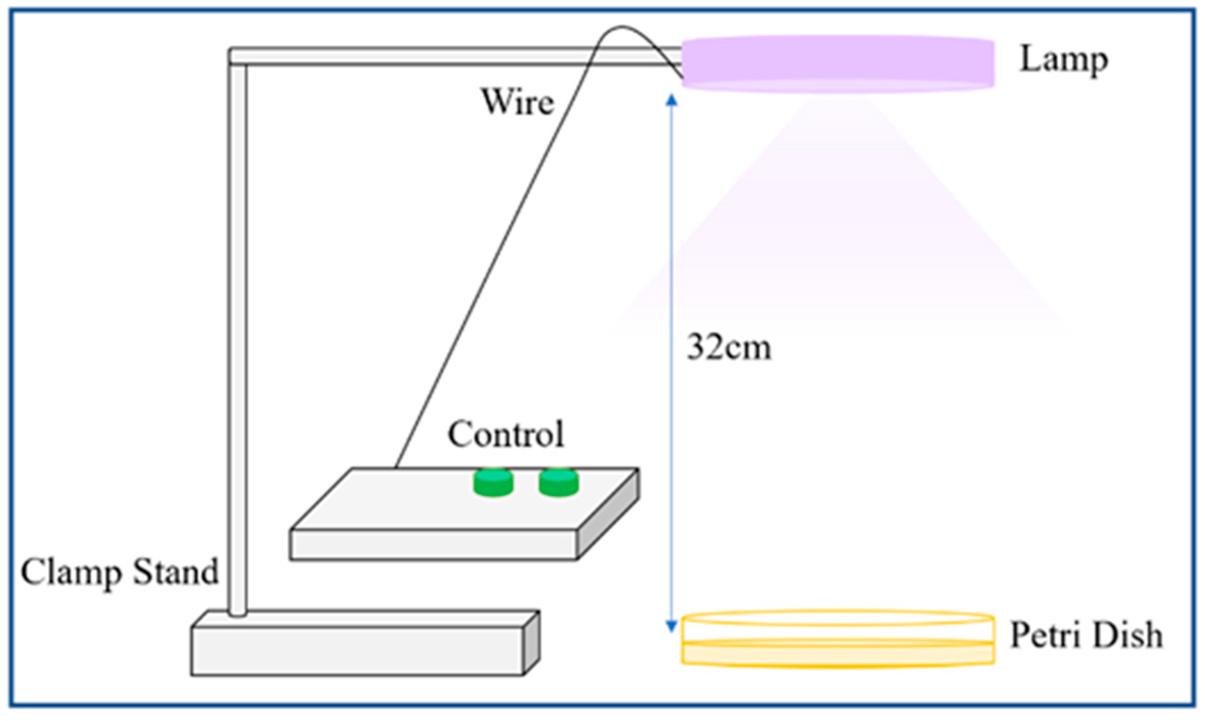
Figure 1. The equipment setup (not drawn to scale). The equipment is contained within a blue housing unit that is situated inside a lightbox. The control panel features two green buttons for activating the UVA and UVC switches. The lamp was positioned 32 cm away from the Petri dish [12].
Aerobiology 01 00007 g001
Figure 2. The equipment setup (not drawn to scale). The control panel features two green buttons for activating the UVA and UVC switches. The lamp was positioned 32 cm away from a plate with infected cells [13].
Aerobiology 01 00007 g002
Figure 3. The percentage difference (%) in bacterial loading between UV light and natural light conditions (blue dot marker), as well as the dark conditions (grey square marker), with the distance of the lamp from the bacteria. E. coli K12 was used as a representative model organism [12].
Aerobiology 01 00007 g003
Figure 4. Tissue culture infectious dose 50 (TCID50) of HCoV-229E treated with UVA and UVC using Protocols 1, 2, and 2a. Control is considered as “0 cycles”. Data shown represent the mean of two experiments with error bars of standard deviation.
Aerobiology 01 00007 g004
Figure 5. Tissue culture infectious dose 50 (TCID50) of HCoV-229E treated with UVA and UVC using Protocol 1. Control is defined as “0 cycles”. Data shown represent the mean of two experiments with error bars of standard deviation.
Aerobiology 01 00007 g005
Figure 6. Tissue culture infectious dose 50 (TCID50) of HCoV-229E treated with UVA and UVC using Protocol 2a. Control is defined as “0 cycles”. Data shown represent the mean of two experiments with error bars of standard deviation.
Aerobiology 01 00007 g006
Table 1. ATP measurements were taken for B. subtilis, E. coli K12, and S. epidermidis, where RLU is relative light unit, under various conditions, including dark, natural light, and UV light, with different power levels for UVA and UVC (F, 42 mW, and 65 mW).
StrainSetting for UVASetting for UVCTime
(h)
ATP (RLU)
DarkUV LightNatural Light
B. subtilis65 mW65 mW12243103893
117 mW117 mW12758047937
42 mW65 mW129551062
E. coli K12117 mW117 mW12700406031
65 mW65 mW12538907496
S. epidermidis117 mW117 mW12770108811
65 mW65 mW12827208899
42 mW65 mW12829703561
Table 2. Reduction in Colony-forming Unit (CFU) of bacteria under natural/dark light with and without UV exposure.
StrainConditionsReduction in CFU
E. coli K12Natural light32 ± 3%
Dark35 ± 8%
S. epidermidisNatural light34 ± 5%
Dark36 ± 2%
Disclaimer/Publisher’s Note: The statements, opinions and data contained in all publications are solely those of the individual author(s) and contributor(s) and not of MDPI and/or the editor(s). MDPI and/or the editor(s) disclaim responsibility for any injury to people or property resulting from any ideas, methods, instructions or products referred to in the content.