


J. Dev. Biol. 2026, 14(1), 14; https://doi.org/10.3390/jdb14010014 - 11 Mar 2026
Abstract
►
Show Figures
Bone tissue is among the most commonly transplanted tissues worldwide. The treatment of critical-sized bone defects remains a significant challenge, as there is currently no universally accepted experimental model or therapeutic standard. Recent advances in fundamental cell biology are driving a paradigm shift
[...] Read more.
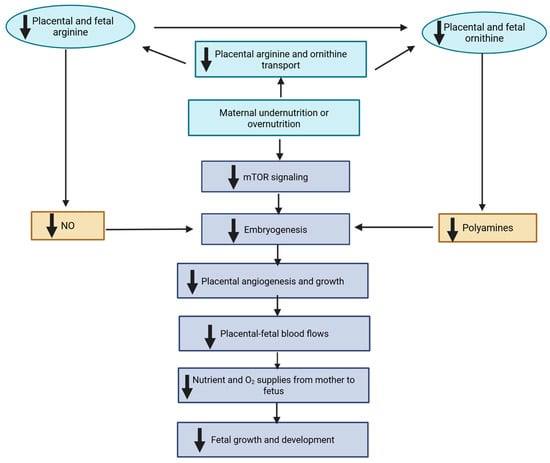
Bone tissue is among the most commonly transplanted tissues worldwide. The treatment of critical-sized bone defects remains a significant challenge, as there is currently no universally accepted experimental model or therapeutic standard. Recent advances in fundamental cell biology are driving a paradigm shift in approaches to bone regeneration, highlighting the transformative potential of biofabrication technologies that integrate tissue engineering with personalized regenerative strategies. Three-dimensional (3D) bioprinting technology enables precise control over the architecture and spatial distribution of cellular and biologically active components, facilitating the creation of complex, personalized bone constructs. Central to this process are bioinks and biomaterials that mimic the extracellular matrix (ECM) and provide an optimal microenvironment for cellular function. Despite the substantial body of accumulated data, a comprehensive theoretical framework for functional bone biofabrication has not yet been fully established, emphasizing both the challenges and the innovative potential of the field. This integrative review synthesizes current knowledge on bone biology—from embryogenesis and cell–matrix interactions to molecular and neural regulation—and links it to the opportunities offered by biofabrication. Particular attention is given to bioinks as mediators between cell biology and engineering sciences, as well as to strategies for creating biomimetic ECM, optimizing scaffold design, and guiding future research toward clinically translatable bone regeneration.
Full article

Figure 1


