


Vision 2026, 10(2), 19; https://doi.org/10.3390/vision10020019 - 31 Mar 2026
Abstract
►
Show Figures
Childhood myopia progression remains a major global public health concern, and spectacle lenses designed to induce peripheral myopic defocus have emerged as a non-pharmacological strategy for myopia control; however, real-world evidence from European populations remains limited. This retrospective observational study evaluated the 12-month
[...] Read more.
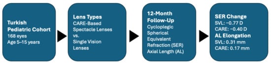
Childhood myopia progression remains a major global public health concern, and spectacle lenses designed to induce peripheral myopic defocus have emerged as a non-pharmacological strategy for myopia control; however, real-world evidence from European populations remains limited. This retrospective observational study evaluated the 12-month real-world effectiveness of cylindrical annular refractive element spectacle lenses in a Turkish pediatric cohort. Children aged 5–15 years who wore myopia-control spectacle lenses from the CARE platform or single-vision lenses were included. Cycloplegic spherical equivalent refraction (SER) and axial length (AL) were measured at baseline and at 12 months. The primary outcomes were 12-month changes in SER and AL. Multivariable generalized estimation equations were applied to account for inter-eye correlation and to adjust for age and gender. A total of 168 eyes were analyzed (85 with single-vision lenses; 83 with myopia-control lenses). After 12 months, the myopia-control group demonstrated significantly slower progression than the single-vision group, with mean SER changes of −0.40 ± 0.92 D versus −0.77 ± 0.74 D and axial elongation of 0.17 ± 0.25 mm versus 0.31 ± 0.30 mm, respectively. Treatment group remained a significant predictor of both refractive progression (p = 0.008) and axial elongation (p = 0.003). Age was independently associated with axial length change (p < 0.001), whereas gender was not. These findings provide real-world European evidence supporting the role of defocus-modulating spectacle lenses in pediatric myopia management.
Full article
Graphical abstract


