Article

Safety and Immunogenicity of a Chimeric Subunit Vaccine against Shiga Toxin-Producing Escherichia coli in Pregnant Cows

1
Programa de Microbiología y Micología, Instituto de Ciencias Biomédicas, Facultad de Medicina, Universidad de Chile, Santiago 8380453, Chile
2
Instituto Milenio de Inmunología e Inmunoterapia, Facultad de Medicina, Universidad de Chile, Santiago 8380453, Chile
3
Programa de Inmunología, Instituto de Ciencias Biomédicas, Facultad de Medicina, Universidad de Chile, Santiago 8380453, Chile
4
Departamento de Microbiología, Facultad de Ciencias Biológicas, Universidad de Concepción, Concepción 4070386, Chile
5
Facultad de Ciencias Veterinarias, Universidad Nacional del Centro de la Provincia de Buenos Aires (UNCPBA), Tandil 7000, Argentina
6
Centro de Investigación Veterinaria de Tandil (CIVETAN), Universidad Nacional del Centro de la Provincia de Buenos Aires (UNCPBA), Tandil 7000, Argentina
7
Departamento de Clínica, Grupo de Medicina Veterinaria Traslacional (MEVET), Facultad de Ciencias Veterinarias, Universidad Nacional del Centro de la Provincia de Buenos Aires (UNCPBA), Tandil 7000, Argentina
*
Authors to whom correspondence should be addressed.
Int. J. Mol. Sci. 2023, 24(3), 2771; https://doi.org/10.3390/ijms24032771
Submission received: 12 December 2022 / Revised: 21 January 2023 / Accepted: 24 January 2023 / Published: 1 February 2023

Abstract

:
Shiga toxin-producing Escherichia coli (STEC) is a zoonotic pathogen that causes gastroenteritis and Hemolytic Uremic Syndrome. Cattle are the main animal reservoir, excreting the bacteria in their feces and contaminating the environment. In addition, meat can be contaminated by releasing the intestinal content during slaughtering. Here, we evaluated the safety and immunogenicity of a vaccine candidate against STEC that was formulated with two chimeric proteins (Chi1 and Chi2), which contain epitopes of the OmpT, Cah and Hes proteins. Thirty pregnant cows in their third trimester of gestation were included and distributed into six groups (n = 5 per group): four groups were administered intramuscularly with three doses of the formulation containing 40 µg or 100 µg of each protein plus the Quil-A or Montanide™ Gel adjuvants, while two control groups were administered with placebos. No local or systemic adverse effects were observed during the study, and hematological parameters and values of blood biochemical indicators were similar among all groups. Furthermore, all vaccine formulations triggered systemic anti-Chi1/Chi2 IgG antibody levels that were significantly higher than the control groups. However, specific IgA levels were generally low and without significant differences among groups. Notably, anti-Chi1/Chi2 IgG antibody levels in the serum of newborn calves fed with colostrum from their immunized dams were significantly higher compared to newborn calves fed with colostrum from control cows, suggesting a passive immunization through colostrum. These results demonstrate that this vaccine is safe and immunogenic when applied to pregnant cows during the third trimester of gestation.

1. Introduction

Shiga toxin-producing Escherichia coli (STEC) is a zoonotic food-borne pathogen that causes diarrhea, dysentery and Hemolytic Uremic Syndrome (HUS), mainly in children under five years of age. STEC O157:H7 is by far the serotype most frequently implicated in severe disease and HUS worldwide. However, several non-O157 serotypes have emerged and are also an important cause of human disease in many countries [1].
The pathogenicity of STEC is mainly determined by the Shiga toxins (Stx), which cause inflammation in the intestinal mucosa. Once the Stx reach the bloodstream, they can cause tissue damage in the kidneys and central nervous system, as well as cause other unusual severe diseases complicated by multi-organ failure [2,3]. Furthermore, a number of different virulence factors are involved in the adherence of STEC to intestinal epithelial cells and colonization of the large intestine. For instance, the pathogenicity island (PAI) called the Locus of Enterocyte Effacement (LEE) encodes a type three secretion system (T3SS) that translocates effector proteins into the human enterocytes, leading to the lesion known as “attaching and effacing” (A/E). The A/E lesion is characterized by the loss of intestinal microvilli which leads to diarrhea [4]. The majority of the STEC strains associated with severe disease in humans harbor the LEE PAI. However, in the absence of the LEE, STEC strains can acquire and accumulate other PAIs, such as the Locus of Adhesion and Autoaggregation (LAA) [5], Subtilase-Encoding Pathogenicity Island (SE-PAI) [6] and Locus of Proteolysis Activity (LPA) [6], demonstrating the high genome plasticity and a wide variety of virulence factors that these pathogens possess. In fact, the LAA PAI promotes the intestinal colonization of STEC and has been identified among the LEE-negative strains associated with disease in humans [7].
STEC may be a resident or transient member of the gastrointestinal microbiota of several mammals, mainly livestock species. Of note, animals capable of maintaining STEC carriage without continuous exposure to the bacteria are defined as reservoirs [8]. Among them, cattle are considered the main reservoir and can intermittently shed STEC through their feces, contaminating the environment [9]. STEC strains have also been isolated from wild animals (e.g., rabbits, birds and rodents). However, it is not clear if they act as transient carriers or as reservoirs [10,11].
In general, intestinal STEC carriage in animals is asymptomatic because most of them lack vascular receptors for Stx; therefore, these toxins cannot be endocytosed and transported to extraintestinal tissues [12]. Occasionally, STEC can cause diarrhea in newborn calves. However, this pathology is thought to be associated with the A/E lesion and with an extensive bacterial colonization leading to the sloughing of enterocytes, rather than a direct cytotoxic action of the Stx [13,14]. Although cattle have receptors for Stx in intestinal epithelial cells, these toxins have been shown to have an immunosuppressive effect in these animals, apparently favoring intestinal colonization [15,16].
STEC carriage in cattle is determined by the ability of the bacteria to adhere to and colonize the large intestine. In particular, STEC O157:H7 have a tissue tropism for the recto-anal junction (RAJ), but non-O157:H7 serotypes may have tropism for other intestinal tissues [17,18]. In experimental infections of cattle and calves with STEC O157:H7, a number of LEE-encoded virulence factors, including Intimin and structural proteins of the T3SS, as well as non-LEE-encoded type III secreted effectors, have been shown to promote intestinal colonization [14,19,20]. However, the adherence of non-O157:H7 STEC to bovine intestinal epithelial cells appears to be mediated by mechanism distinct from those used by O157:H7 [21]. Thus, other proteins associated with adhesion phenotypes, such as EhaA, Iha, EspP and Efa-1, could also be important for the colonization of STEC in cattle [9]. Furthermore, there are STEC strains that persist in cattle for long periods of time, while other strains are identified only sporadically [22]. Consequently, the molecular mechanisms behind the ability of different STEC strains to persist in cattle and other reservoirs are not yet fully understood.
Cattle vaccination is a feasible intervention method to reduce the colonization and shedding of STEC and thus lower the risk of zoonotic transmission to humans [23]. Several vaccine candidates have been evaluated in controlled and natural conditions with variable results [24]. To date, two vaccines against STEC O157:H7 have been licensed for use in cattle in Canada and the USA; however, these formulations reduce the shedding but are not capable of completely clearing the colonization of these bacteria [24,25]. Besides, these vaccines have shown no efficacy in reducing the shedding of other STEC serotypes [26].
Previously, we developed a vaccine candidate against STEC based on two chimeric proteins (Chi1 and Chi2), which contain selected epitopes of the OmpT, Cah and Hes proteins. These proteins are ideal targets against STEC because they are immunogenic and widespread among LEE-positive and LEE-negative STEC strains isolated from humans [5,27,28]. Evaluation of this vaccine candidate in mice showed that it confers protection by reducing intestinal colonization of STEC O157:H7 and kidney damage caused by oral challenge with STEC O91:H21 [29]. Here, our objective was to evaluate the safety and immunogenicity of this vaccine candidate in pregnant cows. Overall, our results demonstrate the safety and immunogenicity of this STEC vaccine. These data need to be complemented by effectiveness and challenge studies to formulate a recommendation protocol for use in cattle.

2. Results

2.1. Experimental Design

The study presented here is a double-blind, randomized field trial, the objectives of which were to evaluate the safety and immunogenicity of a vaccine candidate against STEC in pregnant cows. This vaccine was previously shown to be protective in a murine model [29]. Figure 1 shows the experimental design of this study.
We implemented a dose-escalation scheme to determine the optimal safe and immunogenic dose of this vaccine candidate. For this, four different formulations were generated containing two increasing concentrations (40 µg and 100 µg) of the Chi1 and Chi2 proteins and the Quil-A or Montanide™ Gel adjuvants (Figure 1). The placebos contained PBS plus the corresponding adjuvant.
A total of 30 pregnant cows were randomly assigned to six groups (n = 5 per groups) to receive the vaccine formulations (P1, P2, P4 and P5 groups) or placebos (P3 and P6 control groups). The immunization protocol was a three-dose schedule (2 weeks apart) by intramuscular route.

2.2. Safety

All the animals showed good health conditions throughout the field trial. No clinically relevant differences were documented with respect to local and systemic adverse events between animals that received the highest vaccine dose (P2 and P5 groups) and animals that received the standard dose (P1 and P4 groups) or placebos (P3 and P6 groups).
Additionally, no significant differences were observed in rectal temperatures and weights after administration of the different treatments (Table S1). The hematological and blood biochemical parameters evaluated remained within the reference range and no significant differences were observed between groups at any time (Tables S2 and S3).
The calves were born at term, healthy and with normal weight, except for one calf from the P1 group that died during parturition due to fetal dystocia and one from the P3 group that was aborted (Table S1). Thus, these results indicate that all vaccine formulations were safe and well tolerated.

2.3. Serum Antibody Response

Before the first immunization, fecal samples of all animals were analyzed by PCR for the presence of STEC, and the herd was found to be positive (result not shown). Consistent with the above, pre-immune sera from all animals were seropositive to STEC, having a baseline of Chi1/Chi2-specific IgG and IgA antibodies. However, the levels of these antibodies were similar among the groups (Figure 2).
After three immunizations with the vaccine formulations, a significant increase in serum IgG levels against both proteins (Figure 2a,b), but not the IgA antibodies (Figure 2c,d), was observed compared to the control groups. The increased level of specific IgG in sera was similar between animals receiving the standard or the highest dose of the formulation, regardless of the adjuvant used. In fact, no significant differences were observed in groups receiving vaccine formulations containing the Quil-A adjuvant (P1 and P2) compared to groups receiving vaccine formulations containing MontanideTM Gel adjuvant (P4 and P5). Therefore, all vaccine formulations triggered specific IgG antibodies in sera at similar levels.

2.4. Colostrum Antibody Response

Bovine colostrum may contain antibodies against important virulence factors of STEC [30]. Therefore, we determined the levels of Chi1/Chi2-specific antibodies in the colostrum of immunized cows. As a result, we found a slight but significant difference in the specific IgG levels between the P2 group and the P3 control group (Figure 3a). Additionally, although there were no significant differences, a trend towards higher specific IgG levels was observed in the P4 and P5 groups compared to the P6 control group (Figure 3b). By contrast, specific IgA antibody levels in colostrum were variable, and no significant differences were observed among the groups.
It has been reported that STEC-specific colostral antibodies are transferred to newborn calves after feeding them with colostrum [31,32]. Thus, passive immunization of newborn calves could reduce STEC colonization and therefore constitutes an interesting strategy for the control of this pathogen.
Notably, our results showed that calves fed with colostrum from their immunized dams had significantly higher levels of anti-Chi1/Chi2 IgG antibodies in serum compared with calves fed with colostrum from control cows (Figure 4). This was especially evident in the P4 and P5 groups compared to the P6 control group. Regarding the anti-Chi1/Chi2 IgA antibodies in serum, a significantly higher level was found in the newborn calves in the P4 group compared to the P6 control group (Figure 4b). In the other groups, there were no differences in the levels of specific IgA in serum.

3. Discussion

Most of the vaccine candidates against STEC that have been evaluated in cattle are based on virulence factors encoded on the LEE PAI [24,25,32,33,34]. By contrast, the vaccine candidate evaluated here contains epitopes of OmpT, Cah and Hes proteins. To our knowledge, no other vaccine candidate has evaluated the use of these antigens.
These three proteins are suitable targets to develop preventive therapies against STEC infections because: (i) their encoding genes are conserved, widely distributed and highly frequent in both LEE-positive (OmpT, Cah) and LEE-negative STEC strains (Hes, Cah and OmpT) [27]; (ii) they are expressed in vivo during infection in humans and are reactive to hyperimmune sera from HUS patients; and (iii) they have been shown to participate in different colonization-associated phenotypes in vitro assays and in infections of human epithelial cells [5,35,36]. However, it is important to note that the role, if any, of these proteins in the colonization of cattle is currently unknown.
Previously, this vaccine candidate was evaluated in a murine model of STEC infection, with promising results [29]. In the present study, we tested the safety and immunogenicity of this vaccine in pregnant cows. For this, the vaccine was formulated with two different concentrations, a standard dose of 80 µg and a higher dose of 200 µg of the Chi1 and Chi2 proteins. We used the Quil-A and the Montanide™ Gel adjuvants because they are safe and have adjuvant activity in cattle [37,38].
Our results indicate that all four vaccine formulations were well tolerated and did not cause adverse reactions, fetal malformations or abortions. The abortion observed during this study occurred in the P3 control group, and its cause was not determined; therefore, it was not associated with the vaccine administration. In this respect, pregnancy loss rate varies between 15% and 23% in livestock and may be due to a variety of causes and combination of factors [39,40,41,42]. This result demonstrates the safety of the vaccine formulations when administered to pregnant cows in their third trimester and following the described immunization protocol.
Based on PCR detection, we found that the cows included in this study were colonized by STEC before the first immunization (not shown). Cattle in Buenos Aires province have a prevalence of STEC as high as 63% [43]; therefore, cattle under natural conditions in this region are highly likely to be colonized with STEC. Thus, it is expected that cows in our study have been exposed to a plethora of diverse STEC strains during their lifetime.
In line with this, we found pre-existing anti-Chi1/Chi2 antibodies in the preimmune sera from the cows (Figure 2). This suggests that, as occurs in humans, the OmpT, Cah and Hes proteins are also expressed by STEC during cattle infection. Many studies have also found a baseline of STEC-specific antibodies in cattle naturally infected with STEC [24,44]; however, despite the pre-existence of these antibodies, cattle are susceptible to being colonized by STEC, often resulting in persistent shedding of these bacteria. Thus, circulating STEC-specific antibody levels should be analyzed with caution, as they should not be considered an absolute correlate of immunity.
Therefore, for the optimal evaluation of the immunogenicity of a vaccine against STEC, both the determination of systemic antibodies and secreted antibodies at the site of colonization must be considered, together with cellular immune responses [16]. Nevertheless, the objective of this work was to carry out an initial evaluation of the immunogenicity and optimal dose of our vaccine candidate.
In this sense, there are some results regarding the immunogenicity of our vaccine that deserve to be highlighted. First, vaccine formulations containing 80 µg of chimeric proteins elicited significant levels of specific IgG antibodies that were similar to those obtained with formulations containing 200 µg (Figure 2a,b). This would favor the scaling and costs of production of this vaccine.
Second, a trend for a higher level of anti-Chi1/Chi2 IgG antibodies in colostrum was observed in the immunized groups compared to the control groups (Figure 3). This difference was statistically significant only between the P2 group and the P3 control group. However, there was a high variability in the levels of antibodies in colostrum, which possibly affected the power of the statistical test used. In particular, we consider that this variability could be attributed to the technical difficulty of taking colostrum samples from cows kept under natural conditions.
Third, although the humoral responses in colostrum were inconclusive, the levels of anti-Chi1/Chi2 IgG antibodies in the sera of newborn calves from immunized cows were significantly higher than those found in sera of newborn calves from control cows (Figure 4). Remarkably, significant levels of specific IgA were also observed in the serum of calves from the P4 group compared to the P6 control group. In humans and other primates, the transfer of IgG from the mother to the neonate occurs prenatally (across the placenta) and postnatally (through lactation). In ruminants, the placenta transmits little or no IgG antibodies [45,46]. Instead, there is exhaustive evidence showing that the mammary gland of these species secretes large amounts of IgG during colostrum formation [45]. Consistent with these studies, it has been shown that STEC-specific antibodies in colostrum from naturally infected or vaccinated cows are efficiently transferred to the newborn calves by feeding with hyperimmune colostrum [31,32,34,47]. Thus, the higher levels of anti-Chi1/Chi2 IgG antibodies in the serum of newborn calves from immunized cows strongly suggest that a passive immunization through colostrum occurred. Since cattle during the first months of life are rapidly colonized by STEC [48], passive immunization of calves may be an alternative strategy to prevent the early colonization by these bacteria [32].
In conclusion, taken together, the results of this study demonstrate the safety of this vaccine candidate against STEC and provide information on its immunogenicity when administered to pregnant cows in their third trimester of gestation.

4. Materials and Methods

4.1. Animals

All animal experiments and protocols were approved (Approval Code: Acta de Bienestar Animal ResCA 087/02) by the Animal Welfare Commission at the National University of the Center of the Province of Buenos Aires, Tandil, Argentina. A total of 30 Aberdeen Angus pregnant cows (third trimester of pregnancy) were obtained, housed and fed with standard food and drink on a cattle farm located in Tandil, Argentina. Before the first immunization, fecal samples were taken and analyzed by PCR (using protocol described in [49]) for the presence of STEC, and the herd was found to be positive. The general characteristics of the cows are shown in Table S1.

4.2. Production of Vaccine Formulations and Immunization Protocol

Chimera 1 (Chi1) and Chimera 2 (Chi2) proteins were produced and purified as described previously [29]. The vaccine formulations and placebos were sterilized by filtration and packaged at a veterinary pharmaceutical laboratory under good manufacturing practices. The vaccine formulations contained 80 (40 µg Chi1 + 40 µg Chi2) or 200 µg (100 µg Chi1 + 100 µg Chi2) of chimeric proteins plus the Quil-A or Montanide™ Gel adjuvants, while placebos contained phosphate-buffered saline (PBS) solution plus the corresponding adjuvant. The animals were ear-tagged identified and randomly assigned to receive a vaccine formulation or placebo in a three-dose schedule (2 weeks apart), which were administered intramuscularly in the cervical region. Four groups (P1, P2, P4 and P5; n = 5 per group) received the vaccine formulations and two control groups (P3 and P6; n = 5 per group) received adjuvants only (Figure 1, Table S1).
Basic clinical observation and general adverse reactions were evaluated by measuring rectal temperature, weight and loss of appetite. Local adverse reactions at the injection site such as swellings or pain were evaluated by palpation. Local inflammation was examined by palpation of the prescapular lymph node.

4.3. Sample Collection

Blood samples (13 mL) were collected by venipuncture into EDTA tubes before each immunization and 2 weeks after the last boost. These samples were used to determine hematological and blood biochemical parameters. To obtain sera, blood samples (7 mL) were collected into non-anticoagulant tubes and then left at 37 °C for 30 min and centrifuged at 1000× g for 10 min, and the supernatants (sera) were collected and stored at −20 °C until use. Additionally, blood samples were taken from the newborn calves during the first 3 days of life and subsequently processed to obtain sera. Colostrum samples were obtained during the prodromal state of labor or within the first 24 h after parturition. Colostrum samples were delipidated by centrifugation (1000× g at 4 °C for 45 min), and the watery phase was stored at −20 °C until use.

4.4. Hematological and Blood Biochemical Parameters

Hematological and blood biochemical parameters were determined by using the Hemax330 hematology analyzer (B&E Scientific Instrument Co., Ltd., Yantai, China) and the CM 250 Wiener Lab® automatic clinical chemistry analyzer (Wiener Lab Group, Santa Fe, Argentina), respectively. Hematological parameters analyzed were red blood cell count, hematocrit, hemoglobin, mean corpuscular volume, mean corpuscular hemoglobin (MCH) mean corpuscular hemoglobin concentration (MCHC), white blood cell count, immature neutrophils, segmented neutrophils, lymphocytes, monocytes, eosinophils and basophils. Biochemical parameters evaluated were total proteins, albumins, globulins, albumin/globulin ratio, alkaline phosphatase, creatinine, alanine aminotransferase and blood urea nitrogen.

4.5. Humoral Immune Responses

The humoral immune responses to the vaccine formulations were determined by indirect ELISA. For this, 96-well ELISA plates (Nunc-Immuno Plates, ThermoFisher, Waltham, MA, USA) were incubated with a mixture of 1 µg of Chi1 and Chi2 proteins diluted in 100 µL of phosphate saline buffer (PBS; 1X, pH 7.2) overnight at 4 °C. Plates were washed 3 times with PBS (400 µL/well) containing 0.05% Tween 20 (T-PBS). Plates were then incubated with 300 µL/well of blocking solution (T-PBS + 0.5% bovine serum albumin) for 15 min at room temperature. Animal sera were diluted 1:50 to 1:1600 (for IgA measurement) or 1:250 to 1:16,000 (for IgG measurement) in blocking solution (100 µL/well) and incubated for 60 min at 37 °C. Colostrum samples were diluted 1:200 (for IgA measurement) or 1:1000 (for IgG measurement) and incubated as described above. After 5 washes with T-PBS (400 µL/well), anti-Bovine IgA (Fc)-HRP (NB773, Novus Biologicals, Centennial, CO, USA) or anti-Bovine IgG (Fc)-HRP (SAB3700020, Sigma-Aldrich, St. Louis, MO, USA), diluted 1:1000 in blocking solution (100 µL/well), were incubated for 60 min at 37 °C. After 5 washes with T-PBS (400 µL/well), plates were incubated with the 3,3′,5,5′-tetramethylbenzidine (TMB) liquid substrate (T0440, Sigma-Aldrich, USA) for 10 min at room temperature. Absorbance was determined at 405 nm using a Synergy HT microplate reader (Biotek Instruments, Winooski, VT, USA). Each sample was determined in duplicate and with at least three independent replicates.

4.6. Statistical Analysis

Statistical differences in anti-Chi1/Chi2 IgG sera and IgA antibody were analyzed by a two-way ANOVA, followed by Tukey’s multiple comparison test. Statistical differences in colostrum anti-Chi1/Chi2 IgG and IgA antibodies were analyzed by the Kruskal−Wallis test, followed by Dunn’s multiple comparison test. For all statistical tests, a p value of <0.05 was considered significant.

Supplementary Materials

The following supporting information can be downloaded at: https://www.mdpi.com/article/10.3390/ijms24032771/s1.

Author Contributions

Conceptualization and experimental design: R.M.V., D.A.M., F.D.C., J.C.S. and A.O. Data acquisition: C.A., A.A., N.L.P., H.M., A.E., D.F., V.V., M.G., R.C., M.S., D.A.M. and R.M.V. Data analysis and interpretation: R.M.V., D.A.M., F.D.C., J.C.S. and A.O. Writing—original draft: D.A.M. and R.M.V.; Review: R.M.V., D.A.M., F.D.C., J.C.S., A.O., N.L.P., R.C. and H.M. Final Edition: R.M.V. and D.A.M. All authors have read and agreed to the published version of the manuscript.

Funding

This study was supported by National FONDEF ID16I10140 grant awarded to R. Vidal.

Institutional Review Board Statement

The animal study protocol was approved by the Animal Welfare Commission at the National University of the Center of the Province of Buenos Aires, Tandil, Argentina (Approval Code: Acta de Bienestar Animal ResCA 087/02).

Informed Consent Statement

Not applicable.

Data Availability Statement

The data that support the findings of this study are available on request from the corresponding author R.V. Amino acid sequences of Chimeric proteins and identified epitopes are not publicly available due to legal restrictions and an ongoing international patent application.

Acknowledgments

We thank Helen Lowry for the careful revision and edition of the manuscript.

Conflicts of Interest

Currently, an application for an international national patent process had been presented for the chimeric antigens developed and uses thereof.

References

  1. Majowicz, S.E.; Scallan, E.; Jones-Bitton, A.; Sargeant, J.M.; Stapleton, J.; Angulo, F.J.; Yeung, D.H.; Kirk, M.D. Global incidence of human Shiga toxin–producing Escherichia coli infections and deaths: A systematic review and knowledge synthesis. Foodborne Pathog. Dis. 2014, 11, 447–455. [Google Scholar] [CrossRef] [PubMed]
  2. Lee, M.S.; Tesh, V.L. Roles of shiga toxins in immunopathology. Toxins 2019, 11, 212. [Google Scholar] [CrossRef] [PubMed]
  3. Wijnsma, K.L.; Schijvens, A.M.; Rossen, J.W.A.; Kooistra-Smid, A.M.D.; Schreuder, M.F.; van de Kar, N.C.A.J. Unusual severe case of hemolytic uremic syndrome due to Shiga toxin 2d-producing E. coli O80:H2. Pediatr. Nephrol. 2017, 32, 1263–1268. [Google Scholar] [CrossRef] [PubMed]
  4. Gaytán, M.O.; Martínez-Santos, V.I.; Soto, E.; González-Pedrajo, B. Type Three Secretion System in Attaching and Effacing Pathogens. Front. Cell Infect. Microbiol. 2016, 6, 129. [Google Scholar] [CrossRef]
  5. Montero, D.A.; Velasco, J.; Del Canto, F.; Puente, J.L.; Padola, N.L.; Rasko, D.A.; Farfán, M.; Salazar, J.C.; Vidal, R. Locus of Adhesion and Autoaggregation (LAA), a pathogenicity island present in emerging Shiga Toxin-producing Escherichia coli strains. Sci. Rep. 2017, 7, 7011. [Google Scholar] [CrossRef]
  6. Michelacci, V.; Tozzoli, R.; Caprioli, A.; Martínez, R.; Scheutz, F.; Grande, L.; Sánchez, S.; Morabito, S. A new pathogenicity island carrying an allelic variant of the Subtilase cytotoxin is common among Shiga toxin producing Escherichia coli of human and ovine origin. Clin. Microbiol. Infect. 2013, 19, E149–E156. [Google Scholar] [CrossRef] [PubMed]
  7. Montero, D.A.; Del Canto, F.; Velasco, J.; Colello, R.; Padola, N.L.; Salazar, J.C.; Martin, C.S.; Oñate, A.; Blanco, J.; Rasko, D.A.; et al. Cumulative acquisition of pathogenicity islands has shaped virulence potential and contributed to the emergence of LEE-negative Shiga toxin-producing Escherichia coli strains. Emerg. Microbes Infect. 2019, 8, 486–502. [Google Scholar] [CrossRef]
  8. Persad, A.K.; LeJeune, J.T. Animal Reservoirs of Shiga Toxin-Producing Escherichia coli. Microbiol. Spectr. 2014, 2, 1–14. [Google Scholar] [CrossRef]
  9. Etcheverría, A.I.; Padola, N.L. Shiga toxin-producing Escherichia coli: Factors involved in virulence and cattle colonization. Virulence 2013, 4, 366–372. [Google Scholar] [CrossRef]
  10. Jahan, N.A.; Lindsey, L.L.; Larsen, P.A. The Role of Peridomestic Rodents as Reservoirs for Zoonotic Foodborne Pathogens. Vector-Borne Zoonotic Dis. 2021, 21, 133–148. [Google Scholar] [CrossRef]
  11. Blanco Crivelli, X.; Rumi, M.V.; Carfagnini, J.C.; Degregorio, O.; Bentancor, A.B. Synanthropic rodents as possible reservoirs of shigatoxigenic Escherichia coli strains. Front. Cell Infect. Microbiol. 2012, 2, 134. [Google Scholar] [CrossRef] [PubMed]
  12. Pruimboom-Brees, I.M.; Morgan, T.W.; Ackermann, M.R.; Nystrom, E.D.; Samuel, J.E.; Cornick, N.A.; Moon, H.W. Cattle lack vascular receptors for Escherichia coli 0157:H7 Shiga toxins. Proc. Natl. Acad. Sci. USA 2000, 97, 10325–10329. [Google Scholar] [CrossRef] [PubMed]
  13. Moxley, R.A.; Smith, D.R. Attaching-effacing Escherichia coli Infections in Cattle. Vet. Clin. N. Am. Food Anim. Pract. 2010, 26, 29–56. [Google Scholar] [CrossRef] [PubMed]
  14. Dean-Nystrom, E.A.; Bosworth, B.T.; Moon, H.W.; O’Brien, A.D. Escherichia coli O157:H7 requires intimin for enteropathogenicity in calves. Infect. Immun. 1998, 66, 4560–4563. [Google Scholar] [CrossRef] [PubMed]
  15. Menge, C. The Role of Escherichia coli Shiga Toxins in STEC Colonization of Cattle. Toxins 2020, 12, 607. [Google Scholar] [CrossRef]
  16. Hoffman, M.A.; Menge, C.; Casey, T.A.; Laegreid, W.; Bosworth, B.T.; Dean-Nystrom, E.A. Bovine immune response to Shiga-toxigenic Escherichia coli O157:H7. Clin. Vaccine Immunol. 2006, 13, 1322–1327. [Google Scholar] [CrossRef]
  17. Naylor, S.W.; Low, J.C.; Besser, T.E.; Mahajan, A.; Gunn, G.J.; Pearce, M.C.; McKendrick, I.J.; Smith, D.G.E.; Gally, D.L. Lymphoid follicle-dense mucosa at the terminal rectum is the principal site of colonization of enterohemorrhagic Escherichia coli O157:H7 in the bovine host. Infect. Immun. 2003, 71, 1505–1512. [Google Scholar] [CrossRef]
  18. Hamm, K.; Barth, S.A.; Stalb, S.; Geue, L.; Liebler-Tenorio, E.; Teifke, J.P.; Lange, E.; Tauscher, K.; Kotterba, G.; Bielaszewska, M.; et al. Experimental Infection of Calves with Escherichia coli O104:H4 outbreak strain. Sci. Rep. 2016, 6, 32812. [Google Scholar] [CrossRef]
  19. Cornick, N.A.; Booher, S.L.; Moon, H.W. Intimin facilitates colonization by Escherichia coli O157:H7 in adult ruminants. Infect. Immun. 2002, 70, 2704–2707. [Google Scholar] [CrossRef]
  20. Dziva, F.; van Diemen, P.M.; Stevens, M.P.; Smith, A.J.; Wallis, T.S. Identification of Escherichia coli O157:H7 genes influencing colonization of the bovine gastrointestinal tract using signature-tagged mutagenesis. Microbiology 2004, 150, 3631–3645. [Google Scholar] [CrossRef] [Green Version]
  21. Kudva, I.T.; Hovde, C.J.; John, M. Adherence of Non-O157 shiga toxin-producing Escherichia coli to bovine recto-anal junction squamous epithelial cells appears to be mediated by mechanisms distinct from those used by O157. Foodborne Pathog. Dis. 2013, 10, 375–381. [Google Scholar] [CrossRef]
  22. Barth, S.A.; Menge, C.; Eichhorn, I.; Semmler, T.; Wieler, L.H.; Pickard, D.; Belka, A.; Berens, C.; Geue, L. The accessory genome of Shiga toxin-producing Escherichia coli defines a persistent colonization type in cattle. Appl. Environ. Microbiol. 2016, 82, 5455–5464. [Google Scholar] [CrossRef]
  23. Matthews, L.; Reeve, R.; Gally, D.L.; Low, J.C.; Woolhouse, M.E.J.; McAteer, S.P.; Locking, M.E.; Chase-Topping, M.E.; Haydon, D.T.; Allison, L.J.; et al. Predicting the public health benefit of vaccinating cattle against Escherichia coli O157. Proc. Natl. Acad. Sci. USA 2013, 110, 16265–16270. [Google Scholar] [CrossRef]
  24. Walle, K.V.; Vanrompay, D.; Cox, E. Bovine innate and adaptive immune responses against Escherichia coli O157: H7 and vaccination strategies to reduce faecal shedding in ruminants. Vet. Immunol. Immunopathol. 2013, 152, 109–120. [Google Scholar] [CrossRef]
  25. O’Ryan, M.; Vidal, R.; del Canto, F.; Carlos Salazar, J.; Montero, D. Vaccines for viral and bacterial pathogens causing acute gastroenteritis: Part II: Vaccines for Shigella, Salmonella, enterotoxigenic E. coli (ETEC) enterohemorragic E. coli (EHEC) and Campylobacter jejuni. Hum. Vaccin. Immunother. 2015, 11, 601–619. [Google Scholar] [CrossRef]
  26. Cernicchiaro, N.; Renter, D.G.; Cull, C.A.; Paddock, Z.D.; Shi, X.; Nagaraja, T.G. Fecal shedding of non-O157 serogroups of shiga toxin-producing Escherichia coli in feedlot cattle vaccinated with an Escherichia coli O157:H7 SRP vaccine or fed a lactobacillus-based direct-fed microbial. J. Food Prot. 2014, 77, 732–737. [Google Scholar] [CrossRef]
  27. Montero, D.; Orellana, P.; Gutiérrez, D.; Araya, D.; Salazar, J.C.; Prado, V.; Oñate, A.; Del Canto, F.; Vidal, R. Immunoproteomic analysis to identify Shiga toxin-producing Escherichia coli outer membrane proteins expressed during human infection. Infect. Immun. 2014, 82, 4767–4777. [Google Scholar] [CrossRef]
  28. Colello, R.; Vélez, M.V.; González, J.; Montero, D.A.; Bustamante, A.V.; Del Canto, F.; Etcheverría, A.I.; Vidal, R.; Padola, N.L. First report of the distribution of Locus of Adhesion and Autoaggregation (LAA) pathogenicity island in LEE-negative Shiga toxin-producing Escherichia coli isolates from Argentina. Microb. Pathog. 2018, 123, 259–263. [Google Scholar] [CrossRef]
  29. Montero, D.A.; Del Canto, F.; Salazar, J.C.; Céspedes, S.; Cádiz, L.; Arenas-Salinas, M.; Reyes, J.; Oñate, Á.; Vidal, R.M. Immunization of mice with chimeric antigens displaying selected epitopes confers protection against intestinal colonization and renal damage caused by Shiga toxin-producing Escherichia coli. NPJ Vaccines 2020, 5, 20. [Google Scholar] [CrossRef]
  30. Vilte, D.A.; Larzábal, M.; Cataldi, Á.A.; Mercado, E.C. Bovine colostrum contains immunoglobulin G antibodies against intimin, EspA, and EspB and inhibits hemolytic activity mediated by the type three secretion system of attaching and effacing Escherichia coli. Clin. Vaccine Immunol. 2008, 15, 1208–1213. [Google Scholar] [CrossRef] [Green Version]
  31. Widiasih, D.A.; Matsuda, I.; Omoe, K.; Hu, D.L.; Sugii, S.; Shinagawa, K. Passive transfer of antibodies to shiga toxin-producing Escherichia coli O26, O111 and O157 antigens in neonatal calves by feeding colostrum. J. Vet. Med. Sci. 2004, 66, 213–215. [Google Scholar] [CrossRef] [PubMed]
  32. Rabinovitz, B.C.; Gerhardt, E.; Tironi Farinati, C.; Abdala, A.; Galarza, R.; Vilte, D.A.; Ibarra, C.; Cataldi, A.; Mercado, E.C. Vaccination of pregnant cows with EspA, EspB, γ-intimin, and Shiga toxin 2 proteins from Escherichia coli O157: H7 induces high levels of specific colostral antibodies that are transferred to newborn calves. J. Dairy Sci. 2012, 95, 3318–3326. [Google Scholar] [CrossRef] [PubMed]
  33. Martorelli, L.; Garimano, N.; Fiorentino, G.A.; Vilte, D.A.; Garbaccio, S.G.; Barth, S.A.; Menge, C.; Ibarra, C.; Palermo, M.S.; Cataldi, A. Efficacy of a recombinant Intimin, EspB and Shiga toxin 2B vaccine in calves experimentally challenged with Escherichia coli O157:H7. Vaccine 2018, 36, 3949–3959. [Google Scholar] [CrossRef] [PubMed]
  34. Rabinovitz, B.C.; Vilte, D.A.; Larzábal, M.; Abdala, A.; Galarza, R.; Zotta, E.; Ibarra, C.; Mercado, E.C.; Cataldi, A. Physiopathological effects of Escherichia coli O157: H7 inoculation in weaned calves fed with colostrum containing antibodies to EspB and Intimin. Vaccine 2014, 32, 3823–3829. [Google Scholar] [CrossRef] [PubMed]
  35. Torres, A.N.; Chamorro-Veloso, N.; Costa, P.; Cádiz, L.; Del Canto, F.; Venegas, S.A.; López Nitsche, M.; Coloma-Rivero, R.F.; Montero, D.A.; Vidal, R.M. Deciphering Additional Roles for the EF-Tu, l-Asparaginase II and OmpT Proteins of Shiga Toxin-Producing Escherichia coli. Microorganisms 2020, 8, 1184. [Google Scholar] [CrossRef]
  36. Torres, A.G.; Perna, N.T.; Burland, V.; Ruknudin, A.; Blattner, F.R.; Kaper, J.B. Characterization of Cah, a calcium-binding and heat-extractable autotransporter protein of enterohaemorrhagic Escherichia coli. Mol. Microbiol. 2002, 45, 951–966. [Google Scholar] [CrossRef]
  37. Dalsgaard, K. Saponin adjuvants. Arch. Fuer Die Gesamte Virusforsch. 1974, 44, 243–254. [Google Scholar] [CrossRef]
  38. Parker, R.; Deville, S.; Dupuis, L.; Bertrand, F.; Aucouturier, J. Adjuvant formulation for veterinary vaccines: MontanideTM Gel safety profile. Procedia Vaccinol. 2009, 1, 140–147. [Google Scholar] [CrossRef]
  39. Mellado, M.; López, R.; de Santiago, Á.; Veliz, F.G.; Macías-Cruz, U.; Avendaño-Reyes, L.; García, J.E. Climatic conditions, twining and frequency of milking as factors affecting the risk of fetal losses in high-yielding Holstein cows in a hot environment. Trop. Anim. Health Prod. 2016, 48, 1247–1252. [Google Scholar] [CrossRef]
  40. Daniel Givens, M.; Marley, M.S.D. Infectious causes of embryonic and fetal mortality. Theriogenology 2008, 70, 270–285. [Google Scholar] [CrossRef]
  41. Albuja, C.; Ortiz, O.; López, C.; Hernández-Cerón, J. Economic impact of pregnancy loss in an intensive dairy farming system. Vet. Mex. 2019, 6, 1–8. [Google Scholar] [CrossRef]
  42. Diskin, M.G.; Waters, S.M.; Parr, M.H.; Kenny, D.A. Pregnancy losses in cattle: Potential for improvement. Reprod. Fertil. Dev. 2016, 28, 83–93. [Google Scholar] [CrossRef]
  43. Padola, N.L.; Sanz, M.E.; Blanco, J.E.; Blanco, M.; Blanco, J.; Etcheverria, A.I.; Arroyo, G.H.; Usera, M.A.; Parma, A.E. Serotypes and virulence genes of bovine Shigatoxigenic Escherichia coli (STEC) isolated from a feedlot in Argentina. Vet. Microbiol. 2004, 100, 3–9. [Google Scholar] [CrossRef]
  44. Asper, D.J.; Karmali, M.A.; Townsend, H.; Rogan, D.; Potter, A.A. Serological response of shiga toxin-producing Escherichia coli type III secreted proteins in sera from vaccinated rabbits, naturally infected cattle, and humans. Clin. Vaccine Immunol. 2011, 18, 1052–1057. [Google Scholar] [CrossRef]
  45. Baintner, K. Transmission of antibodies from mother to young: Evolutionary strategies in a proteolytic environment. Vet. Immunol. Immunopathol. 2007, 117, 153–161. [Google Scholar] [CrossRef]
  46. Chucri, T.M.; Monteiro, J.M.; Lima, A.R.; Salvadori, M.L.B.; Junior, J.R.K.; Miglino, M.A. A review of immune transfer by the placenta. J. Reprod. Immunol. 2010, 87, 14–20. [Google Scholar] [CrossRef]
  47. Albanese, A.; Sacerdoti, F.; Seyahian, E.A.; Amaral, M.M.; Fiorentino, G.; Fernandez Brando, R.; Vilte, D.A.; Mercado, E.C.; Palermo, M.S.; Cataldi, A.; et al. Immunization of pregnant cows with Shiga toxin-2 induces high levels of specific colostral antibodies and lactoferrin able to neutralize E. coli O157:H7 pathogenicity. Vaccine 2018, 36, 1728–1735. [Google Scholar] [CrossRef]
  48. Engelen, F.; Thiry, D.; Devleesschauwer, B.; Mainil, J.; De Zutter, L.; Cox, E. Occurrence of ‘gang of five’ Shiga toxin-producing Escherichia coli serogroups on Belgian dairy cattle farms by overshoe sampling. Lett. Appl. Microbiol. 2021, 72, 415–419. [Google Scholar] [CrossRef]
  49. Fernández, D.; Sanz, M.E.; Parma, A.E.; Padola, N.L. Short communication: Characterization of Shiga toxin-producing Escherichia coli isolated from newborn, milk-fed, and growing calves in Argentina. J. Dairy Sci. 2012, 95, 5340–5343. [Google Scholar] [CrossRef]
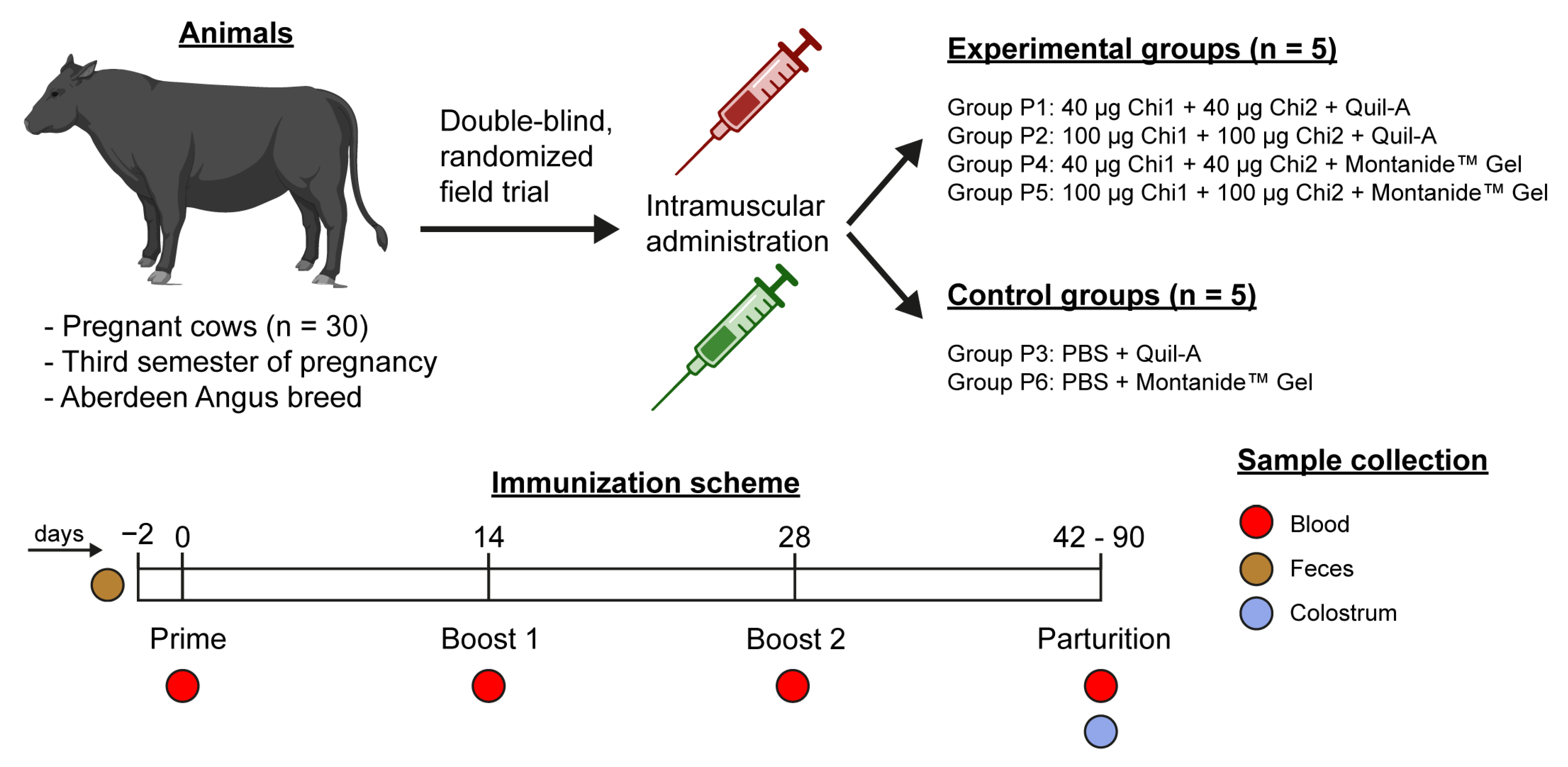
Figure 1. Experimental design of this field trial. The different groups of animals, vaccine formulations and placebos and immunization schedule are shown.
Ijms 24 02771 g001
Figure 2. Serum antibody response in cows immunized with the vaccine formulations. Sera obtained before the first immunization and after the last immunization were used for the determination of anti-Chi1/Chi2 IgG and IgA antibodies. Samples were analyzed in duplicate, and the results are expressed as means ± SEM of absorbance values at 405 nm for each serum dilution, n = 5 animals per group. Anti-Chi1/Chi2 IgG (a) and IgA (c) antibodies from P1, P2 and P3 groups. Anti-Chi1/Chi2 IgG (b) and IgA (d) antibodies from P4, P5 and P6 groups. Statistical analysis was performed using a two-way ANOVA, followed by Tukey’s multiple comparison test. p < 0.05 was considered significant. Asterisks (*) indicate significant differences between the group immunized with the formulation containing 40 µg of each chimeric protein and the control group. * p < 0.05, ** p < 0.005, *** p < 0.0005. Number signs (#) indicate significant differences between the group immunized with the formulation containing 100 µg of each chimeric protein and the control group. # p < 0.05, ## p < 0.005, ### p < 0.0005.
Ijms 24 02771 g002
Figure 3. Chi1/Chi2—specific antibody levels in colostrum. (a) Colostrum from P1, P2 and P3 groups. (b) Colostrum from P4, P5 and P6 groups. Data analysis was by Kruskal−Wallis test, followed by Dunn’s multiple comparison test. p < 0.05 was considered significant. * p < 0.05.
Ijms 24 02771 g003
Figure 4. Chi1/Chi2—specific antibody levels present in the serum of newborn calves. (a) Newborn calves from groups P1, P2 and P3. (b) Newborn calves from groups P4, P5 and P6. Data analysis was by a two-way ANOVA, followed by Tukey’s multiple comparison test. p < 0.05 was considered significant. Asterisks (*) indicate significant differences between the group immunized with the formulation containing 40 µg of each chimeric antigen and the control group (adjuvant alone). * p < 0.05, ** p < 0.005, *** p < 0.0005. Number signs (#) indicate significant differences between the group immunized with the formulation containing 100 µg of each chimeric antigen and the control group (adjuvant alone). # p < 0.05, ### p < 0.0005.
Ijms 24 02771 g004
Disclaimer/Publisher’s Note: The statements, opinions and data contained in all publications are solely those of the individual author(s) and contributor(s) and not of MDPI and/or the editor(s). MDPI and/or the editor(s) disclaim responsibility for any injury to people or property resulting from any ideas, methods, instructions or products referred to in the content.