Article

Analysis of ANRIL Isoforms and Key Genes in Patients with Severe Coronary Artery Disease

by
Francisco Rodríguez-Esparragón
1,2,3,4,*,
Laura B. Torres-Mata
1,2,5,
Sara E. Cazorla-Rivero
1,2,6,
Jaime A. Serna Gómez
1,2,7,
Jesús M. González Martín
1,2,8,
Ángeles Cánovas-Molina
1,2,9,
José A. Medina-Suárez
1,2,5,
Ayose N. González-Hernández
1,2,10,
Lidia Estupiñán-Quintana
1,2,
María C. Bartolomé-Durán
1,2,
José C. Rodríguez-Pérez
11 and
Bernardino Clavo Varas
1,2,3,4,9,12,13,14,*
1
Research Unit, Hospital Universitario de Gran Canaria Dr. Negrín, 35010 Las Palmas de Gran Canaria, Gran Canaria, Spain
2
Fundación Canaria Instituto de Investigación Sanitaria de Canarias (FIISC), Hospital Universitario de Gran Canaria Dr. Negrín, 35010 Las Palmas de Gran Canaria, Gran Canaria, Spain
3
Instituto Universitario de Enfermedades Tropicales y Salud Pública de Canarias de la Universidad de La Laguna, 38296 San Cristobal de La Laguna, Tenerife, Spain
4
CIBER de Enfermedades Infecciosas (CIBERINFEC), Instituto de Salud Carlos III, 28029 Madrid, Spain
5
Department of Specific Didactics, University of Las Palmas de Gran Canaria, 35004 Las Palmas de Gran Canaria, Gran Canaria, Spain
6
Department of Internal Medicine, University of La Laguna, 38200 La Laguna, Tenerife, Spain
7
Deparment of Cardiovascular Surgery, Hospital Universitario de Gran Canaria Dr. Negrín, 35010 Las Palmas de Gran Canaria, Gran Canaria, Spain
8
CIBER de Enfermedades Respiratorias, Instituto de Salud Carlos III, 28029 Madrid, Spain
9
Chronic Pain Unit, Hospital Universitario de Gran Canaria Dr. Negrín, 35010 Las Palmas de Gran Canaria, Gran Canaria, Spain
10
Deparment of Neurology and Clinical Neurophysiology, Hospital Universitario de Gran Canaria Dr. Negrín, 35010 Las Palmas de Gran Canaria, Gran Canaria, Spain
11
Vice Chancellor of Research, Universidad Fernando Pessoa Canarias, 35002 Santa María de Guía de Gran Canaria, Gran Canaria, Spain
12
Radiation Oncology Department, Hospital Universitario de Gran Canaria Dr. Negrín, 35010 Las Palmas de Gran Canaria, Gran Canaria, Spain
13
Universitary Institute for Research in Biomedicine and Health (iUIBS), Molecular and Translational Pharmacology Group, University of Las Palmas de Gran Canaria, 35016 Las Palmas de Gran Canaria, Gran Canaria, Spain
14
Spanish Group of Clinical Research in Radiation Oncology (GICOR), 28290 Madrid, Spain
*
Authors to whom correspondence should be addressed.
Int. J. Mol. Sci. 2023, 24(22), 16127; https://doi.org/10.3390/ijms242216127
Submission received: 29 September 2023 / Revised: 24 October 2023 / Accepted: 7 November 2023 / Published: 9 November 2023
(This article belongs to the Section Molecular Genetics and Genomics)

Abstract

:
ANRIL (Antisense Noncoding RNA in the INK4 Locus), also named CDKN2B-AS1, is a long non-coding RNA with outstanding functions that regulates genes involved in atherosclerosis development. ANRIL genotypes and the expression of linear and circular isoforms have been associated with coronary artery disease (CAD). The CDKN2A and the CDKN2B genes at the CDKN2A/B locus encode the Cyclin-Dependent Kinase inhibitor protein (CDKI) p16INK4a and the p53 regulatory protein p14ARF, which are involved in cell cycle regulation, aging, senescence, and apoptosis. Abnormal ANRIL expression regulates vascular endothelial growth factor (VEGF) gene expression, and upregulated Vascular Endothelial Growth Factor (VEGF) promotes angiogenesis by activating the NF-κB signaling pathway. Here, we explored associations between determinations of the linear, circular, and linear-to-circular ANRIL gene expression ratio, CDKN2A, VEGF and its receptor kinase insert domain-containing receptor (KDR) and cardiovascular risk factors and all-cause mortality in high-risk coronary patients before they undergo coronary artery bypass grafting surgery (CABG). We found that the expression of ANRIL isoforms may help in the prediction of CAD outcomes. Linear isoforms were correlated with a worse cardiovascular risk profile while the expression of circular isoforms of ANRIL correlated with a decrease in oxidative stress. However, the determination of the linear versus circular ratio of ANRIL did not report additional information to that determined by the evaluation of individual isoforms. Although the expressions of the VEFG and KDR genes correlated with a decrease in oxidative stress, in binary logistic regression analysis it was observed that only the expression of linear isoforms of ANRIL and VEGF significantly contributed to the prediction of the number of surgical revascularizations.

1. Introduction

Cardiovascular diseases (CVDs) are the leading cause of global mortality and a major contributor to disability [1]. Coronary artery disease (CAD) is the most prevalent type of CVD [2]. Damage to the vascular endothelium is the first step in the cascade of events that lead to CAD.
The first series of genome-wide association studies (GWAS) for CAD identified a novel CAD risk locus on chromosome 9p21.3 [3,4,5,6]. To date, the 9p21.3 locus is the most robust and frequently replicated risk locus of CAD, among the numerous CAD risk loci identified by GWAS [7,8]. Chr9p21 risk is independent of classically known CAD risk determinants, such as dyslipidemia, diabetes mellitus, age, and sex [9]. However, the causative gene or genes for CAD at the 9p21.3 locus is at least partially unknown [10].
The CDKN2A/B locus at the 9p21.3 region encompasses three major tumor suppressor genes that are joined in a gene cluster: p16INK4a, p15INK4b, and p14ARF [11]. The cyclin-dependent kinase inhibitor 2A (CDKN2A) gene, using a different reading frame, encodes the p16INK4a and p14ARF proteins. The CDKN2B gene encodes the p15INK4b. CDKN2B-AS1, known as antisense non-coding RNA in the INK4 locus (ANRIL), is transcribed from the opposite strand to the CDKN2B gene and encodes a long noncoding RNA (lncRNA) [12]. Single nucleotide polymorphisms (SNPs) that alter the expression of CDKN2B-AS are associated with CAD and with a human healthy life expectancy, as well as diabetes and cancers [12,13].
Multiple alternatives of ANRIL-processed transcripts, some of which may take the form of circular RNA molecules, have been detected [14,15,16]. The circular ANRIL isoforms originate from the linear ANRIL through a back-splicing mechanism [17]. The expression of certain linear and circular ANRIL transcripts shows tissue-specific patterns; however, the majority of studies have found that linear ANRIL isoforms significantly contribute to atherosclerosis development and CAD [12,13,15,18,19,20]. In contrast, circular ANRIL isoforms have been shown to confer an atheroprotective role [18,19,21,22,23].
Several molecular mechanisms have been described that explain how both linear and circular ANRIL isoforms might influence CAD and the progression of atherosclerosis. Thus, it has been observed that an altered ANRIL expression or dysfunction can influence the expression of the CDKN2A and CDKN2B genes [11]. The cyclins encoded by these genes play crucial roles in cell cycle regulation and apoptosis, subsequently influencing cell proliferation and senescence [24,25]. Further emphasizing its importance, altered ANRIL expression, especially in the context of modulating apoptosis and senescence, has been linked to an increased vulnerability of atherosclerotic plaques [12]. This is believed to be due to its effects on vascular smooth muscle cells and macrophages within these plaques [26]. Additionally, it has been described that ANRIL can recruit Polycomb repressive complexes (PRC1 and PRC2) to specific genomic sites [27]. These complexes trigger epigenetic modifications that suppress the transcription of numerous genes, some of which are involved in atherosclerotic development pathways [12,27]. Also, there is emerging evidence suggesting that ANRIL is involved in inflammatory pathways, potentially via modulating the NF-κB signaling cascade or influencing the activities of inflammatory cells, such as macrophages [28]. Moreover, there are described ANRIL interactions with specific microRNAs (miRNAs) that are pivotal in atherosclerosis. By acting as a “sponge” for these miRNAs, ANRIL may affect their regulatory capabilities on target genes, subsequently modulating atherosclerotic processes [29,30,31,32,33]. Finally, it has been shown that ANRIL regulates VEGF expression. VEGF is an important regulator of angiogenesis, lymphopoiesis and lymphangiogenesis, oxidative stress, lipid metabolism, and inflammation [34].
In this study, we analyzed the associations between the linear, circular, and linear-to-circular expression ratios of the ANRIL gene, in conjunction with CDKN2A, VEGF, and its kinase insert domain-containing receptor (KDR). Our focus was on evaluating their correlations with cardiovascular risk parameters and overall mortality rates in high-risk coronary patients who were slated for coronary artery bypass grafting surgery (CABG).

2. Results

One hundred sixty-three patients were recruited during the study period. Clinical, biochemical, and analyzed gene expression variables of the analyzed patients are depicted in Table 1. Table 2 shows the distribution of medical conditions and habits of evaluated patients, stratified by gender.

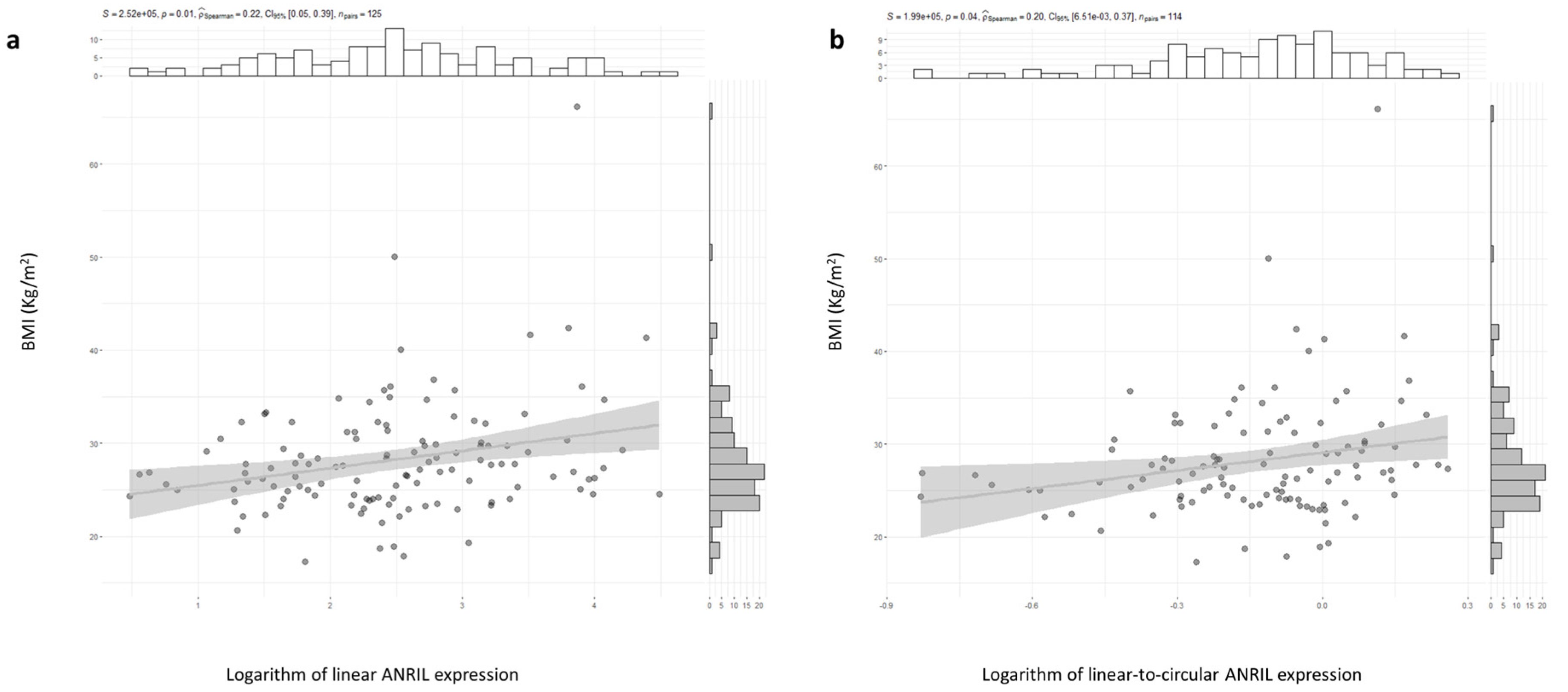
2.1. ANRIL and CDKN2A Gene Expression and CAD Risk Factor

Males had higher linear and linear-to-circular ANRIL expression values than women. For ANRIL expression, a median value of 153 (IQR 33–754) was found for men and 84 (IQR 17–278) for women (p = 0.04). For linear-to-circular ANRIL expression, a median value of 2 (IQR 0.32–17) was found for men and 0.42 (IQR 0.48–5.7) for women (p = 0.01). No differences were observed for circular isoforms and CDKN2A expression values.
There were statistically significant differences in circular ANRIL expression and the linear-to-circular expression ratio, according to hypertensive status. The expression of linear isoforms and the expression ratio between linear and circular isoforms of ANRIL were found to be different in hypertensive individuals compared to normotensive ones. The median value of linear isoforms of ANRIL in hypertensives was 123 (IQR 25–397) and 323 (IQR 67–757) in normotensives (p = 0.03). The median expression value of the ratio between linear and circular isoforms of ANRIL was 1.2 (IQR 0.2–7.5) in hypertensives and 7.4 (IQR 1.2–25) in normotensives (p = 0.05). There was a positive and significant correlation between linear ANRIL expression and BMI values (ρ = 0.224, p = 0.012, N = 125). Also, there was a positive and significant correlation between BMI values and linear-to-circular ANRIL expression values (ρ = 0.231, p = 0.04, N = 114) (Figure 1a,b). The expression levels of ANRIL linear isoforms differed according to a previously diagnosed of obesity. The median value was 748 (IQR 262–2742) in obese patients and 254 (IQR 58–897) in non-obese patients (p-value = 0.0057). When categorizing patients based on their mean IMC value, a significant difference was observed. Thus, the median linear ANRIL value was 107 (IQR 14–487) in patients with an IMC of 27 Kg/m2 or lower, while it was 166 (IQR 64–660) in those with an IMC higher than 27 Kg/m2 (p-value = 0.01023). No differences in the linear, circular, linear-to-circular ratio, and CDKN2A gene expression were found according to diabetic or dyslipidemic conditions, nor with respect to tobacco consumption. There were negative and significant correlations between circular ANRIL expression and CDKN2A gene expression and TBARS levels (rho = −0.2404738, S = 567281, p-value = 0.004211 and rho = −0.1871373, S = 628466, p-value = 0.02323, respectively).

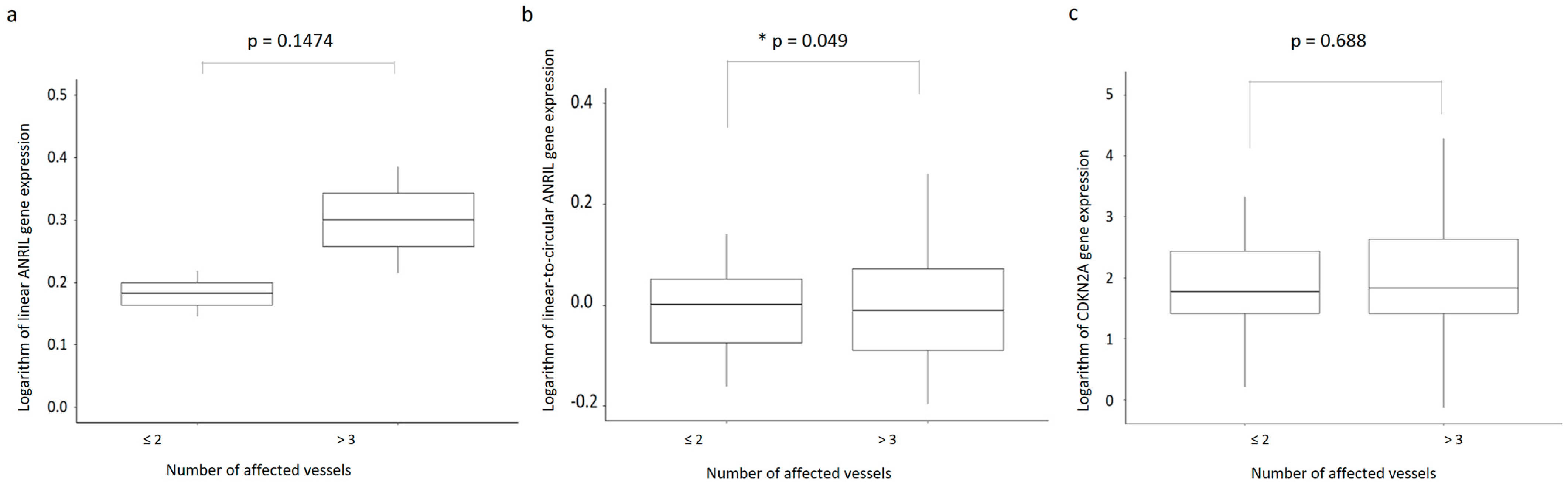
2.2. ANRIL and CDKN2A Gene Expression and CAD Severity

The linear, circular, and linear-to-circular ANRIL expression ratio and CDKN2A gene expression were analyzed, with respect to CAD severity. We found that, among patients stratified by the presence or absence of left ventricular dysfunction, there was a significant difference in the ANRIL linear-to-circular expression ratio. The median value was 0.74 (IQR 0.53–1) in patients with normal ventricular function, whereas a median value of 0.89 (IQR 0.6–1.18) was found in patients with ventricular dysfunction (p = 0.03).
There was a significant correlation between the patient’s number of affected vessels and the number of surgical revascularizations that were performed (ρ = 0.525, p < 0.0001, N = 163). In Figure 2a–c, the boxplots illustrate the expression pattern of linear ANRIL isoforms, the expression ratio of linear-to-circular, and the expression of CDKN2A in accordance with the number of affected vessels. Figure 3a–c presents boxplots depicting the same parameters, but relative to the number of revascularizations. Interestingly, when dichotomized values of affected vessel numbers were analyzed, they bordered the threshold of significance in relation to the linear-to-circular expression ratio (W = 1335, p = 0.49) (Figure 2b). Nonetheless, when considering individual expressions of linear or circular ANRIL isoforms or CDKN2A gene expression, no variations were discerned regarding either vessel number. However, the linear ANRIL expression and the ANRIL expression ratio showed significant differences with respect to the number of surgical revascularizations (χ2 = 5, p = 0.025 and χ2 = 4.3, p = 0.036, respectively) (Figure 3b).
Furthermore, no differences in linear or circular ANRIL isoforms expression or CDKN2A gene expression were found when considering the presence or absence of aortic trunk lesion.
Patients were divided into high- and low-expression categories based on medium and median values of linear and circular ANRIL and CDKN2A gene expression levels. Despite this segmentation, no association was observed between all-cause mortality and the analyzed gene expressions of ANRIL and CDKN2A.

2.3. Plasma VEGF, VEGF, and KDR Gene Expression and CAD Risk Factor

No differences were observed in VEGF and KDR gene expression according to sex. The median VEGF concentration was 31.12 (IQR 9.6–52), being significantly different between men and women (p = 0.049) with a median value for men of 37 (IQR 12.5–55), while for women it was 23 (IQR 7–32). However, no correlation was observed between VEGF expression and VEGF plasma levels in fully evaluated patients, also after stratifying for sex. VEGF and KDR gene expression were negatively correlated with TBARS levels (ρ = −0.253, p = 0.002, N = 147 and ρ = −0.202, p = 0.03, N = 116, respectively). The median VEGF concentration differed significantly between patients with and without a previous diagnosis of dyslipidemia (p = 0.016). For those with dyslipidemia, the median value was 37 (IQR 12.5–55), whereas for those without dyslipidemia at the time of diagnosis, the median was 19 (IQR 10–52). However, no differences in VEGF or KDR gene expression were observed with respect to diabetes and hypertension status, nor with respect to tobacco consumption.

2.4. Plasma VEGF, VEGF, and KDR Gene Expression and CAD Severity

VEGF gene expression correlated with the number of surgical revascularizations (ρ = 0.219, p = 0.007, N = 153) and with linear and circular ANRIL gene isoforms expression (ρ = 0.304, p < 0.001, N = 152 and ρ = 0.465, p < 0.0001, N = 141). Also, VEGF expression positively correlated with CDKN2A gene expression (ρ = 0.522, p < 0.001, N = 131). However, no associations were observed between VEGF and KDR gene expression levels with respect to evaluated CAD severity parameters. According to the medium and median values of both VEGF and KDR gene expression levels, all patients were divided into high- and low-expression groups. No association with mortality for all causes was observed for analyzed gene expressions.

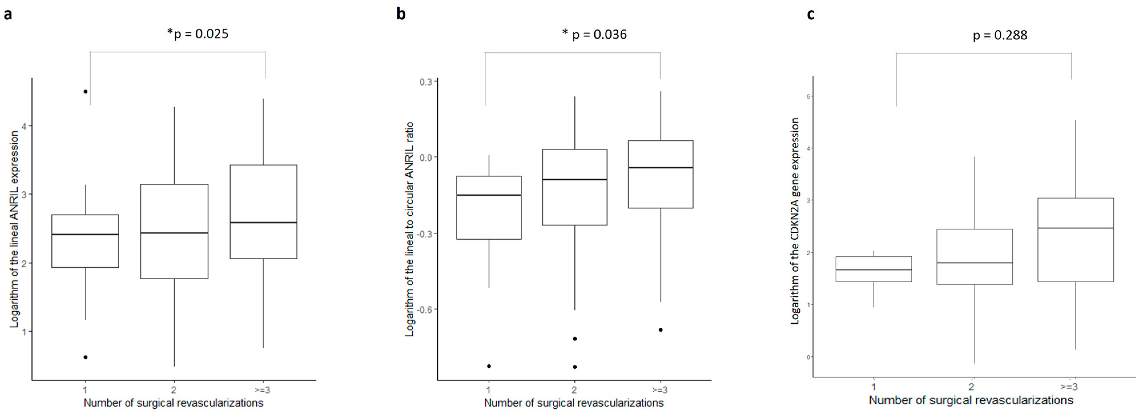
2.5. Gene Expressions as Predictors of Surgical Revascularizations

The contribution of the evaluated gene expressions, as well as clinical, biochemical, and anthropometric variables, were analyzed using logistic binary models. As dependent variables, we evaluated the number of affected vessels, the number of grafting procedures, and all-cause mortality.
Table 3, Table 4 and Table 5 shows the results, considering univariate models, a multivariate model, and the optimal model for each evaluated dependent variable.
No significant predictors were obtained either in single- or multivariate analysis of the number of affected vessels. On the contrary, a significant univariate contribution was obtained for the linear-to-circular ANRIL expression ratio and VEGF gene expression as predictors of the number of surgical revascularizations performed. Also, considering the CABG number as a dependent variable, the optimal multivariate model shows that linear ANRIL and VEFG gene expressions were significant predictors.
Neither univariate nor multivariate models were obtained with significant predictors of all-cause mortality. Non-prospective assessment of gene expression does not appear to contribute significantly to all-cause mortality.

3. Discussion

ANRIL is subjected to a variety of splicing patterns producing multiple isoforms [12]. Differences in the biological effects, according to the differential expression of a particular ANRIL linear isoform and cell types, have been previously described [13,14,15,35,36,37]. Also, it has been observed that, in coronary arteries and peripheral blood mononuclear cells (PBMCs) from CAD patients, the expressed ANRIL linear isoforms result from specific exon combinations [13,14,15]. Increased ANRIL linear isoform expression increases, in turn, proatherogenic cell activities like proliferation and reduced apoptosis, as well as the differential expression of hundreds of genes, without affecting the expression of CDKN2A and CDKN2B genes [12,38]. In proliferative cells, linear ANRIL isoforms prevent cell senescence by repressing INK4 genes through the recruitment of Polycomb-group proteins [21].
Although there have been some discordant results [20,39], most linear isoforms have been associated with an increased risk of atherosclerosis and CAD severity [12,19,35,38]. In addition to the presence of risk SNPS that are also ANRIL gene transcription modifiers [29], conflictive results are thought to occur because ANRIL isoforms’ abundance and functions depend on the tissue and/or cell type [13,15] and on the existence of circular forms [19,31,37]. Circular ANRIL isoforms (circANRIL) are formed from linear isoforms by back-splicing [12,18,31] and, contrarily to linear ANRIL isoforms, circANRIL confer atheroprotection by regulating ribosomal RNA (rRNA) maturation [16,31] and microRNA sponging [23,32,33]. Accordingly, the atherogenic or atheroprotective roles of ANRIL seem to depend, among other factors, on the predominant expression of particular isoforms in different cell types and the balance of linear and circANRIL expression [14,23], as well as the metabolic control that they exert on genes, microRNAs, and proteins, which in turn are involved in cell proliferation or senescence.
VEGF is an important regulator of angiogenesis, lymphopoiesis and lymphangiogenesis, oxidative stress, lipid metabolism, and inflammation [40]. VEGF accelerates vascular injury by promoting endothelial cell migration, proliferation, and vascular permeability [40,41]. ANRIL expression is increased under inflammatory stimuli [28] and increased ANRIL levels regulate in turn VEGF expression, activating the NF-κB pathway, recruiting PCRC2 or p300 and regulating the miR-200b expression [34,38].
In the analyzed blood samples of patients with severe atherosclerosis collected before undergoing coronary artery bypass grafting (CABG), a positive and significant correlation between the values of the expression ratio of linear-to-circular isoforms of ANRIL and linear ANRIL isoforms was found. In partial agreement with previous observations, we found in univariate analysis that the linear-to-circular expression ratio and circANRIL isoforms differed with respect to hypertensive status. Accordingly, lower levels of ANRIL in hypertensive CAD patients, with respect to non-hypertensive CAD patients, have been observed [23]. Transcriptome studies have also discovered the contributions of ANRIL, AK098656, MEG3, H19, PAXIP1-AS1, TUG1, GAS5, CASC2, and CPS1-IT, among other long non-coding RNAs, to the pathophysiology of hypertension [42]. We also found a significant correlation between BMI and linear and circular ANRIL isoforms, and differences in linear and circANRIL, according to previously diagnosed obesity. As previously reported, there is a significant described role for several lncRNAs, including linear ANRIL isoforms, in the regulation of inflammatory pathways associated with obesity [43,44]. Also, univariate analysis showed that the ANRIL expression ratio was a predictor of the number of affected vessels and the CABG number, whereas VEGF gene expression was a predictor of the number of surgical revascularizations.
We have evaluated an ANRIL amplicon that is in common with at least 17 of the 28 ANRIL linear transcripts and therefore represents the most frequent linear isoforms found in PBMCs from CAD patients. Additionally, the cirANRIL amplicon tested represents the most frequent circular ANRIL form described. Disease severity in CAD patients was evaluated by testing for the significant contributions of genes and clinical, biochemical, and anthropometric variables to the number of affected vessels, the number of CABGs, and all-cause mortality. We observed that the number of surgical revascularizations performed with internal mammary grafts was significantly correlated with the number of affected vessels in the patients evaluated. However, the joint analysis of gene expression and the different variables analyzed resulted in better and more precise multivariate models in the classification of the number of revascularizations than those obtained for the number of vessels or in the evaluation of mortality for all causes. Moreover, in the logistic binary regression analysis, both linear ANRIL and VEGF gene expression were predictors of the number of surgical revascularization procedures. However, no univariate or multivariate logistic models were obtained in which genes were significant predictors of affected vessels and all-cause mortality.
Nevertheless, these results should be considered with caution, considering our study limitations. Among others, this was a small sample study without a control group. A long-term assessment of overall survival would have been more informative than the assessment of all-cause mortality. Moreover, some ANRIL isoforms are predominant in specific cell types that are relevant in the atherosclerosis process, such as endothelial cells. Thus, the use only of peripheral blood mononuclear cells to determine the expression of the evaluated genes constitutes another significant limitation of the study. Furthermore, the prospective evaluation of gene expression would probably have also provided information of interest.
In conclusion, this study suggests that the assessment of the expression of linear and circular isoforms of ANRIL expression and VEGF are useful predictors of the CAGB number and CAD severity. However, linear ANRIL expression and the linear-to-circular expression ratio were found to be highly correlated, suggesting that a single evaluation of the ANRIL expression ratio does not provide more information than that obtained from the sole determination of the linear expression of ANRIL.

4. Materials and Methods

4.1. Patients

Blood samples were collected from 163 men and women undergoing coronary artery bypass grafting (CABG) at Hospital Universitario de Gran Canaria Dr. Negrín (HUGCDN), between April 2017 and April 2022. CABG indication was based on standard clinical and angiographic criteria. The study approval was granted by the Ethics Committee of the HUGCDN (reference 140157), and informed consent was obtained from all individual donors in accordance with Spanish legislation. Research was carried out in compliance with the Helsinki Declaration (http://www.wma.net/e/policy/b3.htm, accessed on 1 September 2023). CAD severity data were collected from medical records at the end of the study period. In that temporary evaluation it was observed that ten patients were exitus.

4.2. Biochemical Determinations

The reactive oxygen species bypass product malondialdehyde was measured in serum samples by thiobarbituric acid reactive substances (TBARS) assay. Serum VEGF levels were measured by an ELISA assay, as described by the manufacturer (Thermo Fisher, Waltham, MA, USA).

4.3. Gene Expression

Total RNAs were isolated from patients’ blood samples using Trizol reagent (Thermo Fisher Scientific, Madrid, Spain), according to the manufacturer’s instructions. cDNA synthesis was performed using the iScript™ kit (Biorad, Hercules, CA, USA). Analysis of the relative gene expression of CDKN2B-AS1 (ANRIL) was performed, using previously described primers with the sequences: 5′-TCACTGTTAGGTGTGCTGGAAT-3′ and 5′-CCTCTGATGGTTTCTTTGGAGT-3′. These primers amplified exon 6 of the actual annotated ANRIL exons structure. Circular ANRIL was amplified with previously described primers [11]. These primers amplified a fragment of the predominant circANRIL5–7 isoform, which consisted of exons 5, 6 and 7, where exon 7 was non-canonically spliced to exon 5. A 150bp fragment of the VEGFA gene was amplified with the primers 5′-TGGGCCTTGCTCAGAGCGGA-3′ and 5′-GCTCACCGCCTCGGCTTGTC-3′. The primer sequences for CDKN2A and KDR amplifications were as previously described [45,46].

4.4. Statistical Analysis

Data were presented as means ± standard deviations (SD) for continuous variables and as frequencies and percentages for categorical variables. The normality of continuous variables was assessed using a Kolmogorov–Smirnov test. Correlations and group comparisons were conducted using parametric or non-parametric tests, based on the normality test results. Analyzed variables included hypertension, diabetes, dyslipidemia, and obesity. Tobacco consumption was categorized as current smokers, including ex-smokers who had quit within one year, and non-smokers. Ventricular function was categorized as normal or as mild/moderate/severe dysfunction. The presence or absence of an aortic trunk lesion and the number of surgical revascularization procedures were recorded. The number of affected vessels was coded as one or two versus three or more. The grafting revascularization number was coded as one affected vessel or more than one vessel. Gene expression comparisons based on categorical values used the Wilcoxon test for dummy variables or the Kruskal–Wallis test for categorical variables with more than two categories. The multivariable association between specific coronary risk factors and evaluated gene expression was assessed using binary logistic regression. To identify independent predictors of the number of affected vessels, the number of CABG procedures, and all-causes mortality, stepwise logistic regression analyses were performed, computing both forward stepwise and backward selection to choose an optimal simple model without compromising accuracy. A value of p < 0.05 was considered statistically significant.

Author Contributions

Conceptualization: F.R.-E., L.B.T.-M., S.E.C.-R. and J.A.S.G.; investigation: F.R.-E., L.B.T.-M., S.E.C.-R., J.A.S.G. and B.C.V.; methodology: F.R.-E., L.B.T.-M., S.E.C.-R., L.E.-Q., M.C.B.-D., J.M.G.M. and Á.C.-M.; writing—original draft preparation: F.R.-E., L.B.T.-M., S.E.C.-R., J.A.S.G., B.C.V. and J.A.M.-S.; writing—review and editing: F.R.-E., L.B.T.-M., S.E.C.-R., J.A.S.G., A.N.G.-H., J.C.R.-P. and B.C.V.; funding acquisition: F.R.-E., B.C.V. and J.C.R.-P. All authors have read and agreed to the published version of the manuscript.

Funding

This research was funded by a grant to F.R.-E. from the Canary Islands Health Research Foundation (FIISC, PIFUN33/17), and by grants from DISA Foundation (2018) and Mapfre-Guanarteme Foundation (2018). S.C.-R. is a recipient of a Margarita Salas postdoctoral grant [Ministerio de Universidades (UNI/551/2021); Fondos Next Generation EU; Universidad de la Laguna].

Institutional Review Board Statement

The study was conducted in line with the principles of the Helsinki Declaration. Approval was granted by the Ethics Committee of the Hospital Universitario de Gran Canaria Dr. Negrín (Approval 140157).

Informed Consent Statement

Informed consent was obtained from all subjects involved in the study.

Data Availability Statement

All data needed to evaluate the conclusions of the paper are present and tabulated in the main text. This article is the result of an original analysis of data from patients with coronary artery disease. The corresponding author has full access to all data in the study and has the final responsibility for the integrity of the data, the accuracy of the data analysis, and the decision to submit for publication. All data associated with the article are available if required in Excel or R, and as pictures in .tif or .jpg formats.

Acknowledgments

We want to express our gratitude to our patients and their families. Thanks also to the funding agencies for their support of the research activity.

Conflicts of Interest

The authors declare no conflict of interest.

References

  1. Roth, G.A.; Mensah, G.A.; Johnson, C.O.; Addolorato, G.; Ammirati, E.; Baddour, L.M.; Barengo, N.C.; Beaton, A.Z.; Benjamin, E.J.; Benziger, C.P.; et al. Global Burden of Cardiovascular Diseases and Risk Factors, 1990–2019. J. Am. Coll. Cardiol. 2020, 76, 2982–3021. [Google Scholar] [CrossRef] [PubMed]
  2. Ozaki, K.; Tanaka, T. Molecular Genetics of Coronary Artery Disease. J. Hum. Genet. 2016, 61, 71–77. [Google Scholar] [CrossRef] [PubMed]
  3. Burton, P.R.; Clayton, D.G.; Cardon, L.R.; Craddock, N.; Deloukas, P.; Duncanson, A.; Kwiatkowski, D.P.; McCarthy, M.I.; Ouwehand, W.H.; Samani, N.J.; et al. Genome-Wide Association Study of 14,000 Cases of Seven Common Diseases and 3000 Shared Controls. Nature 2007, 447, 661–678. [Google Scholar] [CrossRef]
  4. McPherson, R.; Pertsemlidis, A.; Kavaslar, N.; Stewart, A.; Roberts, R.; Cox, D.R.; Hinds, D.A.; Pennacchio, L.A.; Tybjaerg-Hansen, A.; Folsom, A.R.; et al. A Common Allele on Chromosome 9 Associated with Coronary Heart Disease. Science 2007, 316, 1488–1491. [Google Scholar] [CrossRef] [PubMed]
  5. Samani, N.J.; Erdmann, J.; Hall, A.S.; Hengstenberg, C.; Mangino, M.; Mayer, B.; Dixon, R.J.; Meitinger, T.; Braund, P.; Wichmann, H.-E.; et al. Genomewide Association Analysis of Coronary Artery Disease. N. Engl. J. Med. 2007, 357, 443–453. [Google Scholar] [CrossRef]
  6. Helgadottir, A.; Thorleifsson, G.; Manolescu, A.; Gretarsdottir, S.; Blondal, T.; Jonasdottir, A.; Jonasdottir, A.; Sigurdsson, A.; Baker, A.; Palsson, A.; et al. A Common Variant on Chromosome 9p21 Affects the Risk of Myocardial Infarction. Science 2007, 316, 1491–1493. [Google Scholar] [CrossRef]
  7. Schunkert, H.; Götz, A.; Braund, P.; McGinnis, R.; Tregouet, D.-A.; Mangino, M.; Linsel-Nitschke, P.; Cambien, F.; Hengstenberg, C.; Stark, K.; et al. Repeated Replication and a Prospective Meta-Analysis of the Association Between Chromosome 9p21.3 and Coronary Artery Disease. Circulation 2008, 117, 1675–1684. [Google Scholar] [CrossRef]
  8. Aragam, K.G.; Jiang, T.; Goel, A.; Kanoni, S.; Wolford, B.N.; Atri, D.S.; Weeks, E.M.; Wang, M.; Hindy, G.; Zhou, W.; et al. Discovery and Systematic Characterization of Risk Variants and Genes for Coronary Artery Disease in over a Million Participants. Nat. Genet. 2022, 54, 1803–1815. [Google Scholar] [CrossRef]
  9. Johnson, A.D.; Hwang, S.-J.; Voorman, A.; Morrison, A.; Peloso, G.M.; Hsu, Y.-H.; Thanassoulis, G.; Newton-Cheh, C.; Rogers, I.S.; Hoffmann, U.; et al. Resequencing and Clinical Associations of the 9p21.3 Region. Circulation 2013, 127, 799–810. [Google Scholar] [CrossRef]
  10. Musunuru, K. Enduring Mystery of the Chromosome 9p21.3 Locus. Circ. Cardiovasc. Genet. 2013, 6, 224–225. [Google Scholar] [CrossRef]
  11. Kong, Y.; Hsieh, C.-H.; Alonso, L.C. ANRIL: A LncRNA at the CDKN2A/B Locus With Roles in Cancer and Metabolic Disease. Front. Endocrinol. 2018, 9, 405. [Google Scholar] [CrossRef]
  12. Razeghian-Jahromi, I.; Karimi Akhormeh, A.; Zibaeenezhad, M.J. The Role of ANRIL in Atherosclerosis. Dis. Markers 2022, 2022, 8859677. [Google Scholar] [CrossRef]
  13. Holdt, L.M.; Beutner, F.; Scholz, M.; Gielen, S.; Gäbel, G.; Bergert, H.; Schuler, G.; Thiery, J.; Teupser, D. ANRIL Expression Is Associated with Atherosclerosis Risk at Chromosome 9p21. Arterioscler. Thromb. Vasc. Biol. 2010, 30, 620–627. [Google Scholar] [CrossRef] [PubMed]
  14. Holdt, L.M.; Hoffmann, S.; Sass, K.; Langenberger, D.; Scholz, M.; Krohn, K.; Finstermeier, K.; Stahringer, A.; Wilfert, W.; Beutner, F.; et al. Alu Elements in ANRIL Non-Coding RNA at Chromosome 9p21 Modulate Atherogenic Cell Functions through Trans-Regulation of Gene Networks. PLoS Genet. 2013, 9, e1003588. [Google Scholar] [CrossRef]
  15. Holdt, L.M.; Teupser, D. Long Noncoding RNA ANRIL: Lnc-Ing Genetic Variation at the Chromosome 9p21 Locus to Molecular Mechanisms of Atherosclerosis. Front. Cardiovasc. Med. 2018, 5, 145. [Google Scholar]
  16. Holdt, L.M.; Stahringer, A.; Sass, K.; Pichler, G.; Kulak, N.A.; Wilfert, W.; Kohlmaier, A.; Herbst, A.; Northoff, B.H.; Nicolaou, A.; et al. Circular Non-Coding RNA ANRIL Modulates Ribosomal RNA Maturation and Atherosclerosis in Humans. Nat. Commun. 2016, 7, 12429. [Google Scholar] [CrossRef] [PubMed]
  17. Wilusz, J.E. Circular RNAs: Unexpected Outputs of Many Protein-Coding Genes. RNA Biol. 2017, 14, 1007–1017. [Google Scholar] [CrossRef] [PubMed]
  18. Hu, Y.; Hu, J. Diagnostic Value of Circulating LncRNA ANRIL and Its Correlation with Coronary Artery Disease Parameters. Brazilian J. Med. Biol. Res. 2019, 52, e8309. [Google Scholar] [CrossRef]
  19. Qin, Z.; Liu, D. Circulating LncRNA ANRIL Level Positively Correlates with Disease Risk, Severity, Inflammation Level and Poor Prognosis of Coronary Artery Disease. Int. J. Clin. Exp. Med. 2019, 12, 8964–8970. [Google Scholar]
  20. Liu, Z.F.; Hu, W.W.; Li, R.; Gao, Y.; Yan, L.L.; Su, N. Expression of LncRNA-ANRIL in Patients with Coronary Heart Disease before and after Treatment and Its Short-Term Prognosis Predictive Value. Eur. Rev. Med. Pharmacol. Sci. 2020, 24, 376–384. [Google Scholar] [CrossRef]
  21. Muniz, L.; Lazorthes, S.; Delmas, M.; Ouvrard, J.; Aguirrebengoa, M.; Trouche, D.; Nicolas, E. Circular ANRIL Isoforms Switch from Repressors to Activators of P15/CDKN2B Expression during RAF1 Oncogene-Induced Senescence. RNA Biol. 2021, 18, 404–420. [Google Scholar] [CrossRef] [PubMed]
  22. Qin, Q.; Zheng, P.; Tu, R.; Huang, J.; Cao, X. Integrated Bioinformatics Analysis for the Identification of Hub Genes and Signaling Pathways Related to CircANRIL. PeerJ 2022, 10, e13135. [Google Scholar] [CrossRef] [PubMed]
  23. Razeghian-Jahromi, I.; Zibaeenezhad, M.J.; Karimi Akhormeh, A.; Dara, M. Expression Ratio of Circular to Linear ANRIL in Hypertensive Patients with Coronary Artery Disease. Sci. Rep. 2022, 12, 1802. [Google Scholar] [CrossRef] [PubMed]
  24. Wagner, K.-D.; Wagner, N. The Senescence Markers P16INK4A, P14ARF/P19ARF, and P21 in Organ Development and Homeostasis. Cells 2022, 11, 1966. [Google Scholar] [CrossRef]
  25. Collado, M.; Blasco, M.A.; Serrano, M. Cellular Senescence in Cancer and Aging. Cell 2007, 130, 223–233. [Google Scholar] [CrossRef] [PubMed]
  26. González-Navarro, H.; Abu Nabah, Y.N.; Vinué, Á.; Andrés-Manzano, M.J.; Collado, M.; Serrano, M.; Andrés, V. P19ARFDeficiency Reduces Macrophage and Vascular Smooth Muscle Cell Apoptosis and Aggravates Atherosclerosis. J. Am. Coll. Cardiol. 2010, 55, 2258–2268. [Google Scholar] [CrossRef] [PubMed]
  27. Kotake, Y.; Nakagawa, T.; Kitagawa, K.; Suzuki, S.; Liu, N.; Kitagawa, M.; Xiong, Y. Long Non-Coding RNA ANRIL Is Required for the PRC2 Recruitment to and Silencing of P15INK4B Tumor Suppressor Gene. Oncogene 2011, 30, 1956–1962. [Google Scholar] [CrossRef]
  28. Zhou, X.; Han, X.; Wittfeldt, A.; Sun, J.; Liu, C.; Wang, X.; Gan, L.M.; Cao, H.; Liang, Z. Long Non-Coding RNA ANRIL Regulates Inflammatory Responses as a Novel Component of NF-ΚB Pathway. RNA Biol. 2016, 13, 98–108. [Google Scholar] [CrossRef]
  29. Xu, B.; Xu, Z.; Chen, Y.; Lu, N.; Shu, Z.; Tan, X.; Franco, N.R.; Massi, M.C.; Ieva, F.; Manzoni, A.; et al. Genetic and Epigenetic Associations of ANRIL with Coronary Artery Disease and Risk Factors. BMC Med. Genom. 2021, 14, 240. [Google Scholar] [CrossRef]
  30. Han, T.S.; Hur, K.; Cho, H.S.; Ban, H.S. Epigenetic Associations between LncRNA/CircRNA and MiRNA in Hepatocellular Carcinoma. Cancers 2020, 12, 2622. [Google Scholar] [CrossRef]
  31. Maguire, E.M.; Xiao, Q. Noncoding RNAs in Vascular Smooth Muscle Cell Function and Neointimal Hyperplasia. FEBS J. 2020, 287, 5260–5283. [Google Scholar] [CrossRef]
  32. Zeng, Z.; Xia, L.; Fan, S.; Zheng, J.; Qin, J.; Fan, X.; Liu, Y.; Tao, J.; Liu, Y.; Li, K.; et al. Circular RNA CircMAP3K5 Acts as a MicroRNA-22-3p Sponge to Promote Resolution of Intimal Hyperplasia Via TET2-Mediated Smooth Muscle Cell Differentiation. Circulation 2021, 143, 354–371. [Google Scholar] [CrossRef] [PubMed]
  33. Thum, T.; Condorelli, G. Long Noncoding RNAs and MicroRNAs in Cardiovascular Pathophysiology. Circ. Res. 2015, 116, 751–762. [Google Scholar] [CrossRef] [PubMed]
  34. Zhang, B.; Wang, D.; Ji, T.-F.; Shi, L.; Yu, J.-L. Overexpression of LncRNA ANRIL Up-Regulates VEGF Expression and Promotes Angiogenesis of Diabetes Mellitus Combined with Cerebral Infarction by Activating NF-ΚB Signaling Pathway in a Rat Model. Oncotarget 2017, 8, 17347–17359. [Google Scholar] [CrossRef]
  35. Cho, H.; Shen, G.Q.; Wang, X.; Wang, F.; Archacki, S.; Li, Y.; Yu, G.; Chakrabarti, S.; Chen, Q.; Wang, Q.K. Long Noncoding RNA ANRIL Regulates Endothelial Cell Activities Associated with Coronary Artery Disease by Up-Regulating CLIP1, EZR, and LYVE1 Genes. J. Biol. Chem. 2019, 294, 3881–3898. [Google Scholar] [CrossRef]
  36. Cho, H.; Li, Y.; Archacki, S.; Wang, F.; Yu, G.; Chakrabarti, S.; Guo, Y.; Chen, Q.; Wang, Q.K. Splice Variants of LncRNA RNA ANRIL Exert Opposing Effects on Endothelial Cell Activities Associated with Coronary Artery Disease. RNA Biol. 2020, 17, 1391–1401. [Google Scholar] [CrossRef]
  37. Hu, D.-J.; Li, Z.-Y.; Zhu, Y.-T.; Li, C.-C. Overexpression of Long Noncoding RNA ANRIL Inhibits Phenotypic Switching of Vascular Smooth Muscle Cells to Prevent Atherosclerotic Plaque Development in Vivo. Aging 2021, 13, 4299–4316. [Google Scholar] [CrossRef]
  38. Chen, L.; Qu, H.; Guo, M.; Zhang, Y.; Cui, Y.; Yang, Q.; Bai, R.; Shi, D. ANRIL and Atherosclerosis. J. Clin. Pharm. Ther. 2020, 45, 240–248. [Google Scholar] [CrossRef]
  39. Vausort, M.; Wagner, D.R.; Devaux, Y. Long Noncoding RNAs in Patients with Acute Myocardial Infarction. Circ. Res. 2014, 115, 668–677. [Google Scholar] [CrossRef]
  40. Dabravolski, S.A.; Khotina, V.A.; Omelchenko, A.V.; Kalmykov, V.A.; Orekhov, A.N. The Role of the VEGF Family in Atherosclerosis Development and Its Potential as Treatment Targets. Int. J. Mol. Sci. 2022, 23, 931. [Google Scholar] [CrossRef]
  41. Li, X.; Wang, J.; Wang, L.; Feng, G.; Li, G.; Yu, M.; Li, Y.; Liu, C.; Yuan, X.; Zang, G.; et al. Impaired Lipid Metabolism by Age-Dependent DNA Methylation Alterations Accelerates Aging. Proc. Natl. Acad. Sci. USA 2020, 117, 4328–4336. [Google Scholar] [CrossRef] [PubMed]
  42. Ghafouri-Fard, S.; Shirvani-Farsani, Z.; Hussen, B.M.; Taheri, M.; Samsami, M. The Key Roles of Non-Coding RNAs in the Pathophysiology of Hypertension. Eur. J. Pharmacol. 2022, 931, 175220. [Google Scholar] [CrossRef]
  43. Wijesinghe, S.N.; Nicholson, T.; Tsintzas, K.; Jones, S.W. Involvements of Long Noncoding RNAs in Obesity-associated Inflammatory Diseases. Obes. Rev. 2021, 22, e13156. [Google Scholar] [CrossRef] [PubMed]
  44. Yau, M.; Xu, L.; Huang, C.-L.; Wong, C.-M. Long Non-Coding RNAs in Obesity-Induced Cancer. Non-Coding RNA 2018, 4, 19. [Google Scholar] [CrossRef] [PubMed]
  45. Tagawa, S.; Nakanishi, C.; Mori, M.; Yoshimuta, T.; Yoshida, S.; Shimojima, M.; Yokawa, J.; Kawashiri, M.; Yamagishi, M.; Hayashi, K. Determination of Early and Late Endothelial Progenitor Cells in Peripheral Circulation and Their Clinical Association with Coronary Artery Disease. Int. J. Vasc. Med. 2015, 2015, 674213. [Google Scholar] [CrossRef]
  46. Varela-Eirín, M.; Carpintero-Fernández, P.; Sánchez-Temprano, A.; Varela-Vázquez, A.; Paíno, C.L.; Casado-Díaz, A.; Continente, A.C.; Mato, V.; Fonseca, E.; Kandouz, M.; et al. Senolytic Activity of Small Molecular Polyphenols from Olive Restores Chondrocyte Redifferentiation and Promotes a Pro-Regenerative Environment in Osteoarthritis. Aging 2020, 12, 15882–15905. [Google Scholar] [CrossRef] [PubMed]
Figure 1. (a) Linear ANRIL expression and (b) linear-to-circular ANRIL expression ratio correlated with BMI in CAD patients. Each position of a point on the graph is determined by the values of both variables for that observation. The points with darker colors occur when observations overlap.
Ijms 24 16127 g001
Figure 2. Boxplots of the (a) logarithm of the ANRIL linear expression, (b) logarithm of linear-to-circular ANRIL expression ratio, and (c) logarithm of the CDKN2A gene expression according to the number of affected vessels. * p < 0.005.
Ijms 24 16127 g002
Figure 3. Boxplots of the (a) logarithm of the ANRIL linear expression, (b) logarithm of linear-to-circular ANRIL expression ratio, and (c) logarithm of the CDKN2A gene expression according to the number of revascularization procedures. * p < 0.05.
Ijms 24 16127 g003
Table 1. Baseline characteristics of CAD patients.
N.MeanSt. Dev.Min.Max.
Age (years)16370.9639.43835101
BMI (Kg/m2) 13528.2066.11517.29066.120
SBP (mmHg)161129.01221.42985200
DBP (mmHg)16172.45314.31248169
Cholesterol (mg/dL)141139.55341.88159355
HDL cholesterol (mg/dL)11840.86913.91417.000138.000
LDL cholesterol (mg/dL)10477.61828.20322.900151.000
Triglycerides (mg/dL)141145.35271.78855.000585.000
TBARS (µM)1532.2851.1821.1187.302
Plasma VEGF (pg/mL) 4040.95145.4454.432228.102
Glycemia (mg/dL)162143.29657.38667442
Urea (mg/dL) 14854.20936.08613258
Urate (mg/dL)545.2441.8571.18011.140
Creatinine (mg/dL)1611.2320.9310.5008.400
GFR (CKD-EPI) (ml/min−1)16170.17726.4025.480118.660
GFR (MDRD-IMDS) (ml/min−1)16072.65730.5496.200166.880
Folic acid (ng/mL)236.9964.3811.80018.800
Vitamin B12 (pg/mL)29357.828197.4551591021
Leukocytes (×103/µL)16210.0744.7891.99031.710
Neutrophils (×103/µL)1627.2144.7321.26030.310
Lymphocytes (×103/µL)1621.8320.8840.2405.080
Monocytes (×103/µL)1620.7770.3940.0303.200
Eosinophils (×103/µL)1620.2050.1920.0000.950
Basophils (×103/µL) 1620.0430.0310.0000.180
Red blood cells (×103/µL)1624.0000.8162.4406.060
BMI: body mass index; SBP: systolic blood pressure; DBP: diastolic blood pressure; HDL cholesterol: high-density lipoprotein cholesterol; TBARS: thiobarbituric acid reactive substances; plasma VEGF: plasma vascular endothelial growth factor; GRF (CKD-EPI): glomerular filtration rate, chronic kidney disease epidemiology collaboration; GFR (MDRD-IMDS): glomerular filtration rate, modification of diet in renal disease study group equation.
Table 2. Medical conditions and habits of patients by gender.
Condition/HabitTotal (%)Male (%)Female (%)p Value
Number of Patients16313132-
Hypertensive patients82.881.687.50.434
Diabetic patients60.757.2750.065
Patients with dyslipidemia79.777.887.50.224
Smokers47.254.218.8<0.001
Patients with overweight15.915.218.70.630
Patients with LVD *58.355.768.70.180
Patients with aortic trunk lesion3334.628.10.485
Patients with ≥2 affected vessels9293.984.40.075
Patients with ≥2 revascularizations82.88765.60.004
All causes of death26.426.7250.843
* LVD: left ventricular dysfunction.
Table 3. Univariate and multivariate analysis of predictors of the number of affected vessels in CAD patients.
VariablesUnivariateMultivariateOptimal Multivariate Model
NBetaSE ORCI 95p-ValueBetaSEORCI 95p-ValueBetaSEORCI 95p-Value
Intercept 2.962.4919.3180.157–3574.7190.2352.831.116.9262.91–328.830.01
Female sex 77−0.580.590.560.18–1.860.325−0.90.80.4050.08–1.9580.26-----
Hypertension77−1.511.080.220.01–1.250.161−1.491.190.2260.011–1.6510.211−1.571.090.2090.011–1.210.15
Diabetes770.460.521.590.57–4.490.3750.720.642.0630.599–7.6570.258-----
Dyslipidemia77−0.170.640.850.21–2.810.7950.210.811.2390.234–5.990.791-----
Smoker770.170.521.180.42–3.350.7510.20.751.2260.28–5.5060.785-----
LVD 77−0.450.520.640.22–1.780.392−0.530.610.590.174–1.9320.385-----
Aortic trunk77−0.820.530.440.15–1.240.122−1.30.660.2740.069–0.9650.051−0.850.540.4260.14–1.210.113
BMI770.010.041.010.93–1.10.857−0.030.050.9720.884–1.0830.57-----
Lineal ANRIL770.320.351.380.7–2.810.360.680.581.9780.66–6.6810.239-----
Circular ANRIL77−0.140.310.870.48–1.660.646−0.170.380.8410.401–1.7980.645-----
Ratio ANRIL771.261.063.530.15–86.80.43----------
CDKN2A770.010.321.010.53–1.930.983−0.560.580.5710.169–1.7520.335-----
VEGF770.040.291.040.59–1.90.890.110.411.1120.499–2.5630.794-----
KDR770.180.191.190.81–1.740.3570.110.221.1150.719–1.7180.618-----
AUC ROC 0.7509 0.643
Factors predicting the number of affected vessels were analyzed using univariate and multivariate logistic regression. The reference categories are as follows: for hypertension, diabetes, and dyslipidemia, it was ‘previously diagnosed disease’; for ‘smoker’, it was ‘current smoker’; for LVD, it was ‘mild to severe dysfunction’; and for ‘aortic lesion’, it was ‘presence of aortic trunk lesion’. LVD: left ventricular dysfunction; BMI: body mass index; CDKN2A: cyclin dependent kinase inhibitor 2A; VEGF: vascular endothelial growth factor; KDR: kinase insert domain receptor; AUC ROC: area under the ROC curve.
Table 4. Univariate and multivariate analysis of predictors of the number of surgical revascularizations in CAD patients.
VariablesUnivariateMultivariateOptimal Multivariate Model
NBetaSEORCI 95p-ValueBetaSEORCI 95p-ValueBetaSEORCI 95p-Value
Intercept------−2.383.160.0920–36.0390.451−1.412.370.2450.001–19.760.554
Femalesex77−10.650.37 0.1–1.4 0.128−1.591.280.2040.013–2.2410.212−1.850.990.1570.018–0.9880.062
Hypertension77−0.911.090.4 0.02–2.38 0.4030.031.611.0260.025–22.300.987-----
Diabetes770.60.611.82 0.54–6.26 0.3282.071.127.9531.17–109.030.0631.770.875.8511.185–39.8240.043
Dyslipidemia770.070.721.07 0.22–4.09 0.9241.91.216.6620.689–93.450.1161.931.126.8760.809–75.7080.085
Smoker770.090.611.1 0.33–3.75 0.8810.11.141.110.122–12.430.927-----
LVD770.410.621.5 0.45–5.44 0.513−0.190.940.8260.115–5.230.838-----
Aortictrunk770.950.72.59 0.71–12.38 0.1760.990.982.6990.433–22.510.31-----
BMI77−0.010.040.99 0.91–1.09 0.768−0.140.080.870.724–1.020.092−0.140.070.8720.75–1.0090.06
LinearANRIL770.820.452.27 0.99–5.86 0.0671.690.885.4191.184–42.450.0551.570.724.81.314–23.8960.029
CircularANRIL77−0.290.340.75 0.39–1.54 0.396−0.990.630.3710.087–1.10.118−0.850.50.4290.14–1.070.09
RatioANRIL773.281.9326.54 0.66–1436.8 0.09----------
CDKN2A770.280.391.32 0.62–2.96 0.481−1.260.80.2840.046–1.1820.118−0.920.630.3990.105–1.3180.143
VEGF770.850.432.33 1.07–5.85 0.0482.731.0615.3692.65–183.230.012.350.8410.5162.378–67.6350.005
KDR770.30.221.34 0.87–2.06 0.1720.380.321.4590.783–2.850.237-----
AUCROC-0.8870.863
Factors predicting the number of surgical revascularizations were analyzed using univariate and multivariate logistic regression. The reference categories are as follows: for hypertension, diabetes, and dyslipidemia, it was ‘previously diagnosed disease’; for ‘smoker’, it was ‘current smoker’; for LVD, it was ‘mild to severe dysfunction’; and for ‘aortic lesion’, it was ‘presence of aortic trunk lesion’. LVD: left ventricular dysfunction; BMI: body mass index; CDKN2A: cyclin dependent kinase inhibitor 2A; VEGF: vascular endothelial growth factor; KDR: kinase insert domain receptor; AUC ROC: area under the ROC curve.
Table 5. Univariate and multivariate analysis of predictors of all causes of mortality in CAD patients.
VariablesUnivariateMultivariateOptimal Multivariate Model
NBetaSEORCI 95p-ValueBetaSEORCI 95p-ValueBetaSEORCI 95p-Value
Intercept------2.772.9416.0190.064–79380.3464.12.0460.0871.399–47120.045
Femalesex7710.612.73 0.79–9.08 0.1031.390.944.0170.654–28.50.141.160.723.180.772–13.60.108
Hypertension77−0.290.740.75 0.19–3.73 0.696−0.040.970.9580.151–7.5210.965-----
Diabetes77−0.110.570.89 0.29–2.8 0.8420.30.721.3490.339–5.9320.676-----
Dyslipidemia77−1.380.610.25 0.07–0.85 0.024−1.350.870.2610.044–1.4370.122−1.490.720.2250.052–0.910.038
Smoker77−0.220.570.8 0.26–2.43 0.6990.760.852.1290.414–12.5010.375-----
LVD770.480.571.62 0.54–5.07 0.3950.520.711.680.418–7.2260.467-----
Aortictrunk77−0.870.630.42 0.11–1.36 0.17−0.750.750.4740.097–1.990.321-----
IMC77−0.210.080.81 0.69–0.93 0.007−0.180.080.8350.692–0.9680.031−0.180.080.8380.708–0.960.022
LinealANRIL77−0.350.380.71 0.33–1.47 0.363−0.460.730.630.141–2.5130.527-----
CircularANRIL770.090.341.09 0.53–2.05 0.7970.10.431.1050.451–2.5690.818-----
RatioANRIL77−1.931.740.15 0–4.31 0.267----------
CDKN2A770.080.351.09 0.54–2.16 0.8130.720.782.0470.46–9.9310.356-----
VEGF770.350.311.42 0.77–2.63 0.2610.190.521.2110.424–3.4530.714-----
KDR77−0.130.210.88 0.59–1.34 0.537−0.130.260.8750.53–1.4960.606-----
AUCROC-0.81350.7684
Factors predicting mortality were analyzed using univariate and multivariate logistic regression. The reference categories are as follows: for hypertension, diabetes, and dyslipidemia, it was ‘previously diagnosed disease’; for ‘smoker’, it was ‘current smoker’; for LVD, it was ‘mild to severe dysfunction’; and for ‘aortic lesion’, it was ‘presence of aortic trunk lesion’. LVD: left ventricular dysfunction; BMI: body mass index; CDKN2A: cyclin dependent kinase inhibitor 2A; VEGF: vascular endothelial growth factor; KDR: kinase insert domain receptor; AUC ROC: area under the ROC curve.
Disclaimer/Publisher’s Note: The statements, opinions and data contained in all publications are solely those of the individual author(s) and contributor(s) and not of MDPI and/or the editor(s). MDPI and/or the editor(s) disclaim responsibility for any injury to people or property resulting from any ideas, methods, instructions or products referred to in the content.

