Review

Acute Erythroid Leukemia: From Molecular Biology to Clinical Outcomes

by
Priyanka Fernandes
1,†,
Natalie Waldron
1,†,
Theodora Chatzilygeroudi
2,‡,
Nour Sabiha Naji
2,‡ and
Theodoros Karantanos
1,2,*
1
Johns Hopkins School of Medicine, Baltimore, MD 21205, USA
2
Department of Oncology, Johns Hopkins School of Medicine, Baltimore, MD 21205, USA
*
Author to whom correspondence should be addressed.
These authors contributed equally to this work.
Int. J. Mol. Sci. 2024, 25(11), 6256; https://doi.org/10.3390/ijms25116256
Submission received: 12 April 2024 / Revised: 30 May 2024 / Accepted: 3 June 2024 / Published: 6 June 2024
(This article belongs to the Section Molecular Pathology, Diagnostics, and Therapeutics)

Abstract

:
Acute Erythroid Leukemia (AEL) is a rare and aggressive subtype of Acute Myeloid Leukemia (AML). In 2022, the World Health Organization (WHO) defined AEL as a biopsy with ≥30% proerythroblasts and erythroid precursors that account for ≥80% of cellularity. The International Consensus Classification refers to this neoplasm as “AML with mutated TP53”. Classification entails ≥20% blasts in blood or bone marrow biopsy and a somatic TP53 mutation (VAF > 10%). This type of leukemia is typically associated with biallelic TP53 mutations and a complex karyotype, specifically 5q and 7q deletions. Transgenic mouse models have implicated several molecules in the pathogenesis of AEL, including transcriptional master regulator GATA1 (involved in erythroid differentiation), master oncogenes, and CDX4. Recent studies have also characterized AEL by epigenetic regulator mutations and transcriptome subgroups. AEL patients have overall poor clinical outcomes, mostly related to their poor response to the standard therapies, which include hypomethylating agents and intensive chemotherapy. Allogeneic bone marrow transplantation (AlloBMT) is the only potentially curative approach but requires deep remission, which is very challenging for these patients. Age, AlloBMT, and a history of antecedent myeloid neoplasms further affect the outcomes of these patients. In this review, we will summarize the diagnostic criteria of AEL, review the current insights into the biology of AEL, and describe the treatment options and outcomes of patients with this disease.

1. Introduction

Acute Erythroid Leukemia (AEL) is a rare but aggressive subtype of Acute Myeloid Leukemia (AML) and constitutes 2% of all AML cases [1,2]. AEL is often associated with a complex karyotype, as well as the biallelic loss of TP53. The aggressive biology of this disease and its high-risk molecular features render AEL a particularly difficult disease to manage and cure [1]. As a result, the median survival of AEL ranges from 3 to 9 months, though studies incorporating the newest myeloid neoplasms classifications have noted that most AEL patients fall on the shorter end of that spectrum [3].
Most AEL cases arise in a de novo fashion, though a subgroup arises from antecedent myelodysplastic syndrome (MDS) or from other chronic myeloid neoplasms [2]. There are no definitive associations of genetic and environment risk factors with the development of AEL. A number of studies conducted before the newest myeloid diseases classifications have highlighted a possible implication of germline variants and environmental factors such as benzene toxin that could increase the risk of AEL [1,4].
In this review, we will discuss the various classification systems used in the definition of AEL, most notably the World Health Organization (WHO) and the International Census Classification (ICC) systems, and will describe the differing degrees of emphasis on disease etiology, aspirate percentages, and genetic markers [5,6,7]. Though classification systems differ in the naming convention of this disease, our review will use the most recent 2022 WHO naming terminology of “AEL” when discussing this disorder. Diagnostic features and work up will also be discussed, emphasizing the non-specific nature of clinical presentation, bone marrow aspirate features, flow cytometry markers that demonstrate erythroblast lineage, and additional genetic mutations linked to AEL. We will describe the current knowledge of the molecular biology of this disease, which is critical for the introduction of novel therapeutic strategies that are urgently needed for AEL. Finally, we will analyze the current therapeutic approaches for this disease, including chemotherapy, hypomethylating agents, and bone marrow transplantation.

2. Definitions

The condition referred to as erythroleukemia was first discovered in 1917 by Giovanni Di Guglielmo, who noted the large number of erythrocyte, platelet, and granule precursors. Di Guglielmo noted that this disorder could be subclassified as a pure acute, chronic, or mixed phenotype. In 1976, the French–American–British cooperative group defined this disease as AML-M6 based on the differentiation features of the leukemia cells [8]. In 2001, the WHO further categorized the FAB AML-M6 into the M6a and M6b subtypes. The M6a classification became known as the erythroid/myeloid leukemia subtype and required erythroblasts to comprise ≥50% of the total nucleated bone marrow cells and myeloblasts to compromise ≥20% of the remaining non-erythroid cells [8]. This subtype was removed in 2016 from the WHO classification system, and its previous criteria became encompassed by other subtypes, such as Myelodysplastic Syndrome and Myelodysplasia-Related Changes (MRC) AML. Contrastingly, the M6b subtype became known as Pure Erythroid Leukemia (PEL). This subtype required >80% erythroid precursors and ≥30% proerythroblasts in the bone marrow. There continues to be many classification criteria to redescribe this aggressive subtype of AML, most notably from the WHO and the ICC organizations.

2.1. World Health Organization

The WHO has revised their classification of AEL several times over the last few decades, making it more stringent with a higher cellularity and blast qualifying criteria. Their most recent 2022 classification defines AEL as a subset of differentiated AML that has (1) a bone marrow sample of ≥30% proerythroblasts and (2) pro-erythroid precursors that account for ≥80% of cellularity [5]. This definition can be challenging in practice, however, since many bone marrow biopsies in AEL are suboptimal and require cytology or immunohistochemistry to aid in diagnosis. As such, some cases with <80% cellularity are now being recognized within the AEL classification category [7]. This change is the main differentiating factor from the previous 2016 WHO criteria.
The WHO discusses the cellular features of AEL, noting maturation arrest, complex karyotype, and biallelic TP53 mutations as supporters of the diagnosis. The presence of these findings does not change the classification label assigned to AEL. However, the findings of previous myeloid neoplasm/MDS or defining germline predispositions require a different and more specific classification of AEL [5]. Of note, the denominator used for calculating the blast percentage in myeloid neoplasms has changed to all nucleated BM cells, not just the “non-erythroid cells”, as in 2016 [9]. As a result, most cases previously diagnosed as erythroid leukemias are now classified as MDS with excess blasts.

2.2. International Census Classification

The 2022 International Census Classification (ICC) system emphasized both blast percentage and genomic factors when discussing the diagnosis of AEL/PEL and classifies this disorder within the broad category of myeloid neoplasms with mutated TP53 due to their similarly aggressive clinical behavior [6]. One subcategory of this broad grouping is AML with mutated TP53. The criteria for this ICC diagnosis entail (1) ≥20% bone marrow or blood blasts or meeting the criteria for pure erythroid leukemia and (2) any form of a somatic TP53 mutation with a variant allele frequency (VAF) >10% [6]. As determined by blast percentages, AEL/PEL, which commonly harbors a biallelic TP53 mutation and a complex karyotype, now falls under the umbrella category of AML with mutated TP53 [10]. It should be noted that the definition of AML with mutated TP53 is broad and regularly includes non-AEL cases. It is also important to note that this classification is independent of the disease origin (de novo or secondary or treatment-related), unlike the 2022 WHO criteria.

3. Diagnosis

3.1. Presentation

The median age of AEL diagnosis is 67 years old, though some studies have demonstrated a bimodal age of diagnosis with a small peak at around 20 years old and a larger second peak in the early 70s [1,10]. This disease also demonstrates a slight male-to-female predominance (2.4:1) [11]. The clinical presentation of AEL can be non-specific in nature, as the most prominent symptoms and findings at diagnosis are fever and pallor, anemia (median hemoglobin of 7.5 g/L), hepatosplenomegaly, and evidence of hemolysis [1]. Clinicians should also ascertain a history of antecedent MDS, myeloproliferative neoplasm, and erythropoietin level, as this information impacts the diagnostic classification.
Diagnosis typically requires a bone marrow biopsy, though studies have raised the concern of the suboptimal quality of biopsies in AEL, as well as the frequent lack of blasts in peripheral blood, which makes the diagnosis challenging. Nevertheless, the most typical features in the bone marrow biopsy are hypercellularity, dyserythropoiesis, and a high percentage of erythroid precursors. Features frequently observed in the peripheral blood smear of AEL patients are leukopenia, basophilic stippling, and abnormal red blood cell morphology; however, these are not characteristic or diagnostic features of AEL [1].

3.2. Immunohistochemistry and Flow Cytometry

Immunohistochemistry and flow cytometry markers are frequently used for the better characterization of AEL cases. CD71 is a surface transferrin receptor that is present on most erythroid progenitors and is typically overexpressed in AEL blasts and erythroid malignant precursors [12]. Immature blasts in AEL may express Gerbich blood group (Gero) antigens, E-cadherin, carbonic anhydrase 1, CD36, and CD68 antigens, as well [1,7]. A dim expression of hemoglobin and Glycoprotein-A can also be found in these cells [1,13]. Additionally, other markers, such as GLUT1, have been shown to stain positively in cells of erythroid lineage [7,13]. It is important to note that Myeloperoxidase, HLA-DR, and CD33, which are known markers of myeloid lineage, are typically negative in the majority of cells in the biopsy [1,7,12]. CD13 and CD117 are highly variable among AEL cases, further highlighting some heterogeneity of this disease [7].
The differentiation pattern of cells based on phenotypic markers has been shown to be of prognostic value in AEL. Particularly, a higher percentage of proerythroblasts is associated with poorer AEL outcomes [3]. Additionally, a recent study showed that de novo AEL is associated with a better median survival (3.9 months) compared to patients with a history of MDS (2.6 months) or therapy-related AEL (2.3 months) [14]. The study notes that secondary AEL is characterized by a high incidence of refractory disease and early resistance to the current standard treatments, thereby leading to worse outcomes [15]. It should be highlighted that these conclusions are almost exclusively based on the former WHO/ICC categories.

3.3. Cytogenetic Characteristics

A complex karyotype is defined as at least three cytogenetic abnormalities and is an almost uniform feature of AEL [14]. In the setting of AEL, deletions in 5q and 7q, monosomy 5 and 7, and trisomy 8 are the most common abnormalities detected [1]. Additionally, a karyotypic abnormality that may be present on chromosome 17 (17p13) has been linked to the p53 loss of function described in a significant percentage of AEL patients [10,16].
Cytogenetic variation amongst patients with AEL when stratified by age has been described. One study that focused on AEL cytogenetic characteristics examined 31 patients and found that patients under 45 years old had more cytogenic abnormalities compared to patients who were over 45 years old (66.7% vs. 54.5%) [17]. The significance of these results was mitigated by the small sample size.
Additionally, several studies have commented on the prognostic factors associated with each cytogenetic feature. t(8;21), t(15;17), inv(16), and del(20q) have all been associated with better outcomes among AEL patients [18]. Poorer outcomes are associated with the −5, −7, and abnormal 3q mutations. It is important to note, however, that most AEL patients have poor outcomes and that it is very challenging to identify chromosomal abnormalities that can potentially be linked with a better prognosis due to the severity and rarity of this disease. Many studies also contradict each other when discussing the prognosis associated with each of these alterations [1,19]. Moreover, the majority of these studies were conducted before the newest WHO/ICC changes, and as such, it may be difficult to extrapolate these results for the newest definitions. Regardless of this, most studies conclude that the absence of complex karyotype, even if it is very rare in AEL, could be associated with better outcomes and more prolonged survival [20,21].

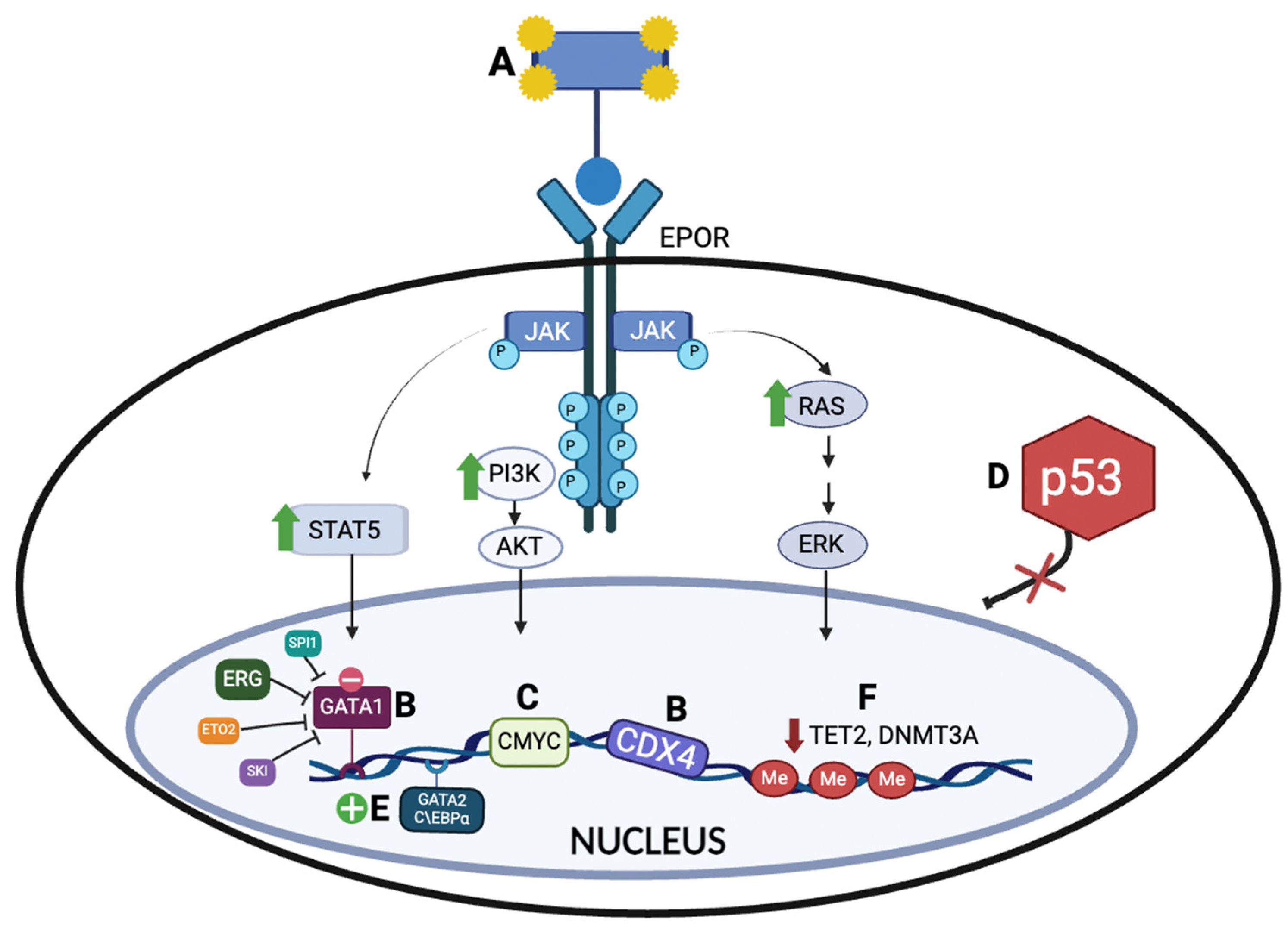
4. Molecular Biology and Genomic Features of AEL

Based on the data from human samples and murine models, attempts have been made to elucidate the pathogenesis of AEL. Several potential mechanisms implicated in the pathobiology of AEL have been explored (Figure 1).

4.1. Erythropoietin Receptor (EPOR) Activation and Downstream JAK2 Signaling Pathway

Viruses inducing erythroleukemia phenotypes served as the first models to study the multistage nature of the disease [22]. The relation of constitutive EPOR activation with erythroleukemia pathogenesis was introduced early during the 1990s, as the erythroblastosis-inducing spleen focus-forming virus (SFFV) was found to activate EPOR [23], while activating point mutations of the EPOR also promoted tumorigenesis [24]. According to very recent data comparing the transcriptional data of human primary AEL tumors with other types of AML, EPOR is one of the upregulated genes in this type of leukemia [25]. Moreover, activation of the EPOR downstream signaling effectors, such as signal transcription activators (STATs), PI3K/AKT, and mitogen-activated protein (MAP) or extracellular signal-regulated (ERK) kinases [22,26,27], promote preleukemic development of proerythroblasts [26].
Genomic alterations in oncogenic signaling, such as the JAK2 and RAS pathways, have been described in AEL patients. Grossman et al. reported that only 3 out of 92 AEL patients carry mutations in NRAS, KRAS, or FLT3 [20]. However, in a recent analysis by Takeda et al., gains and amplifications involving EPOR, JAK2, and/or ERG/ETS2 were recurrently detected in AEL patients [28]. Additionally, another recent study found at least one of these mutations to be present in 5 of the 35 AEL cases [28]. The gains and amplifications of EPOR and JAK2 were found to be more highly enriched in AEL than erythroid/myeloid leukemia cases [28]. These specific AEL cases showed enhanced cell proliferation, as well as sensitivity to ruxolitinib, in in vitro and xenograft models. As such, JAK2 inhibition could be a potential therapeutic target in AEL in patients with JAK2 gains and amplifications [28,29]. Similarly, several of these gene mutations were also found among the 41 AEL patients analyzed by the Mayo clinic group [10]. Particularly, 30% of the patients were found to have JAK2 mutations, and 10% of the patients were found to have NRAS or CSF3R mutations, suggesting that kinase signaling pathways are possibly associated with the progression of AEL.

4.2. Erythroid Transcriptional Regulators

4.2.1. GATA Binding Protein 1 (GATA1)

GATA1 is a zinc finger transcription factor with a central role in erythropoiesis and acts by modulating complexes with TAL1, LMO2, LDB1, RUNX1, ETO, and ETS family proteins [22]. GATA1 undergoes several posttranslational modifications, including phosphorylation, resulting mainly from EPO activation [30]. Recent AEL patient transcriptomic data show alterations of transcription or downstream signaling factors that mediate GATA1 activity in more than 25% of the cases [31]. Ectopic expression of these physical or functional interactors of the GATA1 transcriptional complexes (ERG, ETO2, SKI, and SPI1) in murine erythroid progenitors resulted in decreased chromatin accessibility at GATA1-binding sites and promoted proliferation with the immature phenotype [31]. Mouse models have confirmed that reduced GATA1 expression, as well as ectopic expression of GATA1 interactors [32,33,34], can promote erythroleukemic phenotypes [35]. Conversely, other AEL mouse models exhibited upregulated erythroid transcription factors (GATA1, FOG-1, and KLF1) and erythroid chromatin access, resulting in ectopic erythroid potential [36]. Mutational analyses of AEL have enlightened, even though rare, the occurrence of fusion genes or mutations affecting GATA1 itself or proteins of the GATA1 complexes (e.g., NFIA-ETO2 and MYB-GATA1) [37,38,39,40,41,42]. Mouse AEL tumors established by CRISPR/Cas9 of HSPCs with Trp53 and Bcor mutations had a gene expression profile recapitulating human AEL tumors with an overexpression of erythroid transcription factors such as Gata1, Gata2, and Klf1 [25]. Overall, these findings support the main role of altered GATA1 activity in AEL molecular biology through the diverse effects of erythroid favoritism and the inhibition of normal erythroid differentiation [22].

4.2.2. ETS Transcription Factors (ERG, SPI1, and FLI1)

The E-twenty-six/E26 (ETS) family genes were originally discovered within the erythroleukemia-causing avian retrovirus E26 through the viral ets (v-ets) oncogene, which was found to have been transduced from homologous genes in the chicken genome to encode part of a hybrid viral protein [43]. ETS transcription factors contain a highly conserved ETS DNA-binding domain that interacts together with other transcription factors to enhance the elements [44]. ERG cooperates with GATA1 to regulate the main hematopoietic transcription factors, such as SCL/TAL1 [45], and is known to promote hemopoietic stem cell (HSC) maintenance and control erythromegakaryocytic differentiation [46]. Higher ERG levels have been related to an unfavorable prognosis in AML [47], and ERG cooperation with GATA1 was found to immortalize hematopoietic progenitor cells [48], further indicating its role in AEL progression. Another ETS transcription factor, SPI1, overexpressed in transgenic mouse models, induced hepatosplenomegaly with erythroblast infiltration and tumor cells in peripheral blood. Nevertheless, these malignant proerythroblasts were partially blocked in differentiation and strictly dependent on erythropoietin for their proliferation both in vivo and in vitro [32]. Interestingly, SPI1 is also known to have a role in HSC maintenance [49]. The FLI1 gene, encoding for another ETS transcription factor, was also first studied in erythroleukemia virus models, and its overexpression reduces GATA1 expression and impairs erythroid differentiation [50]. FLI1 expression is upregulated by SPI1, implying a synergistic action in AEL [50]. Moreover, engineering mice with an inducible expression of the fusion EWS/FLI-1 resulted in the rapid development of erythroleukemia expressing GATA1 [51]. Mouse models with ectopic expression of ERG, SPI1, and FLI1 confirm that these transcription factors can induce AEL phenotypes [32,33,34], converging on the GATA1 lead to the development of the disease.

4.2.3. Caudal-Type Homeobox 4 (CDX4)

The caudal-type homeobox family consists of CDX1, CDX2, and CDX4, which are developmental regulators of HOX gene expression [52]. Essentially, CDX4 is expressed normally in early hematopoietic progenitors; however, it is expressed aberrantly in around 25% of AML patient samples [53]. In a retroviral transduction/bone marrow transplant model, the onset of AML in mice, induced by MLL-AF9, was substantially delayed when CDX4 was absent [54]. In vitro, the retroviral overexpression of CDX4 induced aberrant self-renewal in mice HSC cells, and similarly, in vivo, transplantation induced an AML-like disease in around 50% of mice [53]. This emphasizes that CDX4 is essential for normal hematopoiesis, and its aberrant expression mediates leukemogenesis. However, its direct applicability to human disease remains in question, as a transcriptome analysis of human AEL samples has not identified CDX4 alterations [31].

4.3. Expression of Master Oncogenes

Proto-oncogenes, particularly c-MYC, have a substantial role in erythroleukemia differentiation [55]. This has been studied particularly through mouse models. Murine erythroleukemia (MEL) cells are erythroid progenitors, with halted erythroid differentiation due to transformation with the Friend virus complex. Consequently, they have been employed as a model to explore the molecular biology of cellular differentiation, since DMSO induces terminal erythroid differentiation in these cells [55]. Erythroid differentiation in MEL cells is associated with a decreased expression of the MYC proto-oncogene, while MYC overexpression inhibits differentiation [56]. In vivo, Leder et al. generated transgenic mice harboring the human c-MYC proto-oncogene under the control of key mouse GATA-1 regulatory sequences. Tumor cells displayed proerythroblast morphology and expressed erythroid lineage markers, including EPOR and β-globin [57]. Thus, aberrant MYC activation at a vulnerable phase of erythroid differentiation likely triggers erythroleukemia. Additionally, H-Ras and K-Ras oncogenes are upregulated in MEL cells [58]. Leder et al. developed another transgenic mouse model (Tg.AC) in which the embryonic zeta-globin promoter was fused to the v-Ha-RAS oncogene, driving its expression [59,60]. These transgenic mice developed multiple mesenchymal and epithelial neoplasms, with a few (estimated to be <5%) developing hepatosplenomegaly with erythroblast infiltration [61]. Peripheral blood results showed a marked increase in metarubricytes and other less differentiated erythroid progenitor cells, including leukemic cells stained positive for GATA-1 [61]. Similarly, in vitro, EPO-induced differentiation was inhibited when a constitutively active RAS mutant (RAS12V) was expressed in SKT6 cells, a Friend murine erythroleukemic cell line. This suggests that aberrant RAS activation can drive erythroid transformation.

4.4. Impaired TP53 Activity in AEL Biology

AEL is characterized by a high prevalence of biallelic TP53 mutations in both de novo and secondary cases [14,20]. The ICC especially emphasizes this feature, as its 2022 definition of AEL stipulates a TP53 mutation as a required characteristic to establish a diagnosis. Most recent studies have similarly noted the association of TP53 mutation with AEL.
A retrospective study of 92 patients by Grossman et al. found that TP53 was mutated in only 43.5% of patient cases [20]. Another study of 58 AEL patients only reported TP53 mutations to be present in 12 patients (36.3%). This study also demonstrated these individuals to have an average of 4.41 mutations per sample, as well as a higher cytogenetic risk and poorer outcome. These studies classified patients according to the less specific 2008 WHO criteria, which could explain the lower proportion of TP53-mutated AEL in their cohort [31].
More recently, studies have begun using the 2016 and 2022 WHO criteria for AEL classification. In a retrospective analysis by the Mayo Clinic, biallelic TP53 alterations were present in all 41 (100%) AEL cases [10]. Similarly, a study published by the MD Anderson Cancer Center group also reported 100% incidence of TP53 mutations, most commonly a mutation in one allele and deletion in the other allele in 21 AEL cases [14]. These newer, specific, and stringent definitions for AEL appear to be associated with a higher rate of TP53 mutations. As such, it is important to note which criteria for AEL diagnosis is being used when evaluating previous studies.
TP53 mutations represent by far the most common genomic alteration in AEL; however, the specifics of functional involvement in erythroid differentiation are not yet thoroughly clarified [22]. During normal erythropoiesis, p53 and GATA1 were found to interact through GATA1 DNA-binding domains to promote erythroid cell development and survival [62]. Moreover, recently, p53 activation during ribosome biogenesis was found to regulate normal erythropoiesis [63]. TP53 mutations in AEL and other subtype AML patients promote the proliferation and survival of hematopoietic stem cells and progenitor cells (HSPCs), accumulating additional DNA damage, and are associated with poor prognosis [64,65]. Nevertheless, at least 80% of TP53-mutated AML patients present more than one genetic alteration, suggesting that additional abnormalities are required for development of the disease [66]. Indeed, TP53 mutations cooperate with multiple alternate pathways to produce the AEL phenotype. The RAS signaling pathway is shown to cooperate with impaired p53 activity, as KRAS and NRAS mutations are shown to produce AML and AEL phenotypes, respectively, when combined with the loss of p53 activity in mice [67,68]. Recent transcriptomic analyses showed that, in TP53-mutated AEL samples, transcription factor ERG is upregulated, and transplanting purified ERG-transduced TP53-mutated HSPC erythroblasts resulted in fatal erythroleukemia within 60 days [31]. Thus, TP53 mutations can cooperate with high ERG expression to enhance the proliferation of erythroid progenitors and development of AEL. Similarly, various other genetic lesions have been found to cooperate with TP53 loss of function mutations, such as JAK2V617F, NTRK1H498R, the t(1;16)(p31;q24) chromosomal translocation (NFIA-ETO2 fusion gene), and loss of function mutations of BCOR with or without DNMT3A mutations [25,37,69,70,71].
Additionally, the analysis of edited sites in leukemia mice models established by CRISPR/Cas 9 genome editing showed that concomitant TP53 and Bcor mutations are central drivers of erythroleukemia [25]. These TP53-mutated tumors also acquired secondary mutations in signaling pathway genes, including Ptpn11, Kit, Kras, Nras, and Csf1r1, in addition to cell cycle regulators and DNA repair genes [25]. Meanwhile, bone marrow samples from mice that did not develop tumors were enriched with gRNAs targeting Tet2, Dnmt3a, Stag2, and Asxl1 but not Nfx1, Rb1, TP53, or Bcor (apart from one mouse). The latter further emphasizes the essential role of TP53 and Bcor co-mutation, as mutations in other listed genes alone were insufficient to drive leukemogenesis [25].

4.5. Impaired C/EBPα Function

GATA2 and C\EBPα

GATA2 and C\EBPα are two essential transcription factors involved in hematopoiesis. On their role in AEL, there was a reported significant association between GATA2 mutations and biallelic C\EBPα mutations in a group of 55 AEL patients [72]. In fact, biallelic C\EBP and GATA-2 zinc finger 1 (ZF1) mutations synergize in leukemia progression [73]. This was also evident by a mouse model that showed biallelic C\ebpα led to myeloid leukemia development, and the addition of Gata2 mutation to the latter promoted leukemia progression, with 40% of triple transgenic mice developing leukemia with both erythroid and myeloid features. Biallelic C\ebpα mutations enhanced erythroid genes expression, while Gata2 mutations induced chromatin accessibility at erythroid transcription factor motifs such as Gata1, Zfpm1, and Klf1 and reduced it at myeloid transcription factor motifs [36]. In addition, published data on GATA2 mutants revealed that there is an increased expression of erythroid-related antigens Ter-119, β-globin, and βh1-globin and increased hemoglobin positivity in GATA2 mutants compared to controls [72]. The above findings confirm that mutant GATA2 activity controls the abnormal chromatin accessibility at crucial loci regulated by erythroid transcription factors, leading to this erythroid phenotype.

4.6. Epigenetic Dysregulation in Erythroleukemia

AEL is also associated with other somatic mutations in epigenetic and transcriptional regulators. Fagnan et al. found that a significant percentage of AEL patients carry mutations in genes encoding epigenetic regulators (33.3% of the cohort). These were mainly TET2 nonsense mutations (n = 8) and DNMT3A (n = 5) mutations [31]. The samples had an average of 5.72 mutations. Several patients carrying TET2 and DNMT3A mutations also had mutations in SRSF2 or IDH2. DNA methylation plays an essential role in erythroid malignancies, and it is regulated by several factors, including TET2 and DNMT3A/B [74,75]. In mice models, inactivating mutations of Tet2 and Dnmt3a/b promotes hematopoietic stem cell (HSC) renewal and inhibits differentiation, leading to leukemic transformation [76,77]. In HSCs, inactivation of these two genes led to an increase in myeloid and decrease in erythroid gene expression, with erythroid progenitors accumulating in mice. Similarly, in human AEL samples, DNMT3A/B and/or TET2 mutations were present in some AEL patients, and the combination of TET2 and DNMT3A was reported to upregulate erythroid transcription factor KLF1 and EPOR in HSCs [31]. Recently, changes in the promoter methylation and gene expression of erythroid transcription factors (GATA1, KLF1, TAL1, JAK2, etc.), as well as factors implicated in protein binding (MEF2c, BRAF, RCOR1, LIFR, and CTNNA1), were observed in AEL mouse models. Moreover, Dnmt3a-mutated/Tet2 wild-type leukemia models, or Dnmt3a-mutant/Tet2 heterozygous models, were of the erythroid phenotype and exhibited hypomethylation and overexpression of genes regulating erythrocyte differentiation and homeostasis, including Gata1, Klf1, and Kit, and hypermethylation and low expression of genes involved in leukocyte activation and differentiation (e.g., Myb, Tnfaip3, Ikzf3, and Cd74). These recent data further highlight the role of DNA methylation in AEL molecular biology [25] and support the use of cytidine analogs such as 5-Azacytidine or Decitabine as potential therapeutic options [78].

4.7. Other Less Frequent Genetic Alterations

The study by Fagnan et al. also found a subset of AEL cases (n = 10, or 30.4% of their cohort) with a significantly lower mutational burden, having less mutations per sample on average than TP53-mutated or epigenetic regulator subgroups. These AEL patients had an average of 1.60 mutations per sample, which was significantly lower than their TP53-mutated or epigenetic regulator subgroups [31]. As this study used the 2008 WHO definition of AEL, some of their samples may be categorized differently than the newer classification models.
In a 2013 study, Grossman et al. found that NPM1 was mutated in 15 out of 92 patients (16.3%). Other less frequent mutations noted in this study were mutations in ASXL1 (8.0%), RUNX1 (8.7%), MLL-PTD (7.8%), IDH1 (7.5%), IDH2 (4.7%), NRAS (3.3%), KRAS (3.3%), FLT3-ITD (3.3%), FLT3-TKD (3.5%), SF3B1 (2.5%), and CEBPA (1.1%) [20]. This study demonstrated that the mutation load was similar across all of the gene mutations analyzed [20]. Again, it is important to note that this study used the old definitions of AEL and that mutation frequencies may differ according to the new criteria. The recent analysis of 41 AEL patients by the Mayo Clinic with the updated AEL definitions demonstrated that 20% of patients had mutations in TET2 and 10% of patients had mutations in ASXL1, IDH2, and DNMT3A [10].
In addition, the NUP98 gene encodes a nucleoporin protein that acts as a transcription activator and has been linked to over 28 hematologic malignancies, including AEL [79]. When analyzing a cohort of pediatric cases, NUP98 fusions appeared to be highly enriched in AEL patients compared to those with other AML types (31.8 vs. 6.7%) [80]. This percentage was even higher in the AEL category, as three out of five patients (60%) exhibited this fusion. Of the seven total patients with NUP98 fusions, four had KDM5A as the fusion partner, two had NSD1, and one had SET [80]. Those with this NUP98 fusion appeared to have higher OS scores; however, this finding was not statistically significant due to the limited sample size (OS = 45 ± 28 no fusion, OS = 65 ± 33 with NUP98 fusion).

5. Treatments and Clinical Outcomes

Due to the rarity of AEL diagnosis and the relatively poor understanding of the disease biology, treatment options are limited at this time (Table 1). The current first-line treatment strategies include intensive chemotherapy (ICT) and hypomethylating agents (HMAs). Allogeneic bone marrow transplant (AlloBMT) is the only potential curative approach, but it requires deep remission prior to its initiation, which proves challenging in this disease.

5.1. Intensive Chemotherapy

ICT is often used as a front-line treatment for AEL patients who are eligible for intensive therapy [3]. In a large retrospective multinational study of 217 patients with AEL by Almeida et al., 122 patients were treated with ICT. Of the 119 whose response data was available, daunorubicin (45 or 60 mg/m2 × 3 days) with cytarabine (100 mg/m2 bid × 7 days) was the most frequently used induction regimen (n = 81, 66%). Other regimens used included idarubicin 12 mg/m2 (n = 25, 20%) or mitoxantrone 12 mg/m2 (n = 8, 7%) in combination with cytarabine. The ICT group had a median age of 60 at diagnosis, compared to 69 for the HMA group. The objective response rate (ORR) was 72%, according to the ELN criteria. Complete response (CR) occurred in 79 patients (66%), partial response (PR) in 7 (6%), stable disease (SD) in 16 (13%), and primary disease progression (PPD) in 17 (14%). Following treatment with ICT, 23 (18.8%) patients received an allogeneic bone marrow transplant. Patients treated with ICT followed by a transplant experienced a median OS of 5.9 months. In the ICT group overall, the authors reported a median OS of 10.5 months but did not find a significant difference in OS between IHMA-treated and ICT-treated patients with MRC intermediate-risk cytogenetics (29.3 vs. 16.9 months, p = 0.277). In patients with MRC high cytogenetic risk, the median OS was significantly higher in the first-line HMA group compared to ICT (13.3 vs. 7.5 months, p = 0.039) [3]. However, as the definitions have changed to become more stringent, these data may not be as relevant to AEL as it is currently defined, and many of these patients would likely be classified as MDS with excess blasts or AML.
Treatment with chemotherapy and younger age have also been associated with significantly higher overall survival in AEL [3,15]. A recent 2023 retrospective analysis by Gera et al. demonstrated an inverse relationship between age and OS in patients with AEL, with a median OS of 69, 18, 8, 3, and 1 month for age groups <18, 18–49, 50–64, 65–79, and 80+, respectively [15]. Pediatric AEL shows a distinct genomic profile, which could partially explain the more favorable outcomes in this age group [15]. Additionally, in many hematologic malignancies, the pediatric chemotherapy regimen is more aggressive, as children are better able to tolerate this treatment compared to adults [19]. While the effect of chemotherapy is heightened amongst pediatric patients, the use of chemotherapy appears to be associated with increased survival in both pediatric and adult populations. The OS in this study was 152 vs. 2 months in children who received chemotherapy vs. those who did not, and the OS was 8 vs. 1 month in adults receiving chemotherapy vs. not [15]. It should be noted that the retrospective nature of these analyses mitigates the significance of these conclusions.

5.2. Hypomethylating Agents

The HMAs azacitidine and decitabine are commonly used in AML patients who are not eligible for ICT [15]. HMAs work by reversing the DNA methylation that often silences tumor suppressor genes involved in cancer pathogenesis signaling. Although HMAs can induce a promising initial response in a subset of AEL patients, resistance emerges in nearly all the patients with this disease [82]. This can either occur through primary resistance, in which there is no improvement after four to six cycles of HMAs, or secondary resistance, in which patients progress after an initial response to HMA [82]. Almeida et al. also found that the median progression-free survival (PFS) was longer for first-line HMA treatment compared to second-line or later HMA (9.4 vs. 3.4 months, respectively) [3], although the AEL definition has changed since the study was published.
However, a recent retrospective study of 41 patients with AEL described no benefit to using one treatment regimen over another. The regimens reported in this study included HMA alone, HMA with venetoclax, ICT, and best supportive care [10]. As such, more studies need to be conducted to better understand the varying degrees of efficacy amongst HMA treatments for AEL as defined by the newer 2022 criteria.

Venetoclax

Venetoclax is a B-cell lymphoma 2 (BCL-2) inhibitor that has been shown to improve the remission rates and OS of patients with AML who are ineligible for ICT [83]. However, erythroid/megakaryocytic AML subtypes are associated with resistance to venetoclax [84]. This is presumably driven by erythroid/megakaryocytic differentiation, in addition to TP53 mutations. p53 loss has been linked to venetoclax resistance, along with a concomitant compensatory BCL-XL upregulation [84]. In fact, Kuusanmäki et al. recently found that AML cells exhibiting erythroid/megakaryocytic differentiation depend on BCL-XL rather than BCL-2 for their survival [84]. Thus, BCL-2 inhibition can exhibit limited therapeutic efficacy in AEL.

5.3. Allogeneic Bone Marrow Transplant

Allogeneic bone marrow transplantation (AlloBMT) is the only potentially curative approach for AEL, but it requires deep remission of the disease, which is rarely achieved in these patients [15]. It has been previously found that the median survival of AEL patients who received AlloBMT was 66 months from transplant [81]. However, it should be noted that the definition of AEL for this analysis was based on the 2008 WHO classification. The median OS of AlloBMT recipients was 89 months, compared to 5 months for those who did not undergo AlloBMT [81]. In one study, twelve patients underwent AlloBMT, representing 28% of the AEL cohort, and 71% of those were in complete remission (CR1 or CR2) [81]. Thus, using AlloBMT as a consolidation therapy may improve outcomes in AEL. A successful initial treatment to achieve remission is currently a requirement to undergo AlloBMT. This stipulation remains a challenge, as only a small proportion of patients reach AlloBMT.

5.4. Chimeric Antigen Receptor T-Cell Therapy

Genetically engineered T cells, such as chimeric antigen receptor (CAR) T cells, have started to be used therapeutically in cancers such as leukemias and lymphomas by targeting distinct antigens present on malignant cells [85,86]. Recently, some studies have examined potential targets of CAR-T therapy in AML. In a 2023 study, Gottschlich et al. investigated RNA sequencing data of single cells from individuals with AML and healthy tissue to determine epitopes expressed selectively on malignant cells. Through computational analysis and subsequent validation, the authors found that colony-stimulating factor 1 receptor and cluster of differentiation 86 could be used as CAR-T therapy targets in AML [85]. In 2019, Gomes-Silva and colleagues found that CD7, which is expressed by the malignant blasts and progenitor cells of a subset of AML patients, acted as a target for CAR-T therapy [87]. Overall, the variety of molecular epitopes that have been identified in CAR-T therapy for AML could represent beneficial future applications for single-cell cancer treatment.
While there have been promising advances in CAR-T cell therapy for the treatment of some leukemias, including AML, studies investigating single-cell analysis in AEL remain limited. As clinical outcomes for AEL are poor and there are few treatment options, CAR-T therapy should be further investigated as a treatment modality for AEL.

5.5. Future Directions

Given the overall poor outcomes of AEL patients, it is evident that clinical trials represent the best option for these individuals. The current trials aimed at treating AEL patients specifically are limited due to the rarity of this condition, especially with the newest definitions and classifications.
Additionally, it was recently reported that the survival rates for AEL have not improved over the last twenty years, despite the introduction of novel targeted therapies for the treatment of AML and the advancements made in the process of AlloBMT resulting in improved non-relapse mortality [15]. It is paramount to continue seeking novel therapeutic advancements to improve these clinical outcomes. Among the challenges is that the main drivers of this disease appear to be transcriptional factors that are not easily targetable and that JAK2 inhibition alone does not seem to be able to control this disease or change its natural history.
Recently, Iacobucci et al. demonstrated a sensitivity to CDK7 and CDK9 inhibition for TP53-, BCOR-, and DNMT3A-mutated AEL models and further confirmed CDK9 efficacy in AEL samples with combinatorial mutations in TP53, BCOR, NFIX, and RB1 [25]. Furthermore, the authors found that TP53-mutated AEL tumors were shown to have high sensitivity to PARP inhibition (talazoparib) in the absence of DNMT3A mutation [25]. As drugs such as decitabine increase the presence of PARP1 at DNA damage sites, this amplifies the cytotoxic effects and, thus, could represent an effective targeted therapy for TP53-mutated AEL [25,88]. Lastly, the efficacy of gemcitabine and bromodomain inhibitors was demonstrated in NUP98::KDM5A leukemia [25].
Furthermore, the molecule BCL-XL has shown promise as a therapeutic target in AEL. BCL-XL is an antiapoptotic protein that has been shown through CRISPR screens to exhibit high expression in erythroid/megakaryocytic AML, thus conferring their survival [84]. A 2023 study by Kuusanmäki and colleagues found BCL-XL-selective inhibitors, rather than BCL-2 inhibitors such as venetoclax, to be highly effective against erythroid/megakaryoblastic leukemia cell lines. When these inhibitors were combined with the JAK inhibitor ruxolitinib, the cell lines showed a synergistic response to the therapy [84]. Moreover, it was demonstrated ex vivo that BCL-XL inhibition successfully eliminated blasts from patients with AML who had erythroid or megakaryocytic differentiation, as well as decreased tumor burden, in a mouse model of an erythroleukemia xenograft [84]. A recent review discussed that navitoclax, which specifically inhibits BCL-XL and BCL-2, displayed therapeutic benefit and antineoplastic effects [89,90]. The combined use of BCL-XL inhibitors with chemotherapy has also been shown to be an effective treatment against acute leukemia [89,91]. As such, compounds that target the BCL-XL molecule may be promising in the future of AEL treatment.
JAK2 inhibition also demonstrates clinical utility in the treatment of AEL. A recent study by Li and colleagues found that biallelic TP53 inactivation leads to leukemic transformation in PEL and that JAK2/TRP53-mutated PEL exhibits DNA damage and persistent copy number variations [92]. The authors further demonstrated that PEL demonstrated high sensitivity to the inhibition of molecules involved in DNA repair, such as WEE1 and poly(ADP-ribose) polymerase (PARP), which could offer a prospective therapeutic approach to this disease [92]. Another recent study found a high frequency of gains and amplifications encompassing EPOR/JAK2 in TP53-mutated cases of AEL, which were frequently associated with poor prognosis [29]. However, these samples exhibited a high sensitivity to ruxolitinib, a JAK2 inhibitor, in both in vitro and xenograft models. This further supports the potential role of JAK2 inhibition in the targeted treatment of AEL cases with altered JAK2 signaling.
Examining future prospects, there are also ongoing and recently completed clinical trials examining the sensitivity of cohesin-mutated AML and MDS with excess blasts to talazoparib (#NCT03974217), as well as evaluating the use of gemtuzumab ozogamicin in CD33+ relapsed or refractory AML (#NCT04207190) [25]. A recent phase I clinical trial studied the use of decitabine with talazoparib in 25 patients with refractory/relapsed AML and found complete remission with incomplete count recovery in 8% (n = 2), as well as hematologic improvement in 12% (n = 3) [93]. Clinical trials investigating drug responses in patients with AEL remain limited, although recent AEL models have shown promising responses [25,84,92]. Thus, future directions include targeting AEL based on a better understanding of the molecular biology of the disease, as well as the confirmation of preclinical findings using patient samples.

6. Conclusions

Overall, AEL is a very aggressive and rare disorder that is associated with poor clinical outcomes. The WHO and ICC differ slightly on their nomenclature and criteria for defining this condition; however, both acknowledge the presence of the hypercellularity of the bone marrow and predominance of erythroblast precursors, as well as a supporting TP53 mutation with evidence of biallelic inactivation [6,11]. In addition to bone marrow biopsy, next-generation sequencing, flow cytometry, and immunohistochemistry are all supporting measures used to aid the diagnosis of AEL [1].
Because of the high-risk features of this disease, clinical outcomes remain poor. HMAs, intensive chemotherapy, and AlloBMT have all been used, with overall limited efficacy. Younger age and the use of chemotherapy aid in survival time; however, AlloBMT is the only potentially curative approach for this disease [3,15].
In conclusion, the improvement of the outcomes of this rare and aggressive disease requires a better understanding of its molecular biology, introduction of novel therapeutic strategies, and treatment of these patients in multi-institutional clinical trials that will allow the evaluation of therapies in enlarged cohorts. These next steps remain challenging given the stricter definitions of AEL.

Author Contributions

Conceptualization, T.K.; methodology, P.F., N.W., T.C., N.S.N. and T.K.; investigation, P.F., N.W., T.C., N.S.N. and T.K.; writing—original draft preparation, P.F., N.W., T.C., N.S.N. and T.K.; writing—review and editing, P.F., N.W., T.C., N.S.N. and T.K.; visualization, P.F.; supervision, T.K.; funding acquisition, T.K. All authors have read and agreed to the published version of the manuscript.

Funding

This research was funded by the National Cancer Institute/NIH (K08HL168777) and the MacMillan Pathway to Independence Program Award (Johns Hopkins University). Theodora Chatzilygeroudi would like to acknowledge funding through the I.K.Y.-Fulbright Partnership Award for PhD research scholarship through both the Greek State Scholarships Foundation (I.K.Y.) and the Fulbright Foundation.

Conflicts of Interest

The authors declare no conflicts of interest. The funders had no role in the design of the study; in the collection, analyses, or interpretation of the data; in the writing of the manuscript; or in the decision to publish the results.

References

  1. Zuo, Z.; Polski, J.M.; Kasyan, A.; Medeiros, L.J. Acute Erythroid Leukemia. Arch. Pathol. Lab. Med. 2010, 134, 1261–1270. [Google Scholar] [CrossRef] [PubMed]
  2. Cervera, N.; Guille, A.; Adélaïde, J.; Hospital, M.-A.; Garciaz, S.; Mozziconacci, M.-J.; Vey, N.; Gelsi-Boyer, V.; Birnbaum, D. Erythroleukemia: Classification. EJHaem 2023, 4, 450–453. [Google Scholar] [CrossRef] [PubMed]
  3. Almeida, A.M.; Prebet, T.; Itzykson, R.; Ramos, F.; Al-Ali, H.; Shammo, J.; Pinto, R.; Maurillo, L.; Wetzel, J.; Musto, P.; et al. Clinical Outcomes of 217 Patients with Acute Erythroleukemia According to Treatment Type and Line: A Retrospective Multinational Study. Int. J. Mol. Sci. 2017, 18, 837. [Google Scholar] [CrossRef] [PubMed]
  4. Novik, Y.; Marino, P.; Makower, D.F.; Wiernik, P.H. Familial Erythroleukemia: A Distinct Clinical and Genetic Type of Familial Leukemias. Leuk. Lymphoma 1998, 30, 395–401. [Google Scholar] [CrossRef] [PubMed]
  5. Alaggio, R.; Amador, C.; Anagnostopoulos, I.; Attygalle, A.D.; Araujo, I.B.d.O.; Berti, E.; Bhagat, G.; Borges, A.M.; Boyer, D.; Calaminici, M.; et al. The 5th Edition of the World Health Organization Classification of Haematolymphoid Tumours: Lymphoid Neoplasms. Leukemia 2022, 36, 1720–1748. [Google Scholar] [CrossRef]
  6. Arber, D.A.; Orazi, A.; Hasserjian, R.P.; Borowitz, M.J.; Calvo, K.R.; Kvasnicka, H.-M.; Wang, S.A.; Bagg, A.; Barbui, T.; Branford, S.; et al. International Consensus Classification of Myeloid Neoplasms and Acute Leukemias: Integrating Morphologic, Clinical, and Genomic Data. Blood 2022, 140, 1200–1228. [Google Scholar] [CrossRef] [PubMed]
  7. Alexander, C. A History and Current Understanding of Acute Erythroid Leukemia. Clin. Lymphoma Myeloma Leuk. 2023, 23, 583–588. [Google Scholar] [CrossRef] [PubMed]
  8. Balduini, C.L. 100-Year Old Haematologica Images: Di Guglielmo Disease or Pure Erythroid Leukemia. Haematologica 2020, 105, 525. [Google Scholar] [CrossRef] [PubMed]
  9. Arber, D.A.; Orazi, A.; Hasserjian, R.; Thiele, J.; Borowitz, M.J.; Le Beau, M.M.; Bloomfield, C.D.; Cazzola, M.; Vardiman, J.W. The 2016 Revision to the World Health Organization Classification of Myeloid Neoplasms and Acute Leukemia. Blood 2016, 127, 2391–2405. [Google Scholar] [CrossRef]
  10. Reichard, K.K.; Tefferi, A.; Abdelmagid, M.; Orazi, A.; Alexandres, C.; Haack, J.; Greipp, P.T. Pure (Acute) Erythroid Leukemia: Morphology, Immunophenotype, Cytogenetics, Mutations, Treatment Details, and Survival Data among 41 Mayo Clinic Cases. Blood Cancer J. 2022, 12, 147. [Google Scholar] [CrossRef]
  11. Kasyan, A.; Medeiros, L.J.; Zuo, Z.; Santos, F.P.; Ravandi-Kashani, F.; Miranda, R.; Vadhan-Raj, S.; Koeppen, H.; Bueso-Ramos, C.E. Acute Erythroid Leukemia as Defined in the World Health Organization Classification Is a Rare and Pathogenetically Heterogeneous Disease. Mod. Pathol. 2010, 23, 1113–1126. [Google Scholar] [CrossRef]
  12. Acharya, S.; Kala, P.S. Role of CD71 in Acute Leukemia- An Immunophenotypic Marker for Erythroid Lineage or Proliferation? Indian J. Pathol. Microbiol. 2019, 62, 418–422. [Google Scholar] [CrossRef] [PubMed]
  13. Kaumeyer, B.A.; Fidai, S.S.; Thakral, B.; Wang, S.A.; Arber, D.A.; Cheng, J.X.; Gurbuxani, S.; Venkataraman, G. GLUT1 Immunohistochemistry Is a Highly Sensitive and Relatively Specific Marker for Erythroid Lineage in Benign and Malignant Hematopoietic Tissues. Am. J. Clin. Pathol. 2022, 158, 228–234. [Google Scholar] [CrossRef]
  14. Fang, H.; Wang, S.A.; Khoury, J.D.; El Hussein, S.; Kim, D.H.; Tashakori, M.; Tang, Z.; Li, S.; Hu, Z.; Jelloul, F.Z.; et al. Pure Erythroid Leukemia Is Characterized by Biallelic TP53 Inactivation and Abnormal P53 Expression Patterns in de Novo and Secondary Cases. Haematologica 2022, 107, 2232–2237. [Google Scholar] [CrossRef] [PubMed]
  15. Gera, K.; Martir, D.; Xue, W.; Wingard, J.R. Survival after Pure (Acute) Erythroid Leukemia in the United States: A SEER-Based Study. Cancers 2023, 15, 3941. [Google Scholar] [CrossRef]
  16. Campo, E.; Cymbalista, F.; Ghia, P.; Jäger, U.; Pospisilova, S.; Rosenquist, R.; Schuh, A.; Stilgenbauer, S. TP53 Aberrations in Chronic Lymphocytic Leukemia: An Overview of the Clinical Implications of Improved Diagnostics. Haematologica 2018, 103, 1956–1968. [Google Scholar] [CrossRef]
  17. Mazzella, F.M.; Kowal-Vern, A.; Shrit, M.A.; Wibowo, A.L.; Rector, J.T.; Cotelingam, J.D.; Collier, J.; Mikhael, A.; Cualing, H.; Schumacher, H.R. Acute Erythroleukemia: Evaluation of 48 Cases with Reference to Classification, Cell Proliferation, Cytogenetics, and Prognosis. Am. J. Clin. Pathol. 1998, 110, 590–598. [Google Scholar] [CrossRef]
  18. Hasserjian, R.P.; Zuo, Z.; Garcia, C.; Tang, G.; Kasyan, A.; Luthra, R.; Abruzzo, L.V.; Kantarjian, H.M.; Medeiros, L.J.; Wang, S.A. Acute Erythroid Leukemia: A Reassessment Using Criteria Refined in the 2008 WHO Classification. Blood 2010, 115, 1985–1992. [Google Scholar] [CrossRef] [PubMed]
  19. Neaga, A.; Jimbu, L.; Mesaros, O.; Bota, M.; Lazar, D.; Cainap, S.; Blag, C.; Zdrenghea, M. Why Do Children with Acute Lymphoblastic Leukemia Fare Better Than Adults? Cancers 2021, 13, 3886. [Google Scholar] [CrossRef]
  20. Grossmann, V.; Bacher, U.; Haferlach, C.; Schnittger, S.; Pötzinger, F.; Weissmann, S.; Roller, A.; Eder, C.; Fasan, A.; Zenger, M.; et al. Acute Erythroid Leukemia (AEL) Can Be Separated into Distinct Prognostic Subsets Based on Cytogenetic and Molecular Genetic Characteristics. Leukemia 2013, 27, 1940–1943. [Google Scholar] [CrossRef]
  21. Linu, J.A.; Udupa, M.N.; Madhumathi, D.S.; Lakshmaiah, K.C.; Babu, K.G.; Lokanatha, D.; Babu, M.S.; Lokesh, K.N.; Rajeev, L.K.; Rudresha, A.H. Study of Clinical, Haematological and Cytogenetic Profile of Patients with Acute Erythroid Leukaemia. Ecancermedicalscience 2017, 11, 712. [Google Scholar] [CrossRef] [PubMed]
  22. Fagnan, A.; Piqué-Borràs, M.-R.; Tauchmann, S.; Mercher, T.; Schwaller, J. Molecular Landscapes and Models of Acute Erythroleukemia. HemaSphere 2021, 5, e558. [Google Scholar] [CrossRef] [PubMed]
  23. Ney, P.A.; D’Andrea, A.D. Friend Erythroleukemia Revisited. Blood 2000, 96, 3675–3680. [Google Scholar] [CrossRef] [PubMed]
  24. Yoshimura, A.; Longmore, G.; Lodish, H.F. Point Mutation in the Exoplasmic Domain of the Erythropoietin Receptor Resulting in Hormone-Independent Activation and Tumorigenicity. Nature 1990, 348, 647–649. [Google Scholar] [CrossRef] [PubMed]
  25. Iacobucci, I.; Qu, C.; Varotto, E.; Janke, L.J.; Yang, X.; Seth, A.; Shelat, A.; Friske, J.D.; Fukano, R.; Yu, J.; et al. Modeling and Targeting of Erythroleukemia by Hematopoietic Genome Editing. Blood 2021, 137, 1628–1640. [Google Scholar] [CrossRef] [PubMed]
  26. Rimmelé, P.; Kosmider, O.; Mayeux, P.; Moreau-Gachelin, F.; Guillouf, C. Spi-1/PU.1 Participates in Erythroleukemogenesis by Inhibiting Apoptosis in Cooperation with Epo Signaling and by Blocking Erythroid Differentiation. Blood 2007, 109, 3007–3014. [Google Scholar] [CrossRef] [PubMed]
  27. Kosmider, O.; Denis, N.; Lacout, C.; Vainchenker, W.; Dubreuil, P.; Moreau-Gachelin, F. Kit-Activating Mutations Cooperate with Spi-1/PU.1 Overexpression to Promote Tumorigenic Progression during Erythroleukemia in Mice. Cancer Cell 2005, 8, 467–478. [Google Scholar] [CrossRef]
  28. Takeda, J. Molecular pathogenesis and therapeutic targets in acute erythroid leukemia. Rinsho Ketsueki 2022, 63, 121–133. [Google Scholar] [CrossRef] [PubMed]
  29. Takeda, J.; Yoshida, K.; Nakagawa, M.M.; Nannya, Y.; Yoda, A.; Saiki, R.; Ochi, Y.; Zhao, L.; Okuda, R.; Qi, X.; et al. Amplified EPOR/JAK2 Genes Define a Unique Subtype of Acute Erythroid Leukemia. Blood Cancer Discov. 2022, 3, 410–427. [Google Scholar] [CrossRef]
  30. Gutiérrez, L.; Caballero, N.; Fernández-Calleja, L.; Karkoulia, E.; Strouboulis, J. Regulation of GATA1 Levels in Erythropoiesis. IUBMB Life 2020, 72, 89–105. [Google Scholar] [CrossRef]
  31. Fagnan, A.; Bagger, F.O.; Piqué-Borràs, M.-R.; Ignacimouttou, C.; Caulier, A.; Lopez, C.K.; Robert, E.; Uzan, B.; Gelsi-Boyer, V.; Aid, Z.; et al. Human Erythroleukemia Genetics and Transcriptomes Identify Master Transcription Factors as Functional Disease Drivers. Blood 2020, 136, 698–714. [Google Scholar] [CrossRef]
  32. Moreau-Gachelin, F.; Wendling, F.; Molina, T.; Denis, N.; Titeux, M.; Grimber, G.; Briand, P.; Vainchenker, W.; Tavitian, A. Spi-1/PU.1 Transgenic Mice Develop Multistep Erythroleukemias. Mol. Cell. Biol. 1996, 16, 2453–2463. [Google Scholar] [CrossRef]
  33. Carmichael, C.L.; Metcalf, D.; Henley, K.J.; Kruse, E.A.; Di Rago, L.; Mifsud, S.; Alexander, W.S.; Kile, B.T. Hematopoietic Overexpression of the Transcription Factor Erg Induces Lymphoid and Erythro-Megakaryocytic Leukemia. Proc. Natl. Acad. Sci. USA 2012, 109, 15437–15442. [Google Scholar] [CrossRef] [PubMed]
  34. Li, Y.; Luo, H.; Liu, T.; Zacksenhaus, E.; Ben-David, Y. The Ets Transcription Factor Fli-1 in Development, Cancer and Disease. Oncogene 2015, 34, 2022–2031. [Google Scholar] [CrossRef]
  35. Shimizu, R.; Kuroha, T.; Ohneda, O.; Pan, X.; Ohneda, K.; Takahashi, S.; Philipsen, S.; Yamamoto, M. Leukemogenesis Caused by Incapacitated GATA-1 Function. Mol. Cell. Biol. 2004, 24, 10814–10825. [Google Scholar] [CrossRef] [PubMed]
  36. Di Genua, C.; Valletta, S.; Buono, M.; Stoilova, B.; Sweeney, C.; Rodriguez-Meira, A.; Grover, A.; Drissen, R.; Meng, Y.; Beveridge, R.; et al. C/EBPα and GATA-2 Mutations Induce Bilineage Acute Erythroid Leukemia through Transformation of a Neomorphic Neutrophil-Erythroid Progenitor. Cancer Cell 2020, 37, 690–704. [Google Scholar] [CrossRef]
  37. Iacobucci, I.; Wen, J.; Meggendorfer, M.; Choi, J.K.; Shi, L.; Pounds, S.B.; Carmichael, C.L.; Masih, K.E.; Morris, S.M.; Lindsley, R.C.; et al. Genomic Subtyping and Therapeutic Targeting of Acute Erythroleukemia. Nat. Genet. 2019, 51, 694–704. [Google Scholar] [CrossRef] [PubMed]
  38. Micci, F.; Thorsen, J.; Haugom, L.; Zeller, B.; Tierens, A.; Heim, S. Translocation t(1;16)(P31;Q24) Rearranging CBFA2T3 Is Specific for Acute Erythroid Leukemia. Leukemia 2011, 25, 1510–1512. [Google Scholar] [CrossRef] [PubMed]
  39. Panagopoulos, I.; Micci, F.; Thorsen, J.; Haugom, L.; Buechner, J.; Kerndrup, G.; Tierens, A.; Zeller, B.; Heim, S. Fusion of ZMYND8 and RELA Genes in Acute Erythroid Leukemia. PLoS ONE 2013, 8, e63663. [Google Scholar] [CrossRef]
  40. Micci, F.; Thorsen, J.; Panagopoulos, I.; Nyquist, K.B.; Zeller, B.; Tierens, A.; Heim, S. High-Throughput Sequencing Identifies an NFIA/CBFA2T3 Fusion Gene in Acute Erythroid Leukemia with t(1;16)(P31;Q24). Leukemia 2013, 27, 980–982. [Google Scholar] [CrossRef]
  41. Matsuzaki, T.; Aisaki, K.; Yamamura, Y.; Noda, M.; Ikawa, Y. Induction of Erythroid Differentiation by Inhibition of Ras/ERK Pathway in a Friend Murine Leukemia Cell Line. Oncogene 2000, 19, 1500–1508. [Google Scholar] [CrossRef] [PubMed]
  42. Linnik, Y.; Pastakia, D.; Dryden, I.; Head, D.R.; Mason, E.F. Primary Central Nervous System Erythroid Sarcoma with NFIA-CBFA2T3 Translocation: A Rare but Distinct Clinicopathologic Entity. Am. J. Hematol. 2020, 95, E299–E301. [Google Scholar] [CrossRef] [PubMed]
  43. Ducker, C.; Shaw, P.E. Ubiquitin-Mediated Control of ETS Transcription Factors: Roles in Cancer and Development. Int. J. Mol. Sci. 2021, 22, 5119. [Google Scholar] [CrossRef] [PubMed]
  44. Beck, D.; Thoms, J.A.I.; Perera, D.; Schütte, J.; Unnikrishnan, A.; Knezevic, K.; Kinston, S.J.; Wilson, N.K.; O’Brien, T.A.; Göttgens, B.; et al. Genome-Wide Analysis of Transcriptional Regulators in Human HSPCs Reveals a Densely Interconnected Network of Coding and Noncoding Genes. Blood 2013, 122, e12–e22. [Google Scholar] [CrossRef] [PubMed]
  45. Wilson, N.K.; Foster, S.D.; Wang, X.; Knezevic, K.; Schütte, J.; Kaimakis, P.; Chilarska, P.M.; Kinston, S.; Ouwehand, W.H.; Dzierzak, E.; et al. Combinatorial Transcriptional Control In Blood Stem/Progenitor Cells: Genome-Wide Analysis of Ten Major Transcriptional Regulators. Cell Stem Cell 2010, 7, 532–544. [Google Scholar] [CrossRef] [PubMed]
  46. Knudsen, K.J.; Rehn, M.; Hasemann, M.S.; Rapin, N.; Bagger, F.O.; Ohlsson, E.; Willer, A.; Frank, A.-K.; Søndergaard, E.; Jendholm, J.; et al. ERG Promotes the Maintenance of Hematopoietic Stem Cells by Restricting Their Differentiation. Genes Dev. 2015, 29, 1915–1929. [Google Scholar] [CrossRef] [PubMed]
  47. Baldus, C.D.; Burmeister, T.; Martus, P.; Schwartz, S.; Gökbuget, N.; Bloomfield, C.D.; Hoelzer, D.; Thiel, E.; Hofmann, W.K. High Expression of the ETS Transcription Factor ERG Predicts Adverse Outcome in Acute T-Lymphoblastic Leukemia in Adults. J. Clin. Oncol. 2006, 24, 4714–4720. [Google Scholar] [CrossRef] [PubMed]
  48. Stankiewicz, M.J.; Crispino, J.D. ETS2 and ERG Promote Megakaryopoiesis and Synergize with Alterations in GATA-1 to Immortalize Hematopoietic Progenitor Cells. Blood 2009, 113, 3337–3347. [Google Scholar] [CrossRef] [PubMed]
  49. Iwasaki, H.; Somoza, C.; Shigematsu, H.; Duprez, E.A.; Iwasaki-Arai, J.; Mizuno, S.-I.; Arinobu, Y.; Geary, K.; Zhang, P.; Dayaram, T.; et al. Distinctive and Indispensable Roles of PU.1 in Maintenance of Hematopoietic Stem Cells and Their Differentiation. Blood 2005, 106, 1590–1600. [Google Scholar] [CrossRef]
  50. Athanasiou, M.; Mavrothalassitis, G.; Sun-Hoffman, L.; Blair, D.G. FLI-1 Is a Suppressor of Erythroid Differentiation in Human Hematopoietic Cells. Leukemia 2000, 14, 439–445. [Google Scholar] [CrossRef]
  51. Torchia, E.C.; Boyd, K.; Rehg, J.E.; Qu, C.; Baker, S.J. EWS/FLI-1 Induces Rapid Onset of Myeloid/Erythroid Leukemia in Mice. Mol. Cell. Biol. 2007, 27, 7918–7934. [Google Scholar] [CrossRef] [PubMed]
  52. Lengerke, C.; Daley, G.Q. Caudal Genes in Blood Development and Leukemia. Ann. N. Y. Acad. Sci. 2012, 1266, 47–54. [Google Scholar] [CrossRef] [PubMed]
  53. Bansal, D.; Scholl, C.; Fröhling, S.; McDowell, E.; Lee, B.H.; Döhner, K.; Ernst, P.; Davidson, A.J.; Daley, G.Q.; Zon, L.I.; et al. Cdx4 Dysregulates Hox Gene Expression and Generates Acute Myeloid Leukemia Alone and in Cooperation with Meis1a in a Murine Model. Proc. Natl. Acad. Sci. USA 2006, 103, 16924–16929. [Google Scholar] [CrossRef] [PubMed]
  54. Koo, S.; Huntly, B.J.; Wang, Y.; Chen, J.; Brumme, K.; Ball, B.; McKinney-Freeman, S.L.; Yabuuchi, A.; Scholl, C.; Bansal, D.; et al. Cdx4 Is Dispensable for Murine Adult Hematopoietic Stem Cells but Promotes MLL-AF9-Mediated Leukemogenesis. Haematologica 2010, 95, 1642–1650. [Google Scholar] [CrossRef] [PubMed]
  55. Lachman, H.M. C-Myc Protooncogene Expression in Mouse Erythroleukemia Cells. Environ. Health Perspect. 1989, 80, 161–172. [Google Scholar] [CrossRef] [PubMed]
  56. Lachman, H.M.; Skoultchi, A.I. Expression of C-Myc Changes during Differentiation of Mouse Erythroleukaemia Cells. Nature 1984, 310, 592–594. [Google Scholar] [CrossRef] [PubMed]
  57. Skoda, R.C.; Tsai, S.F.; Orkin, S.H.; Leder, P. Expression of C-MYC under the Control of GATA-1 Regulatory Sequences Causes Erythroleukemia in Transgenic Mice. J. Exp. Med. 1995, 181, 1603–1613. [Google Scholar] [CrossRef] [PubMed]
  58. Robert-Lézénès, J.; Meneceur, P.; Ray, D.; Moreau-Gachelin, F. Protooncogene Expression in Normal, Preleukemic, and Leukemic Murine Erythroid Cells and Its Relationship to Differentiation and Proliferation. Cancer Res. 1988, 48, 3972–3976. [Google Scholar] [PubMed]
  59. Tennant, R.W.; Stasiewicz, S.; Eastin, W.C.; Mennear, J.H.; Spalding, J.W. The Tg.AC (v-Ha-Ras) Transgenic Mouse: Nature of the Model. Toxicol. Pathol. 2001, 29, 51–59. [Google Scholar] [CrossRef]
  60. Leder, A.; Kuo, A.; Cardiff, R.D.; Sinn, E.; Leder, P. V-Ha-Ras Transgene Abrogates the Initiation Step in Mouse Skin Tumorigenesis: Effects of Phorbol Esters and Retinoic Acid. Proc. Natl. Acad. Sci. USA 1990, 87, 9178–9182. [Google Scholar] [CrossRef]
  61. Trempus, C.S.; Ward, S.; Farris, G.; Malarkey, D.; Faircloth, R.S.; Cannon, R.E.; Mahler, J.F. Association of V-Ha-Ras Transgene Expression with Development of Erythroleukemia in Tg.AC Transgenic Mice. Am. J. Pathol. 1998, 153, 247–254. [Google Scholar] [CrossRef] [PubMed]
  62. Trainor, C.D.; Mas, C.; Archambault, P.; Di Lello, P.; Omichinski, J.G. GATA-1 Associates with and Inhibits P53. Blood 2009, 114, 165–173. [Google Scholar] [CrossRef] [PubMed]
  63. Le Goff, S.; Boussaid, I.; Floquet, C.; Raimbault, A.; Hatin, I.; Andrieu-Soler, C.; Salma, M.; Leduc, M.; Gautier, E.-F.; Guyot, B.; et al. P53 Activation during Ribosome Biogenesis Regulates Normal Erythroid Differentiation. Blood 2021, 137, 89–102. [Google Scholar] [CrossRef] [PubMed]
  64. Asai, T.; Liu, Y.; Bae, N.; Nimer, S.D. The P53 Tumor Suppressor Protein Regulates Hematopoietic Stem Cell Fate. J. Cell. Physiol. 2011, 226, 2215–2221. [Google Scholar] [CrossRef] [PubMed]
  65. Prokocimer, M.; Molchadsky, A.; Rotter, V. Dysfunctional Diversity of P53 Proteins in Adult Acute Myeloid Leukemia: Projections on Diagnostic Workup and Therapy. Blood 2017, 130, 699–712. [Google Scholar] [CrossRef] [PubMed]
  66. Schneider, R.K.; Schenone, M.; Ferreira, M.V.; Kramann, R.; Joyce, C.E.; Hartigan, C.; Beier, F.; Brümmendorf, T.H.; Germing, U.; Platzbecker, U.; et al. Rps14 Haploinsufficiency Causes a Block in Erythroid Differentiation Mediated by S100A8 and S100A9. Nat. Med. 2016, 22, 288–297. [Google Scholar] [CrossRef] [PubMed]
  67. Zhang, J.; Kong, G.; Rajagopalan, A.; Lu, L.; Song, J.; Hussaini, M.; Zhang, X.; Ranheim, E.A.; Liu, Y.; Wang, J.; et al. P53−/− Synergizes with Enhanced NrasG12D Signaling to Transform Megakaryocyte-Erythroid Progenitors in Acute Myeloid Leukemia. Blood 2017, 129, 358–370. [Google Scholar] [CrossRef] [PubMed]
  68. Zhao, Z.; Zuber, J.; Diaz-Flores, E.; Lintault, L.; Kogan, S.C.; Shannon, K.; Lowe, S.W. P53 Loss Promotes Acute Myeloid Leukemia by Enabling Aberrant Self-Renewal. Genes Dev. 2010, 24, 1389–1402. [Google Scholar] [CrossRef] [PubMed]
  69. Tsuruta-Kishino, T.; Koya, J.; Kataoka, K.; Narukawa, K.; Sumitomo, Y.; Kobayashi, H.; Sato, T.; Kurokawa, M. Loss of P53 Induces Leukemic Transformation in a Murine Model of Jak2 V617F-Driven Polycythemia Vera. Oncogene 2017, 36, 3300–3311. [Google Scholar] [CrossRef]
  70. Piqué-Borràs, M.-R.; Jevtic, Z.; Bagger, F.O.; Seguin, J.; Sivalingam, R.; Bezerra, M.F.; Louwagie, A.; Juge, S.; Nellas, I.; Ivanek, R.; et al. The NFIA-ETO2 Fusion Blocks Erythroid Maturation and Induces Pure Erythroid Leukemia in Cooperation with Mutant TP53. Blood 2023, 141, 2245–2260. [Google Scholar] [CrossRef]
  71. Rampal, R.; Ahn, J.; Abdel-Wahab, O.; Nahas, M.; Wang, K.; Lipson, D.; Otto, G.A.; Yelensky, R.; Hricik, T.; McKenney, A.S.; et al. Genomic and Functional Analysis of Leukemic Transformation of Myeloproliferative Neoplasms. Proc. Natl. Acad. Sci. USA 2014, 111, E5401–E5410. [Google Scholar] [CrossRef] [PubMed]
  72. Ping, N.; Sun, A.; Song, Y.; Wang, Q.; Yin, J.; Cheng, W.; Xu, Y.; Wen, L.; Yao, H.; Ma, L.; et al. Exome Sequencing Identifies Highly Recurrent Somatic GATA2 and CEBPA Mutations in Acute Erythroid Leukemia. Leukemia 2017, 31, 195–202. [Google Scholar] [CrossRef] [PubMed]
  73. Fasan, A.; Haferlach, C.; Alpermann, T.; Jeromin, S.; Grossmann, V.; Eder, C.; Weissmann, S.; Dicker, F.; Kohlmann, A.; Schindela, S.; et al. The Role of Different Genetic Subtypes of CEBPA Mutated AML. Leukemia 2014, 28, 794–803. [Google Scholar] [CrossRef] [PubMed]
  74. Belizaire, R.; Wong, W.J.; Robinette, M.L.; Ebert, B.L. Clonal Haematopoiesis and Dysregulation of the Immune System. Nat. Rev. Immunol. 2023, 23, 595–610. [Google Scholar] [CrossRef] [PubMed]
  75. Lyko, F. The DNA Methyltransferase Family: A Versatile Toolkit for Epigenetic Regulation. Nat. Rev. Genet. 2018, 19, 81–92. [Google Scholar] [CrossRef] [PubMed]
  76. Lio, C.-W.J.; Yuita, H.; Rao, A. Dysregulation of the TET Family of Epigenetic Regulators in Lymphoid and Myeloid Malignancies. Blood 2019, 134, 1487–1497. [Google Scholar] [CrossRef] [PubMed]
  77. Zhang, X.; Su, J.; Jeong, M.; Ko, M.; Huang, Y.; Park, H.J.; Guzman, A.; Lei, Y.; Huang, Y.-H.; Rao, A.; et al. DNMT3A and TET2 Compete and Cooperate to Repress Lineage-Specific Transcription Factors in Hematopoietic Stem Cells. Nat. Genet. 2016, 48, 1014–1023. [Google Scholar] [CrossRef] [PubMed]
  78. Castillo-Aguilera, O.; Depreux, P.; Halby, L.; Arimondo, P.B.; Goossens, L. DNA Methylation Targeting: The DNMT/HMT Crosstalk Challenge. Biomolecules 2017, 7, 3. [Google Scholar] [CrossRef] [PubMed]
  79. Gough, S.M.; Slape, C.I.; Aplan, P.D. NUP98 Gene Fusions and Hematopoietic Malignancies: Common Themes and New Biologic Insights. Blood 2011, 118, 6247–6257. [Google Scholar] [CrossRef] [PubMed]
  80. Chisholm, K.M.; Heerema-McKenney, A.E.; Choi, J.K.; Smith, J.; Ries, R.E.; Hirsch, B.A.; Raimondi, S.C.; Alonzo, T.A.; Wang, Y.-C.; Aplenc, R.; et al. Acute Erythroid Leukemia Is Enriched in NUP98 Fusions: A Report from the Children’s Oncology Group. Blood Adv. 2020, 4, 6000–6008. [Google Scholar] [CrossRef]
  81. Alkhateeb, H.B.; Damlaj, M.; Hefazi, M.; Dias, A.; Hashmi, S.K.; Hogan, W.J.; Litzow, M.R.; Patnaik, M.S. Allogeneic Hematopoietic Stem Cell Transplant Outcomes in Patients with Acute Erythroleukemia. Biol. Blood Marrow Transplant. 2016, 22, S195–S196. [Google Scholar] [CrossRef]
  82. Stomper, J.; Rotondo, J.C.; Greve, G.; Lübbert, M. Hypomethylating Agents (HMA) for the Treatment of Acute Myeloid Leukemia and Myelodysplastic Syndromes: Mechanisms of Resistance and Novel HMA-Based Therapies. Leukemia 2021, 35, 1873–1889. [Google Scholar] [CrossRef] [PubMed]
  83. DiNardo, C.D.; Jonas, B.A.; Pullarkat, V.; Thirman, M.J.; Garcia, J.S.; Wei, A.H.; Konopleva, M.; Döhner, H.; Letai, A.; Fenaux, P.; et al. Azacitidine and Venetoclax in Previously Untreated Acute Myeloid Leukemia. N. Engl. J. Med. 2020, 383, 617–629. [Google Scholar] [CrossRef] [PubMed]
  84. Kuusanmäki, H.; Dufva, O.; Vähä-Koskela, M.; Leppä, A.-M.; Huuhtanen, J.; Vänttinen, I.; Nygren, P.; Klievink, J.; Bouhlal, J.; Pölönen, P.; et al. Erythroid/Megakaryocytic Differentiation Confers BCL-XL Dependency and Venetoclax Resistance in Acute Myeloid Leukemia. Blood 2023, 141, 1610–1625. [Google Scholar] [CrossRef] [PubMed]
  85. Gottschlich, A.; Thomas, M.; Grünmeier, R.; Lesch, S.; Rohrbacher, L.; Igl, V.; Briukhovetska, D.; Benmebarek, M.-R.; Vick, B.; Dede, S.; et al. Single-Cell Transcriptomic Atlas-Guided Development of CAR-T Cells for the Treatment of Acute Myeloid Leukemia. Nat. Biotechnol. 2023, 41, 1618–1632. [Google Scholar] [CrossRef]
  86. June, C.H.; Sadelain, M. Chimeric Antigen Receptor Therapy. N. Engl. J. Med. 2018, 379, 64–73. [Google Scholar] [CrossRef] [PubMed]
  87. Gomes-Silva, D.; Atilla, E.; Atilla, P.A.; Mo, F.; Tashiro, H.; Srinivasan, M.; Lulla, P.; Rouce, R.H.; Cabral, J.M.S.; Ramos, C.A.; et al. CD7 CAR T Cells for the Therapy of Acute Myeloid Leukemia. Mol. Ther. 2019, 27, 272–280. [Google Scholar] [CrossRef] [PubMed]
  88. Muvarak, N.E.; Chowdhury, K.; Xia, L.; Robert, C.; Choi, E.Y.; Cai, Y.; Bellani, M.; Zou, Y.; Singh, Z.N.; Duong, V.H.; et al. Enhancing the Cytotoxic Effects of PARP Inhibitors with DNA Demethylating Agents—A Potential Therapy for Cancer. Cancer Cell 2016, 30, 637–650. [Google Scholar] [CrossRef]
  89. Morales-Martínez, M.; Vega, M.I. Roles and Regulation of BCL-xL in Hematological Malignancies. Int. J. Mol. Sci. 2022, 23, 2193. [Google Scholar] [CrossRef]
  90. Poeta, G.; Bruno, A.; Del Principe, M.; Venditti, A.; Maurillo, L.; Buccisano, F.; Stasi, R.; Neri, B.; Luciano, F.; Siniscalchi, A.; et al. Deregulation of the Mitochondrial Apoptotic Machinery and Development of Molecular Targeted Drugs in Acute Myeloid Leukemia. Curr. Cancer Drug Targets 2008, 8, 207–222. [Google Scholar] [CrossRef]
  91. Andersson, A.; Ritz, C.; Lindgren, D.; Edén, P.; Lassen, C.; Heldrup, J.; Olofsson, T.; Råde, J.; Fontes, M.; Porwit-MacDonald, A.; et al. Microarray-Based Classification of a Consecutive Series of 121 Childhood Acute Leukemias: Prediction of Leukemic and Genetic Subtype as Well as of Minimal Residual Disease Status. Leukemia 2007, 21, 1198–1203. [Google Scholar] [CrossRef] [PubMed]
  92. Li, B.; An, W.; Wang, H.; Baslan, T.; Mowla, S.; Krishnan, A.; Xiao, W.; Koche, R.P.; Liu, Y.; Cai, S.F.; et al. BMP2/SMAD Pathway Activation in JAK2/P53-Mutant Megakaryocyte/Erythroid Progenitors Promotes Leukemic Transformation. Blood 2022, 139, 3630–3646. [Google Scholar] [CrossRef] [PubMed]
  93. Baer, M.R.; Kogan, A.A.; Bentzen, S.M.; Mi, T.; Lapidus, R.G.; Duong, V.H.; Emadi, A.; Niyongere, S.; O’Connell, C.L.; Youngblood, B.A.; et al. Phase I Clinical Trial of DNA Methyltransferase Inhibitor Decitabine and PARP Inhibitor Talazoparib Combination Therapy in Relapsed/Refractory Acute Myeloid Leukemia. Clin. Cancer Res. 2022, 28, 1313–1322. [Google Scholar] [CrossRef] [PubMed]
Figure 1. Mechanisms implicated in the pathobiology of AEL. (A) EPOR activation and subsequent constitutional activation of the downstream signaling effectors, such as signal transcription activators (STATs), PI3K/AKT, and mitogen-activated protein (MAP) or extracellular signal-regulated (ERK) kinases, promote the development of proerythroblasts. (B) Reduced GATA1 expression, as well as the ectopic expression of GATA1 interactors/transcriptional complexes (ERG, ETO2, SKI, and SPI1), results in the inhibition of normal erythroid differentiation. An aberrant overexpression of CDX4, a developmental regulator, mediates leukemogenesis. (C) Overexpression of a proto-oncogene, c-MYC, also halts erythroid differentiation. (D) TP53 mutations promote the proliferation and survival of hematopoietic stem cells and progenitor cells, accumulating additional DNA damage. (E) Activating mutations in GATA2 and C/EBPα, enhancing both erythroid genes expression and chromatin accessibility. (F) Epigenetic dysregulation (inactivating mutations of TET2 and DNMT3A/B) promote hematopoietic stem cell renewal and inhibit differentiation. Created with BioRender.com.
Ijms 25 06256 g001
Table 1. Previous clinical studies on Acute Erythroid Leukemia and their respective outcomes.
AuthorsAEL Definition CriteriaParticipantsTreatmentOutcomeReference Number
Almeida et al., 2017WHO 2008 Criteria for AEL217 total patients with AEL pooled from 28 international registries and 8 countries
(1998–2014)
HMA or ICT
  • Median OS: 11.1 mo
  • Median PFS: 7.1 mo
  • 1-Year Survival: 49%
  • No significant difference between OS between groups
  • High risk cytogenic group had higher survival rate with HMA (7.5% vs. 13.3%, p = 0.039)
[3]
88 patients with AEL (mean age 69)HMA
  • 66 primary, 11 secondary, 11 unknown
  • Median OS= 13.7 mo
  • PFS was longer for first line HMA vs. second-line/later (9.4 vs. 3.4 months)
  • 1 year survival: 65.8%
[3]
122 patients with AEL (mean age 60)ICT
  • 81 primary, 17 secondary, 24 unknown
daunorubicin with cytarabine (N = 81)
idarubicin with cytarabine (n = 25)
mitoxantrone with cytarabine (n = 8)
  • Median OS = 10.5 mo
  • Median PFS: 8.0 mo
  • 1-Year survival: 46.7%
[3]
Gera et al., 2023WHO 2001 Criteria for AEL968 patients with PEL from the 2000–2019 SEER database
(Median Age 68 years old, 62% male)
65% of patients were treated with ICT
  • Patients who received ICT had an OS was significantly higher, adults p < 0.0001, children p = 0.004
  • Median OS revealed no change from 2000–2019
  • OS was significantly correlated with younger age (p < 0.0001)
[15]
918 Adults > 18 years of age559 patients treated with ICTAdults
  • Median OS: 5 mo
  • 5-Year survival: 9.47 mo
[15]
50 Children < 18 years of age46 patients treated with ICTChildren
  • Median OS: 69 mo
  • 5-Year survival: 55.01%
[15]
Reichard et al., 2022WHO 2016 Criteria for AEL41 PEL patients (14 de novo, 12 secondary to MDS, 14 therapy-related)
(Mean age 66 years, 71% male)
29 patients had treatment data recordedHMA (n = 5), HMA with Venetoclax (n = 12), ICT (n = 4), and best supportive care (n = 8)
  • All cases expressed biallelic TP53 mutations
  • Overall mean OS: 3.3 mo (Median 2 mo)
    Of the 29 patient subgroup, mean OS = 1.8 mo
[10]
Alkhateeb et al., 2016WHO 2008 Criteria for AEL43 patients at Mayo Clinic
  • 12 underwent HCT
(Median Age 65, 79% male)
Stem Cell Transplant
  • 1 Autologous
  • 11 Allogenic
  • 3 MRD
  • 8MUD
Conditioning regimen
  • 6 Flu/Mel
  • 1 Flu/Blu
  • 5 Cy TBI
  • Median OS: 66 mo
  • 50% acute GHVD, 33% Chronic GHVD
  • Median OS HCT 89 months, no HCT 5 months (p = 0.003)
[81]
Disclaimer/Publisher’s Note: The statements, opinions and data contained in all publications are solely those of the individual author(s) and contributor(s) and not of MDPI and/or the editor(s). MDPI and/or the editor(s) disclaim responsibility for any injury to people or property resulting from any ideas, methods, instructions or products referred to in the content.

