


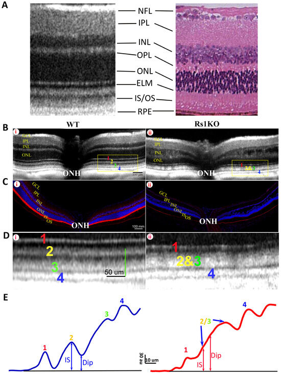
Bioengineering 2024, 11(5), 450; https://doi.org/10.3390/bioengineering11050450 - 01 May 2024
Abstract
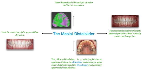
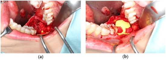
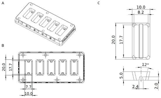
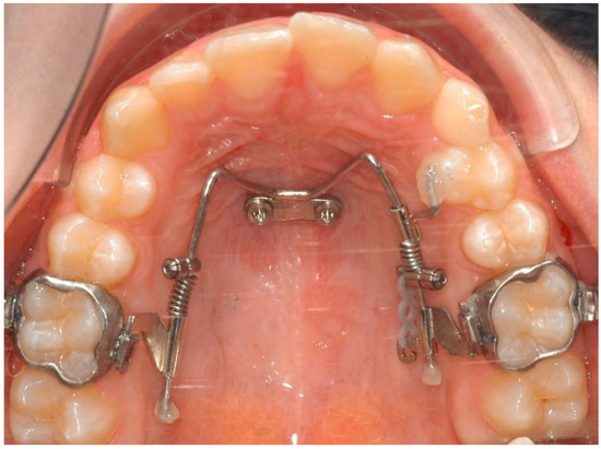
Aim: The purpose of the present study is the three-dimensional (3D) analysis of molar and incisor movements that occur during the correction of the upper midline deviation by using the Mesial-Distalslider appliance. Materials and Methods: A total of 20 consecutive patients (12 women
[...] Read more.
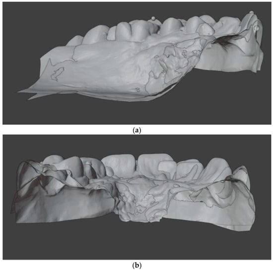
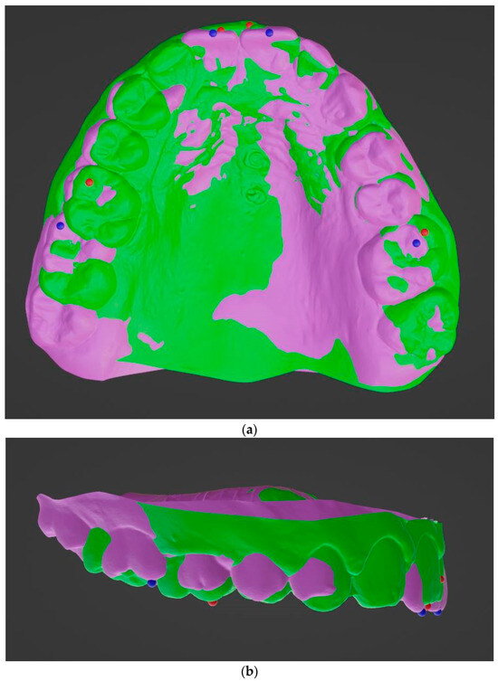
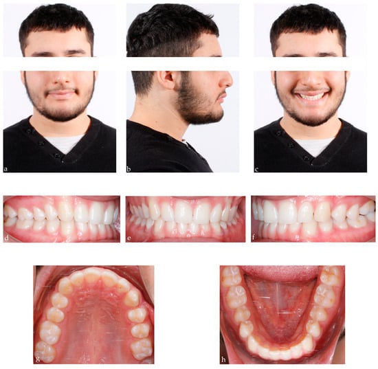
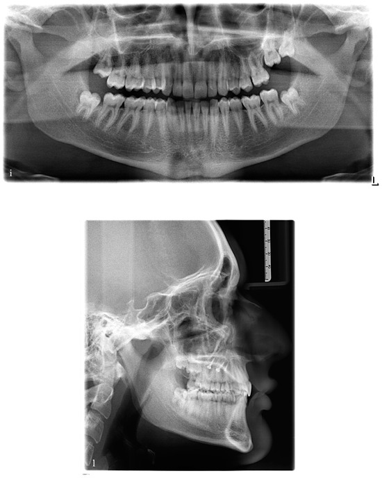
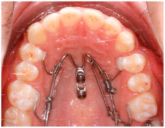
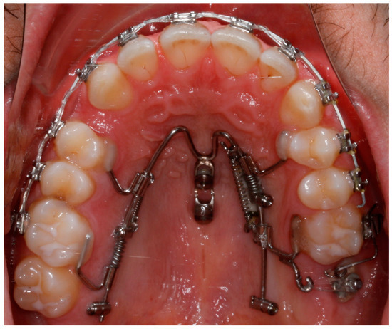
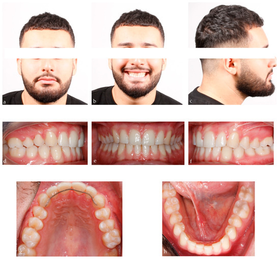
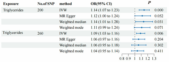
Aim: The purpose of the present study is the three-dimensional (3D) analysis of molar and incisor movements that occur during the correction of the upper midline deviation by using the Mesial-Distalslider appliance. Materials and Methods: A total of 20 consecutive patients (12 women and 8 men; mean age 19.6 ± 11.1 years) were selected from the Orthodontic Department of Heinrich-Heine University of Düsseldorf. To correct the upper midline deviation (>2 mm), the patients were treated with asymmetric mechanics (mesialization on one side and distalization on the contralateral side) with the aid of Mesial-Distalslider. Dental casts were taken for each patient before (T0) and after the treatment (T1). The casts were 3D digitized and the models were superimposed on the palatal anterior region. Three-dimensional molar movements and sagittal incisor movements (proclination and retroclination) were assessed for T0 and T1. Results: At the end of the treatment, the total movements of the molars resulted in 4.5 ± 2.2 mm (antero-posterior direction), −0.4 ± 2.4 mm (transverse direction) and 0.3 ± 0.9 mm (vertical direction) on the mesialization side, and −2.4 ± 1.7 mm (antero-posterior direction), −0.5 ± 1.5 mm (transverse direction) and 0.2 ± 1.4 mm (vertical direction) on the distalization side. Incisor displacement was 0.9 mm ± 1.7 (mesialization side) and 0.6 mm ± 0.7 (distalization side). Conclusion: The Mesial-Distalslider appliance could be considered a valuable tool in orthodontic treatment for upper midline correction. Within the limits of a retrospective study, asymmetric molar movements appeared possible without clinically relevant anchorage loss.
Full article
(This article belongs to the Section Biomedical Engineering and Biomaterials)
►
Show Figures
Graphical abstract


