Article

β-Orcinol Metabolites from the Lichen Hypotrachyna revoluta

1
University of Athens, Department of Pharmacy, Division of Pharmacognosy and Chemistry of Natural Products, Panepistimioupoli Zographou, Athens, 157 71, Greece
2
Department of Food Quality and Chemistry of Naturals Products, Mediterranean Agronomic Institute of Chania, Alsyllion Agrokepiou, Chania, 73100, Crete, Greece
*
Author to whom correspondence should be addressed.
Molecules 2007, 12(5), 997-1005; https://doi.org/10.3390/12050997
Submission received: 27 March 2007 / Revised: 7 May 2007 / Accepted: 11 May 2007 / Published: 12 May 2007

Abstract

:
Four new β-orcinol metabolites, hypotrachynic acid (1), deoxystictic acid (2), cryptostictinolide (3) and 8΄-methylconstictic acid (4) along with the metabolites 8΄-methylstictic acid (5), 8΄-methylmenegazziaic acid (6), stictic acid (7), 8΄-ethylstictic acid (8) and atranorin (9), that have been previously described, were isolated for the first time from the tissue extracts of the lichen Hypotrachyna revoluta (Flörke) Hale. The structures of the new metabolites were elucidated on the basis of extensive spectroscopic analyses. Radical scavenging activity (RSA) of the metabolites isolated in adequate amounts, was evaluated using luminol chemiluminescence and comparison with Trolox®.

Introduction

Lichens are complex symbiotic associations consisted of a fungus and an alga. The secondary metabolites produced by lichens are unique within the higher plants and have shown a wide spectrum of biological properties including antibiotic, antimycobacterial, cytotoxic and antioxidant activities [1,2,3,4,5,6,7,8]. A number of species have been used in various remedies in fork medicine, but the literature concerning the chemical composition of lichens is limited, since comprehensive studies are most of the times inhibited by the difficulties encountered with the collection of adequate amounts of tissue. Lichens frequently contain characteristic phenols such as depsides, depsidones, dibenzofurans and pulvinates [7]. These phenolic compounds are assumed to be fungal metabolites as studies with lichen mycobionts without the algal partner have shown their ability to biosynthesize extraordinary constituents under stress conditions [9]. Owing to the phenolic nature of the structures, these molecules are promising radical scavengers.
In the course of our investigations towards the isolation of new metabolites with antioxidant properties from terrestrial and marine organisms [10,11,12] we started a chemical study on the thalii extract of Hypotrachyna revoluta (Parmeliaceae). This lichen was selected on the basis of a comparative preliminary screening on a number of lichens collected in Southern Greece. In the present investigation we report the isolation of nine metabolites belonging to the β-orcinol depsides and depsidones chemical class from the CH2Cl2-MeOH extract of H. revoluta. The new natural products hypotrachinic acid (1), deoxystictic acid (2), cryptostictinolide (3) and 8΄-methylconstictic acid (4) share the rare, for their chemical class, structural feature of a γ-lactone. The structure elucidation of the new natural products was accomplished by extensive analyses of their spectral data. It is important to mention that these metabolites are the first examples of depsides (compound 1) and depsidones (compounds 2, 3) with a non functionalized α-carbon on the lactone ring. Along with the new molecules, the previously described metabolites 8΄-methylstictic acid (5) [13,14], 8΄-methyl- menegazziaic acid (6) [13], stictic acid (7) [15,16], 8΄-ethylstictic acid (8) [16] and atranorin (9) [7] were isolated from the same extract. Metabolite 6 has been isolated previously from an unidentified Usnea species collected in Sri Lanka [13]. It is noteworthy that compounds 1 - 8 are reported for the first time as metabolites of H. revoluta, since the only study on the chemistry of this species,reported the presence of atranorin and gyrophoric acid in a sample collected in New Zealand [17]. The radical scavenging activity of isolated metabolites, except metabolites 4 and 8 due to a paucity of material, was evaluated by the Co(II)/EDTA induced luminol plateau chemiluminescence assay [18].

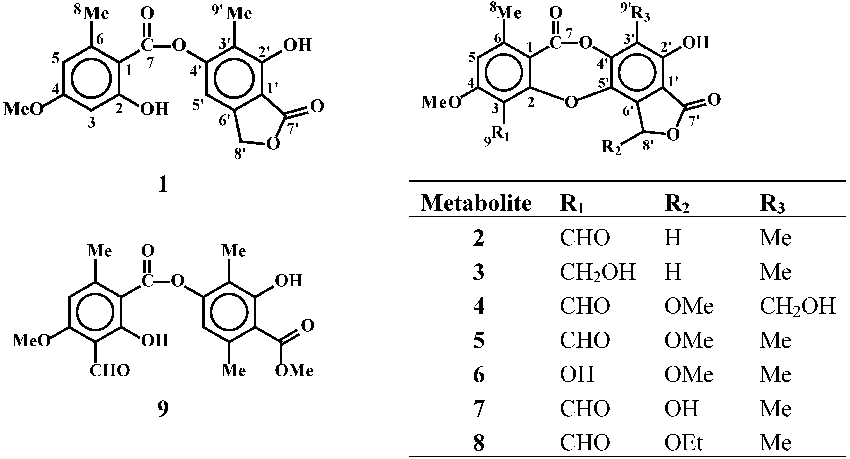
Results and Discussion

Hypotrachynic acid (1) was isolated as a white amorphous powder that by EI and FAB MS was found to have the molecular formula C18H16O7, as indicated by the molecular ion at m/z 344.0897 and the NMR data (Table 1). The eleven degrees of unsaturation along with the UV and IR spectra suggested a tricyclic phenolic metabolite containing ester carbonyls and free hydroxyls. The presence of two ester carbonyls was confirmed by their chemical shifts at 169.3 and 164.4 ppm. Additionally evident were two aromatic methyls at δ 2.44 and 2.20 ppm and a methoxy group at 3.82 ppm that along with a number of oxygenated aromatic quaternary carbons suggested a β-orcinol depsidone or depside chemical class. The only meta coupling observed between two of the three aromatic protons indicated their relative position on the same aromatic ring and together with the isolation of the remaining proton on the second aromatic ring thus suggested a depside skeleton. The downfield shift of the methylene carbon at 58.6 ppm and the heteronuclear correlations of the corresponding protons with aromatic carbons and an ester carbonyl, taking into account the number of remaining carbons suggested its involvement in a γ-lactone ring. Key information to confirm the lactone ring fusion was provided by the correlations between the H-8΄ and C-5΄ and between H-5΄ and C-3΄. The nOe correlations between the proton at 6.58 ppm with the methoxy (3.82 ppm) and the methyl group at 2.44 ppm along with the m-coupling of the protons and the correlation of H-5 with C-6 and C-1 supported the suggested assignment of substituents on the ring. The correlations on the HMBC spectrum between H3-9΄ and C-2΄, C-3΄ and C-4΄ were valuable for the final structure assignment of the second aromatic ring.
Figure 1. Structures of the compounds isolated from the lichen Hypotrachyna revoluta.
Molecules 12 00997 g001
Deoxystictic acid (2) was isolated as a white amorphous powder, that by EI and FAB MS was found to possess the molecular formula C19H14O8, as indicated by the pseudomolecular [M+H]+ ion at m/z 371.0741. The thirteen degrees of unsaturation, the presence of an additional aldehyde carbonyl (δC 187.2 and δH 10.45) and comparison of its NMR data with those of metabolite 1 suggested an additional ring in the structure, thus indicating that 2 belongs to the depsidone class. Evident from the 1H- and 13C-NMR spectra were, as before, the presence of two aromatic methyls on C-6 and C-3΄, the aromatic methoxy at C-4 and the hydroxyl group at C-2΄. The presence of only one aromatic proton was supportive of the ether bridge between C-2 and C-5΄ leading to a depsidone skeleton. The methylene resonance at δC 70.1 ppm and the γ-lactone carbonyl at δC 171.8 ppm clearly suggested that the previously encountered lactone moiety remained unchanged on metabolite 2 as well. Heteronuclear correlations of the aldehyde proton at 10.45 ppm with C-2 and C-3 at 161.3 and 113.6 ppm respectively, safely led to the assignment of the aldehyde carbon on C-3. Comparison of the proposed structure for metabolite 2 with stictic acid showed that the main differences were observed on the γ-lactone ring and especially on the absence of the hydroxyl group on C-8΄. All observed correlations on the HMBC spectrum were in accordance with the proposed structure for metabolite 2.
Table 1. 1H- (400 MHz) and 13C- (50.3 MHz) NMR spectral data of metabolites 1, 2, 3 and 5 in CDCl3 and 4 in CDCl3-CD3OD (1:3; v/v).
1234a5
δCδHδCδHδCδHδC δHδCδH
1114.5 114.4 113.7 113.9
2163.2 161.3 159.3 160.1
3100.96.73 (brs)113.6 121.5 114.6
4161.6 163.33 160.8 163.6
4-OCH3 56.03.82 (s) 56.6 3.96 (s)56.23.91 (s) 56.7 3.98 (s) 56.5 3.95 (s)
5111.36.58 (brs)111.7 6.74 (s)112.66.65 (s)112.4 6.81 (s)112.0 6.73 (s)
6144.9 151.1 144.7 151.7
7169.3 nd nd nd
8 22.62.44 (s) 22.4 2.56 (s)21.52.51 (s) 22.2 2.55 (s) 22.1 2.54 (s)
9 187.210.45 (s)53.64.79 (s)186.9 10.47 (s)187.310.47 (s)
1’107.2 106.9 104.8 107.2
2’152.9 152.2 151.8 152.0
3’120.9 117.7 119.9 121.4
4’150.1 148.9 149.4 149.3
5’101.37.17 (s)134.0 131.6 nd
6’nd 137.1 134.3 132.2
7’164.4 171.8 172.3 169.0
8’ 58.65.31
(d, J=13.2)
5.17
(d, J=13.2)
70.1 5.43 (s)67.15.64 (s)109.4 6.35 (s)102.3 6.38 (s)
9’ 8.92.20 (s) 8.9 2.27 (s)9.02.27 (s) 55.1 4.90 (s) 9.1 2.27 (s)
2’-OH nd 7.95 (brs) 7.87 (brs) nd 7.87 (brs)
8’-OCH3 57.4 3.65 (s)57.5 3.68 (s)
  • nd = not detected
  • a 13C-NMR assignments from indirect HMQC detection, due to insufficient sample quantities.
Cryptostictinolide (3) exhibited spectral characteristics similar to those of metabolite 2, with the two major points of difference being the absence of the aldehyde group and the presence of a new hydroxy methylene functionality (δC 53.6 and δH 4.79). The reduction of the aldehyde on C-3 to the secondary alcohol is in agreement with the molecular formula of C19H16O8 as indicated by the LR EI-MS and the HR FAB MS showing a pseudomolecular [M+H]+ ion at m/z 373.0948. Our literature search revealed that the proposed structure has been reported in the past [14] as a product of catalytic reduction of cryprostictic acid. The limited literature spectral data are now thus completed, with full assignments of protons and carbons of the molecule, which is reported for the first time as a natural product.
Metabolite 4 shared similar spectral features with all previously described molecules. The main differences were found to be the presence of a hydroxyl methylene, an aldehyde and an additional methoxy group on the carbon skeleton. The substitution of the previously consistent lactone methylene by a downfield shifted methine (δC 109.4 and δH 6.35) indicated oxidation of C-8΄ and supporting the presence of an additional methoxy group on this acetal moiety. The molecular formula of C20H16O10 was indicated by the LR EI-MS and was confirmed by the HR FAB MS showing a pseudomolecular [M+H]+ ion at m/z 417.0832. Comparison of the spectral characteristics with the above described metabolites and compounds reported in the literature revealed significant similarities with constictic acid previously isolated from Usnea aciculifera [7]. All spectra, of the minor metabolite 4, were supportive of being the 8΄-methyl analogue of constictic acid or the oxidation derivative of metabolite 5 on C-9΄. The chemical shifts of proton and carbon resonances for metabolite 5 are reported along with the new compounds in Table 1, since careful spectral analyses revealed some erroneous assignments in the previous literature report [13].

Radical scavenging activity

The evaluation of the lichen metabolites by the chemiluminescence method showed most of them to possess noteworthy antioxidant activity, with the highest levels being exhibited by compound 6, which was only seven times less potent than the standard antioxidant Trolox® that was used for comparison reasons (Table 2). The results showed that compounds 6 and 9 that posses an additional hydroxyl group on the aromatic ring are the most active ones and the activity is reduced by half when the hydroxyl of C-3 is replaced by an aldehyde moiety. Finally the scavenging activity of the metabolites possessing an aldehyde group on C-3 seems to be drastically reduced when the methylene of the γ-lactone ring is substituted by a hydroxy or methoxy moiety, as observed in the cases of metabolites 2, 7 and 5. Further studies though, with more sensitive and specialized assays, are planned for the future when more significant quantities of the metabolites will be available from scaled up separations.
Table 2. Hydroxyl radical-scavenging activity (CL) of evaluated metabolites.
MetaboliteRadical scavenging activity
(expressed as Trolox® equivalents) #
185.14 ± 0.60
213.76 ± 1.14
3not active
561.85 ± 1.65
67.63 ± 0.68
7104.34 ± 2.70
911.85 ± 0.64
# mg of metabolite exhibiting the same activity as 1 mg of Trolox®

Experimental

General

UV spectra were obtained on a Waters 996 photodiode array detector. IR spectra were obtained using a Paragon 500 Perkin-Elmer FT spectrophotometer. NMR spectra were recorded using Bruker AC 200 and Bruker DRX 400 spectrometers. Chemical shifts are given in δ (ppm) scale using TMS as internal standard. The 2D experiments (1H–1H COSY, HMQC, HSQC, HMBC) were performed using standard Bruker microprograms. High Resolution Mass Spectra data were provided by the University of Notre Dame, Department of Chemistry and Biochemistry, Notre Dame, Indiana, USA. EI and CI MS data were recorded on a MSQ Thermo Electron Mass Selective Detector. Column chromatography was performed with Kieselgel 60 (Merck), HPLC separations were conducted on Pharmacia LKB with UV detector (columns: Nucleosil 50-7, 7 μm, 250 x 10 mm; Spherisorb, 10 μm, 250 x 10 mm). MPLC separations were realized on a Buchi B-688 system using a silica gel 20 x 2 cm glass column. TLC was performed on Kieselgel 60 F254 aluminum support plates.

Plant material

Specimens of Hypotrachyna revoluta were collected from the barks of Oaks at Hleia Prefecture, in Southern Greece, in March 2004. Identification of the lichen was performed by Dr Alfons Piterans at the Department of Biology, University of Latvia. A specimen of the lichen is deposited at the Herbarium of the Laboratory of Pharmacognosy and Chemistry of Natural Products with number LN1.

Extraction and Isolation

After careful cleanup to remove remaining debris and pieces of the wood substrate air-dried thalli of H. revoluta (298.3 g) were extracted three times by maceration with CH2Cl2-MeOH (2:1) at room temperature for 24 hours each time. Following filtration and evaporation of the solvents under vacuum, the residue (24 g) was submitted to vacuum liquid chromatography (VLC) on silica gel, using CH2Cl2 with increasing amounts of EtOAc and subsequently MeOH to afford twenty five fractions that were analysed by TLC and 1H-NMR. Fraction II (eluted with CH2Cl2-EtOAc, 95:5) was further submitted to a gravity column chromatography on silica gel using mixtures of cyclohexane-CH2Cl2 of increasing polarity as eluent to afford in fractions 6-7 white crystals (202.2 mg) that by spectral analysis were found to be pure atranorin (9).
Fraction V was chromatographed by VLC using CH2Cl2-EtOAc as the mobile phase resulting in 20 subfractions. Subfraction V-7 was further separated by silica gel column chromatography (eluted with CHCl3-EtOAc mixtures of increasing polarity) to afford 30 fractions, the fourth of which when purified by HPLC (Nucleosil 50-7, CH2Cl2- MeOH, 98:2) resulted in the isolation of metabolite 3 (2.2 mg) as an amorphous white powder. Subfraction V-8 was purified by HPLC (Spherisorb, CHCl2-EtOAc, 95:5) to afford as eluted from the column: metabolite 1 (1.8 mg of white crystals), 2 (4.5 mg of amorphous white powder), 6 (3.8 mg of white crystals) and 5 (7.0 mg of white crystals).
Fraction VI was separated by gravity silica gel column chromatography using mixtures of CH2Cl2-EtOAc of increasing polarity to afford 45 subfractions. The subfraction eluted with EtOAc-CH2Cl2 (90:10) was characterized by the presence of stictic acid (7) as the main constituent and was further purified by an additional gravity silica gel column chromatography, isocratically eluted with CHCl3-EtOAc (90:10), to afford in pure form 2.1 mg of metabolite 7 as an amorphous white powder.
Fraction XVI was purified by silica gel Medium Pressure Liquid Chromatography (MPLC), isocratically eluted with CH2Cl2-MeOH-HCOOH (98:1:1), to afford 70 subfractions. Subfractions XVI-13 to XVI-16 were combined and submitted to HPLC separation (Spherisorb, CHCl3-EtOAc, 80:20) resulting in the isolation of 1.4 mg of pure metabolite 8 as an amorphous powder. Fraction XVII was submitted to silica gel MPLC isocratically eluted with CH2Cl2-MeOH-HCOOH (98:1:1) to afford 30 subfractions. Subfractions XVII-15 to XVII-17 were combined and separated by an additional MPLC isocratically eluted with CH2Cl2-MeOH-HCOOH (96:3:1) to afford 0.9 mg of pure metabolite 4 as an amorphous white powder.
Hypotrachynic acid (1): C18H16O7; white amorphous powder; UV (MeOH) λmax 314, 246; IR (MeOH) νmax: 3750, 3618, 1747, 1715, 1660, 1457; for NMR spectroscopic data, see Table 1; HREIMS: 344.0897 (M+, calcd. for C18H16O7, 344.0896).
Deoxystictic acid (2): C19H14O8; white amorphous powder; UV (MeOH) λmax 308, 246; IR (MeOH) νmax: 3421, 1737, 1716, 1691, 1681, 1455, 1439; for NMR spectroscopic data, see Table 1; HREIMS: 371.0741 ([M+H]+, calcd. for C19H15O8, 371.0766).
Cryptostictinolide (3): C19H16O8; white amorphous powder; UV (MeOH) λmax: 306, 253; IR (MeOH) νmax: 3510, 3260, 3212, 1745, 1730, 1714, 1652, 1459; for NMR spectroscopic data, see Table 1; HREIMS: 373.0948([M+H]+, calcd. for C19H17O8, 373.0918).
8΄-Methylconstictic acid (4): C20H16O10; white amorphous powder; UV (MeOH) λmax: 309. 252; IR (MeOH) νmax: 3448, 1740, 1712, 1690, 1685, 1451; for NMR spectroscopic data, see Table 1; HREIMS: 417.0832 ([M+H]+, calcd. for C20H18O10, 417.0816).

Hydroxyl radical - scavenging activity (CL)

Three dilutions of each sample were prepared using methanol as a solvent. The concentration of the dilutions depended on the activity of the metabolite and ranged from 0.012 – 0.017 mg/mL. The chemiluminescence assays were carried out on a Jenway 6200 Fluorimeter (Leeds, U.K.), with the lamp off, using only the photomultiplier of the apparatus. Boric acid buffer solution (0.05 M adjusted to pH 9 with NaOH 1M, 1 mL) containing CoCl2. 6H2O (0.2 mg/mL) and EDTA (1 mg/mL) was vortexed for 15 sec with luminol solution (5.6 x 10⁻⁴ M, 100 μL) in borate buffer (0.05 M adjusted to pH 9 with 1M NaOH) in a test tube. Then aqueous H2O2 solution (5.4 x 10⁻³ M, 25 μL) were added and the mixture was vortexed again for 15 sec, quickly placed in a glass cuvette with a Pasteur pipette and the chemiluminescence intensity was measured when the plateau was reached (Io) with a lifetime of about 30 sec. When the light intensity was stabilized, the sample solution (25 μL) was added and mixed with a Pasteur pipette. The instantaneous decrease of the plateau was recorded (I) [18]. The ratio Io/I was plotted against mg of algae extract/mL for three dilutions of each extract and the quantity required to reduce Io by 50% (IC50 value) was calculated thereof [18] and compared against Trolox® standard. The evaluation of antioxidant activity of tested metabolites with the chemiluminescence method showed metabolite 6 to be the most active with an IC50 value of 0.389 mg/mL.

Acknowledgments

We wish to thank Dr Alfons Piterans (Department of Biology, University of Latvia) for the identification of the lichen Hypotrachyna revoluta. The financial support to this study by the 01ED146 PENED program of the Greek Secretariat for Research and Development is gratefully acknowledged.

References

  1. Vartia, K.O. The Lichens; Ahmadjian, V., Hale, M.E., Eds.; Academic Press: New York, 1974; p. 547. [Google Scholar]
  2. Rundel, P.W. The Ecological Roleof Secondary Lichen Substances. Biochem. Syst. Ecol. 1978, 6, 157–170. [Google Scholar] [CrossRef]
  3. Ingólfsdóttir, K.; Chung, G.A.C.; Skúlason, V.G.; Gissurarson, S.R.; Vilhelmsdóttir, M. Antimycobacterial activity of lichen metabolites in vitro. Eur. J. Pharm. Sci. 1998, 6, 141–144. [Google Scholar]
  4. Perry, N.B.; Benn, M.H.; Brennan, N.J.; Burgess, E.J.; Ellis, G.; Galloway, D.J.; Lorimer, S.D.; Tangney, R.S. Antimicrobial, antiviral and cytotoxic activity of New Zealand lichens. Lichenologist 1999, 31, 627–636. [Google Scholar] [CrossRef]
  5. Richardson, D.H.S. Handbook of Lichenology; Calun, M., Ed.; CRC Press: Boca Raton, 1988; Vol. 3, p. 93. [Google Scholar]
  6. Richardson, D.H.S. Frontiers in Mycology; Hawksworth, D.L., Ed.; CAB International: Wallingford, 1991; p. 187. [Google Scholar]
  7. Huneck, S.; Yoshimura, I. Identification of Lichen Substances; Springer-Verlag: Berlin, 1996. [Google Scholar]
  8. Hidalgo, M.E.; Fernandez, E.; Quilhot, W.; Lissi, E. Antioxidant Activity of Depsides and Depsidones. Phytochemistry 1994, 37, 1585–1587. [Google Scholar] [CrossRef]
  9. Culberson, C.F. Chemical and Botanical Guide to Lichen Products; The University of North Carolina Press: Chapel -Hill, NC, 1969. [Google Scholar]
  10. Nahas, R.; Abatis, D.; Anagnostopoulou, M. A.; Kefalas, P.; Vagias, C.; Roussis, V. Radical scavenging activity of Aegean sea marine algae. Food Chem. 2007, 102, 577–581. [Google Scholar] [CrossRef]
  11. Guri, A.; Kefalas, P.; Roussis, V. Antioxidant potential of six pine species. Phytother. Res. 2006, 20, 263–266. [Google Scholar]
  12. Tziveleka, L.-A.; Kourounakis, A.P.; Kourounakis, P.N.; Roussis, V.; Vagias, C. Antioxidant potential of natural and synthesized polyprenylated hydroquinones. Bioorg. Med. Chem. 2002, 10, 935–939. [Google Scholar] [CrossRef] [PubMed]
  13. Kathirgamanathar, S.; Williams, D.E.; Andersen, R.J.; Bombuwela, K.; De Silava, D.; Karunaratne, V. β-Orcinol depsidones from the lichen Usnea sp. from Sri Lanka. Nat. Prod. Res. 2005, 19, 695–701. [Google Scholar] [CrossRef]
  14. Shimada, S.; Saitoh, T.; Sankawa, U.; Shibata, S. New depsidones from Lobaria oregano. Phytochemistry 1980, 19, 328–330. [Google Scholar] [CrossRef]
  15. González, A.G.; Barrera, J.B.; Perez, E.M.R.; Padron, C.E.H. Depsidones from Lobaria pulmonaria and their chemotaxonomic importance. Biochem. Syst. Ecol. 1994, 22, 583–586. [Google Scholar]
  16. Huneck, S.; Tabacchi, R. ψ-Esters of depsidones with a lactole ring. Phytochemistry 1987, 26, 1131–1138. [Google Scholar] [CrossRef]
  17. Walker, J.R.L.; Lintott, E.A. A phytochemical register of New Zealand lichens. New Zeal. J. Bot. 1997, 35, 369–384. [Google Scholar]
  18. Parejo, I.; Codina, C.; Petrakis, C.; Kefalas, P. Evaluation of scavenging activity assessed by Co(II)/EDTA-induced luminol chemiluminescence and DPPH (2,2-diphenyl-1-picrylhydrazyl) free radical assay. J. Pharmacol. Toxicol. Methods 2000, 44, 507–512. [Google Scholar] [CrossRef] [PubMed]
  • Sample Availability: Contact the authors.

