Article

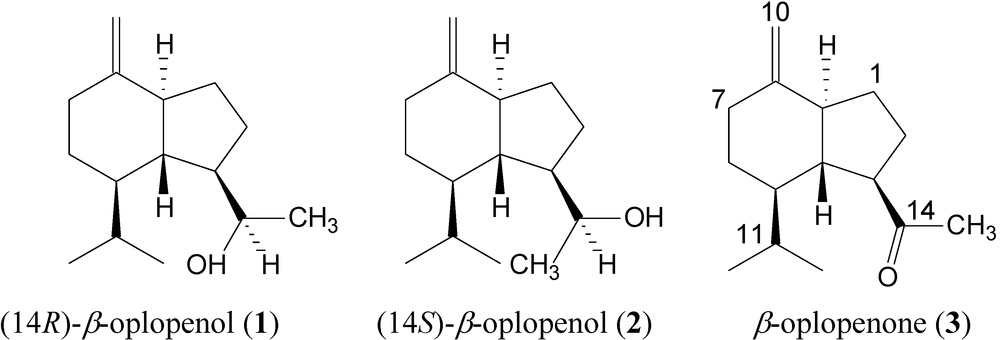
Spectral Data of Two New Asymmetric Sesquiterpene Alcohols: (14R)-β-Oplopenol and (14S)-β-Oplopenol

by
Julien Paolini
*,
Jean-Marie Desjobert
,
Alain Muselli
and
Jean Costa
Université de Corse, UMR-CNRS 6134, Laboratoire de Chimie des Produits Naturels, BP 52, 20250 Corti, France
*
Author to whom correspondence should be addressed.
Molecules 2008, 13(4), 1004-1010; https://doi.org/10.3390/molecules13041004
Submission received: 31 March 2008 / Revised: 28 April 2008 / Accepted: 28 April 2008 / Published: 29 April 2008

Abstract

:
The epimeric sesquiterpene alcohols (14R)-β-oplopenol and (14S)-β-oplopenol were obtained by LiAlH4 reduction of β-oplopenone. The complete 1H- and 13C-NMR assignments of these two new sesquiterpene alcohols have been made using 1D and 2D NMR techniques, including COSY, NOESY, HSQC, HMBC experiments.

Introduction

The oplopane derivatives were well represented from plants of the Senecioneae family [1,2,3,4,5,6,7,8]. The sesquiterpenic ketone β-oplopenone was identified as a major component of the essential oil of Adenostyles briquetii, an endemic species from the island of Corsica [9]. To our knowledge, the spectral data of these asymmetric alcohols: (14R)-β-oplopenol (1) and (14S)-β-oplopenol (2), have never been reported. The aim of this study was to establish the spectral assignments and the relative configuration of these two new epimers using extensive NMR assignments data along with EI-mass spectra and IR spectrum.

Results and Discussion

The LiAlH4 reduction of β-oplopenone (3), a sesquiterpene isolated from the aerial parts of Adenostyles briquetii [9], yielded two new stereoisomers (Figure 1). Their structures were identified as (14R)-β-oplopenol (1) and (14S)-β-oplopenol (2). The retention indice (I Rtx-1), EI-mass spectra, 1H- and 13C-NMR spectral data of β-oplopenone (3) were in agreement with those reported in the literature [10,11,12,13,14,15,16]. Those of compounds 1 and 2 are compiled in Table 1 and Table 2.
Figure 1. Structures of (14R)-β-oplopenol 1, (14S)-β-oplopenol 2 and β-oplopenone 3.
Molecules 13 01004 g001
The EI-mass spectra of alcohols 1 and 2 showed a molecular ion (M.+) at m/z 222, while that of ketone 3 appears at m/z 220. The EI-mass spectra of components 1 and 2 were similar, except for the relative abundance of several signals. Both epimers exhibited a [M-18]+. signal at m/z 204 corresponding to the loss of a hydroxyl group after hydrogen rearrangement. Their IR spectra showed a strong hydroxyl group band (3300-3500 cm-1) and the absence of the carboxyl group one (1710 cm-1) observed for compound 3. The 1H-NMR spectra of the both epimers showed a methyl-15 signal at significantly smaller chemical shift [δ = 1.18 ppm (d) in 1 and δ = 1.13 ppm (d) in 2] while in 3 it appeared at δ = 1.97 ppm (s). The C-14 signal appears in the 13C-NMR spectra at δ = 211.71 ppm (s) in 3, and at δ = 68.71 ppm in 1 and at δ = 69.56 ppm in 2. In the HMBC spectrum, this signal was correlated with the proton signals of C-15, C-4, C-3 and C-2, proving that C-14 was bonded to the hydroxyl group obtained by reduction of the carbonyl group of 3. Finally, the stereochemistry of H-3α, H-4β, H-5α and H-9α in agreement with the structure of 3 was confirmed by 1D and 2D NMR spectrum.
The molecular formulae of 1 and 2 were confirmed from 13C-NMR, 1H-NMR and DEPT data as C15H26O, indicating three degrees of unsaturation. The oplopane sesquiterpene skeleton was directly inferred from its 13C-NMR spectrum with the aid of a DEPT experiment which exhibited fifteen signals comprising three methyl, five methylenes, six methines and one non-protonated carbons atoms. The 13C-NMR assignments of the both epimers 1 and 2 were confirmed from a two dimensional 13C-1H chemical shifts correlation diagram (HSQC) and the assignments of C-3, C-4, C-5 and C-9 were established from a HMBC spectrum and proton-coupled spectrum (COSY). In order to determine the relative configuration of C-14, we have compared the spectral data of both epimers. The H-3 and H-4 proton signals of 1 and 2 differed in the 1H-NMR spectra. The chemical shift at δ = 2.14 ppm (ddt, J = 7.0; 11.0; 3.6 Hz) and δ = 0.87 ppm (q, J = 11.0 Hz) in 2 appears as broadened signals at δ =1.77 ppm (m) and δ = 1.25 ppm (m) in 1, respectively.
Table 1. 1H- and 13C-NMR spectra data, HSQC and HMBC correlations for (14R)-β-oplopenol (1).
Cδ (13C)DEPTδ (1H)Mutiplicity (J, Hz)HMBC COSY
127.79CH21.34m9, 3, 22, 3, 9
1.43m
222.62CH21.62dtd (20.0; 7.4; 12.8)14, 9, 3, 11, 3, 4
1.81m
349.02CH1.77m14, 15, 6, 4, 22, 5, 1
450.23CH1.25m14, 11, 9, 6, 5, 33, 9, 1
550.09CH1.34m13, 12, 11, 7, 6, 46, 3
626.95CH21.10dq (4.4; 13.0)11, 7, 5, 45, 7
1.75m
735.32CH21.95m10, 6, 810, 6
2.35ddd (2.4; 4.3; 13.4)6,
8151.98C--9, 7, 6, 5, 1-
952.50CH1.83m10, 7, 5, 4, 3, 2, 110, 4
10102.70CH24.54q (1.7)9, 79
4.62q (1.7)7
1128.50CH1.95m13, 12, 6, 512, 13
1221.95CH30.95d (6.9)13, 11, 511
1315.85CH30.78d (6.9)12, 11, 511
1468.71CH3.98q (6.3)15, 4, 3, 215
1523.01CH31.18d (6.3)14, 4, 3, 214
Table 2. 1H- and 13C-NMR spectra data, HSQC and HMBC correlations for (14S)-β-oplopenol (2).
Cδ (13C)DEPTδ (1H)Mutiplicity (J, Hz)HMBCCOSY
128.12CH21.33m9, 29, 2
1.72m9, 2
223.44CH21.65dtd (18.1; 7.0; 11.0)14, 13, 1
1.77m
348.79CH2.14ddt (7.0; 11.0; 3.6)15, 4, 2, 12,4,14,
451.72CH0.87q (11.0)9, 6, 2, 19, 5, 3
549.94CH1.34m13, 12, 6, 4, 76, 4
626.95CH21.01dq (13.0; 4.5)11, 77, 5
1.73m
735.18CH21.96m10, 66, 10
6
2.34ddd (2.4; 4.5; 13.0)
8152.03C--9, 7, 1
952.69CH1.86m10, 7, 5, 4, 1, 2, 610, 4
10102.55CH24.54q (1.7)11, 79
4.61q (1.7)7
1128.69CH1.94m13, 1213, 12, 6
1221.95CH30.96d (6.8)13, 611
1315.83CH30.70d (6.8)12, 411
1469.56CH4.03dq (3.9; 6.3)15, 4, 23, 15
1516.89CH31.13d (6.3)1214
Moreover, in 2 the H-14 proton signal was a double quartet at 4.03 ppm (J = 3.6; 6.3 Hz), whereas a quartet was observed at δ = 3.98 ppm (J = 6.32 Hz) in 1. These results proved the presence of a 3J vicinal coupling between H-3 and H-14 in 2, not observed in 1. The 3JH-H coupling constant value measured in 2 (3.6 Hz) and 1 (0 Hz) indicated that the dihedral angle between H-3 and H-14 were close to 110° in 2 and to 90° in 1 (Karplus curve) [17,18]. Furthermore, NOE contacts between H-14, H-11 and H-4 were observed in the NOESY spectra of both epimers, proving that the relative configurations of H-14 and H-11 were β. The variation of the 13C-NMR chemical shifts in 1 and 2, were quite similar (Δδ always lower than 0.33 ppm) except for the C-2 (Δδ = 0.82 ppm), C-14 (Δδ = 0.85 ppm), C-4 (Δδ = 1.49 ppm) and particularly for the C-15 (Δδ = 6.12 ppm). The strong shielding of the C-15 carbon signal (δ = 16.89 ppm) in 2 may be ascribed to a steric interaction and proved that the hydrogenated carbons C-2 and C-15 were in the γ-gauche position relative to each other. This γ-steric effect was not observed for the C-15 signal in 1 (δ = 23.01 ppm). These results, in combination with the NOE results, allowed us to establish the relative configuration of C-14 as (14R)- for 1 and (14S)- for 2. Furthermore the configuration at C-14 was predictable since the isopropyl group causes a marked steric effect on one side of the carbonyl group impeding its easy reduction, favoring formation of alcohol 1.

Experimental

General

GC analysis were carried out using a Perkin-Elmer (Waltham, MA, USA) Autosystem XL GC apparatus equipped with a dual flame ionization detection (FID) system and two fused-silica capillary columns (60 m ° 0.22 mm I.D., film thickness 0.25 μm), Rtx-1 (polydimethylsiloxane) and Rtx-wax (polyethyleneglycol). The oven temperature was programmed from 60 °C to 230 °C at 2 °C/min and then held isothermally at 230 °C for 35 min. Injector and detector temperature was maintained at 280 °C. Samples were injected in the split mode (1/50), using helium as carrier gas (1 mL/min); injection volume, 0.1 μL of pure component. Retention indices (I) of compounds were determined relative to the retention times of series of n-alkanes (C5-C30) with linear interpolation, using Van den Dool and Kratz equation [19] and software from Perkin-Elmer. Components relative concentrations were calculated based on GC peak areas without using correction factors. GC/MS samples were analysed with a Perkin-Elmer Turbo mass detector (quadrupole), coupled to a Perkin-Elmer Autosystem XL, equipped with fused-silica capillary columns Rtx-1 and Rtx-Wax. Carrier gas: helium (1mL/min), ion source temperature: 150°C, oven temperature programmed from 60°C to 230°C at 2°C/min and then held isothermally at 230°C (35 min), injector temperature: 280°C, energy ionization: 70 eV, electron ionization mass spectra were acquired over the mass range 35-350 Da, split: 1/80, injection volume: 0.1 μL of pure component. IR spectra were obtained on a FT-IR spectrometer Perkin-Elmer spectrum RX 1. The components were analysed by 13C-NMR in deuterated chloroform on a Bruker (Wissembourg, France) Avance 400 Fourier Transform spectrometer operating at 100.13 MHz, equipped with a 5 mm probe. All shifts are referred to internal tetramethylsilane (TMS). 13C-NMR spectra were recorded with the following parameters: pulse width = 4 µs (flip angle 45°); acquisition time = 2.7 s for 128 000 data table with a spectral width of 25 000 Hz (250 ppm); CPD mode decoupling; digital resolution = 0.183 Hz/pt. The number of accumulated scans was 5000 for each sample (around 40 mg of pure component in 0.5 mL of CDCl3). Exponential line broadening multiplication (1 Hz) of the free induction decay was applied before Fourier transformation. 1H-NMR spectra were recorded with the following parameters: flip angle 30°; acquisition time = 2.56 s for 32 000 data table with a spectral width of 7000 Hz (17.5 ppm).

Reduction

β-Oplopenone (3, 140 mg, 99% purity by GC) dissolved in dry Et2O (10 mL) was carefully added to a suspension of LiAlH4 (40 mg) in dry Et2O (15 mL) at 0°C. The mixture was stirred at room temperature and then refluxed for 3 h. The reaction mixture was hydrolysed by addition of a solution of 15% NaOH (1 mL) and cold water (1 mL). The organic layer was separated, washed with water to neutrality, dried over Na2SO4 and conc. in vacuum. The mixture (120 mg) contained as major components β-oplopenone (3, GC: 31%) and two alcohols: (14R)-β-oplopenol (1, GC: 55%, characterized after isolation), and (14S)-β-oplopenol (2, GC: 12%, characterized after isolation). The two epimers 1 and 2 and the startuing material 3 were isolated by column chromatography (CC: SiO2, 63-200 μm). β-Oplopenone (3, 40 mg, 99% purity by GC) was eluted with a mixture n-C5H12/Et2O = 95/5, (14R)-β-oplopenol (1, 60 mg, 98% purity by GC) with n-C5H12/Et2O = 90/10 and (14S)-β-oplopenol (2, 15 mg, 95% purity on GC) with n-C5H12/Et2O = 85/15.
(14R)-β-oplopenol (1). GC: 98%; I Rtx-1 = 1644; I Rtx-Wax = 2216; EIMS (70 e/v): 222 (3), 204 (17), 177 (18), 161 (83), 135 (58), 119 (38), 105 (57), 91 (82), 79 (69), 67 (48), 55 (39), 45 (71), 43 (93), 41 (100); IR (KBr) υmax cm-1: 3427, 3076, 2932, 1651, 1460, 1375, 886; 1H- and 13C-NMR: see Table 1.
(14S)-β-oplopenol (2). GC: 95%; I Rtx-1 = 1670; I Rtx-Wax = 2296; EIMS (70 e/v): 222 (2), 204 (14), 177 (12), 161 (69), 135 (51), 119 (34), 105 (52), 91 (80), 79 (71), 67 (47), 55 (39), 45 (98), 43 (97), 41 (100); IR (KBr) υmax cm-1: 3349, 3076, 2932, 1652, 1461, 1378, 886; 1H- and 13C-NMR: see Table 2.
β-oplopenone (3) [4]. GC: 99%; I Rtx-1 = 1596; I Rtx-Wax = 2050; EIMS (70 e/v): 220 (4), 177 (31.6), 107 (16), 91 (20), 79 (17), 67 (14), 55 (12), 43 (100), 41 (34); IR (KBr) υmax cm-1 (CCl4): 3070, 2955, 2874, 1710, 1652, 1462, 1358, 887; 1H-NMR: δ 0.65, 0.90 (3H, 2d, J= 6.8 Hz, Me-12 and Me-13), 1.97 (3H, s, Me-15), 2.34 (1H, ddd, J= 13.4 Hz; 2.6 Hz; 4.3 Hz, H-7β), 2.70 (1H, ddd, J= 5.1 Hz; 12.0 Hz; 10.5 Hz, H-3), 4.66 (1H, q, J= 1.6 Hz, H-10), 4.55 (1H, q, J= 1.6 Hz, H-10’) and 1.10 (1H, dq, 13.8 Hz; J= 4.3 Hz, H-6β); 13C-NMR: 27.34 (C-1), 28.48 (C-2), 56.06 (C-3), 52.08 (C-4), 49.26 (C-5), 26.55 (C-6), 35.27 (C-7), 150.86 (C-8), 51.80 (C-9), 103.52 (C-10), 29.58 (C-11), 21.93 (C-12), 15.66 (C-13), 211.71 (C-14), 28.87 (C-15).

Acknowledgements

The authors are grateful to the laboratory “Chimie et Biomasse” (University of Corsica) for recording the NMR spectra. Furthermore, we would like to thank the European Community for partial financial support (PIC INTERREG IIIA).

References

  1. Takeda, K.; Minato, H.; Ishikawa, M. Studies on sesquiterpenoids-XII- Structure and absolute configuration of oplopanone, a new sesquiterpene from oplopanax japonicus (Nakai). Tetrahedron Suppl. 1966, 7, 219–225. [Google Scholar] [CrossRef]
  2. David, J.R. The biology and chemistry of the Compositae; Heywood, V.H., Harborne, J.B., Turner, B.L, Eds.; Academic Press: London-New York-San Francisco, 1977; Vol. 2, pp. 831–850. [Google Scholar]
  3. Abdel Aal, M.; Bohlmann, F.; Sarg, T.; El-Domiaty, M.; Nordenstam, B. Oplopane derivatives from Acrisione denticulata. Phytochemistry 1988, 27, 2599–2602. [Google Scholar]
  4. Joseph-Nathan, P.; Villagómez, J.R.; Román, L.U.; Hernández, J.D. An oplopane from Senecio mexicanus. Phytochemistry 1989, 28, 1207–1209. [Google Scholar] [CrossRef]
  5. Joseph-Nathan, P.; Villagómez, J.R.; Román, L.U.; Hernández, J.D. Oplopanes from the leaves of Senecio mexicanus. Phytochemistry 1990, 29, 977–979. [Google Scholar] [CrossRef]
  6. Hayashi, K. Oplopane sesquiterpenes from Petasites palmatus. Phytochemistry 1989, 28, 3373–3376. [Google Scholar] [CrossRef]
  7. Arciniegas, A.; Pérez-Castorena, A.L.; Reyes, S.; Contreras, J.L.; De Vivar, A.R. New oplopane and eremophilane derivatives from Robinsonecio gerberifolius. J. Nat. Prod. 2003, 66, 225–229. [Google Scholar]
  8. Xie, W.D.; Zhang, Q.; Li, P.L.; Jia, Z.J. Two triterpenoids and other constituents from Petasites tricholobus. Phytochemistry 2005, 66, 2340–2345. [Google Scholar] [CrossRef]
  9. Paolini, J.; Nasica, E.; Desjobert, J.M.; Muselli, A.; Bernardini, A.F.; Costa, J. Analysis of volatile constituents isolated by hydrodistillation and headspace-solid phase microextraction from Adenostyles briquetii Gamisans. Phytochem. Ana 2007, in press. [Google Scholar] [CrossRef]
  10. Bohlmann, F.; Zdero, C. Neue furanoeremophilane und andere sesquiterpene aus vertretern der gattung Euryops. Phytochemistry 1978, 17, 1135–1153. [Google Scholar] [CrossRef]
  11. Wratten, S.J.; Faulkner, D.J. Metabolites of the red alga Laurencia subopposita. J. Org. Chem. 1977, 42, 3343–3349. [Google Scholar] [CrossRef]
  12. De Pascual, J.; Feliciano, A.S.; Miguel de Corral, J.M.; Barrero, A.F. Terpenoids from Juniperus Sabina. Phytochemistry 1983, 22, 300–301. [Google Scholar] [CrossRef]
  13. Piers, E.; Gavai, A.V. Convenient (Z)-ethylidenecyclopentane annulation sequences. Total synthesis of (±)-oplopanone, (±)-8-epi-oplopanone, and (±)-anhydro-oplopanone. Tetrahedron Lett. 1986, 27, 313–316. [Google Scholar] [CrossRef]
  14. Joseph-Nathan, P.; Villagomez, J.R.; Rojas-Gardida, M.; Roman, L.U.; Hernandez, J.D. Minor oplopanes from Senecio mexicanus. Phytochemistry 1989, 28, 2397–2401. [Google Scholar]
  15. Piers, E.; Gavai, A.V. A (Z)-Ethylidenecyclopentane annulation method. Total syntheses of (±)-Anhydrooplopanone, (±)-Oplopanone, and (±)-8-epi-Oplopanone. J. Org. Chem. 1990, 55, 2380–2390. [Google Scholar] [CrossRef]
  16. König, W.A.; Hochmuth, D.H.; Joulain, D. Terpenoids and Related Constituents of Essential Oils; MassFinder 2.1 Library; Institute of Organic Chemistry: Hamburg, Germany, 2001. [Google Scholar]
  17. Karplus, M.J. Contact Electron-Spin Coupling of Nuclear Magnetic Moments. Chem. Phys. 1959, 30, 11–15. [Google Scholar] [CrossRef]
  18. Karplus, M.J. Vicinal Proton Coupling in Nuclear Magnetic Resonance. J. Am. Chem. Soc. 1963, 85, 2870–2871. [Google Scholar] [CrossRef]
  19. Van Den Dool, H.; Kratz, P.D. A generalization of the retention index system including linear temperature programmed gas-liquid partition chromatography. J. Chromatogr. 1963, 11, 463–471. [Google Scholar] [CrossRef]
  • Sample Availability: Contact the authors.

