Isolation and Characterization of Phenolic Compounds from the Leaves of Salix matsudana

Abstract
:Introduction

Results and Discussion
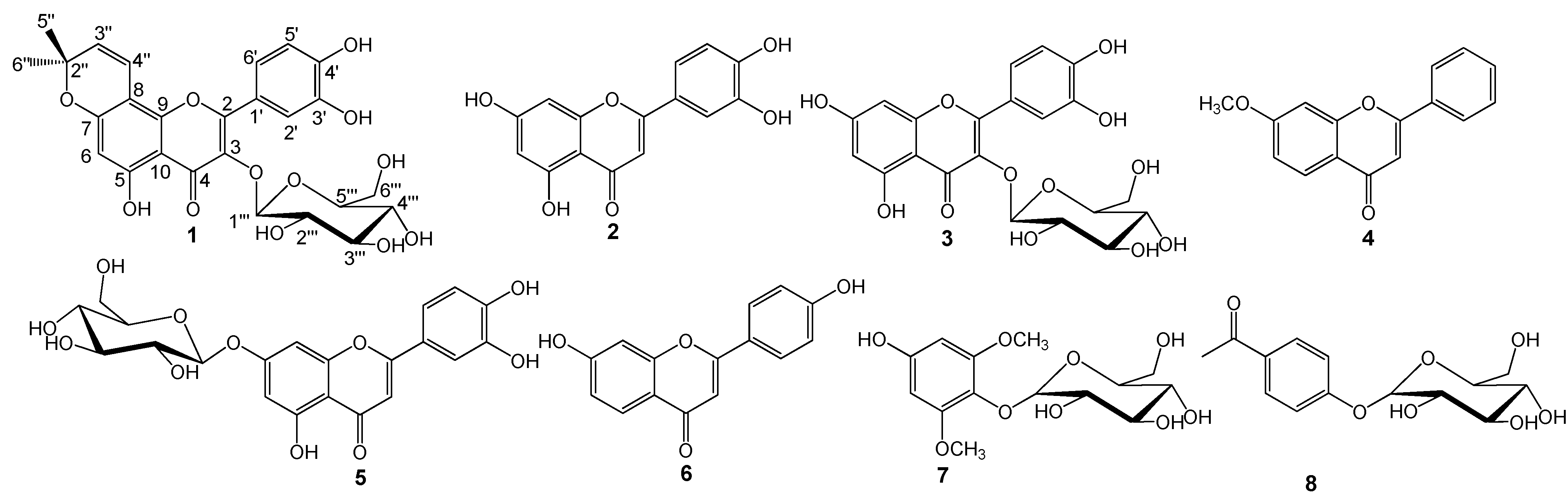
Characterization of compounds 1–8

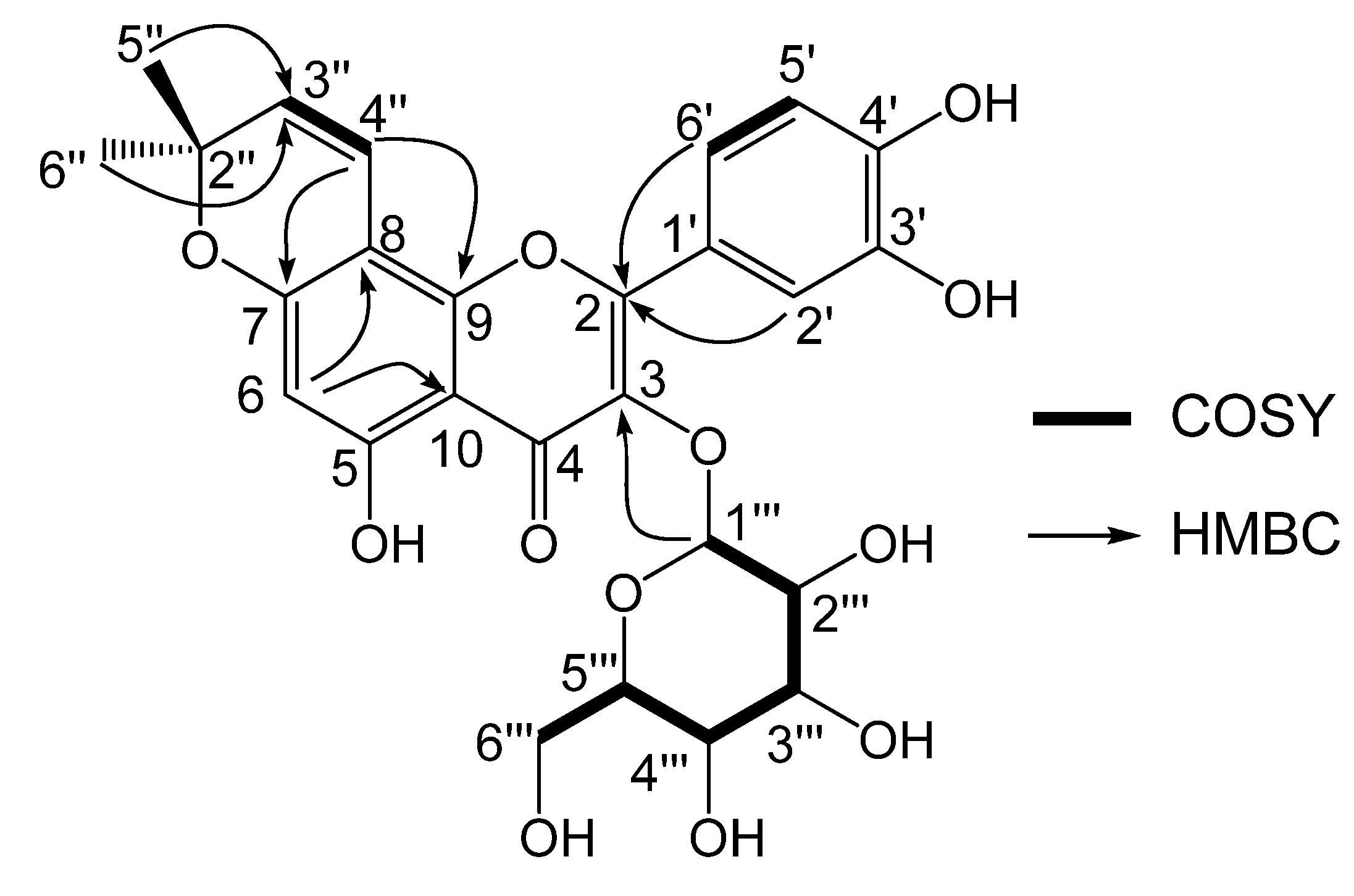
| Position | 1H-NMR | J (Hz) | 13C-NMR | DEPT | COSY | HMBC | 13C-NMR b |
|---|---|---|---|---|---|---|---|
| 2 | - | - | 155.2 | C | - | - | 155.4 |
| 3 | - | - | 142.7 | C | - | - | 137.2 |
| 4 | - | - | 178.9 | C | - | - | 177.5 |
| 5 | - | - | 155.0 | C | - | - | 154.8 |
| 6 | 6.40, s | - | 95.3 | CH | - | C-8, C-10 | 94.1 |
| 7 | - | - | 160.0 | C | - | - | 158.0 |
| 8 | - | - | 105.1 | C | - | - | 103.8 |
| 9 | - | - | 153.9 | C | - | - | 154.7 |
| 10 | - | - | 105.6 | C | - | - | 104.6 |
| 1’ | - | - | 122.1 | C | - | - | 120.0 |
| 2’ | 7.67, d | 1.7 | 117.2 | CH | H-6' | C-2, C-3', C-4', C-6' | 115.1 |
| 3’ | - | - | 145.3 | C | - | - | 144.6 |
| 4’ | - | - | 147.6 | C | - | - | 148.3 |
| 5’ | 6.95, d | 8.5 | 114.7 | CH | H-6' | C-1', C-3' | 115.0 |
| 6’ | 7.38, dd | 8.5, 1.7 | 120.7 | CH | H-2', H-6' | C-1', C-2, C-2', C-4' | 120.0 |
| 2” | - | - | 78.1 | C | - | - | 77.4 |
| 3” | 5.57, d | 10.0 | 125.7 | CH | H-4'' | C-5''', C-6''', C-7, C-8 | 128.5 |
| 4” | 6.56, d | 10.0 | 116.3 | CH | H-3'' | C-5''', C-6''', C-7, C-9 | 113.9 |
| 5” | 1.47, s | - | 27.3 | CH3 | - | C-2''', C-3''' | 27.2 |
| 6” | 1.47, s | - | 27.3 | CH3 | - | C-2''', C-3''' | 27.2 |
| 1’’’ | 5.15, d | 7.6 | 102.2 | CH | H-2''' | C-3, C-3''' | - |
| 2’’’ | 3.18, m | - | 72.7 | CH | - | - | - |
| 3’’’ | 3.2 – 3.5, m | - | 77.5 | CH | - | - | - |
| 4’’’ | 3.2 – 3.5, m | - | 70.6 | CH | - | - | - |
| 5’’’ | 3.2 – 3.5, m | - | 76.9 | CH | - | - | - |
| 6’’’ | 3.65, d | 11.5 | 62.1 | CH2 | H-6''' | C-4''', C-5''' | - |
| -OMe | - | - | - | - | - | - | 59.1 |


Bioactivity Results
| # | 1 | 2 | 3 | 4 | 5 | 6 | 7 | 8 | Aspirin |
|---|---|---|---|---|---|---|---|---|---|
| IC50 of COX-1 (µM) | 153.1 | I.A.b | 102.7 | 99.1 | 92.3 | I.A. | 195.4 | 216.9 | 21.7 |
| IC50 of COX-2 (µM) | 27.3 | I.A. | I.A. | 169.0 | 39.1 | 58.8 | 199.7 | I.A. | 19.0 |
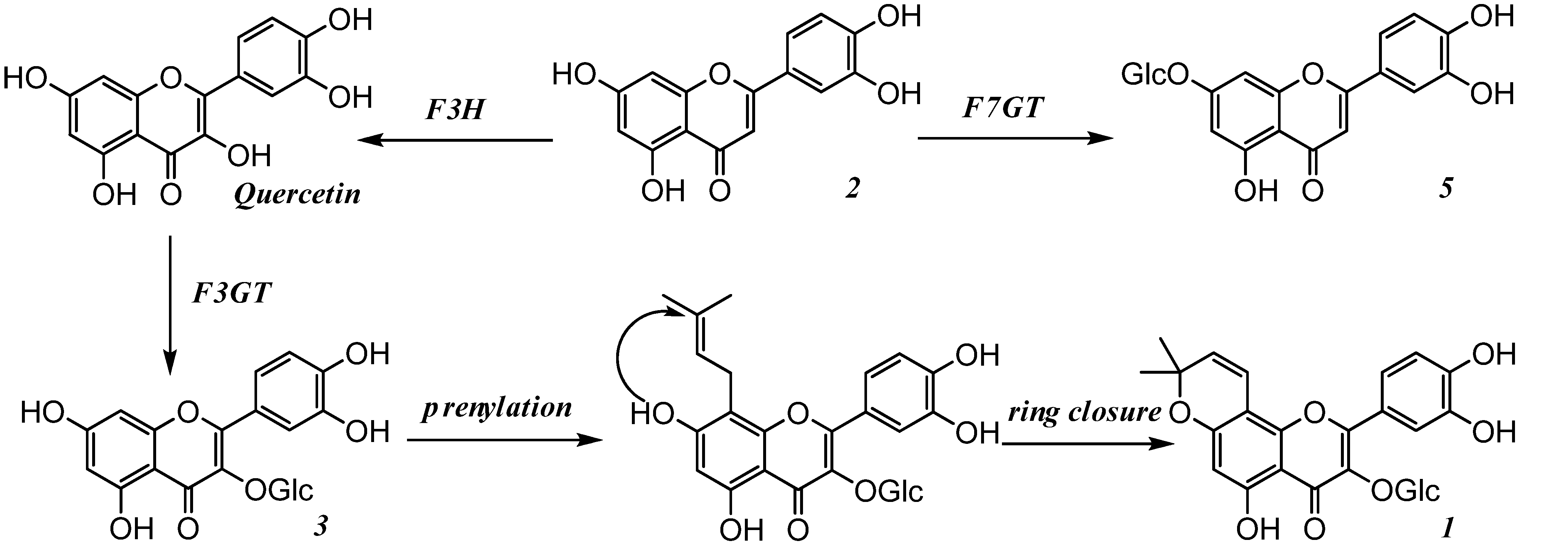
Conclusions
Experimental
General
Chemicals and reagents
Extraction and isolation
Acid hydrolysis of compounds 1, 3, 5, 7 and 8
 -59.1 (c 0.5, MeOH); mp 349.1 – 349.9 ºC; UV (MeOH) λmax: 246, 285 (sh), 325 nm; HRESI-MS [+]: m/z = 531.1517 [M+H]+ (calcd. for C26H27O12, 531.1503); 1H and 13C-NMR data, see Table 1.
Effect on Cyclooxygenase-1 and -2
Acknowledgements
References and Notes
- Kuzovkina, Y.; Quigley, M. Willows beyond wetlands: uses of Salix L. species for environmental projects. Water Air Soil Poll. 2005, 162, 183–204. [Google Scholar] [CrossRef]
- Li, S. Ben Cao Gang Mu; (in Chinese). The People’s Health Press: Bei Jing, P.R. China, 1982; pp. 2039–2040. [Google Scholar]
- Jiangsu New Medical College. Dictionary of Traditional Chinese Medicine; (in Chinese). Jiangsu Technology Press: Nan Jing, China, 1977; pp. 1123–1124. [Google Scholar]
- Zheng, Y.; Zhang, J.; Han, L.; Sekiya, K.; Kimura, Y.; Okuda, H. Effects of compounds in leaves of Salix matsudana on arachidonic acid metabolism. Yakugaku Zasshi 2005, 125, 1005–1008. [Google Scholar]
- Xu, C.; Zheng, Y.; Yang, X.; Li, X.; Li, X.; Chen, Q. Raddeanalin, a new flavonoid glycoside from the leaves of Salix raddeana Laksh. J. Asian Nat. Prod. Res. 2007, 9, 415–419. [Google Scholar] [CrossRef]
- Wang, J.; Zheng, S.; Shen, T.; Shen, X.; Li, Y. Two New Acyclic Diterpene-γ-lactones from the Leaves of Salix matsudana. Chin. Chem. Lett. 2002, 5, 432–435. [Google Scholar]
- Zhang, J.; Zheng, Y.; Han, L. Studies on chemical constituents of leaves of Salix matsudana Koidz and their influence on lipolysis. Zhong Guo Zhong Yao Za Zhi 2000, 25, 538–541. [Google Scholar]
- Zhang, J.; Zheng, Y.; Han, L. Isolation of resisting thrombus and arteriosclerosis compounds in leaves of Salix matsudana. Zhong Yao Cai 1999, 22, 131–133. [Google Scholar]
- Han, L.; Sumiyoshi, M.; Zhang, J.; Liu, M.; Zhang, X.; Zheng, Y.; Okuda, H.; Kimura, Y. Anti-obesity action of Salix matsudana leaves (Part 1). Anti-obesity action by polyphenols of Salix matsudana in high fat-diet treated rodent animals. Phytother. Res. 2003, 17, 1188–1194. [Google Scholar]
- Han, L.; Sumiyoshi, M.; Zheng, Y.; Okuda, H.; Kimura, Y. Anti-obesity action of Salix matsudana leaves (Part 2). Isolation of anti-obesity effectors from polyphenol fractions of Salix matsudana. Phytother. Res. 2003, 17, 1195–1198. [Google Scholar]
- Wu, Q.; Wang, S.; Du, L.; Zhang, S.; Yang, J.; Xiao, P. Chromone glycosides and flavonoids from Hypericum Japonicum. Phytochemistry 1998, 49, 1417–1420. [Google Scholar]
- Peters, N.; Frost, J.; Long, S. A plant flavone, luteolin, induces expression of Rhizobium meliloti dodulation gene. Science 1986, 233, 977–980. [Google Scholar]
- Pakudina, Z.; Leontev, V.; Kamaev, F.; Sadykov, A. Structure and PMR spectra of isoquercitrin and hirsutrin. Chem. Nat. Comp. 1973, 5, 572–574. [Google Scholar]
- Ambrozin, A.; Vieira, P.; Fernandes, J.; Silva, M.; Albuquerque, S. Trypanocidal activity of Meliaceae and Rutaceae plant extracts. Mem. Inst. Oswaldo Cruz 2004, 99, 227–231. [Google Scholar] [CrossRef]
- Chiruvella, K.; Mohammed, A.; Dampuri, G.; Ghanta, R.; Raghavan, S. Phytochemical and antimicrobial studies of methyl angolensate and luteolin-7-O-glucoside isolated from callus cultures of Soymida febrifuga. Int. J. Biomed. Sci. 2007, 4, 269–278. [Google Scholar]
- Tsetsegmaa, S.; Batsurén, D.; Dungerdorzh, D.; Batirov, E.; Malikov, V. A chemical study of plants of the Mongolian flora the flavonoids of two species of Oxytropis. Chem. Nat. Comp. 1993, 28, 628–629. [Google Scholar]
- Otsuka, H.; Takeuchi, M.; Inoshiri, S.; Sato, T.; Yamasaki, K. Phenolic compounds from Coix jachryma-jobi var. Ma-yuen. Phytochemistry 1989, 28, 883–886. [Google Scholar]
- Mitrocots, D.; Skaltsounis, A.; Mitaku, S.; Harvala, C.; Tillequin, F. Flavonoid and terpene glycosides from European Ebenus species. Biochem. Syst. Ecol. 1999, 27, 305–307. [Google Scholar]
- Tanaka, Y.; Sasaki, N.; Ohmiya, A. Biosynthesis of plant pigments: anthocyanins, betalains and carotenoids. Plant J. 2008, 54, 733–749. [Google Scholar] [CrossRef]
- Sample Availability: Samples are available from the co-author Xiang Li ([email protected]).
© 2008 by the authors. Licensee Molecular Diversity Preservation International, Basel, Switzerland. This article is an open-access article distributed under the terms and conditions of the Creative Commons Attribution license ( http://creativecommons.org/licenses/by/3.0/).
Share and Cite
Li, X.; Liu, Z.; Zhang, X.-f.; Wang, L.-j.; Zheng, Y.-n.; Yuan, C.-c.; Sun, G.-z. Isolation and Characterization of Phenolic Compounds from the Leaves of Salix matsudana. Molecules 2008, 13, 1530-1537. https://doi.org/10.3390/molecules13081530
Li X, Liu Z, Zhang X-f, Wang L-j, Zheng Y-n, Yuan C-c, Sun G-z. Isolation and Characterization of Phenolic Compounds from the Leaves of Salix matsudana. Molecules. 2008; 13(8):1530-1537. https://doi.org/10.3390/molecules13081530
Chicago/Turabian StyleLi, Xiang, Zhi Liu, Xin-feng Zhang, Li-juan Wang, Yi-nan Zheng, Chang-chun Yuan, and Guang-zhi Sun. 2008. "Isolation and Characterization of Phenolic Compounds from the Leaves of Salix matsudana" Molecules 13, no. 8: 1530-1537. https://doi.org/10.3390/molecules13081530