Antioxidant Activity of Some Plant Extracts Towards Xanthine Oxidase, Lipoxygenase and Tyrosinase

Abstract
:Introduction
Results and Discussion

| No. | Plant | Part | Family | Voucher specimen | % yield |
|---|---|---|---|---|---|
| 1 | Agave sisalana Perr. ex Enghlm. | leaves | Agavaceae | M-69 | 1.2 |
| 2 | Alternanthera bettzickiana (Regel) Nicholsen | aerial | Amaranthaceae | M-78 | 5.7 |
| 3 | Antrodia cinnamomea Chang & Chou | carpophore | Polyporeceae | M-75 | 11.3 |
| 4 | Astragalus membranaceus Bge. | roots | Leguminosae | M-76 | 6.9 |
| 5 | Calocedrus macrolepis Kurz var. formosana Folrin | leaves | Cupressaceae | M-83 | 4.1 |
| 6 | Camellia sinensis (L.) O. Ktze. | leaves | Theaceae | M-70 | 3.4 |
| 7 | Camptotheca acuminata Decne. | leaves | Nyssaceae | M-74 | 5.6 |
| 8 | Chamaecyparis formosensis | leaves | Cupressaceae | M-71 | 4.8 |
| 9 | Chamaecyparis obtusa var. formosana (Hayata) Rehder | leaves | Cupressaceae | M-77 | 3.9 |
| 10 | Citrus ponki (Hayata) Hort. ex Tanaka | peel | Rutaceae | M-73 | 7.7 |
| 11 | Cyclocodon lancifolia (Roxb.) Kurz subsp. lancifolia | aerial | Campanulaceae | M-84 | 2.9 |
| 12 | Dendrobium officinale K. KIMURA et MIGO | aerial | Orchidacea | M-81 | 2.3 |
| 13 | Euphorbia formosana Hayata | aerial | Euphorbiaceae | M-59 | 4.7 |
| 14 | Helicia formosana Hemsl. | seed | Proteaceae | M-68 | 10.5 |
| 15 | Garcinia subelliptica Merr. | fruit | Clusiaceae | M-65 | 14.5 |
| 16 | Gynura bicolor (Willd.) DC. | leaves | Asteraceae | M-66 | 8.1 |
| 17 | Koelreuteria henryi Dumme | leaves | Sapindaceae | M-73 | 6.7 |
| 18 | Passiflora edulis Sims | peel | Lauraceae | M-60 | 11.4 |
| 19 | Persea americana Mill | fruit | Passifloraceae | M-64 | 7.8 |
| 20 | Persea americana Mill | leaves | Passifloraceae | M-61 | 5.4 |
| 21 | Phyllanthus urinaria L. | aerial | Euphorbiaceae | M-67 | 3.3 |
| 22 | Pittosporum tobira | leaves | Pittosporaceae | M-63 | 4.6 |
| 23 | Prunus campanulata Maxim | leaves | Rosaceae | M-94 | 4.2 |
| 24 | Rhodiola rosea L. | roots | Crassulaceae | M-91 | 9.0 |
| 25 | Ruellia tuberosa L. | aerial | Acanthaceae | M-48 | 7.9 |
| 26 | Syzygium samarangense (Blume) Merr. & Perry | fruit | Myrtaceae | M-55 | 1.3 |
| 27 | Washingtonia filifera (Linden ex Andre) Wendl. | leaves | Arecaceae | M-75 | 2.9 |
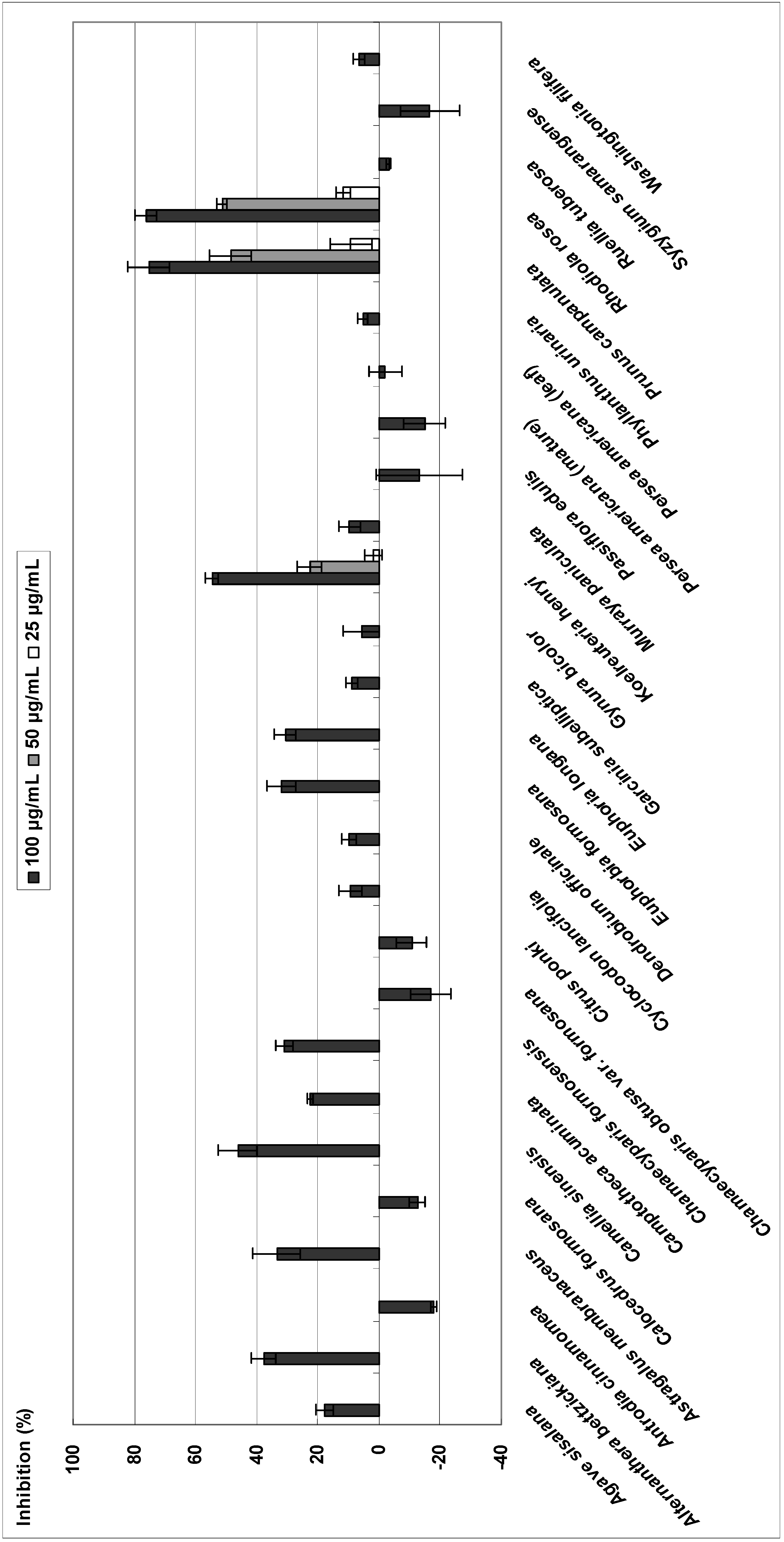
Xanthine oxidase
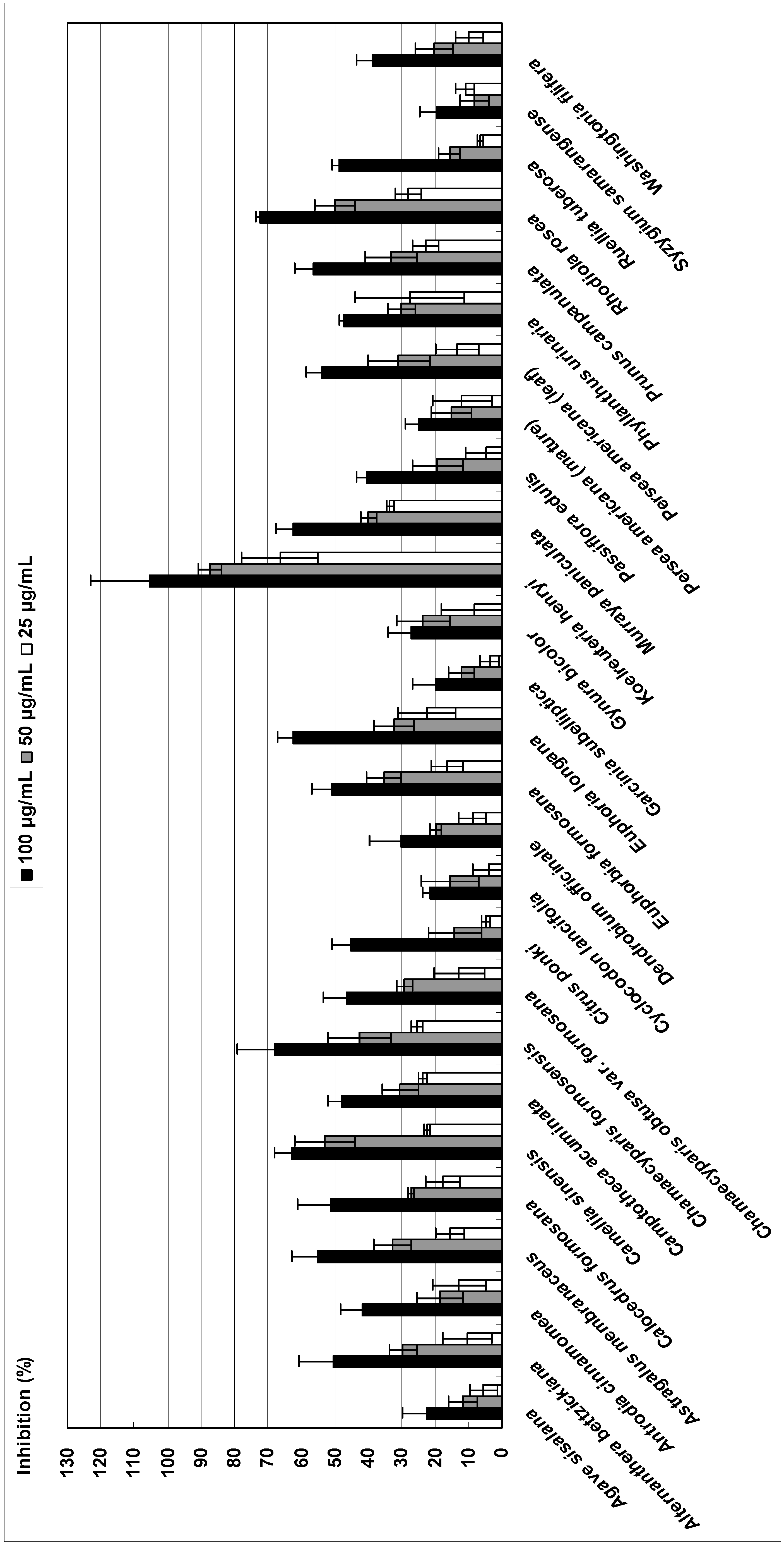
Tyrosinase
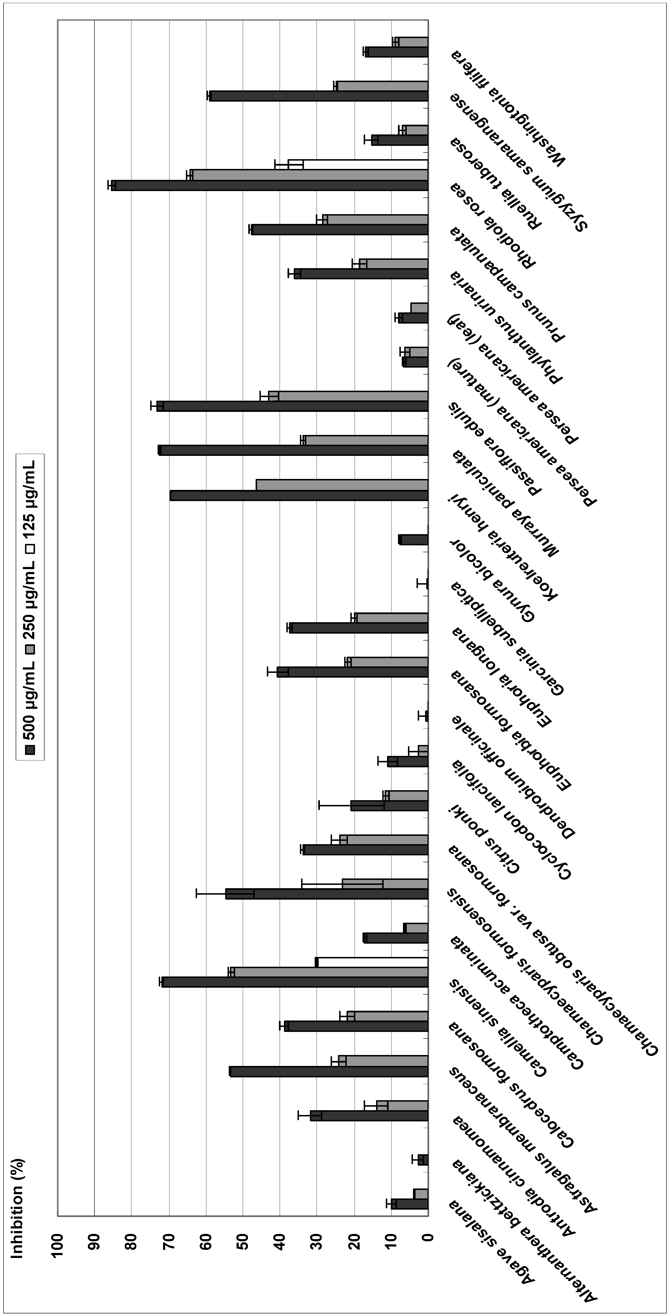
Lipoxygenase


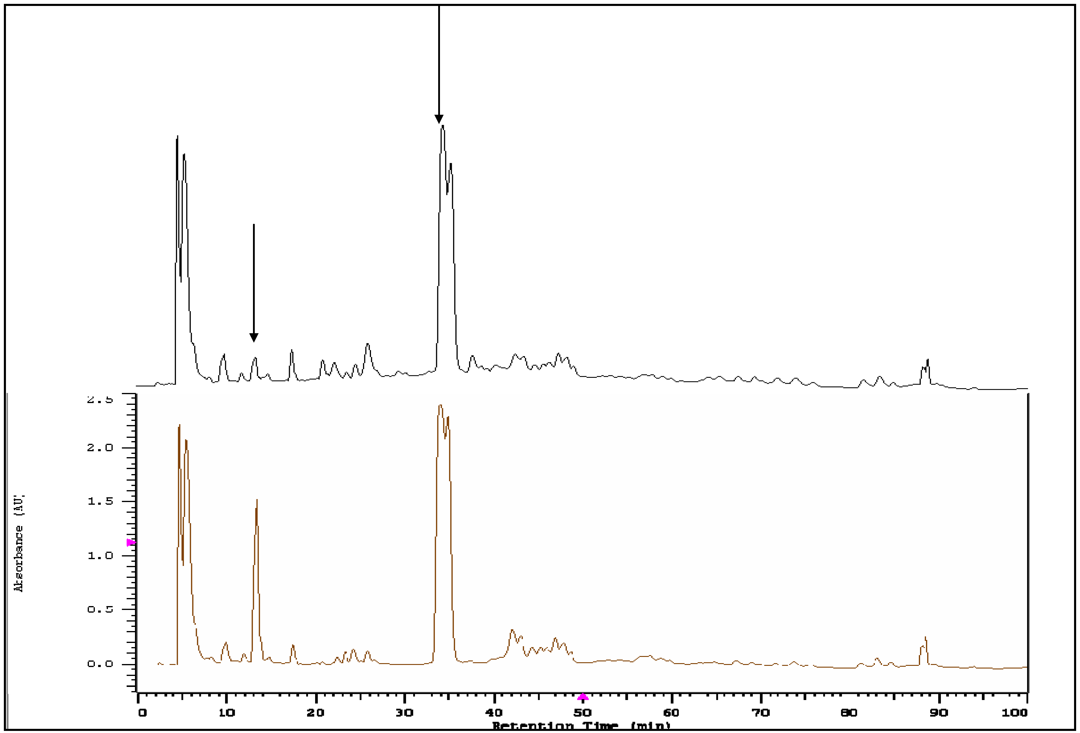
HPLC-DAD for Koelreuteria henryi with tyrosinase

Experimental
General
Plant material
Extraction and isolation
Xanthine oxidase inhibition assay
Tyrosinase inhibition assay
Lipoxygenase inhibition assay
Detection of antioxidant compounds in a plant extract of Koelreuteria henryi by HPLC-DAD
Statistical analysis
Acknowledgements
References and Notes
- Halliwell, B. Antioxidants in human health and disease. Annu. Rev. Nutr. 1996, 16, 33–50. [Google Scholar] [CrossRef]
- Matés, J.M.; Pérez-Gómez, C.; Núñez de Castro, I. Antioxidant enzymes and human diseases. Clin. Biochem. 1999, 32, 595–603. [Google Scholar] [CrossRef]
- Noguchi, N.; Niki, E. Phenolic antioxidants: A rationale for design and evaluation of novel antioxidant drug for atherosclerosis. Free Radic. Biol. Med. 2000, 28, 1538–1546. [Google Scholar] [CrossRef]
- Mayne, S.T. Antioxidant nutrients and chronic disease: use of biomarkers of exposure and oxidative stress status in epidemiologic research. J. Nutr. 133 Suppl. 2003, 3, 933–940. [Google Scholar]
- Pinnell, S.R. Cutaneous photodamage, oxidative stress, and topical antioxidant protection. J. Am. Acad. Dermatol. 2003, 48, 1–19. [Google Scholar] [CrossRef]
- Vinson, J.A.; Hao, Y.; Su, X.; Zubik, L. Phenol antioxidant quantity and quality in foods: vegetables. J. Agric. Food Chem. 1998, 46, 3630–3634. [Google Scholar] [CrossRef]
- Ganthavorn, C.; Hughes, J.S. Inhibition of soybean oil oxidation by extracts of dry beans (Phaseolus vulgaris). J. Am. Oil Chem. Soc. 1997, 74, 1025–1030. [Google Scholar] [CrossRef]
- Jitoe, A.; Masuda, T.; Tengah, I.G.P.; Suprapta, D.N.; Gara, I.W.; Nakatani, N. Antioxidant activity of tropical ginger extracts and analysis of the contained curcuminoids. J. Agric. Food Chem. 1992, 40, 1337–1340. [Google Scholar] [CrossRef]
- Zheng, W.; Wang, S.Y. Antioxidant activity and phenolic compounds in selected herbs. J. Agric. Food Chem. 2001, 49, 5165–5170. [Google Scholar] [CrossRef]
- Nijveldt, R.J.; van Nood, E.; van Hoorn, D.E.; Boelens, P.G.; van Norren, K.; van Leeuwen, P.A. Flavonoids: A review of probable mechanisms of action and potential applications. Am. J. Clin. Nutr. 2001, 74, 418–425. [Google Scholar]
- Pieroni, A.; Janiak, V.; Dürr, C.M.; Lüdeke, S.; Trachsel, E.; Heinrich, M. In vitro antioxidant activity of non-cultivated vegetables of ethnic Albanians in southern Italy. Phytother. Res. 2002, 16, 467–473. [Google Scholar] [CrossRef]
- No, J.K.; Soung, D.Y.; Kim, Y.J.; Shim, K.H.; Jun, Y.S.; Rhee, S.H.; Yokozawa, T.; Chung, H.Y. Inhibition of tyrosinase by green tea components. Life Sci. 1999, 65, PL241–PL246. [Google Scholar]
- Rackova, L.; Oblozinsky, M.; Kostalova, D.; Kettmann, V.; Bezakova, L. Free radical scavenging activity and lipoxygenase inhibition of Mahonia aquifolium extract and isoquinoline alkaloids. J. Inflamm. 2007, 4, 15. [Google Scholar] [CrossRef]
- Bandoniene, D.; Murkovic, M. On-line HPLC-DAD-DPPH screening method for evaluation of radical scavenging phenols extracted from apples (Malus domestica L.). J. Agric. Food Chem. 2002, 50, 2482–2487. [Google Scholar] [CrossRef]
- Masuda, T.; Inaba, Y.; Maekawa, T.; Takeda, Y.; Yamaguchi, H.; Nakamoto, K.; Kuninaga, H.; Nishizato, S.; Nonaka, A. Simple detection method of powerful antiradical compounds in the raw extract of plants and its application for the identification of antiradical plant constituents. J. Agric. Food Chem. 2003, 51, 1831–1838. [Google Scholar] [CrossRef]
- Lee, M.H.; Jiang, C.B.; Juan, S.H.; Lin, R.D.; Hou, W.C. Antioxidant and heme oxygenase-1 (HO-1)-induced effects of selected Taiwanese plants. Fitoterapia 2006, 77, 109–115. [Google Scholar] [CrossRef]
- Gerasimidis, K.; Fillou, D.T.; Babatzimcpoulou, M.; Tassou, K.; Katsikas, H. Preparation of an edible cottonseed protein concentrate and evaluation of its functional properties. Int. J. Food Sci. Nutr. 2007, 58, 486–490. [Google Scholar] [CrossRef]
- Eloff, J.N. Which extractant should be used for the screening and isolation of antimicrobial components from plants? J. Ethnopharmacol. 1998, 60, 1–8. [Google Scholar]
- Cui, T.; Nakamura, K.; Tian, S.; Kayahara, H.; Tian, Y.L. Polyphenolic content and physiological activities of Chinese hawthorn extracts. Biosci. Biotechnol. Biochem. 2006, 70, 2948–2956. [Google Scholar] [CrossRef]
- Hout, S.; Chea, A.; Bun, S.S.; Elias, R.; Gasquet, M.; Timon-David, P.; Balansard, G.; Azas, N. Screening of selected indigenous plants of Cambodia for antiplasmodial activity. J. Ethnopharmacol. 2006, 107, 12–18. [Google Scholar] [CrossRef]
- Sturm, R.A.; Teasdale, R.D.; Box, N.F. Human pigmentation genes: identification, structure and consequences of polymorphic variation. Gene 2001, 277, 49–62. [Google Scholar] [CrossRef]
- Lyckander, I.M.; Malterud, K.E. Lipophilic flavonoids from Orthosíphon spicatus as inhibitors of 15-lipoxygenase. Acta Pharm. Nord. 1992, 4, 159–166. [Google Scholar]
- Sample Availability: Samples of all compounds are available from the authors.
© 2009 by the authors; licensee Molecular Diversity Preservation International, Basel, Switzerland. This article is an open access article distributed under the terms and conditions of the Creative Commons Attribution license ( http://creativecommons.org/licenses/by/3.0/).
Share and Cite
Chen, C.-H.; Chan, H.-C.; Chu, Y.-T.; Ho, H.-Y.; Chen, P.-Y.; Lee, T.-H.; Lee, C.-K. Antioxidant Activity of Some Plant Extracts Towards Xanthine Oxidase, Lipoxygenase and Tyrosinase. Molecules 2009, 14, 2947-2958. https://doi.org/10.3390/molecules14082947
Chen C-H, Chan H-C, Chu Y-T, Ho H-Y, Chen P-Y, Lee T-H, Lee C-K. Antioxidant Activity of Some Plant Extracts Towards Xanthine Oxidase, Lipoxygenase and Tyrosinase. Molecules. 2009; 14(8):2947-2958. https://doi.org/10.3390/molecules14082947
Chicago/Turabian StyleChen, Chin-Hui, Hsiu-Chen Chan, Yi-Tsu Chu, Hsin-Yi Ho, Pi-Yu Chen, Tzong-Huei Lee, and Ching-Kuo Lee. 2009. "Antioxidant Activity of Some Plant Extracts Towards Xanthine Oxidase, Lipoxygenase and Tyrosinase" Molecules 14, no. 8: 2947-2958. https://doi.org/10.3390/molecules14082947