A New Diterpene Glycoside from Stevia rebaudiana

The Coca-Cola Company, Organic Chemistry Department, Research and Technology, One Coca-Cola Plaza, Atlanta, GA 30313, USA
*
Author to whom correspondence should be addressed.
Molecules 2011, 16(4), 2937-2943; https://doi.org/10.3390/molecules16042937
Submission received: 18 February 2011
/
Revised: 29 March 2011
/
Accepted: 1 April 2011
/
Published: 4 April 2011
(This article belongs to the Section Natural Products Chemistry)
Abstract
:From the commercial extract of the leaves of Stevia rebaudiana, a new diterpene glycoside was isolated besides the known steviol glycosides including stevioside, rebaudiosides A-F, rubusoside and dulcoside A. The new compound was identified as 13-[(2-O-β-d-glucopyranosyl-3-O-β-d-glucopyranosyl-β-d-glucopyranosyl)oxy] ent-kaur-16-en-19-oic acid-(2-O-α-l-rhamnopyranosyl-β-d-glucopyranosyl) ester (1) on the basis of extensive spectroscopic (NMR and MS) and chemical studies.
1. Introduction
Stevia rebaudiana (Bertoni) Bertoni is a perennial shrub of the Asteraceae (Compositae) family native to Brazil and Paraguay, which is often referred to as “the sweet herb of Paraguay”. The major constituents in the leaves of S. rebaudiana are the potently sweet diterpenoid glycosides stevioside, rebaudiosides A and D, and dulcoside A. These compounds, which are known as Stevia sweeteners, are glycosides of the diterpene steviol (ent-13-hydroxykaur-16-en-19-oic acid) [1]. As a part of our continuing research to discover natural sweeteners [2,3], we have reported several diterpene glycosides from the commercial extract of S. rebaudiana. In this article, we present the isolation and structure elucidation based on extensive spectroscopic (NMR and MS) and chemical studies of the new diterpenoid glycoside 1, identified from the stevia extract (SG-95) obtained from Pure Circle (Kuala Lumpur, Malaysia).
2. Results and Discussion
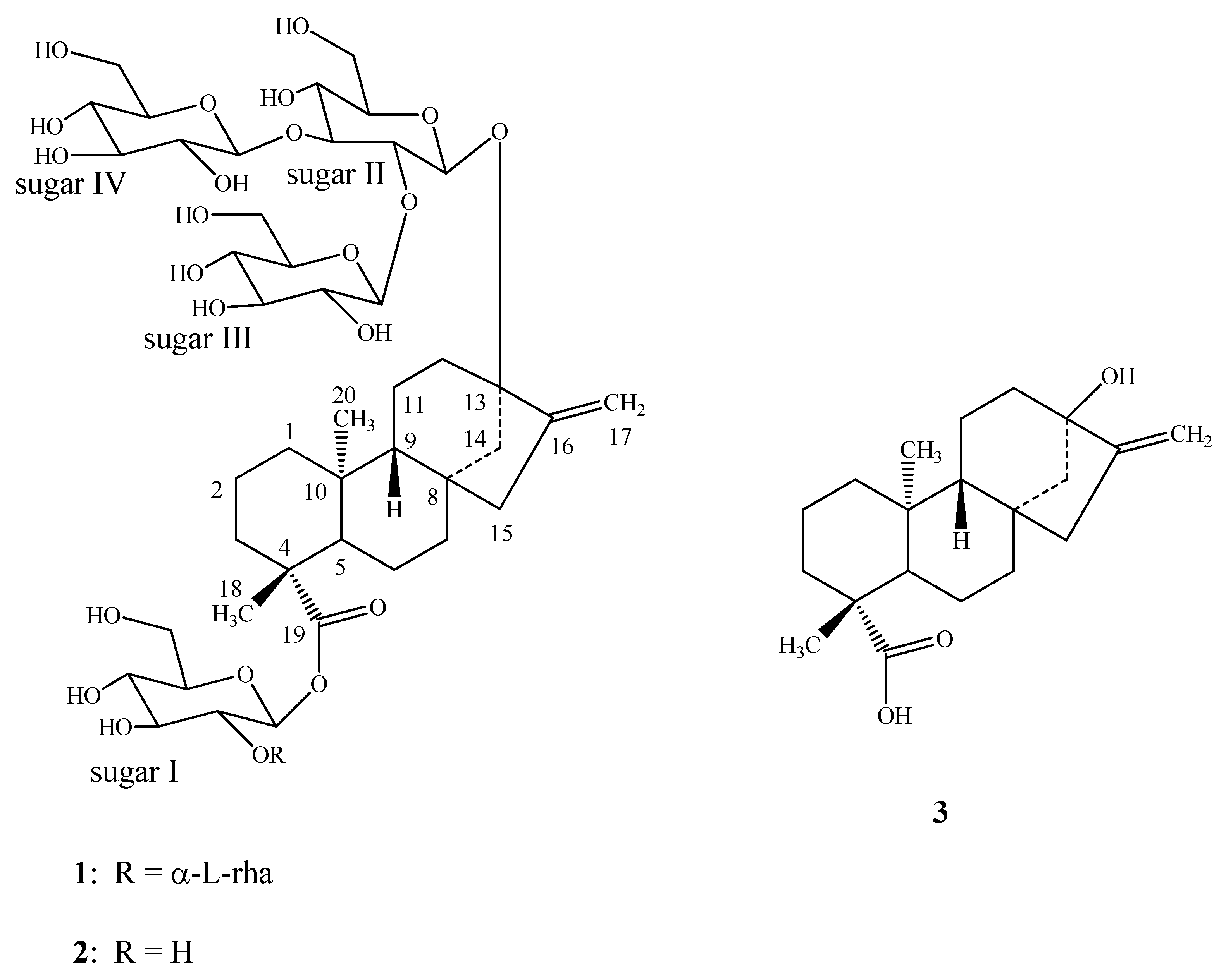
Purification of the commercial extract SG-95 obtained from the leaves of S. rebaudiana resulted in the isolation of the new diterpenoid glycoside 1, and the known steviol glycosides, stevioside, rebaudioside A (2), rebaudiosides B-F, rubusoside and dulcoside A (Figure 1). The structures of all the known compounds were identified by comparison of their retention times with authentic standards using the HPLC-MS method reported earlier [4] and the spectral data reported in the literature [4,5,6,7,8,9,10,11].
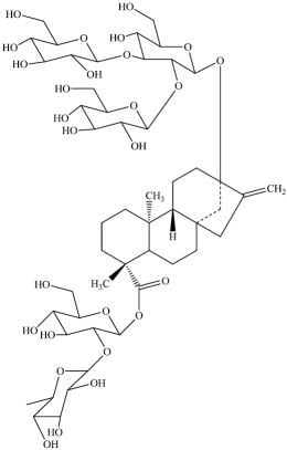
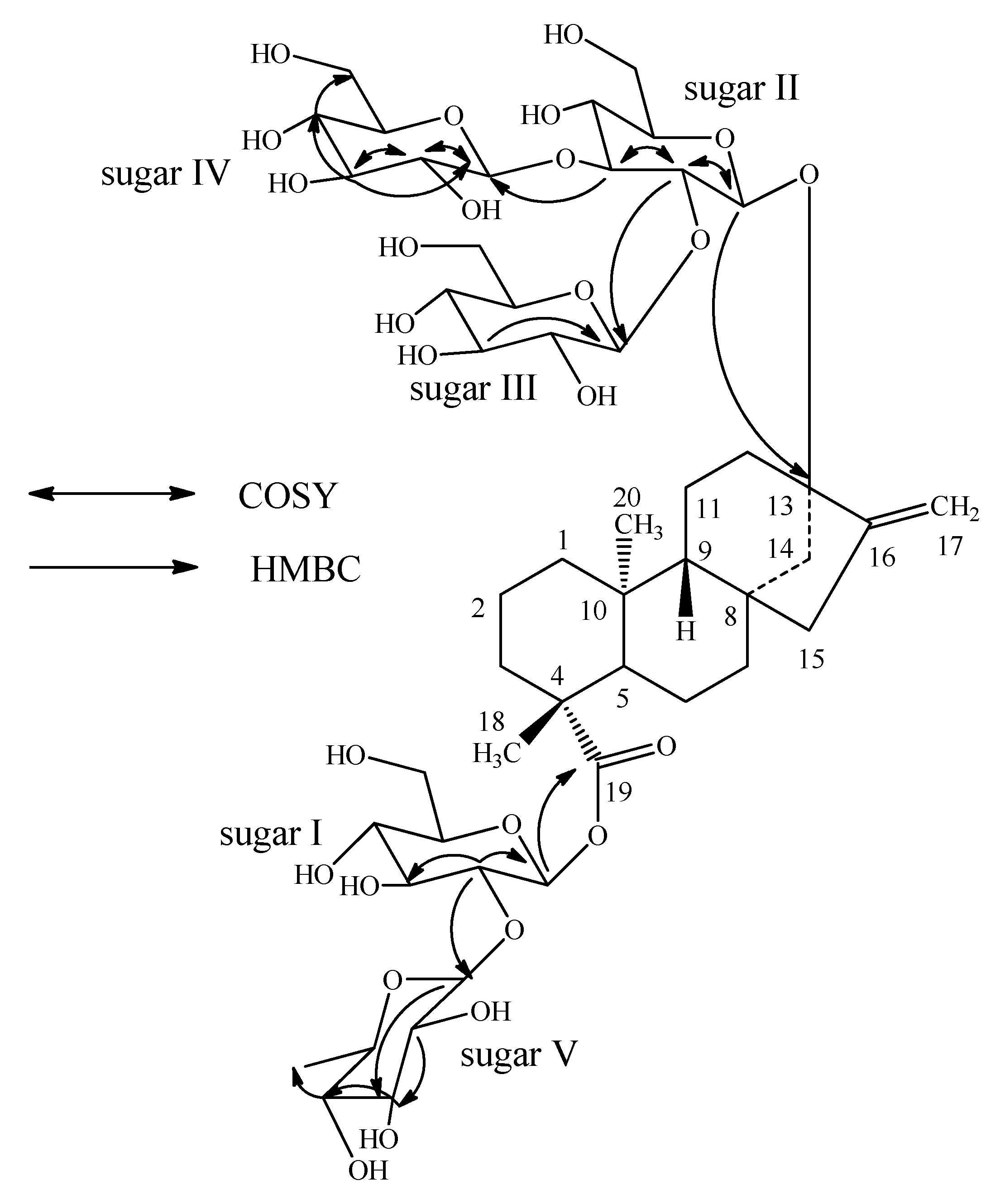
Compound 1 was isolated as a colorless oil and its molecular formula was deduced as C50H80O27 on the basis of its positive ESI mass spectrum, which showed an [M+H]+ ion at m/z 1,113.4977, together with [M+NH4]+ and [M+Na]+ adducts at m/z 1,130.5243 and 1,135.4805, respectively. This composition was supported by 13C-NMR spectral data. The 1H-NMR spectrum of 1 showed the presence of two methyl singlets at δ 0.94 and 1.26, two olefinic protons of an exocyclic double bond as singlets at δ 4.87 and 5.25, nine methylene and two methine protons between δ 0.85–2.27 characteristic for the ent-kaurane diterpenoids isolated earlier from the genus Stevia [7,8,9]. The basic ent-kaurane diterpenoid skeleton was supported by COSY (H-1/H-2; H-2/H-3; H-5/H-6; H-6/H-7; H-9/H-11; H-11/H-12) and HMBC (H-1/C-2, C-10; H-3/C-1, C-2, C-4, C-5, C-18, C-19; H-5/C-4, C-6, C-7, C-9, C-10, C-18, C-19, C-20; H-9/C-8, C-10, C-11, C-12, C-14, C-15; H-14/C-8, C-9, C-13, C-15, C-16 and H-17/C-13, C-15, C-16) correlations. The positive mode ESI MS/MS spectrum of 1 showed fragment ions at m/z 951, 789, 627 and 465, suggesting the presence of four hexose moieties. The fragment ion observed at m/z 951 was further fragmented to an ion at m/z 805, suggesting an additional deoxyhexose unit in its structure. The presence of five sugar units in its structure was supported by the 1H-NMR spectrum, which showed the presence of anomeric protons at δ 4.62, 4.66, 4.86, 5.31, and 5.62.
Enzymatic hydrolysis of 1 furnished an aglycone which was identified as steviol (3) by comparison of 1H-NMR [10] and co-TLC with standard compound. Acid hydrolysis of 1 with 5% H2SO4 afforded d-glucose and L-rhamnose, which were identified by direct comparison with authentic samples by TLC [12,13,14]. The 1H- and 13C-NMR values for all the carbons in 1 were assigned on the basis of COSY, HSQC and HMBC correlations (Table 1). Based on the results from NMR spectral data and hydrolysis experiments of 1, it was concluded that there are four d-glucose and one l-rhamnose moieties in its structure. A close comparison of the 1H- and 13C-NMR spectrum of 1 with rebaudioside A (2) suggested that compound 1 is also a steviol glycoside which has three glucose residues that are attached at the C-13 hydroxyl as a 2,3-branched β-d-glucotriosyl substituent and another glucose moiety in the form of an ester at C-19 leaving the assignment of the additional rhamnose moiety. The downfield shift for both the 1H and 13C chemical shifts at C-2′ suggested that the additional rhamnsoe is attached at this position. This was confirmed by the key HMBC correlations: H-2′/C-1′, C-3′, C-1′′′′′ and H-1′′′′′/C-2′, C-2′′′′′, C-3′′′′′ (Figure 2).
The anomeric proton of the rhamnose residue was observed at δ 5.31 and had a coupling constant of 1.5 Hz, confirming that it has an α-configuration, similar to dulcosides A and B [6]. The large coupling constants observed for the four anomeric protons of the glucose moieties at δ 4.62 (d, J = 7.8 Hz), 4.66 (d, J = 7.8 Hz), 4.86 (d, J = 7.6 Hz), and 5.62 (d, J = 7.4 Hz), suggested their β-orientation as reported for steviol glycosides [5,6,7]. Based on the results from chemical and spectral studies, 1 was assigned as 13-[(2-O-β-d-glucopyranosyl-3-O-β-d-glucopyranosyl-β-d-glucopyranosyl)oxy] ent-kaur-16-en-19-oic acid-(2-O-α-l-rhamnopyranosyl-β-d-glucopyranosyl) ester.
3. Experimental
3.1. General
NMR spectra were acquired on Bruker Avance DRX 500 MHz and Varian Unity Plus 600 MHz instruments using standard pulse sequences. The spectra were referenced to the residual solvent signal (δH 3.30, δC 49.0 for CD3OD), chemical shifts are given in δ (ppm), and coupling constants are reported in Hz. MS and MS/MS data were generated with a Waters Premier Quadrupole Time-of-Flight (Q-TOF) mass spectrometer equipped with an electrospray ionization source operated in the positive-ion mode and ThermoFisher Discovery OrbiTrap in the positive mode electrospray. Samples were diluted with water: acetonitrile (1:1) containing 0.1% formic acid and introduced via infusion using the onboard syringe pump. Preparative HPLC was performed on an Agilent 1100 system using a Phenomenex Prodigy ODS (3) column (250 × 21.2 mm, 5 μm). Semi-preparative HPLC was carried out with a Waters 600E multisolvent delivery system using a Phenomenex Synergi Hydro RP column (250 × 10 mm, 4 μm) column.
3.2. Plant Material
Stevia extract SG-95, the commercial sample consisting of a mixture of diterpenoid glycosides from the leaves of S. rebaudiana was obtained from Pure Circle (Kuala Lumpur, Malaysia). A voucher specimen is deposited at The Coca-Cola Company, No. VSPC-3166-002.
3.3. Isolation
Preliminary separation of the crude stevioside extract was carried out using a preparative HPLC method employing a water/acetonitrile (B) gradient (25% B for 8.5 min, 25 to 29% B over 1.5 min, 29 to 30% B over 5.5 min, 30 to 34% B over 2.0 min, 34% B for 6 min, 34 to 52% B over 2.0 min, 52% B for 3.0 min, 52 to 70% B over 1.0 min, 70% B for 5.5 min) at 20 ml/min. The baseline fraction at 16.8 min was collected and dried by rotary evaporation under reduced pressure as the crude impurity fraction. Final fractionation was then performed using HPLC method by injecting the crude impurity fraction (over several runs) on a Synergi Hydro RP column by semi-preparative HPLC using a gradient of water (0.01156% acetic acid, 0.02844% ammonium acetate) in acetonitrile (25% B for 8.5 min,25 to 29% B over 1.5 min, 29 to 30% B over 5.5 min, 30 to 34% B over 2.0 min, 34% B for 6 min, 34 to 52% B over 2.0 min, 52% B for 3.0 min, 52 to 70% B over 1.0 min, 70% B for 5.5 min) at 5 mL/min to yield 1 (tR 10.6 min, 1.6 mg). All the known compounds were identified in comparison of their retention times with authentic standards using the HPLC-MS method as described previously [4] and the spectral data that were reported in the literature [4,5,6,7,8,9,10,11].
13-[(2-O-β-d-glucopyranosyl-3-O-β-d-glucopyranosyl-β-d-glucopyranosyl)oxy] ent-kaur-16-en-19-oic acid-(2-O-α-l-rhamnopyranosyl-β-d-glucopyranosyl) ester (1). Colorless film; 1H-NMR (CD3OD, δ ppm) and 13C-NMR (CD3OD, δ ppm) spectroscopic data, see Table 1; +ESI TOFMS m/z 1,113.4977 (calcd. for C50H81O27: 1,113.4965).
Enzymatic hydrolysis of 1. A solution of 1 (250 μg) was dissolved in 0.1 M sodium acetate buffer, pH 4.5 (2.5 mL) and crude pectinase from Aspergillus niger (50 µL, Sigma-Aldrich, P2736) was added. The mixture was stirred at 50o C for 48 hr. The product precipitated out during the reaction and was filtered and then crystallized from methanol (MeOH). The resulting steviol (3) was identical to an authentic sample by TLC and 1H-NMR.
Acid Hydrolysis of 1. To a solution of 1 (250 μg) in MeOH (1 mL) was added 5% H2SO4 (1 mL) and the mixture was refluxed for 8 hours. The reaction mixture was then neutralized with saturated sodium carbonate and extracted with ethyl acetate (EtOAc, 2 × 5 mL) to give an aqueous fraction containing sugars and an EtOAc fraction containing the aglycone part. The aqueous phase was concentrated and compared with standard sugars using the TLC system EtOAc/n-butanol/water (2:7:1) and CH2Cl2/MeOH/water (10:6:1) [12,13,14]; the two sugars were identified as l-rhamnose and d-glucose.
4. Conclusions
A new diterpenoid glycoside 1, as well as nine known steviol glycosides – stevioside, rebaudiosides A-F, rubusoside and dulcoside A – were isolated from the commercial extract obtained from the leaves of S. rebaudiana obtained from Pure Circle (Kuala Lumpur, Malaysia). The new compound was identified as 13-[(2-O-β-d-glucopyranosyl-3-O-β-d-glucopyranosyl-β-d-glucopyranosyl)oxy] ent-kaur-16-en-19-oic acid-(2-O-α-l-rhamnopyranosyl-β-d-glucopyranosyl) ester on the basis of 2D NMR and +EI TOF MS, as well as chemical studies.
Acknowledgements
We wish to thank Pure Circle (Kuala Lumpur, Malaysia) for providing the stevia extract SG-95.
References
- Brandle, J.E.; Starrratt, A.N.; Gijen, M. Stevia rebaudiana: Its agricultural, biological and chemical properties. Can. J. Plant Sci. 1998, 78, 527–536. [Google Scholar] [CrossRef]
- Chaturvedula, V.S.P.; Rhea, J.; Milanowski, D.; Mocek, U.; Prakash, I. Two minor diterpene glycosides from the leaves of Stevia rebaudiana. Nat. Prod. Commun. 2011, 6, 175–178. [Google Scholar] [PubMed]
- Chaturvedula, V.S.P.; Clos, J.F.; Rhea, J.; Milanowski, D.; Mocek, U.; DuBois, G.E.; Prakash, I. Minor diterpenoid glycosides from the leaves of Stevia rebaudiana. Phytochem. Lett. 2011, in press. [Google Scholar] [CrossRef]
- Clos, J.F.; DuBois, G.E.; Prakash, I. Photostability of Rebaudioside A and Stevioside in Beverages. J. Agric. Food Chem. 2008, 56, 8507–8513. [Google Scholar] [CrossRef] [PubMed]
- Kohda, H.; Kasai, R.; Yamsaki, K.; Murakami, K.; Tanaka, O. New sweet diterpene glucosides from Stevia rebaudiana. Phytochemistry 1976, 15, 981–983. [Google Scholar] [CrossRef]
- Kobayashi, M.; Horikawa, S.; Degrandi, I.H.; Ueno, J.; Mitsuhashi, H. Dulcosides A and B, new diterpene glycosides from Stevia rebaudiana. Phytochemistry 1977, 16, 1405–1408. [Google Scholar] [CrossRef]
- Starratt, A.N.; Kirby, C.W.; Pocs, R.; Brandle, J.E. Rebaudioside F, a diterpene glycoside from Stevia rebaudiana. Phytochemistry 2002, 59, 367–370. [Google Scholar] [CrossRef]
- Sakamoto, I.; Yamasaki, K.; Tanaka, O. Application of 13C NMR spectroscopy to the chemistry of natural glycosides: Rebaudioside C, a new sweet diterpene glycoside of Stevia rebaudiana. Chem. Pharm. Bull. 1977, 25, 844–846. [Google Scholar] [CrossRef]
- Sakamoto, I.; Yamasaki, K.; Tanaka, O. Application of 13C NMR spectroscopy to chemistry of plant glycosides: Rebaudiosides D and E, new sweet diterpene glucosides of Stevia rebaudiana Bertoni. Chem. Pharm. Bull. 1977, 25, 3437–3439. [Google Scholar] [CrossRef]
- Ohta, M.; Sasa, S.; Inoue, A.; Tamai, T.; Fujita, I.; Morita, K.; Matsuura, F. Characterization of novel steviol glycosides from leaves of Stevia rebaudiana Morita. J. Appl. Glycosci. 2010, 57, 199–209. [Google Scholar] [CrossRef]
- Ohtani, K.; Aikawa, Y.; Kasai, R.; Chou, W.; Yamasaki, K.; Tanaka, O. Minor diterpene glycosides from sweet leaves of Rubus suavissimus. Phytochemistry 1992, 31, 1553–1559. [Google Scholar] [CrossRef]
- Bedir, E.; Toyang, N.J.; Khan, I.A.; Walker, L.A.; Clark, A.M. A new dammarane type triterpene glycoside from Polyscias fulva. J. Nat. Prod. 2001, 64, 95–97. [Google Scholar] [CrossRef] [PubMed]
- Chaturvedula, V.S.P.; Schilling, J.K.; Miller, J.S.; Andriantsiferana, R.; Rasamison, V.E.; Kingston, D.G.I. New cytotoxic oleanane saponis from the infructescences of Polyscias amplifolia from the Madagascar rainforest. Planta Med. 2003, 69, 440–444. [Google Scholar] [PubMed]
- Huan, V.D.; Yamamura, S.; Ohtani, K.; Kasai, R.; Yamasaki, K.; Nham, N.T. Oleanane saponins from Polyscias fructicosa. Phytochemistry 1998, 47, 451–457. [Google Scholar] [CrossRef]
Sample Availability: Samples of the steviol glycosides stevioside, rebaudiosides A-F, rubusoside and dulcoside A are available from the authors. |
Figure 1.
Structure of 1 and other compounds.

Figure 2.
Key COSY and HMBC correlations of 1.


Table 1.
1H- and 13C-NMR spectral data (chemical shifts and coupling constants) for 1 in CD3OD. a
| Position | 1H NMR | 13C NMR |
|---|---|---|
| 1 | 0.85 (m, 1H), 1.88 (m, 1H) | 41.5 |
| 2 | 1.41 (m, 1H), 1.94 (m, 1H) | 20.0 |
| 3 | 1.06 (m, 1H), 2.27 (m, 1H) | 38.5 |
| 4 | 45.1 | |
| 5 | 1.10 (m, 1H) | 58.7 |
| 6 | 1.88 (m, 1H), 1.94 (m, 1H) | 22.6 |
| 7 | 1.43 (m, 1H), 1.55 (m, 1H) | 42.5 |
| 8 | 43.0 | |
| 9 | 1.00 (m, 1H) | 54.8 |
| 10 | 40.5 | |
| 11 | 1.65 (m, 1H), 1.80 (m, 1H) | 21.0 |
| 12 | 1.53 (m, 1H), 1.95 (m, 1H) | 38.4 |
| 13 | 88.5 | |
| 14 | 1.53 (m, 1H), 2.25 (d, J = 12.2, 1H) | 45.2 |
| 15 | 2.05 (m, 1H), 2.14 (d, J = 17.4, 1H) | 48.4 |
| 16 | 153.4 | |
| 17 | 4.87 (s, 1H), 5.25 (s, 1H) | 105.4 |
| 18 | 1.26 (s, 3H) | 29.4 |
| 19 | 176.8 | |
| 20 | 0.94 (s, 3H) | 16.3 |
| 1′ | 5.62 (d, J = 7.4, 1H ) | 93.8 |
| 2′ | 3.59 (m, 1H) | 82.6 |
| 3′ | 3.44 (m, 1H) | 77.9 |
| 4′ | 3.34 (m, 1H) | 71.4 |
| 5′ | 3.36 (m, 1H) | 78.2 |
| 6′ | 3.60 (m, 1H), 3.82 (m, 1H) | 62.3 |
| 1′′ | 4.62 (d, J = 7.8, 1H) | 97.2 |
| 2′′ | 3.63 (m, 1H) | 79.6 |
| 3′′ | 3.72 (m, 1H) | 87.5 |
| 4′′ | 3.38 (m, 1H) | 70.0 |
| 5′′ | 3.30 (m, 1H) | 77.1 |
| 6′′ | 3.60 (m, 1H), 3.82 (m, 1H) | 62.7 |
| 1′′′ | 4.86 (d, J = 7.6, 1H) | 103.5 |
| 2′′′ | 3.18 (t, J = 8.2, 1H) | 76.1 |
| 3′′′ | 3.32 (m, 1H) | 77.8 |
| 4′′′ | 3.13 (m, 1H) | 72.2 |
| 5′′′ | 3.44 (m, 1H) | 78.1 |
| 6′′′ | 3.56 (m, 1H), 3.81 (m, 1H) | 63.1 |
| 1′′′′ | 4.66 (d, J = 7.8, 1H) | 104.0 |
| 2′′′′ | 3.26 (m, 1H) | 75.1 |
| 3′′′′ | 3.42 (m, 1H) | 78.6 |
| 4′′′′ | 3.32 (m, 1H) | 71.1 |
| 5′′′′ | 3.36 (m, 1H) | 77.8 |
| 6′′′′ | 3.62 (m, 1H), 3.80 (m, 1H) | 62.4 |
| 1′′′′′ | 5.31 (d, J=1.5, 1H) | 101.4 |
| 2′′′′′ | 3.89 (m, 1H) | 71.8 |
| 3′′′′′ | 3.63 (m, 1H) | 72.0 |
| 4′′′′′ | 3.36 (m, 1H) | 73.6 |
| 5′′′′′ | 3.75 (m, 1H) | 70.0 |
| 6′′′′′ | 1.24 (d, J = 6.3, 3H) | 18.0 |
a assignments made on the basis of COSY, HSQC and HMBC correlations; b Coupling constants are in Hz; c Chemical shift values are in δ (ppm).
© 2011 by the authors; licensee MDPI, Basel, Switzerland. This article is an open access article distributed under the terms and conditions of the Creative Commons Attribution license (http://creativecommons.org/licenses/by/3.0/).
Share and Cite
MDPI and ACS Style
Prakash Chaturvedula, V.S.; Prakash, I. A New Diterpene Glycoside from Stevia rebaudiana. Molecules 2011, 16, 2937-2943. https://doi.org/10.3390/molecules16042937
AMA Style
Prakash Chaturvedula VS, Prakash I. A New Diterpene Glycoside from Stevia rebaudiana. Molecules. 2011; 16(4):2937-2943. https://doi.org/10.3390/molecules16042937
Chicago/Turabian StylePrakash Chaturvedula, Venkata Sai, and Indra Prakash. 2011. "A New Diterpene Glycoside from Stevia rebaudiana" Molecules 16, no. 4: 2937-2943. https://doi.org/10.3390/molecules16042937