Article

Synthesis, Singlet Oxygen Photogeneration and DNA Photocleavage of Porphyrins with Nitrogen Heterocycle Tails

1
College of Chemistry and Molecular Sciences, Wuhan University, Wuhan 430072, China
2
Key Laboratory for Green Chemical Process of Ministry of Education, Wuhan Institute of Technology, Wuhan 430074, China
*
Authors to whom correspondence should be addressed.
Molecules 2011, 16(5), 3488-3498; https://doi.org/10.3390/molecules16053488
Submission received: 11 March 2011 / Revised: 13 April 2011 / Accepted: 18 April 2011 / Published: 26 April 2011

Abstract

:
Eight novel compounds were prepared by reaction of 5-(bromo- propoxyphenyl)-10,15,20-triphenylporphyrin with oxazole thiols, 1,3,4-oxadiazole thiols and 1,3,4-thiadiazole thiols, and their structures confirmed by UV-vis, IR, 1H-NMR, MS and elemental analysis. The assessment of indirectly measured 1O2 production rates against 5,10,15,20-tetraphenyl porphyrin (H2TPP) were described and the relative singlet oxygen production yields were: porphyrin 5 > porphyrins 1, 3, 4, 6-8, H2TPP > porphyrin 2. Porphyrin 4 and porphyrin 7 showed substantial photocleavage activities toward DNA, with over 75% cleavage observed at 40 µM. It suggested that these those porphyrins with nitrogen heterocycle tails are potential photosensitive agents.

1. Introduction

The human vascular endothelial growth factor (VEGF) gene locates at chromosome 6P21,3, whose length is 28 Kb. The VEGF gene, of 14 Kb length, consists alternately of eight extrons and seven introns. The encoding product is a homogenous dimeric glycoprotein of 34–35 KD [1]. Against a target of VEGFR-2 tyrosine kinase VEGFR-2 plays a main receptor function in tumor angiogenesis mediated by VEGF, which prevents the activation of VEGFR-2 enzyme and limits VEGF signal transduction so as to inhibit tumor growth for cancer treatment. Compounds with oxazole, 1,3,4-oxadiazole and 1,3,4-thiadiazole structures display certain capability of acting in the VEGFR-2 tyrosine kinase domain and of binding VEGFR-2 in competition with ATP to prevent the growth of tumor vessels and cause tumor death [2,3,4]. So far, research on the use of angiogenesis inhibitors against the tumor metastasis has become a major topic.
Meanwhile, porphyrin-based compounds with unique structures have special affinity interactions with tumor cells and they can selectively remain in the tumor tissues [5,6]. Photodynamic therapy (PDT) is a promising therapy against cancer with intrinsic selectivity, which is used to eliminate deviant tissues, such as tumors. While gathering selectively around the tumors, the photosensitizers can absorb visible light under illumination and generate active oxygen (such as the singlet oxygen, 1O2, generated when porphyrins are photoactivated by absorption at the Soret band [7]), which leads to tumor cell death [8,9]. The 1O2 is produced by energy transfer from the photoexcited sensitizer (in its triplet state) to the ground-state (triplet) oxygen. This is known as the Type II photosensitization mechanism, which is considered to be more important in PDT [10,11]. The DNA photocleavage activity of a photosensitizer depends on its 1O2 yield [12,13].
Porphyrins linked with some anticancer drugs can effectively localize in tumor cells and increase the cell cytotoxicity through the synergistic effect of porphyrins and anticancer drugs [14]. To enhance targeted therapy role of pophyrins against the VEGFR-2 tyrosine kinase target some porphyrins with nitrogen heterocycle tails were prepared using as starting materials oxazole thiols, 1,3,4-oxadiazole thiols and 1,3,4-thiadiazole thiols linked by -O(CH2)3-S- spacer group (shown in Scheme 1). Their 1O2 productivity and DNA photocleavage activities were then investigated.

2. Results and Discussion

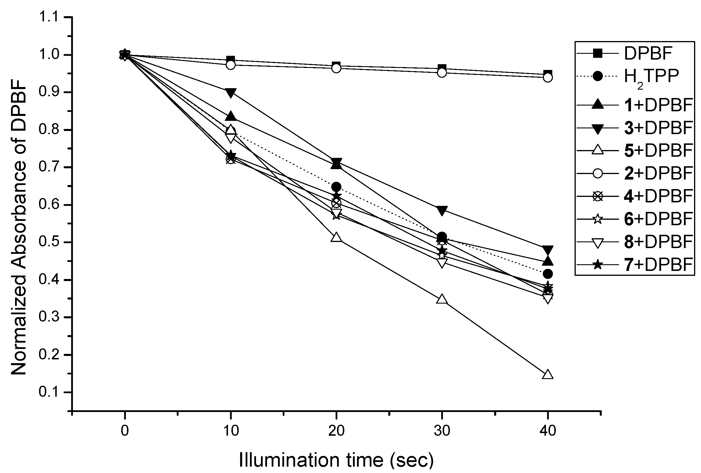
2.1. Photogeneration of 1O2

DNA photocleavage of the porphyrins was the result of generating the singlet oxygen. 1,3-diphenylisobenzofuran (DPBF) was able to capture the 1O2 photogenerated by the porphyrins, which reduced its own light activity [15,16]. The relationship between A/A0 abosorbed by DPBF and illumination time indirectly reflected 1O2 yield of those porphyrins compared with H2TPP.
As shown in Figure 1 for H2TPP and porphyrins 1-8, the absorbance of DPBF at 418 nm decreased in the presence of each porphyrin with increasing illumination time. From the slope of the line, the relative rates of 1O2 photogeneration of the porphyrins could be compared. With the increasing of the line slopes, 1O2 yield was higher. It could be seen that the order was porphyrin 5 > porphyrins 1, 3, 4, 6-8, H2TPP > porphyrin 2. The ability of photogenerating 1O2 by the photosensitizers might be greatly affected through the interaction between the chromophoric groups [17]. The yield of 1O2 photogeneration by porphyrins 1, 3, 4, 6-8 was similar to that of H2TPP. The reason was that the conjugated system was not connected to the structure, but rather flexible chains were used, which made the porphyrins with oxazoles, oxadiazoles and thiadiazole rings show no significant change of 1O2 photogeneration compared to H2TPP. For copper (II) porphyrinate 2 no 1O2 photogenration was seen. This phenomenon has previously been observed in other paramagnetic metalloporphyrins with partially filled d orbitals, such as AgII-TPP [18] and CoII-TCNPP [19], presumably due to a metal-facilitated relaxation of the porphyrin excited triplet state that precludes bimolecular quenching by oxygen. In contrast, Zn-porphyrin 5 showed a greater yield of 1O2, as other Zn (II)-porphyrin derivatives, and this is attributed to their correspondingly high triplet quantum yield, ΦT [20].

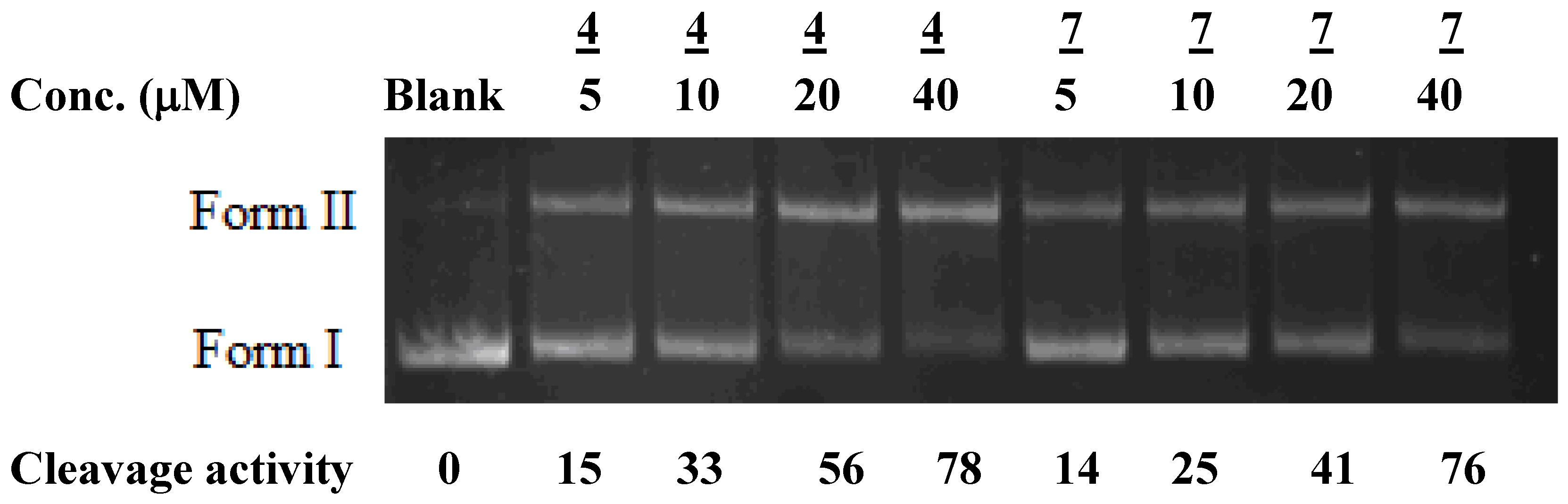
2.2. DNA photocleavage ability

The DNA photocleavage of porphyrins 1-8 and H2TPP was detected by monitoring the conversion of supercoiled form (form I) to the nicked circular form (form II). Only porphyrins 4, 7 with cationic groups showed certain photocleavage activies towards DNA, with over 75% cleavage activities observed at 40 µM (see Figure 2). The reason might be that porphrins 4, 7 with single cationic groups can bind the anionic area of DNA via electrostatic interactions. Although porphyrins 4, 7 have relatively weaker electrostatic interaction with DNA compared with other cationic porphyrins, such as meso-tetrakis(N-methylpyridinium-4-yl)porpyrin, H2TMPyP, (98% cleavage activity at 2.5 µM [19]), porphyrins 4, 7 also showed a good DNA photocleavage activity at higher concentrations. As for the remaining porphyrins and H2TPP, their insignificant DNA photocleavage activities (data not shown) were likely the result of a lack of a binding interaction with DNA and a more effectively oxidative attack from close range despite the high 1O2 yields. This suggests the importance of close-range interactions with the target for the effect of the drugs and the positive charge of porphyrin structures to affect the interaction with DNA [21,22].
In PDT, a good cellular uptake effect depends more on lipophilicity of a photosensitizer than on its 1O2 yield [23]. For example, H2TMPyP showed strong DNA binding and photocleavage activity, but its PDT efficacy was poor due to its poor cellular uptake. Hence, more porphyrins with both hydrophilic and lipophilic groups, such as porphyrins 4 and 7, can be an effective PDT agent.

3. Experimental

3.1. Materials and methods

DMF was distilled from calcium hydride. 5-[4-(3-bromopropoxy)phenyl]-10,15,20-triphenyl- porphyrin and its Zn complex [24], 2-mercapto-5-phenyloxazole [3], 5-(3-pyridyl)-1,3,4-oxadiazole- 2-thione [25], 5-(3-pyridinyl)-1,3,4-thiadiazole-2- thione [26] and 5-(2-hydroxyphenyl)-2-thioxo- 1,3,4-oxadiazoline [27] were prepared according to the corresponding literature procedures. The other reagents were purchased from the China Sinopharm Company. NMR spectra were recorded with a Bruker ARX-300 (300 MHz) NMR spectrometer. Electronic absorption spectra in the UV/Vis region were recorded with a Shimadzu UV-PC 2401 spectrophotometer. The IR spectra (KBr pellets) were recorded with a Shimadzu FT-IR 3000 spectrometer. Melting points were measured with a Tianjing RY-1 melting point apparatus (the thermometer was not corrected). Elemental analyses were performed by using aVario ELIII elemental analyzer. High-resolution mass spectra (+ve mode, CDCl3) were recorded with a Bruker Autoflex MALDI-TOF mass spectrometer. All measurements were performed at ambient temperature (20 ± 2 °C) under atmospheric pressure.

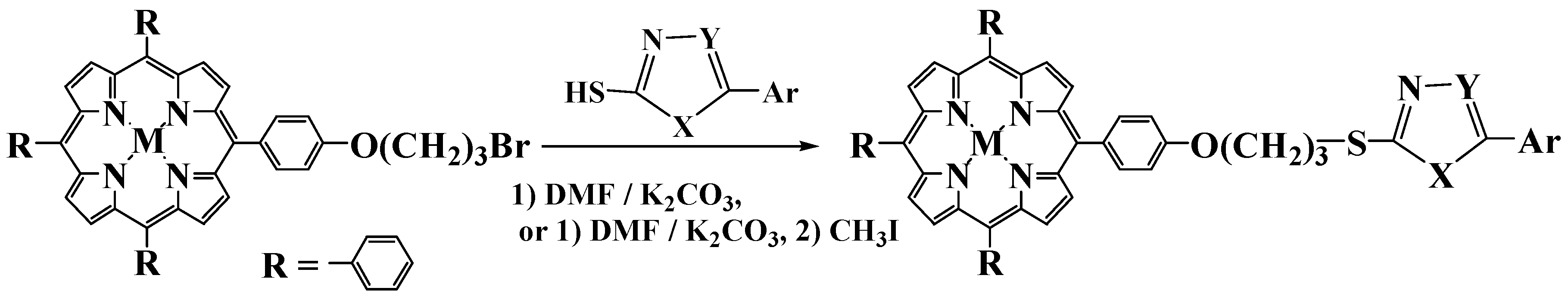
3.2. Synthesis routes and procedures

3.2.1. Preparation of porphyrin 1

5-[4-(3-Bromopropoxy)phenyl]-10,15,20-triphenylporphyrin (106.5 mg, 0.14 mmol) and 2-mercapto-5-phenyloxazole (25 mg, 0.14 mmol) were dissolved in dry DMF (15 mL) and anhydrous K2CO3 (1 g) was added to the solution. Under nitrogen protection, the mixture was heated to 65 °C for 5 h. After cooling to room temperature, the reaction mixture was poured into water saturated with sodium chloride (30 mL) and filtered. The precipitate was purified on a silica gel column eluted with chloroform. The second fraction was the blue-violet title product. Yield: 90 mg, 76%. 1H-NMR (CDCl3) δ: −2.78 (2H, s, pyrrole, NH), 2.50~2.58 (2H, m, -CH2CH2CH2-), 3.57~3.60 (2H, t, J = 7.2 Hz, -CH2S-), 4.42 (2H, t, J = 7.2 Hz, -CH2O-), 7.35~7.38 (6H, m, oxazole-PhH and oxazole-H), 7.62~7.64 (3H, m, PorPhHp), 7.74~7.76 (8H, m, PorPhHm), 8.10~8.12 (2H, m, PorPhHo), 8.19~8.22 (6H, m, PorPhHo), 8.83 (8H, s, Hβ); UV-Vis (CHCl3, 20 °C), λmax (log ε): 422 (5.58), 515 (4.18), 549 (3.85), 588 (3.70), 643 nm (3.34 dm3mol−1cm−1); IR (KBr), ν: 3316, 1600, 1242, 1173, 799 cm−1; HRMS: m/z = 847.3006 [M]+ (C56H41N5O2S: calcd. 847.2981, Δm = 2.92 ppm) Anal. Calcd for C56H41N5O2S: C 79.31, H 4.87, N 8.26; found C 79.42, H 4.97, N 8.33.

3.2.2. Preparation of porphyrin 2

The complex was prepared by heating porphyrin 1 (50 mg, 0.059 mmol) at reflux with an excess amount of copper (II) acetate in CHCl3/methanol for 3 h. It was then purified by column chromatography on silica gel with chloroform as eluent. Yield: 49 mg, 92%. UV-Vis (CHCl3, 20 °C), λmax (log ε): 423 (5.86), 523 (4.83), 550 nm (4.52 dm3 mol−1 cm−1); IR (KBr), ν: 1601, 1508, 1242, 1175, 1000 (CuII, OSMB), 812 cm−1; HRMS: m/z = 908.2150 [M]+ (C56H39N5O2SCu: calcd. 908.2121, Δm = 3.24 ppm). Anal. Calcd for C56H39N5O2SCu: C 73.95, H 4.32, N 7.70; found C 73.84, H 4.39, N 7.61.

3.2.3. Preparation of porphyrin 3

5-[4-(3-Bromopropoxy)phenyl]-10,15,20-triphenylporphyrin (106.5 mg, 0.14 mmol) and 5-(3-pyridyl)-1,3,4-oxadiazole-2-thione (27 mg, 0.14 mmol) were dissolved in dry DMF (15 mL) and anhydrous K2CO3 (1 g) was added to the solution. Under nitrogen protection, the mixture was heated to 65 °C for 5 h. After cooling to room temperature, the reaction mixture was poured into water saturated with sodium chloride (30 mL) and filtered. The precipitate was purified on a silica gel column eluted with chloroform. The second fraction was the blue-violet title product. Yield: 84 mg, 70%. 1H-NMR (CDCl3) δ: −2.78 (2H, s, pyrrole NH), 2.55~2.59 (2H, m, -CH2-), 3.71 (2H, t, J = 7.2 Hz, -CH2S-), 4.43 (2H, t, J = 7.2 Hz, -CH2O-), 7.43~7.47 (3H, m, PorPhHp), 7.74~7.76 (8H, m, PorPhHm), 8.10~8.13 (2H, m, -O-PorPhHo), 8.19~8.22 (6H, m, PorPhHo), 8.32~8.35 (1H, m, PyH6), 8.75~8.76 (2H, m, PyH4-5), 8.83 (8H, s, Hβ), 9.26 (1H, s, PyH2); UV-Vis (CHCl3, 20 °C), λmax (log ε): 429 (5.66), 514 (5.19), 548 (4.76), 588 (4.23), 643 nm (3.68 dm3 mol−1 cm−1); IR (KBr) ν: 3313, 1600, 1242, 1177, 800 cm−1; HRMS: m/z = 849.2923 [M]+ (C54H39N7O2S: calcd. 849.2886, Δm = 4.31 ppm). Anal. Calcd for C54H39N7O2S: C 76.30, H 4.62, N 11.53; found C 76.44, H 4.73, N 11.62.

3.2.4. Preparation of porphyrin 4

Porphyrin 3 (50 mg, 0.059 mmol) was added to CH3I (10 mL). After heating at reflux for 4 h under nitrogen, the solution was concentrated to dryness in vacuum. The precipitate was purified on a silica gel column and eluted with chloroform and methanol (v/v 20:1). The second fraction was the blue-violet title product. Yield: 53 mg, 90%. 1H-NMR (CDCl3) δ: −2.85 (2H, s, Pyrrole NH), 2.53~2.56 (2H, m, -CH2-), 3.73 (2H, t, J = 7.5 Hz, -CH2S-), 4.42 (2H, t, J = 7.5 Hz, -CH2O-), 4.46 (3H, s, -N-CH3), 7.39~7.43 (3H, m, PorPhHp), 7.72~7.75 (8H, m, PorPhHm), 8.11~8.12 (2H, m, -O-PorPhHo), 8.18~8.21 (6H, m, PorPhHo), 8.34~8.36 (1H, m, PyH6), 8.73~8.75 (2H, m, PyH4-5), 8.85 (8H, s, Hβ), 9.65 (1H, s, PyH2); UV-Vis (CHCl3, 20 °C), λmax (log ε): 426 (5.47), 514 (5.16), 548 (4.80), 586 (4.27), 645 nm (3.60 dm3mol−1cm−1); IR (KBr) ν: 3315, 1602, 1504, 1242, 1175, 805 cm−1; HRMS: m/z = 991.2181 [M]+ (C55H42IN7O2S: calcd. 991.2165, Δm = 1.65 ppm). Anal. Calcd for C55H42IN7O2S: C 66.60, H 4.27, N 9.88; found C 66.54, H 4.33, N 9.91.

3.2.5. Preparation of porphyrin 5

Znic(II), 5-[4-(3-bromopropoxy)phenyl]-10,15,20-triphenylporphyrinate (50 mg, 0.061 mmol) and 5-phenyl-2-sulfydryl-1,3,4-oxadiazole (11 mg, 0.061 mmol) were dissolved in dry DMF (20 mL) and anhydrous potassium carbonate (1 g) was added to the solution. Under nitrogen protection, the mixture was heated to 65 °C for 4 h. After cooling to room temperature, the reaction mixture was poured into water saturated with sodium chloride (30 mL) and filtered. The precipitate was purified on a silica gel column and eluted with chloroform. The second fraction was the blue-violet title product. Yield: 40 mg, 71%. 1H-NMR (CDCl3), δ: 2.53~2.59 (2H, m, - CH2CH2CH2-), 3.68 (2H, t, J = 7.5 Hz, -CH2S-), 4.43 (2H, t, J = 5.1 Hz, -CH2O-), 7.28~7.29 (3H, m, oxadiazole-PhHm, p) 7.51~7.52 (3H, m, PorPhHp), 7.73~7.76 (8H, m, PorPhHm), 8.03~8.06 (2H, m, oxadiazole-PhHo), 8.10~8.13 (2H, m, -O-PorPhHo), 8.20~8.22 (6H, m, PorPhHo), 8.92 (8H, s, Hβ); UV-Vis (CHCl3, 20 °C), λmax (log ε): 427 (5.80), 521 (4.45), 554 nm (3.76 dm3 mol−1 cm−1); IR (KBr) ν: 3316, 1598, 1240, 1174, 996 (ZnII, OSMB), 810 cm−1; HRMS: m/z = 910.2103 [M]+ (C55H38N6O2SZn: calcd. 910.2068, Δm = 3.87 ppm). Anal. Calcd for C55H38N6O2SZn: C 72.40, H 4.20, N 9.21; found C 72.53, H 4.31, N 9.25.

3.2.6. Preparation of porphyrin 6

5-[4-(3-Bromopropoxy)phenyl]-10,15,20-triphenylporphyrin (106 mg, 0.14 mmol) and 5-(3-pyridinyl)-1,3,4-thiadiazole-2-thione (27 mg, 0.14 mmol) were dissolved in dry DMF (15 mL) and anhydrous potassium carbonate (1 g) was added to the solution. Under nitrogen protection, the mixture was heated to 65 °C for 6 h. After cooling to room temperature, the reaction mixture was poured into water saturated with sodium chloride (30 mL) and filtered. The precipitate was purified on a silica gel column and eluted with chloroform. The second fraction was the blue-violet title product. Yield: 92 mg, 76%. 1H-NMR (CDCl3) δ: −2.78 (2H, s, pyrrole NH), 2.55~2.59 (2H, m, -CH2-), 3.76~3.81 (2H, t, J = 6.0 Hz, -CH2S-), 4.42~4.45 (2H, t, J = 6.0 Hz, -CH2O-), 7.28~7.31 (5H, m, PorPhHp and PyH4, 5), 7.35~7.40 (1H, m, PyH6), 7.75~7.77 (8H, m, PorPhHm), 8.11~8.14 (2H, m, -O-PorPhHo), 8.20~8.23 (6H, m, PorPhHo), 8.53 (1H, s, PyH2), 8.84 (8H, s, Hβ); UV-Vis (CHCl3, 20 °C), λmax (log ε): 424 (5.66), 513 (4.32), 550 (3.85), 586 (3.77), 633 nm (3.56 dm3 mol−1 cm−1); IR (KBr) ν: 3315, 1601, 1503, 1243, 1176, 803 cm−1; HRMS: m/z = 865.2693 [M]+ (C54H39N7OS2: calcd. 865.2658, Δm = 4.05 ppm). Anal. Calcd for C54H39N7OS2: C 74.89, H 4.54, N 11.32; found C 75.01, H 4.65, N 11.38.

3.2.7. Preparation of porphyrin 7

Porphyrin 6 (50 mg, 0.058 mmol) was added to CH3I (10 mL). After heating at reflux for 4 h under nitrogen, the solution was concentrated to dryness in vacuum. The precipitate was purified on a silica gel column and eluted with chloroform and methanol (v/v 20:1). The second fraction was the blue-violet title product. Yield: 54 mg, 92%. 1H-NMR (CDCl3) δ: −2.82 (2H, s, pyrrole NH), 2.50~2.55 (2H, m, -CH2-), 3.77 (2H, t, J = 7.5 Hz, -CH2S-), 4.40 (2H, t, J = 7.5 Hz, -CH2O-), 4.48 (3H, s, -N-CH3), 7.74~7.76 (11H, m, PorPhHm and PorPhHp), 8.10~8.12 (2H, m, -O-PorPhHo), 8.19~8.22 (6H, m, PorPhHo), 8.83~8.85 (11H, m, Hβ and PyH4-6), 9.70 (1H, s, PyH2); UV-Vis (CHCl3, 20 °C), λmax (log ε): 423 (5.70), 514 (4.40), 548 (3.80), 588 (3.69), 638 nm (3.40 dm3 mol−1 cm−1); IR (KBr) ν: 3313, 1600, 1242, 1177, 805 cm−1; HRMS: m/z = 1007.1962 [M]+ (C55H42IN7OS2: calcd. 1007.1937, Δm = 2.53 ppm). Anal. Calcd for C55H42IN7OS2: C 65.53, H 4.20, N 9.73; found C 65.45, H 4.29, N 9.69.

3.2.8. Preparation of porphyrin 8

5-[4-(3-Bromopropoxy)phenyl]-10,15,20-triphenylporphyrin (106.5 mg, 0.14 mmol) and 5-(2-hydroxyphenyl)-2-thioxo-1,3,4-oxadiazoline (13 mg, 0.14 mmol) were dissolved in dry DMF (25 mL) and anhydrous potassium carbonate (1 g) was added to the solution. Under nitrogen protection, the mixture was heated to 65 °C for 5 h. After cooling to room temperature, the reaction mixture was poured into water saturated with sodium chloride (30 mL) and filtered. The precipitate was purified on a silica gel column and eluted with chloroform. The second fraction was the blue-violet title product. Yield: 130 mg, 61%. 1H-NMR (CDCl3) δ: −2.77 (4H, s, pyrrole NH), 2.44~2.48 (2H, m, -OCH2-CH2-CH2-S-), 2.54~2.57 (2H, m, -OCH2-CH2-CH2-O-), 3.63 (2H, t, J = 6.6 Hz, -OCH2-CH2-CH2-S-), 4.18 (2H, t, J = 5.1 Hz, -OCH2CH2CH2-O-Por), 4.49 (2H, t, J = 6.0 Hz, Por-O-CH2CH2CH2S-), 4.59 (2H, t, J = 6.0 Hz, Por-O-CH2CH2 CH2O-), 7.33~7.35 (4H, m, PhH3-6), 7.54~7.61 (6H, m, PorPhHp), 7.65~7.76 (16H, m, PorPhHm), 7.98~8.00 (4H, m, -O-PorPhHo), 8.05~8.07 (4H, m, -O-PorPhHo), 8.14~8.22 (12H, m, PorPhHo), 8.75~8.89 (16H, m, Hβ); UV-Vis (CHCl3, 20 °C), λmax (log ε): 421 (5.50), 520 (4.18), 552 (3.90), 589 (3.60), 651 nm (3.59 dm3mol−1cm−1); IR (KBr) ν: 3312, 1607, 1240, 1173, 798 cm−1; HRMS: m/z = 1535.5677 [M]+ (C102H74N10O4S: calcd. 1535.5649, Δm = 1.83 ppm). Anal. Calcd for C102H74N10O4S: C 79.77, H 4.86, N 9.12; found C 79.68, H 4.91, N 9.20.

3.3. Measurement of singlet oxygen production rate

1,3-Diphenylisobenzofuran (DPBF) was used as a selective singlet oxygen (1O2) acceptor, which was bleached upon reaction with 1O2. Eight sample solutions of DPBF in DMSO (50 μM) containing, respectively no porphyrin (control sample), H2TPP (1 μM) and porphyrins 1-8 (1 μM) were prepared in the dark. Each sample container was covered with aluminum foil with a yellow filter (with cutoff wavelength <500 nm) on one side. The samples were then exposed to light (50 watt) through the filter. After irradiation, visible spectra of the sample solutions were measured spectrophotometrically. The normalized absorbances of DPBF at 418 nm in these samples were reported as a function of the photo-irradiation time. From this plot, the rates of 1O2 production of porphyrins 1-8 relative to those of H2TPP were determined.

3.4. DNA photocleavage assay

The DNA photocleavage activities of porphyrins 1-8 and H2TPP were measured using the plasmid DNA relaxation assay. Briefly, the plasmid DNA (pBluescript, 0.5 μg), enriched with the covalently-closed circular or supercoiled conformer (Form I), and the one-phor-all plus buffer (10 mM Tris-acetate, 10 mM magnesium acetate, 50 mM potassium acetate, pH 7.5) was vortexed. Aliquots of the DNA were pipetted into different Eppendorf tubes. Various amounts of autoclaved water (control sample) or prophyrins (test sample) were added into the Eppendorf tubes to give a final volume of 20 μL in each sample tube. The sample mixtures were then photo-irradiated at 400–450 nm for 60 min using a transilluminator (Vilber Lourmat) containing 4 × 15 W light tubes (Aqua Lux) with maximum emission at 435 nm. After photo-irradiation, 2 μL of the 6x sample dye solution (which contained 20% glycerol, 0.25% bromophenol blue and 0.25% xylene cyanol FF) was added to each Eppendorf tube and mixed well by centrifugation. The sample mixtures were loaded onto a 0.8% (v/v) agarose gel (Gel dimension: 13 cm × 10 cm), with 1× TBE buffer (89 mM Tris-borate, 1 mM EDTA, pH 8) used as supporting electrolyte, and electrophoresized at 1.3 Vcm−1 for 3 h using a mini gel set (CBS Scientific Co., Model No. MGU-502T). After electrophoresis, the gel was stained with 0.5 μg/mL ethidium bromide solution for 30 min and then destained using deionized water for 10 min. The resulting gel image was viewed under 365 nm and captured digitally using a gel documentation system (BioRad).

4. Conclusions

Eight porphyrins with nitrogen heterocycle tails were synthesized and characterized. Compared with H2TPP, the 1O2 yields of porphyrins 1-8 were measured indirectly and their DNA photocleavage activities were tested. Two porphyrins with cationic groups, 4 and 7, which showed certain singlet oxygen yields and DNA photocleavage activities could be potenial photosensitizers. The in vitro PDT activities against VEGFR-2 receptors and tumor angiogenesis are currently under investigation.

Acknowledgments

The authors are grateful for the financial support of the National Natural Science Foundation of China (grant No.20902071, 20672082) and the Open Funds of Key Laboratory for Green Chemical Process of Ministry of Education in China (grant No. RGCT201003).

References and Notes

  1. Sahni, A.; Francis, C.W. Vascular endothelial growth factor binds to fibrinogen and fibrin and stimulates endothelial cell proliferation. Blood 2000, 96, 3772–3778. [Google Scholar] [PubMed]
  2. Brown, M.L.; Cheung, M.; Dickerson, S.H.; Gauthier, C.; Harris, P.A.; Hunter, R.N., III; Pacofsky, G.; Peel, M.R.; Stafford, J.A. Chemical compounds. WO 2004032882, 22 April 2004. [Google Scholar]
  3. Harris, P.A.; Cheung, M.; Hunter, R.N., III; Brown, M.L.; Veal, J.M.; Nolte, R.T.; Wang, L.P.; Liu, W.; Crosby, R.M.; Johnson, J.H.; Epperly, A.H.; Kumar, R.; Luttrell, D.K.; Stafford, J.A. Discovery and Evaluation of 2-Anilino-5-aryloxazoles as a Novel Class of VEGFR2 Kinase Inhibitors. J. Med. Chem. 2005, 48, 1610–1619. [Google Scholar] [CrossRef] [PubMed]
  4. Kiselyov, A.S.; Semenova, M.; Semenov, V.V.; Milligan, D. Inhibitors of VEGF receptors-1 and -2 based on the 2-((pyridin-4-yl)ethyl)pyridine template. Bioorg. Med. Chem. Lett. 2006, 16, 1913–1919. [Google Scholar] [CrossRef] [PubMed]
  5. Qiu, H.; Liu, Y.Q.; Han, S.T. Synthesis of bis (chlorophenyl porphin)5-fluorouacil compounds (in Chinese). Chem. Reagent 2001, 23, 346–349. [Google Scholar]
  6. Li, D.H.; Diao, J.L.; Yu, K.G.; Zhou, C.H. Synthesis and anticancer activities of porphyrin induced anticancer drugs. Chin. Chem. Lett. 2007, 18, 1331–1334. [Google Scholar] [CrossRef]
  7. Dolmans, D.E.J.G.J.; Fukumura, D.; Jain, R.K. TIMELINE: Photodynamic therapy for cancer. Nat. Rev. Cancer 2003, 3, 380–387. [Google Scholar] [CrossRef] [PubMed]
  8. Cecic, I.; Minchinton, A.I.; Korbelik, M. The impact of complement activation on tumor oxygenation during photodynamic therapy. Photochem. Photobiol. 2007, 83, 1049–1055. [Google Scholar] [CrossRef] [PubMed]
  9. Sternberg, E.D.; Dolphin, D.; Bruckner, C. Porphyrin-based photosensitizers for use in photodynamic therapy. Tetrahedron 1998, 54, 4151–4202. [Google Scholar] [CrossRef]
  10. Juzeniene, A.; Moan, J. The history of PDT in Norway Partone: Identication of basic mechanisms of general PDT. Photodiag. Photodyn. Ther. 2007, 4, 3–11. [Google Scholar] [CrossRef] [PubMed]
  11. Castano, A.P.; Demidova, T.N.; Hamblin, M.R. Mechanisms in photodynamic therapy: Part three – Photosensitizer pharmacokinetics, biodistribution, tumor localization and modes of tumor destruction. Photodiag. Photodyn. Ther. 2005, 2, 91–106. [Google Scholar] [CrossRef]
  12. Hwu, J.R.; Yang, J.R.; Tsay, S.C.; Hsu, M.H.; Chen, Y.C.; Chou, S.S.P. Photo-induced DNA cleavage by (heterocyclo)carbonyl oxime esters of anthraquinone. Tetrahedron Lett. 2008, 49, 3312–3315. [Google Scholar] [CrossRef]
  13. Lobachevsky, P.N.; Martin, R.F. DNA targeted UVA photosensitization: Characterization of an extremely photopotent iodinated minor groove binding DNA ligand. J. Photochem. Photobiol. B Biol. 2006, 83, 195–204. [Google Scholar]
  14. Kamat, J.P.; Devasagayam, T.P.A. Oxidative damage to mitochondria in normal and cancer tissues, and its modulation. Toxicology 2000, 155, 73–82. [Google Scholar] [CrossRef]
  15. Oda, K.; Ogura, S.; Okura, I. Preparation of a water-soluble fluorinated zinc phthalocyanine and its effect for photodynamic therapy. J. Photochem. Photobiol. B Biol. 2000, 59, 20–25. [Google Scholar] [CrossRef]
  16. Ishikawa, Y.; Yamakawa, N.; Uno, T. Potent DNA photocleavage by zinc(II) complexes of cationic bis-porphyrins linked with aliphatic diamine. Bioorg. Med. Chem. 2002, 10, 1953–1960. [Google Scholar] [CrossRef]
  17. Hirakawa, K.; Kawanishi, S.; Matsumoto, J.; Shiragami, T.; Yasuda, M. Guanine-specific DNA damage photosensitized by the dihydroxo(tetraphenylporphyrinato)antimony(V) complex. Photochem. Photobiol. B Biol. 2006, 82, 37–44. [Google Scholar] [CrossRef] [PubMed]
  18. Kunkely, H.; Vogler, A. Photodemetalation of silver(II) tetraphenylporphyrin. Inorg. Chem. Commun. 2007, 10, 479–481. [Google Scholar] [CrossRef]
  19. Wang, K.; Poon, C.T.; Wong, W.K.; Wong, W.Y.; Kwong, D.W.J.; Zhang, H.; Li, Z.Y. Synthesis, Characterization, Singlet Oxygen Photogeneration, DNA Photocleavage and Two-Photon Absorption Properties of Some 4-Cyanophenylporphyrins. Eur. J. Inorg. Chem. 2009, 922–927. [Google Scholar] [CrossRef]
  20. Pineiro, M.; Carvalho, A.L.; Pereira, M.M.; d’A. R. Gonsalves, A.M.; Arnaut, L.G.; Formosinho, S.J. Photoacoustic measurements of porphyrin triplet-state quantum yields and singlet-oxygen efficiencies. Chem. Eur. J. 1998, 4, 2299–2307. [Google Scholar] [CrossRef]
  21. Jia, T.; Jiang, Z.X.; Wang, K.; Li, Z.Y. Binding and photocleavage of cationic porphyrin–phenylpiperazine hybrids to DNA. Biophys. Chem. 2006, 119, 295–302. [Google Scholar] [CrossRef] [PubMed]
  22. Ishikawa, Y.; Yamakawa, N.; Uno, T. Synthetic control of interchromophoric interaction in cationic bis-porphyrins toward efficient DNA photocleavage and singlet oxygen production in aqueous solution. Bioorg. Med. Chem. 2007, 15, 5230–5238. [Google Scholar] [CrossRef] [PubMed]
  23. Ben-Dror, S.; Bronshtein, I.; Wiehe, A.; Roder, B.; Senge, M.O.; Ehreberg, B. On the correlation between hydrophobicity, liposome binding and cellular uptake of porphyrin sensitizers. Photochem. Photobiol. 2006, 82, 695–701. [Google Scholar] [CrossRef] [PubMed]
  24. Li, Z.Y.; Wang, K.; Zhao, Y.M.; Li, H.Y. Synthesis and Structural Characterization of Three Tailed Porphyrins (in Chinese). Chin. J. Org. Chem. 2003, 23, 265–269. [Google Scholar]
  25. Jha, K.K.; Kumar, Y.; Shaharyar, M. Design, synthesis and biological evaluation of 1,3,4-oxadiazole derivatives. Asian J. Chem. 2009, 21, 7403–7410. [Google Scholar] [CrossRef] [PubMed]
  26. Wei, M.X.; Feng, L.; Li, X.Q.; Zhou, X.Z.; Shao, Z.H. Synthesis of new chiral 2,5-disubstituted 1,3,4-thiadiazoles possessing-butenolide moiety and preliminary evaluation of in vitro anticancer activity. Eur. J. Med. Chem. 2009, 44, 3340–3344. [Google Scholar] [CrossRef] [PubMed]
  27. Aboraia, A.S.; Abdel-Rahman, H.M.; Mahfouz, N.M.; EL-Gendy, M.A. Novel 5-(2-hydroxyphenyl)-3-substituted-2,3-dihydro-1,3,4-oxadiazole-2-thione derivatives: Promising anticancer agents. Bioorg. Med. Chem. 2006, 14, 1236–1246. [Google Scholar] [CrossRef] [PubMed]
Sample Availability: Samples of the porphyrin 1-8 and some intermediates are available from the authors.
Scheme 1. Synthesis of eight porphyrins with nitrogen heterocycle tails.
Molecules 16 03488 sch001

PorphyrinMXYAr
12HOCH-Ph
2CuOCH-Ph
32HON Molecules 16 03488 i001
42HON Molecules 16 03488 i002
5ZnON-Ph
62HSN Molecules 16 03488 i003
72HSN Molecules 16 03488 i004
82HON Molecules 16 03488 i005
Figure 1. The relation between illumination time and DPBF’s absorption value ratio (A/A0).
Molecules 16 03488 g001
Figure 2. DNA photocleavage activity of porphyrin 4 (lanes 2-5) and porphyrin 7 (lanes 6-9) as a function of its concentration. Lane 1: DNA (Form I) control; lane 2, 6: 5 μM; lane 3, 7: 10 μM; lane 4, 8: 20 μM; lane 5, 9: 40 μM for porphyrin 4 and 7. Photo-irradiation conditions: λirrad = 455 nm; duration, 60 min.
Molecules 16 03488 g002

