Article

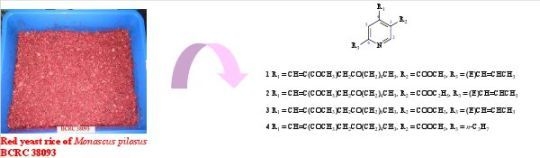
Monasnicotinates A–D, Four New Pyridine Alkaloids from the Fungal Strain Monascus pilosus BCRC 38093

1
Bioresource Collection and Research Center (BCRC), Food Industry Research and Development Institute (FIRDI), Hsinchu 300, Taiwan
2
School of Pharmacy, College of Pharmacy, Kaohsiung Medical University (KMU), Kaohsiung 807, Taiwan
*
Authors to whom correspondence should be addressed.
These authors contributed equally to this work.
Molecules 2011, 16(6), 4719-4727; https://doi.org/10.3390/molecules16064719
Submission received: 4 May 2011 / Revised: 30 May 2011 / Accepted: 31 May 2011 / Published: 7 June 2011

Abstract

:
Four new pyridine derivatives, monasnicotinates A–D (14) were isolated from the red yeast rice of Monascus pilosus BCRC 38093. Their structures were elucidated on the basis of physicochemical evidence, in-depth NMR spectroscopic analysis, and high-resolution mass spectrometry. Their inhibitory effects on NO production was also evaluated.

Graphical Abstract

1. Introduction

Monascus-fermented rice (ang-kak, red koji) has been used for centuries as a natural food colorant and traditional medicine in oriental countries. The species of Monascus produced secondary metabolites such as pigments [1], monacolin K [2], γ-aminobutyric acid [3], dimerumic acid [4], and citrinin [5]. Several secondary metabolites from Monascus sp. have been found to have some beneficial pharmacological effects in decreasing blood pressure [6], lowering plasma cholesterol levels [2,7,8] and antibacterial activity [9]. In Taiwan, M. purpureus, M. pilosus, and M. ruber, are the common species used to make Monascus-fermented red rice, which contains some red pigments and physiological biological active metabolites. M. pilosus is one of the fungi traditionally used in food items in south China, Taiwan, Japan, Korea, Indonesia and other eastern countries. Many metabolites were identified from the Monascus species in previous studies [1,5,10,11,12,13,14,15,16,17,18,19], but knowledge of their biological or toxicological effects is limited. Therefore, characterization of the secondary metabolites of Monascus and their functionality still remain unclear and are worthy of examination.
In a series of studies on the nitric oxide (NO) production inhibitory activity from natural sources, we were especially interested to realize the chemical constituents of red yeast rice, and M. pilosus BCRC 38093 has been found to be one of the active species. Chromatographic purification of the AcOEt-soluble fraction of a 95% EtOH extract of the red yeast rice produced by M. pilosus BCRC 38093 led to the isolation of four new pyridine derivatives, monasnicotinates A–D (14). The structural elucidation of the new natural compounds and the inhibitory effects on NO production by macrophages of the isolates are described herein.
Figure 1. Chemical structures of compounds 14.
Molecules 16 04719 g003

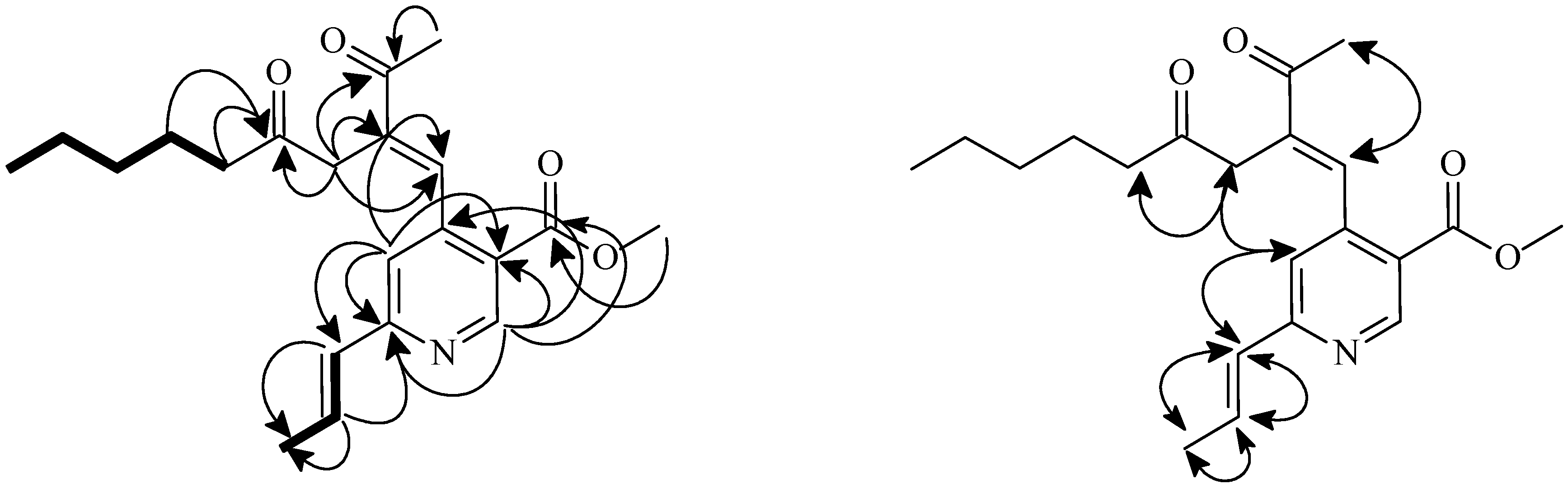
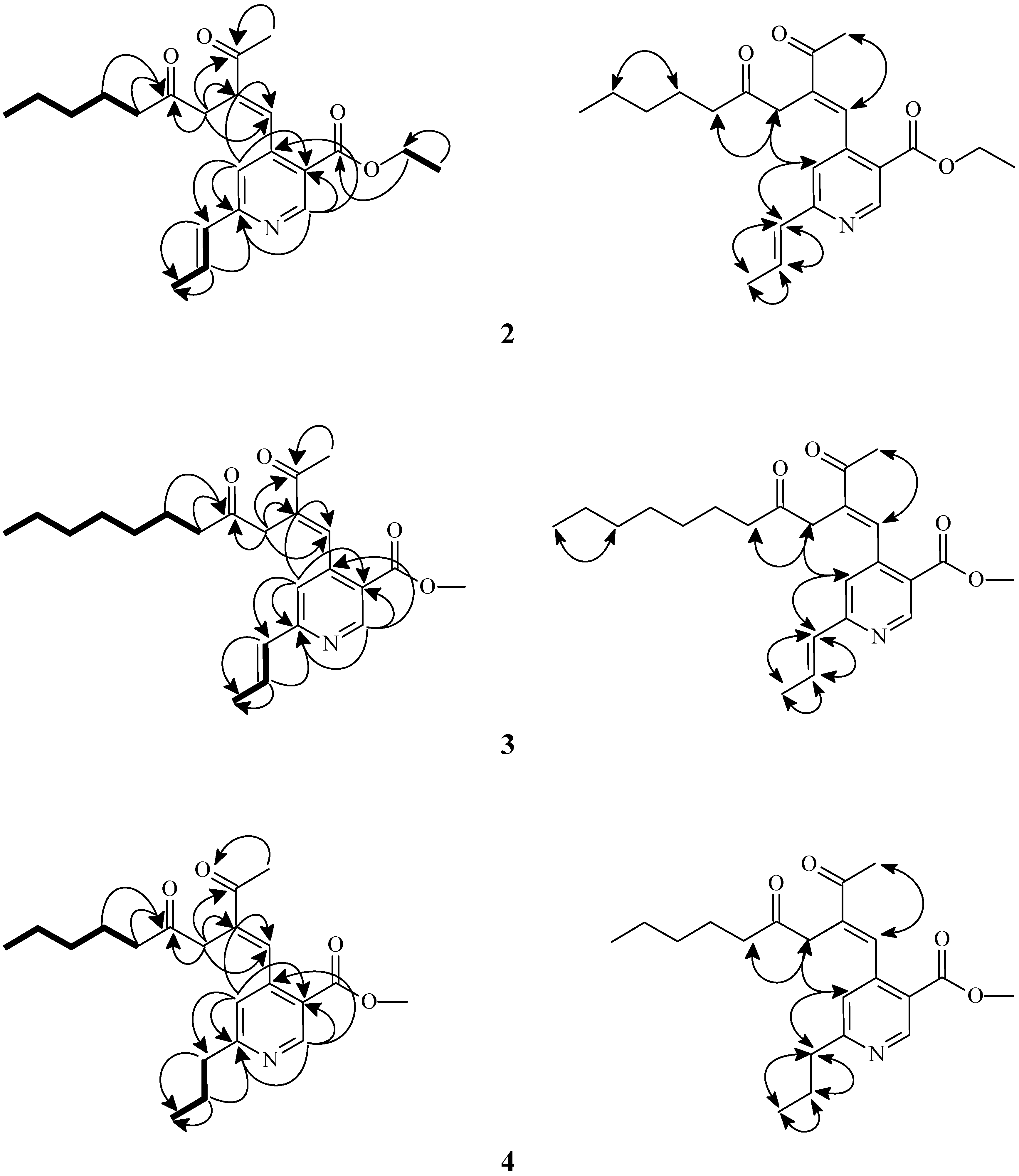
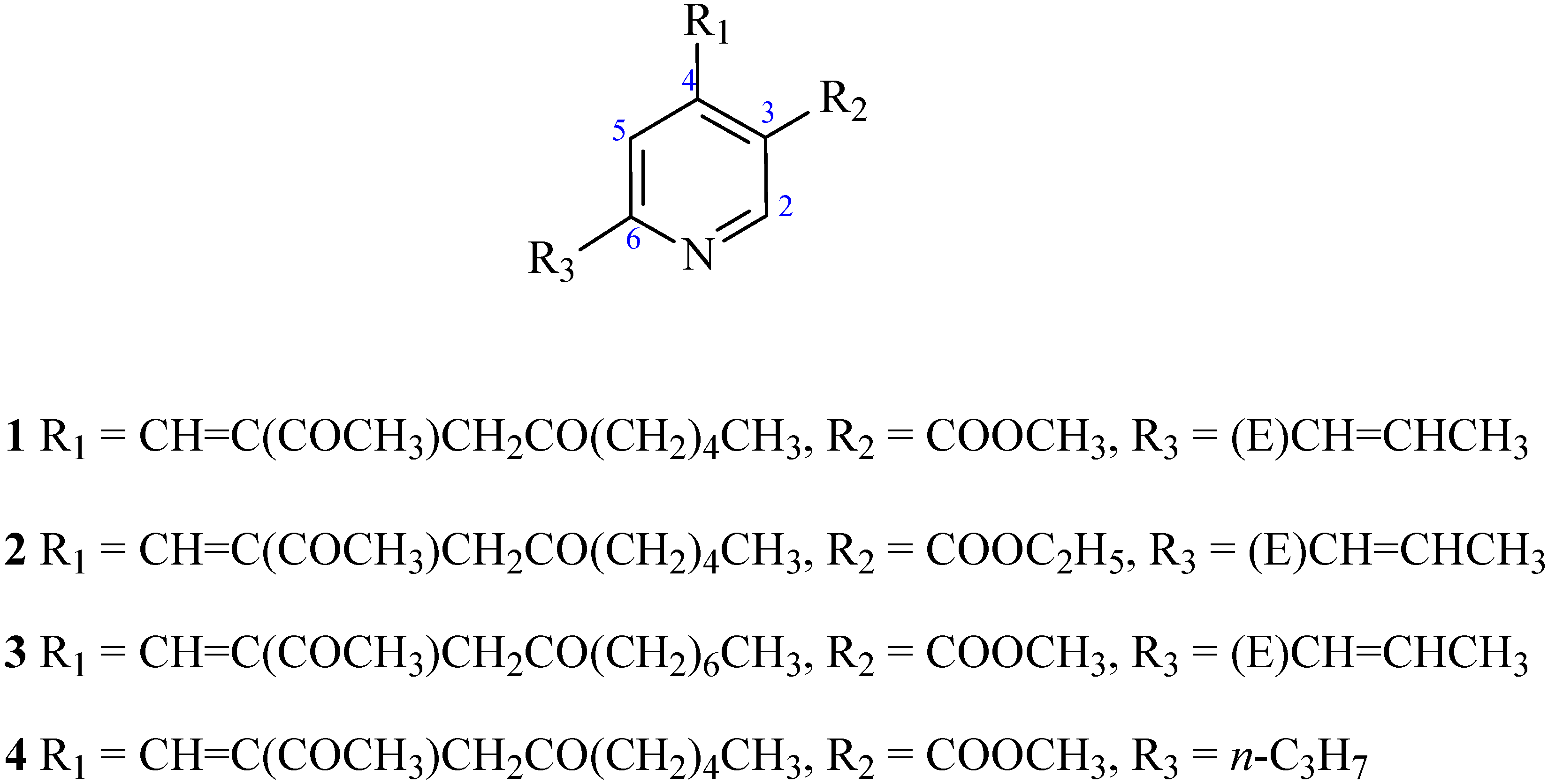
2. Results and Discussion

Compound 1, obtained as a yellowish oil, had the molecular formula C21H27NO4, requiring nine degrees of unsaturation, as determined by HR-ESI-MS data [m/z 380.1837 ([M+Na]+; calc. 380.1835)] in combination with its 1H-NMR, 13C-NMR and DEPT data. The IR spectrum revealed the presence of multiple carbonyls (1712, 1668 cm-1), one of which was by UV spectrum analysis (λmax 253, 280 and 330 nm) in conjugation with a pyridine.
The 1H-/13C-NMR spectra (Table 1) indicated seven quaternary C-atoms, five CH, five CH2, and four Me groups. In the 1H-NMR spectrum, there were typical signals for one OMe groups at δH 3.92 (s, MeO-C(12)), one acetyl moiety at δ 2.51 (3H, s, Me-15), signals of α-methylene protons of a ketone at δ 2.54 (2H, d, J = 7.4 Hz, CH2-18) and 3.27 (2H, s, CH2-16), one β-methylene resonance of a ketone at δ 1.59 (2H, m, CH2-19), two signal for a pyridine olefinic proton at δ 7.33 (1H, s, H-5) and 9.13 (1H, s, H-2), as well as one (E)-double bond signals at δ 6.54 (1H, br d, J = 16.0 Hz, H-7) and 6.93 (1H, dq, J = 16.0, 7.0 Hz, H-8), indicating that 1 was probably a pyridine ring moiety possessing a conjugated carbonyl ester group. The carbons of the pyridine derivative were assigned, from 13C-NMR and DEPT experiments (Table 1), there were resonances for three C=O functions [δ 198.8 (α,β-unsaturated C=O group); 165.5 (ester C=O group), and 208.7 (C=O)], one C=C bond [δC 130.2, 135.4], one vinyl methyl carbon [δC 18.6], one methoxyl groups [δC 52.3], one acetyl methyl moiety [δC 25.4], and three aliphatic methylenes C-atoms [δC 20.0, 31.0, 34.6].
Table 1. 1H- (400 MHz) and 13C-NMR (100 MHz) data of 1 and 2 in CDCl3. δ in ppm, J in Hz.
No.1 2
δHδC δHδC
29.13 (s)151.8 9.14 (s)151.7
3 121.1 121.4
4 145.7 145.7
57.33 (s)120.1 7.24 (s)120.1
6 159.4 159.2
76.54 (dd, J = 16.0, 1.8)130.2 6.54 (dd, J = 16.0, 2.0)130.1
86.93 (dq, J = 16.0, 7.0)135.4 6.92 (dq, J = 16.0, 7.0)135.5
91.96 (dd, J = 7.0, 1.8) 18.6 1.95 (dd, J = 7.0, 2.0)18.7
10 165.5 165.1
MeO-12 3.92 (s) 52.3
12 4.36 (q, J = 7.2)61.4
138.13 (s)140.9
Me-13 1.39 (t, J = 7.2)14.2
C-14 136.9 141.0
C-15 198.8 136.9
Me-152.51 (s) 25.4
Me-16 2.50 (s)25.4
163.27 (s) 40.6 198.8
17 208.7 3.27 (s)40.7
182.54 (d, J = 7.4)43.2 208.7
191.59 (m)23.4 2.54 (t, J = 6.8)43.2
201.32 (m)31.3 1.59 (m)23.5
211.27 (m)22.4 1.32 (m)31.3
Me-220.88 (t, J = 6.8)13.9
22 1.32 (m)22.4
Me-23 0.87 (t, J = 7.2)13.9
The above observation accompanied by the 1H,1H-COSY, and NOESY (Figure 1) spectrum of 1 was constructed by the aid of HMBC spectrum (Figure 2). Thus, the structure of 1 was determined to be methyl 4-((E)-2-acetyl-4-oxonon-1-enyl)-6-((E)-prop-1-enyl)nicotinate, and designated monasnicotinate A.
Figure 1. Key COSY (▬), NOESY (H↔H), and HMBC (H→C) correlations of 1.
Molecules 16 04719 g001
The 1H/13C-NMR spectra of compounds 2–4 (Table 1 and Table 2) were similar to those of the above compound 1, except that the substitution patterns on the pyridine moiety were different (Figure 1). The structures were further confirmed by 13C-NMR, DEPT, COSY, NOESY (Figure 2), HSQC, and HMBC (Figure 2) experiments. Thus, the structures of 2–4 were elucidated to be ethyl 4-[(E)-2-acetyl-4-oxonon-1-enyl]-6-[(E)-prop-1-enyl]nicotinate, methyl 4-[(E)-2-acetyl-4-oxoundec-1-enyl]-6-[(E)-prop-1-enyl)]- nicotinate and (E)-methyl 4-(2-acetyl-4-oxonon-1-enyl)-6-propylnicotinate and named monasnicotinates BD, respectively.
Figure 2. Key COSY (▬), NOESY (H↔H), and HMBC (H→C) correlations of 2–4.
Molecules 16 04719 g002
Table 2. 1H- (400 MHz) and 13C-NMR (100 MHz) data of 3 and 4 in CDCl3. δ in ppm, J in Hz.
3 4
δHδC δHδC
29.14 (s)151.8 9.16 (s)150.9
3 121.1 121.5
4 145.8 146.2
57.24 (s)120.2 7.23 (s)122.8
6 159.4 166.4
76.54 (dq, J = 15.6, 2.0)130.2 2.83 (t, J = 7.2)39.8
86.92 (dq, J = 15.6, 7.0)135.6 1.75 (sext, J = 7.2)22.7
91.95 (dd, J = 7.0, 2.0) 18.7 0.94 (t, J = 7.2) 13.7
10 165.6 165.3
MeO-123.92 (s) 52.4 3.93 (s) 52.5
138.12 (s) 140.9 8.13 (s) 140.5
14 137.0 137.2
15198.8 198.7
Me-152.51 (s) 25.4 2.51 (s)25.4
163.27 (s)40.7 3.26 (s)40.7
17208.7 208.4
182.53 (d, J = 7.2)43.8 2.52 (t, J = 7.2)43.2
191.58 (m)23.8 1.57 (m)23.4
201.26 (m)31.6 1.26 (m)31.3
211.26 (m)22.6 1.26 (m)22.4
221.26 (m)29.1 0.88 (t, J = 7.2)13.9
231.26 (m)22.6
240.87 (t, J = 7.2)14.1
We examined the inhibitory effects of these compounds 14 on the production of NO induced by LPS. Compound 1, 3, and 4 showed stronger inhibition on NO production (IC50 = 6.95, 8.34, and 9.42 μg/mL) than quercetin, used as a positive control (IC50 = 11.01 μg/mL). Cytotoxic effects of compounds 1, 3, and 4 were measured using the MTT assay and these compounds did not show any cytotoxic effects.

3. Experimental

3.1. General

TLC: silica gel 60 F254 precoated plates (Merck). Column chromatography (CC): silica gel 60 (70-230 or 230-400 mesh, Merck). M.p.: Yanaco micro-melting point apparatus; uncorrected. UV Spectra: Jasco UV-240 spectrophotometer; λmax (log ε) in nm. Optical rotation: Jasco DIP-370 polarimeter; in CHCl3. IR Spectra: Perkin-Elmer-2000 FT-IR spectrophotometer; ν in cm-1. 1H-, 13C- and 2D-NMR spectra: Varian-Gemini-200, Varian-Unity-Plus-400 and Varian-Mercury-400 spectrometers; δ in ppm rel. to Me4Si, J in Hz. GC-MS: Trace GC/POLARIS Q Thermo Finnigan; in m/z (rel. %). EI-MS: VG-Biotech Quatro-5022 mass spectrometer; in m/z (rel. %). ESI- and HR-ESI-MS: Bruker APEX-II mass spectrometer; in m/z.

3.2. Fungal Material

Monascus pilosus BCRC 38093 was used throughout this study, and specimens deposited at the Bioresource Collection and Research Center (BCRC) of the Food Industry Research and Development Institute (FIRDI).

3.3. Fungal Material

BCRC 38093 was maintained on potato dextrose agar (PDA; Difco). The strain was cultured on PDA slants at 25°C for 7 days and then the spores were harvested by sterile water. The spores (5 × 10⁵) were seeded into 300 mL shake flasks containing 50 mL RGY medium (3% rice starch, 7% glycerol, 1.1% polypeptone, 3% soybean powder, 0.1% MgSO4, 0.2% NaNO3), and cultivated with shaking (150 rpm) at 25 °C for 3 days. After the mycelia enrichment step, an inoculum mixing 100 mL mycelia broth and 100 mL RGY medium was inoculated into plastic boxes (25 cm × 30 cm) containing 1 kg sterile rice and cultivated at 25 °C for producing red yeast rice. At day 7, 150 mL RGY medium was added for maintaining the growth of cells. After 28 days of cultivation, the red yeast rice was harvested and lyophilized for metabolites extraction.

3.4. Extraction and ISOLATION

The dried red yeast rice extracted three times with 95% EtOH (6 L) at rt. The ethanol syrup extract was partitioned between EtOAc and H2O (1:1) (10 L) to afford the EtOAc soluble fraction (76 g). The EtOAc-soluble fraction was chromatographed over silica gel (75 g, 70-230 mesh) and eluted with hexane/EtOAc: 12:1, 10:1, 8:1, 6:1, 4:1, 2:1, 1:1, EA, EtOAc/MeOH: 8:1, 6:1, 4:1, 2:1, 1:1, and MeOH (each 1 L) to afford thirty-two fractions. Fr. 10 was purified by prep. TLC (hexane/EtOAc, 2:1) to give 1 (36.4 mg). Fr. 11 was purified by preparative TLC (hexane/EtOAc, 2:1) to give 2 (7.8 mg) and 3 (39.2 mg). Fr. 14 was further purified by silica gel column chromatography eluting with hexane/EtOAc: 8:1, 6:1, 4:1, 2:1, 1:1 and EtOAc (each 500 mL) to yield seven fractions (frs 14.1 to 14.7). 4 (3.5 mg) was furnished from fraction 14.3 by prep. TLC (hexane/EtOAc, 2:1).

3.5. Biological Assay

Determination of Nitric Oxide Production. The murine macrophage cells RAW264.7 (BCRC 60001 = ATCC TIB-71) were transferred to 96-well plates at a density of 1x10⁵ cell/well. After 24 hr incubation, the cells were stimulated with 1 μg/mL of LPS (Sigma, Cat no: L-2654) for 24 hr in the presence or absence of the compounds (0, 1, 5, 10 and 20 μg/mL) tested. As a parameter of NO synthesis, nitrite concentration was measured in the supernatant of RAW264.7 cells by the Griess reagent [1:1 mixture of 1% sulfanilamide and 0.1% N-(1-naphthyl)ethyl-enediamine dihydrochloride, each in 2.5% phosphoric acid solution] in a 96-well plate, and incubated for 10 min at room temperature. Nitrite concentration was determined by measuring the absorbance at 540 nm using an ELISA plate reader (μ Quant) [20]. All tests were run in triplicate and averaged. The data were expressed as a mean of three experiments. Statistical comparisons were carried out the Student’s t-test for paired values.
Determination of cell viability. The cell viability was assessed using a MTT [3-(4,5-dimethylthiazol-2-yl)-2,5-diphenyltetrazolium bromide, Merck KGaA, Damstadt, Germany]-based colorimetric assay, as previously described [21]. After sampling the supernatant for the NO assay, 100 μL of fresh medium containing 0.5 mg/mL of MTT was added to each well and incubated for 3 hr at 37 °C. The medium was then removed and the violet for mazan crystals in the viable cells were dissolved in dimethyl sulfoxide. The absorbance of each well was then read at a wavelength of 540 nm using microplate reader (μ Quant, Bio-TEK instruments INC).

3.6. Spectral Data

Monasnicotinate A (1). Yellowish oil. UV λmax (MeOH): 253, 280, 330. IR νmax (Neat): 1668, 1712 (C=O). 1H- and 13C-NMR: see Table 1. ESI-MS: 380 ([M+Na]+). HR-ESI-MS: 380.1837 ([M+Na]+, C21H27NaNO4+; calcd. 380.18353).
Monasnicotinate B (2). Yellowish oil. UV λmax (MeOH): 248, 275, 338. IR νmax (Neat): 1676, 1716 (C=O). 1H- and 13C-NMR: see Table 1. ESI-MS: 394 ([M+Na]+). HR-ESI-MS: 394.1994 ([M+Na]+, C22H29NaNO4+; calcd. 394.1998).
Monasnicotinate C (3). Yellowish oil. UV λmax (MeOH): 251, 282, 327. IR νmax (Neat): 1672, 1724 (C=O). 1H- and 13C-NMR: see Table 2. ESI-MS: 408 ([M+Na]+). HR-ESI-MS: 408.2151 ([M+Na]+, C22H29NaNO4+; calcd. 408.2153).
Monasnicotinate D (4). Yellowish oil. UV λmax (MeOH): 245, 271, 328. IR νmax (Neat): 1665, 1716 (C=O). 1H- and 13C-NMR: see Table 2. ESI-MS: 382 ([M+Na]+). HR-ESI-MS: 382.1994 ([M+Na]+, C21H29NaNO4+; calcd. 382.1994).

4. Conclusions

In this study, we focused on the minor secondary metabolites appearing in the EtOAc-soluble fraction of a 95% EtOH extract of the red yeast rice produced by Monascus purpureus BCRC 38093. Four new natural metabolites 14 were found in this study. In the in vitro NO production inhibitory assay, isolates 1, 3, and 4 showed stronger inhibition on NO production with IC50 value of 6.95, 8.34, and 9.42 μg/mL respectively, while showing no cytotoxicity to normal cells at the same concentration.

Acknowledgements

The first two authors (M.-D. Wu and M.-J. Cheng) contributed equally to this work and should both be regarded as first author of this publication. This investigation was supported by a grant from the Ministry of Economic Affairs of Republic of China (Grant No. 94-EC-17-A-17-R7-0563).

References

  1. Akihisa, T.; Tokuda, H.; Yasukawa, K.; Ukiya, M.; Kiyota, A.; Sakamoto, N.; Suzuki, T.; Tanabe, N.; Nishino, H. Azaphilones, furanoisophthalides, and amino acids from the extracts of Monascus pilosus-fermented rice (red-mold rice) and their chemopreventive effects. J. Agric. Food Chem. 2005, 53, 562–565. [Google Scholar] [CrossRef]
  2. Endo, A.; Monacolin, K. A new hypocholesterolemic agent produced by Monascus species. J. Antibiot. 1979, 32, 852–854. [Google Scholar] [CrossRef]
  3. Kohama, Y.; Matsumoto, S.; Mimura, T.; Tanabe, N.; Inada, A.; Nakanishi, T. Isolation and identification of hypotensive principles in red-mold rice. Chem. Pharm. Bull. 1987, 35, 2484–2489. [Google Scholar] [CrossRef]
  4. Aniya, Y.; Ohtani, I.I.; Higa, T.; Miyagi, C.; Gibo, H.; Shimabukuro, M.; Nakanish, H.; Taira, J. Dimerumic acid as an antioxidantof the mold, Monascus anka. Free Radical Biol. Med. 1999, 286, 999–1004. [Google Scholar]
  5. Blanc, P.J.; Loret, M.O.; Goma, G. Production of citrinin by various species of Monascus. Biotechnol. Lett. 1995, 17, 291–294. [Google Scholar] [CrossRef]
  6. Tsuji, K.; Ichikawa, T.; Tanabe, N.; Abe, S.; Tarui, S.; Nakagawa, Y. Effects of two kinds of Koji on blood pressure in spontaneously hypertensive rats. Nippon. Nogeikagaku Kaishi. 1992, 66, 1241–1246. [Google Scholar]
  7. Endo, A. Compactin (ML-236B) and related compounds as potential cholesterol-lowering agents that inhibit HMG-CoA reductase. J. Med. Chem. 1985, 28, 401–405. [Google Scholar] [CrossRef]
  8. Martinokova, L.; Juzlova, P.; Vesely, D. Biological activity of polyketide pigments produced by the fungus Monascus. J. Appl. Bacteriol. 1995, 79, 609–616. [Google Scholar] [CrossRef]
  9. Wong, H.C.; Bau, Y.S. Pigmentation and antibiobacterial activity of fast neutron-and X-ray induced strains of Monascus purpureus went. Plant Physiol. 1977, 60, 578–581. [Google Scholar] [CrossRef]
  10. Ma, J.; Li, Y.; Ye, Q.; Li, J.; Hua, Y.; Ju, D.; Zhang, D.; Cooper, R.; Chang, M. Constituents of red yeast rice, a traditional Chinese food and medicine. J. Agric. Food Chem. 2000, 48, 5220–5225. [Google Scholar]
  11. Akihisa, T.; Mafune, S.; Ukiya, M.; Kimura, Y.; Yasukawa, K.; Suzuki, T.; Tokuda, H.; Tanabe, N.; Fukuoka, T. (+)- and (–)-syn-2-Isobutyl-4-methylazetidine-2,4-dicarboxylic acids from the extract of Monascus pilosus-fermented rice (red-mold rice). J. Nat. Prod. 2004, 67, 479–480. [Google Scholar] [CrossRef]
  12. Cheng, M.J.; Wu, M.D.; Chen, I.S.; Yuan, G.F. Secondary metabolites from the mycelia of the fungus Monascus pilosus BCRC 38072. Chem. Pharm. Bull. 2008, 56, 394–397. [Google Scholar] [CrossRef]
  13. Huang, Z.; Xu, Y.; Li, L.; Li, Y. Two new Monascus metabolites with strong blue fluorescence isolated from red yeast rice. J. Agric. Food Chem. 2008, 56, 112–118. [Google Scholar] [CrossRef]
  14. Jongrungruangchok, S.; Kittakoop, P.; Yongsmith, B.; Bavovada, R.; Tanasupawat, S.; Lartpornmatulee, N.; Thebtaranonth, Y. Azaphilone pigments from a yellow mutant of the fungus Monascus kaoliang. Phytochemistry 2004, 65, 2569–2575. [Google Scholar] [CrossRef]
  15. Juzlová, P.; Martínková, L.; Kren, V. Secondary metabolites of the fungus Monascus: A review. J. Industrial Microbiol. 1996, 16, 163–170. [Google Scholar] [CrossRef]
  16. Juzlová, P.; Rezanka, T.; Martínková, L.; Kren, V. Long-chain fatty acids from Monascus purpureus. Phytochemistry 1996, 43, 151–153. [Google Scholar]
  17. Sato, K.; Goda, Y.; Sakamoto, S.S.; Shibata, H.; Maitani, T.; Yamada, T. Identification of major pigments containing D-amino acid units in commercial Monascus pigments. Chem. Pharm. Bull. 1997, 45, 227–229. [Google Scholar] [CrossRef]
  18. Wild, D.; Tóth, G.; Humpf, H.U. New Monascus metabolite isolated from red yeast rice (Angkak, red Koji). J. Agric. Food Chem. 2002, 50, 3999–4002. [Google Scholar] [CrossRef]
  19. Wild, D.; Tóth, G.; Humpf, H.U. New Monascus metabolites with a pyridine structure in red fermented rice. J. Agric. Food Chem. 2003, 51, 5493–5496. [Google Scholar] [CrossRef]
  20. Mosman, T. Utilization of purified human monocytes in the agarose droplet assay for measuring migration inhibitory factors. J. Immunol. Methods 1983, 65, 55–63. [Google Scholar] [CrossRef]
  21. Johansson, M.; Kopcke, B.; Anke, H.; Sterner, O. Biologically active secondary metabolites from the ascomycete A111-95. J. Antibiot. 2002, 55, 104–106. [Google Scholar] [CrossRef]
  • Sample Availability: Samples of the compounds 1-4 are available from the authors.

