A Novel Cyclodione Coumarin from the Stem Bark of Mesua beccariana
by
Gwendoline Cheng Lian Ee
1,*, 
Soek Sin Teh
1,
Siau Hui Mah
1,
Mawardi Rahmani
1,
Yun Hin Taufiq-Yap
1 and
Khalijah Awang
2 1
Department of Chemistry, Faculty of Science, Universiti Putra Malaysia, Serdang, Selangor 43400, Malaysia
2
Department of Chemistry, Faculty of Science, University of Malaya, Kuala Lumpur 50603, Malaysia
*
Author to whom correspondence should be addressed.
Molecules 2011, 16(9), 7249-7255; https://doi.org/10.3390/molecules16097249
Submission received: 19 July 2011
/
Revised: 1 August 2011
/
Accepted: 15 August 2011
/
Published: 25 August 2011
(This article belongs to the Special Issue Coumarins and Xanthones)
Abstract
:Our ongoing investigations on the stem bark of Mesua beccariana afforded a novel cyclodione coumarin, beccamarin, together with two known xanthones, mesuarianone, mesuasinone, two anthraquinones, 4-methoxy-1,3,5-trihydroxy-anthraquinone and 2,5-dihydroxy-1,3,4-trimethoxyanthraquinone and one coumarin, mammea A/AB. The structures were elucidated by 1D and 2D NMR and MS techniques.
1. Introduction
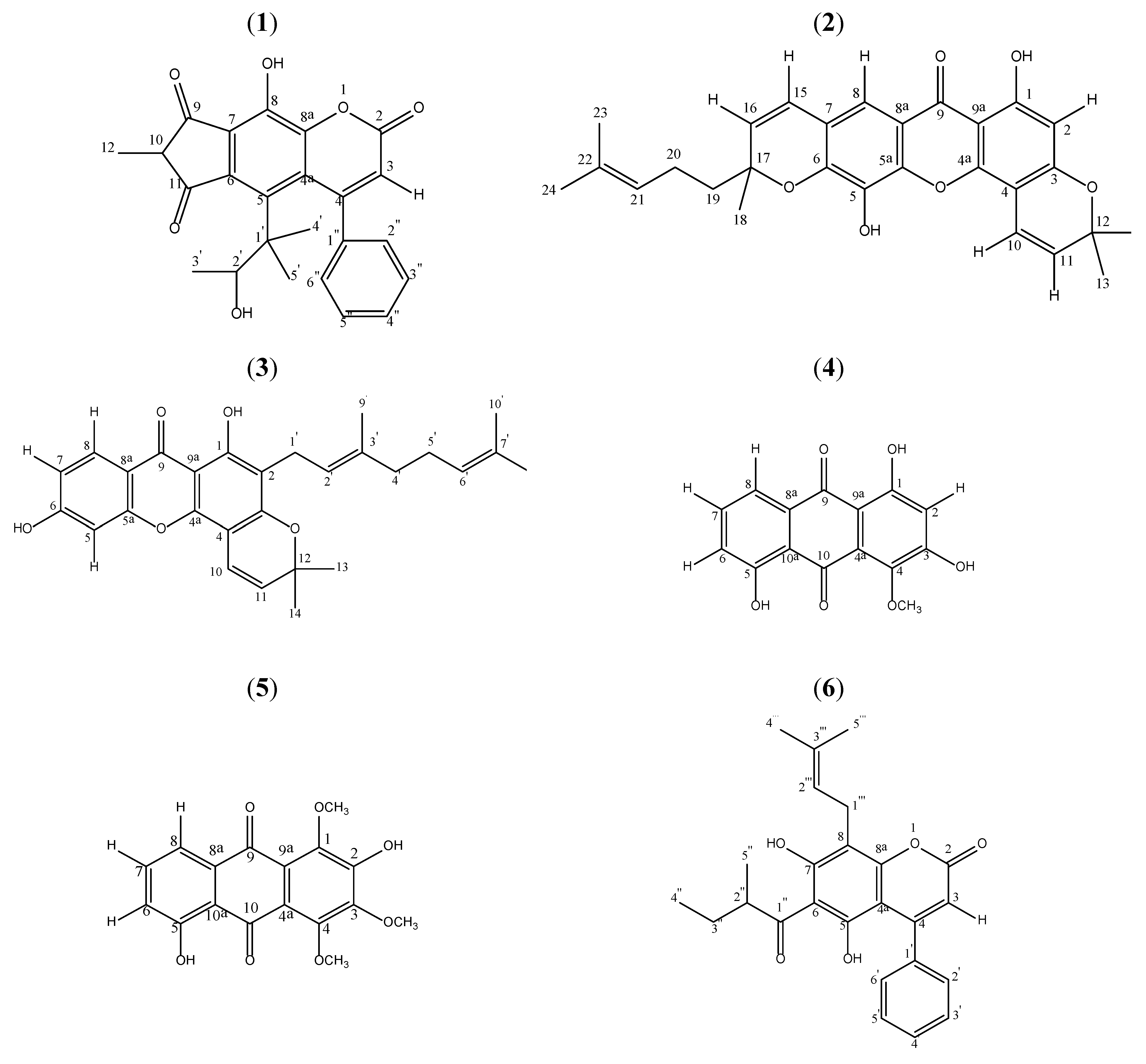
Mesua is a genus of flowering plants in the family of Clusiaceae, native to tropical southern Asia. Common names for Mesua include ironwood and rose chestnut. They are evergreen shrubs or small trees growing up to 13 m tall, with leaves arranged in opposite pairs. The blossoms are white and give off a nice fragrance. Although Mesua species have been investigated before, reports on phytochemical constituents of Mesua species are few [1]. Phytochemical investigations on the genus show the occurrence of xanthones [2,3,4,5,6], coumarins [2,7,8,9,10], terpenoids [3] and essential oil [8,11]. Our recent study on Mesua ferrea identified mesuaferrin A and mesuaferrin B [12]. We report here a novel cyclodione coumarin, beccamarin (1). See Figure 1.
2. Results and Discussion
Beccamarin (1, Figure 1) was isolated as a yellowish solid from the hexane extract of Mesua beccariana. Its melting point was 139.0–139.6 °C. The HRESIMS spectrum revealed a molecular ion peak at 405.1361 [M-H]− (calculated 406.1417), which corresponds to a molecular formula of C24H22O6. The UV spectrum supported the coumarin skeleton due to its maxima absorptions at 209 (5.23), 229 (5.27), 281 (5.36) and 348 (5.45). The FTIR spectrum gave absorptions of chelated hydroxyl (3,400 cm−1), carbonyl (1,741 cm−1), saturated C-H stretch (2971 cm−1) and aromatic ring (1,466 and 1605 cm−1), which reflected similarity to typical IR bands for coumarins.
The 1H-NMR spectrum indicated a monosubstituted phenyl group, deduced by the presence of three sets of triplets at δ 7.38 (t, 1H, J = 5.7 Hz), 7.37 (t, 2H, J = 5.7 Hz, overlapped) and 7.30 (d, 2H, J = 5.7 Hz, overlapped). In the 13C-NMR spectrum, the signals at δ 127.2 (C-2” & C-6”, overlapped), 127.7 (C-3” & C-5”, overlapped) and 128.2 (C-4”) belong to the phenyl group. Meanwhile, δ 7.30 (H-2” & H-6”) was correlated to δ 156.8 (C-4) via a 3J correlation and δ 7.37 (H-3” & H-5”) was correlated to δ 139.5 (C-1”) (3J correlation). All these observations place the phenyl group at C-4 of the main coumarin skeleton.
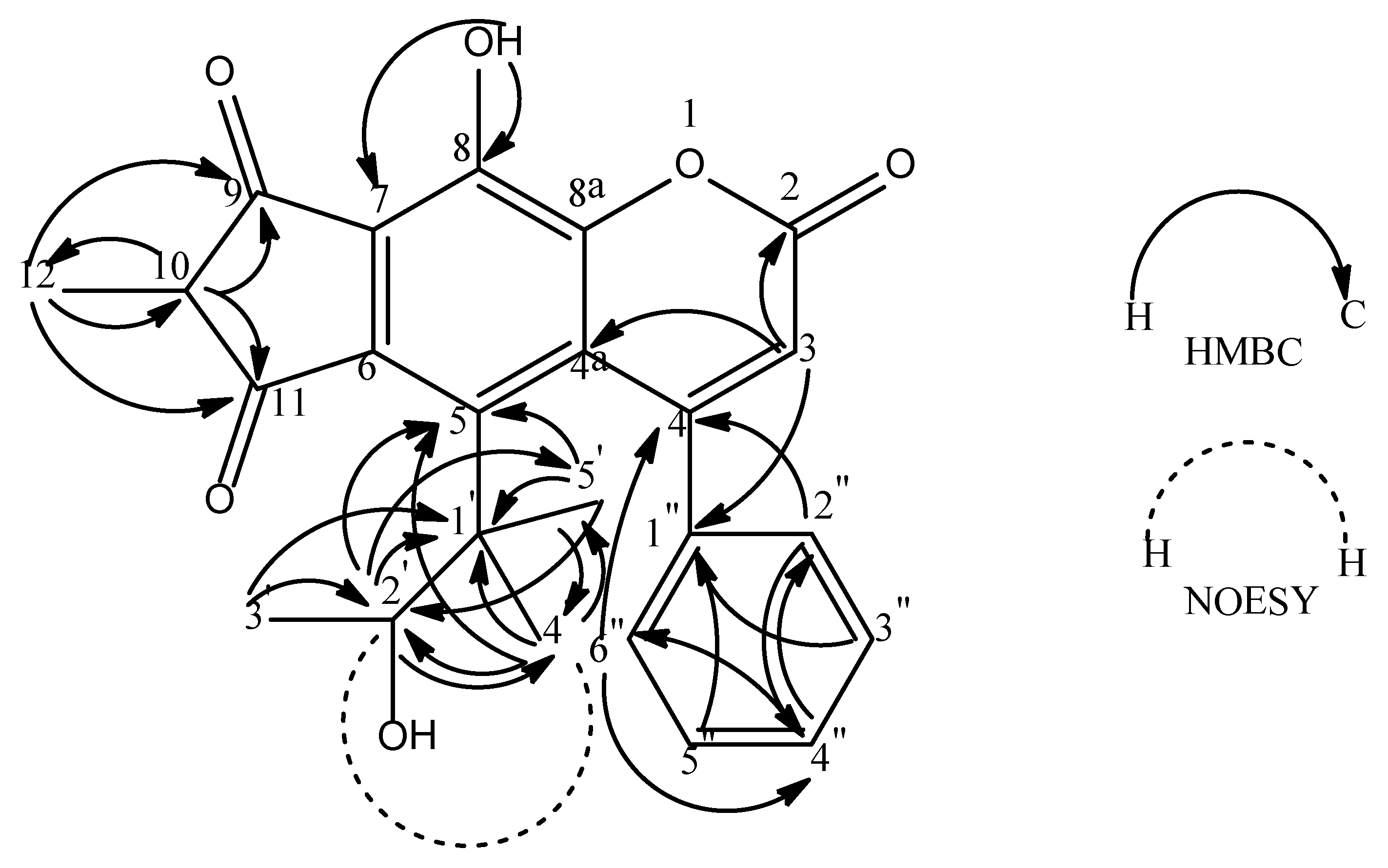
The 1H, 13C and HMBC spectra revealed a low field hydroxyl group (δ 14.50, s, 1 H). This OH group correlated to δ 164.8 (C-8) via a 2J correlation in the HMBC spectrum. A 3J correlation to δ 102.6 (C-7) was also observed. Hence, the hydroxyl group was assigned to C-8. The 1H, HMQC and HMBC experiments gave one sharp singlet proton signal at δ 5.93 and this has direct connectivity to δ 112.1 (C-3) and long range couplings to δ 102.6 (C-4a), 139.5 (C-1”) and 159.7 (C-2). Thus, this singlet proton was positioned at C-3.
The 1H and COSY spectra suggested the occurrence of a methylbutanol moiety with a set of correlated signals which are δ 1.33 (s, 3H, H-5’), 1.46 (d, 3H, J = 6.9, H-3’), 1.58 (s, 3H, H-4’) and 4.61 (q, 1H, J = 6.9, H-2’) with δ 1.46 (d, H-3’) coupling to δ 4.61 (q, H-2’). The HMQC and HMBC spectrum indicated that δ 4.61 (H-2’) has connectivities with δ 21.0 (C-5’), 25.8 (C-4’), 43.6 (C-1’) and 113.7 (C-5); δ 1.46 (H-3’) correlates with δ 43.6 (C-1’) and 91.9 (C-2’); δ 1.58 (H-4’) has connectivities with δ 21.0 (C-5’), 43.6 (C-1’), 91.9 (C-2’) and 113.7 (C-5); δ 1.33 (H-5’) correlates with δ 25.8 (C-4’), 43.6 (C-1’), 91.9 (C-2’) and 113.7 (C-5). This information justifies a 3-methylbutan-2-ol group to be attached to the coumarin ring at position C-5 (δ 113.7) leaving C-6 and C-7 to form a fused ring.
In addition, the 1H-NMR experiment demonstrated the presence of a multiplet and doublet signal at the upfield region (δ 3.77, 1H and δ 1.15, 3H, respectively). The signal at δ 3.77 (H-10) was directly bonded to δ 39.2 (C-10) whereas the signal at δ 1.15 (H-12) was directly bonded to δ 18.8 (C-12) as seen in the HMQC spectrum. The HMBC spectrum showed H-10 to have connectivities with δ 18.8 (C-12), 209.9 (C-11) and 210.1 (C-9); Meanwhile, H-12 has connectivities with δ 39.2 (C-10), 209.9 (C-11) and δ 210.1 (C-9). This implies the presence of 2-methylcyclopentane-1,3-dione. This substituent group has to be placed at C-6 and C-7 since C-5 carries the 3-methylbutan-2-ol group. The left ring of the coumarin is thus substituted at C-7 and C-6 by the above dione substituent group. (Figure 2).
Meanwhile, the NOESY experiment shows correlations between H-4’ and H-2’ suggesting that they have a similar configuration. The HMBC and NOESY correlations for 1 are shown in Figure 2. Therefore, compound 1 was identified as 4-phenyl-8-hydroxy-5-(3-methylbutan-2-ol)-(2-methylcyclopentane-1,3-dione)-[4”,5”:6,7]-chromen-2-one.
3. Experimental
3.1. General
Infrared spectra were measured using the universal attenuated total reflection (UATR) technique on a Perkin-Elmer 100 Series FT-IR spectrometer. EIMS were recorded on a Shimadzu GCMS-QP5050A spectrometer. NMR spectra were obtained on a Unity JEOL 500 MHz FT-NMR spectrometer using CDCl3 as solvent and tetramethylsilane (TMS) as internal standard. Ultraviolet spectra were recorded in EtOH on a Shimadzu UV-160A, UV-Visible Recording Spectrophotometer.
3.2. Plant Material
The stem bark of Mesua beccariana was collected from the Sri Aman district in Sarawak, Malaysia. The plant material was identified by Associate Professor Dr Rusea Go, Biology Department, Faculty of Science, Universiti Putra Malaysia.
3.3. Extraction and Isolation
The three kg of milled, air-dried and powdered sample was defatted with n-hexane and extracted successively with dichloromethane, ethyl acetate and methanol. The extracts were dried under reduced pressure using a rotary evaporator to yield hexane (15.6 g), dichloromethane (21.2 g), ethyl acetate (15.8 g) and methanol (80.5 g) extracts. Each of these extracts was chromatographed over a silica gel column using a stepwise gradient system (hexane/chloroform, chloroform/ethyl acetate, and ethyl acetate/methanol). The hexane extract was subjected to vacuum column chromatography over silica gel with a stepwise gradient of hexane/dichloromethane (hexane/CH2Cl2) and dichloromethane/ethyl acetate (CH2Cl2/EA). The eluted fraction A (CH2Cl2/EA-80:20, 3.5 g) was then subjected to a flash column chromatography using hexane/CH2Cl2 to obtain fraction B (hex/ CH2Cl2-30:70, 36 mg) to give beccamarin. Beccamarin was crystallized by repeated crystallizations from hexane. The hexane extract provided beccamarin (1, 20 mg), mesuarianone (2, 110 mg), mesuasinone (3, 76 mg) while the ethyl acetate extract gave two anthraquinones, 4-methoxy-1,3,5-trihydroxyanthraquinone (4, 9 mg) and 2,5-dihydroxy-1,3,4-trimethoxyanthraquinone (5, 8 mg) and a coumarin, mammea A/AB (6, 11 mg).
3.4. Spectral Data
Beccamarin (1). Yellow solid. UV (EtOH) λmax nm (log ε): 209 (5.23), 229 (5.27), 281 (5.36) and 348 (5.45). IR νmax (cm-1): 3400, 2971, 1741, 1605 and 1466. MS m/z (rel. int.): 406 [M+] (12), 392 (28), 377 (16), 350 (24), 349 (100), 293 (19), 43 (18). For 1H and 13C-NMR spectra, see Table 1.
Mesuarianone (2). Yellow solid. UV (EtOH) λmax nm (log ε): 208, 280 and 333.8. IR νmax (cm−1): 3392, 2971, 2923, 1639, 1574, 1475. MS m/z (rel. int.): 460 [M+] (39), 445 (100), 377 (89), 361 (10), 347 (11), 323 (12), 203 (8), 181 (61), 91 (6), 77 (5), 69 (27), 55 (9). The 1H- and 13C-NMR (CDCl3) spectral data are consistent with published data [1].
Mesuasinone (3). Yellow solid. UV (EtOH) λmax nm (log ε): 208, 254, 274 and 334. IR νmax (cm−1): 3229, 2925, 2865, 1640, 1576, 1496. MS m/z (rel. int.): 446 [M+] (14), 431 (4), 391 (8), 363 (100), 307 (21), 154 (12), 69 (10). The 1H and 13C NMR (CDCl3) spectral data are consistent with published data[1].
4-Methoxy-1,3,5-trihydroxyanthraquinone (4). Orange solid. UV (MeOH) λmax nm (log ε): 279, 320, 425, 470 and 485. IR νmax (cm−1): 3420, 2920, 2860, 1720, 1630, 1470. MS m/z (rel. int.): 286 [M+] (100), 268 (87), 257 (10), 243 (38), 212 (27), 180 (30). The 1H and 13C NMR (CDCl3) spectral data are consistent with published data[13].
2,5-Dihydroxy-1,3,4-trimethoxyanthraquinone (5). Orange solid. UV (MeOH) λmax nm (log ε): 218, 276 and 410. IR νmax (cm−1): 3400, 2920, 2840, 1660, 1630, 1540. MS m/z (rel. int.): 330 [M+] (100), 315 (60), 312 (5), 297 (20), 287 (22), 272 (24), 227 (20), 58 (23). The 1H and 13C NMR (CDCl3) spectral data are consistent with published data[13].
Mammea A/AB (6). Colourless solid. UV (EtOH) λmax nm (log ε): 283 and 337. IR νmax (cm−1): 3296, 2930, 1706, 1621. MS m/z (rel. int.): 406 [M+] (2), 392 (18), 377 (10), 349 (100), 293 (10). The 1H and 13C NMR (CDCl3) spectral data are consistent with published data[10].
4. Conclusions
A novel cyclodione coumarin, beccamarin (1), along with two xanthones, two anthraquinones and another coumarin were isolated from the stem bark of Mesua beccariana.
Acknowledgments
The authors would like to thank Jegak Uli for collection of plant samples. Financial support from UPM under the RUGS research fund is gratefully acknowledged.
References
- Teh, S.S.; Ee, G.C.L.; Rahmani, M.; Sim, W.C.; Mah, S.H.; Teo, S.H. Two new pyranoxanthones from Mesua beccariana. Molecules 2010, 15, 6733–6742. [Google Scholar] [CrossRef]
- Bandaranayak, W.M.; Selliah, S.S.; Sultanbawa, M.U.S. Xanthones and 4-phenylcoumarins of Mesua thwaitesii. Phytochemistry 1975, 14, 265–269. [Google Scholar] [CrossRef]
- Ee, G.C.L.; Lim, C.K.; Cheow, Y.L.; Sukari, M.A. Xanthones and triterpenoids from Mesua daphnifolia and Garcinia maingayi. Malays. J. Sci. 2005, 24, 183–185. [Google Scholar]
- Ee, G.C.L.; Lim, C.K.; Ong, G.P.; Sukari, M.A.; Lee, H.L. Daphnifolin, a new xanthone from Mesua daphnifolia. J. Asian Nat. Prod. Res. 2006, 8, 567–570. [Google Scholar] [CrossRef] [PubMed]
- Govindachari, T.R.; Pai, B.R.; Subramaniam, P.S.; Rao, U.R.; Muthukumaraswa, N. Constituents of Mesua ferrea L.-mesuaxanthone A and mesuaxanthone B. Tetrahedron 1967, 23, 243–248. [Google Scholar] [CrossRef]
- Walia, S.; Mukerjee, S.K. Ferrxanthone,a 1,3,5,6-tetraoxygenated xanthone from Mesua ferrea. Phytochemistry 1984, 23, 1816–1817. [Google Scholar] [CrossRef]
- Awang, K.; Chan, G.; Litaudon, M.; Ismail, N.H.; Martin, M.T.; Gueritte, F. 4-Phenylcoumarins from Mesua elegans with acetylcholinesterase inhibitory activity. Bioorg. Med. Chem. 2010, 18, 7873–7877. [Google Scholar] [CrossRef] [PubMed]
- Bala, K.R.; Seshadri, T.R. Isolation and synthesis of some coumarin components of Mesua ferrea seed oil. Phytochemistry 1971, 10, 1131–1134. [Google Scholar] [CrossRef]
- Morel, C.; Guilet, D.; Oger, J.M.; Seraphin, D.; Sevenet, T.; Wiart, C.; Hadi, A.H.A.; Richomme, P.; Bruneton, J. 6-Acylcoumarins from Mesua racemosa. Phytochemistry 1999, 50, 1243–1247. [Google Scholar] [CrossRef]
- Verotta, L.; Lovaglio, E.; Vidari, G.; Finzi, P.V.; Neri, M.G.; Raimondi, A.; Parapini, S.; Taramelli, D.; Riva, A.; Bombardelli, E. 4-Alkyl- and 4-phenylcoumarins from Mesua ferrea as promising multidrug resistant antibacterials. Pytochemistry 2004, 65, 2867–2879. [Google Scholar] [CrossRef] [PubMed]
- Dennis, T.J.; Kumar, K.A. Constituents of Mesua ferrea. Fitoterapia 1998, 69, 291–304. [Google Scholar]
- Teh, S.S.; Ee, G.C.L.; Rahmani, M.; Yap, Y.H.T.; Go, R.; Mah, S.H. Pyranoxanthones from Mesua ferrea. Molecules 2011, 16, 5647–5654. [Google Scholar] [CrossRef]
- Wijnsma, R.; Verpoorte, R.; Mulder-Krieger, T.; Svendsen, A.B. Anthraquinones in callus cultures of Cinchona ledgeriana. Phytochemistry 1984, 23, 2307–2311. [Google Scholar] [CrossRef]
Sample Availability: Samples of the compounds 1-6 are available from the authors. |
Figure 1.
Structures of Compounds. Compounds 2 and 3 were isolated from the hexane extract while compounds 4-6 were found from the ethyl acetate extracts.
Figure 1.
Structures of Compounds. Compounds 2 and 3 were isolated from the hexane extract while compounds 4-6 were found from the ethyl acetate extracts.

Figure 2.
Selected HMBC (2J and 3J) and NOESY correlations for compound 1.


Table 1.
1H-NMR (500 MHz, CDCl3) and 13C-NMR (125 MHz, CDCl3) data for beccamarin (1).
| Position | 1H(δ) | 13C(δ) | HMBC |
|---|---|---|---|
| 1 | - | - | - |
| 2 | - | 159.7 | - |
| 3 | 5.93 (s, 1H) | 112.1 | 102.6 (C-4a), 139.5 (C-1”), 159.7 (C-2) |
| 4 | - | 156.8 | - |
| 4a | - | 102.6 | - |
| 5 | - | 113.7 | - |
| 6 | - | 103.0 | - |
| 7 | - | 102.6 | - |
| 8 | - | 164.8 | - |
| 8a | - | 156.4 | - |
| 9 | - | 210.1 | - |
| 10 | 3.77 (m, 1H) | 39.2 | 18.8 (C-12), 209.9 (C-11), 210.1 (C-9) |
| 11 | - | 209.9 | - |
| 12 | 1.15 (d, 3H) | 18.8 | 39.2 (C-10), 209.9 (C-11), 210.1 (C-9) |
| 1’ | - | 43.6 | - |
| 2’ | 4.61 (q, 1H, 6.9) | 91.9 | 21.0 (C-5’), 25.8 (C-4’), 43.6 (C-1’), 113.7 (C-5) |
| 3’ | 1.46 (d, 3H, 6.9) | 14.5 | 43.6 (C-1’), 91.9 (C-2’) |
| 4’ | 1.58 (s, 3H) | 25.8 | 21.0 (C-5’), 43.6 (C-1’), 91.9 (C-2’), 113.7 (C-5) |
| 5’ | 1.33 (s, 3H) | 21.0 | 25.8 (C-4’), 43.6 (C-1’), 91.9 (C-2’), 113.7 (C-5) |
| 1” | - | 139.5 | - |
| 2” | 7.30 (d, 1H, 5.7) | 127.2 | 128.2 (C-4”), 156.8 (C-4) |
| 3” | 7.37 (t, 1H, 5.7) | 127.7 | 139.5 (C-1”) |
| 4” | 7.38 (t, 1H, 5.7) | 128.2 | 127.2 (C-2”& C-6”) |
| 5” | 7.37 (t, 1H, 5.7) | 127.7 | 139.5 (C-1”) |
| 6” | 7.30 (d, 1H, 5.7) | 127.2 | 128.2 (C-4”), 156.8 (C-4) |
| 8-OH | 14.50 (s, 1H) | - | 102.6 (C-7), 164.8 (C-8) |
| 2’-OH | 4.61 (s, 1H) | - | - |
© 2011 by the authors; licensee MDPI, Basel, Switzerland. This article is an open access article distributed under the terms and conditions of the Creative Commons Attribution license (http://creativecommons.org/licenses/by/3.0/).
Share and Cite
MDPI and ACS Style
Ee, G.C.L.; Teh, S.S.; Mah, S.H.; Rahmani, M.; Taufiq-Yap, Y.H.; Awang, K. A Novel Cyclodione Coumarin from the Stem Bark of Mesua beccariana. Molecules 2011, 16, 7249-7255. https://doi.org/10.3390/molecules16097249
AMA Style
Ee GCL, Teh SS, Mah SH, Rahmani M, Taufiq-Yap YH, Awang K. A Novel Cyclodione Coumarin from the Stem Bark of Mesua beccariana. Molecules. 2011; 16(9):7249-7255. https://doi.org/10.3390/molecules16097249
Chicago/Turabian StyleEe, Gwendoline Cheng Lian, Soek Sin Teh, Siau Hui Mah, Mawardi Rahmani, Yun Hin Taufiq-Yap, and Khalijah Awang. 2011. "A Novel Cyclodione Coumarin from the Stem Bark of Mesua beccariana" Molecules 16, no. 9: 7249-7255. https://doi.org/10.3390/molecules16097249