Article

A New Triterpenoid Saponin from Abrus precatorius Linn

1
Key Laboratory of Marine Bio-Resources Sustainable Utilization, South China Sea Institute of Oceanology, Chinese Academy of Sciences, Guangzhou 510301, China
2
Graduate University of Chinese Academy of Sciences, 19 Yuquan Road, Beijing 100049, China
3
Department of Biochemistry and Molecular Biology, College of Basic Medical Sciences, Second Military Medical University, Shanghai 200433, China
*
Author to whom correspondence should be addressed.
Molecules 2012, 17(1), 295-302; https://doi.org/10.3390/molecules17010295
Submission received: 30 November 2011 / Revised: 20 December 2011 / Accepted: 23 December 2011 / Published: 30 December 2011
(This article belongs to the Section Natural Products Chemistry)

Abstract

:
A new triterpenoid saponin, 3-O-β-D-glucopyranosyl-(1→2)-β-D-glucopyranosyl subprogenin D (1), together with six known triterpenoids: subprogenin D (2), abrusgenic acid (3), triptotriterpenic acid B (4), abruslactone A (5), abrusogenin (6) and abrusoside C (7) were isolated from the leaves and stems of Abrus precatorius. Their structures were elucidated on the basis of physical and NMR analysis, respectively. Compounds 5 and 6 showed moderate cytotoxicity against MCF-7, SW1990, Hela, and Du-145 cell lines. Compounds 1, 2 and 4 were isolated from this plant for the first time.

1. Introduction

Abrus precatorius Linn belongs to the family Leguminosae. Its seeds, known as Xiang-si-zi, have been used in China as an insecticide and for treatment of some skin diseases since ancient times [1]. Besides, the leaves and roots are sweetish and traditionally used to cure fever, stomatitis, asthma and bronchititis [2]. Several groups of biologically active secondary compounds including alkaloids [3], flavones [4], triterpenoids [5] and isoflavano-quinones [6] have been isolated from this plant, some of which possess anti-inflammatory [7], antibiosis [8], antiplatelet [9], and anti-implantation [10] properties. In our research on bioactive compounds from Abrus precatorius collected from the mangrove wetlands of Hainan Island, China, a new 3-O-β-D-glucopyranosyl-(1→2)-β-D-glucopyranosyl subprogenin D (1), as well as six known ones (compounds 27) were obtained. The structure of the new compound was elucidated using 1D, 2D NMR and MS experiments, while the configuration of 1 was defined by NOESY spectroscopy. Compounds 27 were identified as subprogenin D (2) [11], abrusgenic acid (3) [12], triptotriterpenic acid B (4) [13], abruslactone A (5) [14], abrusogenin (6) [15] and abrusoside C (7) [15], respectively, by comparison of their spectroscopic data with those reported in the literature. Compounds 1, 2 and 4 were isolated from this plant for the first time.

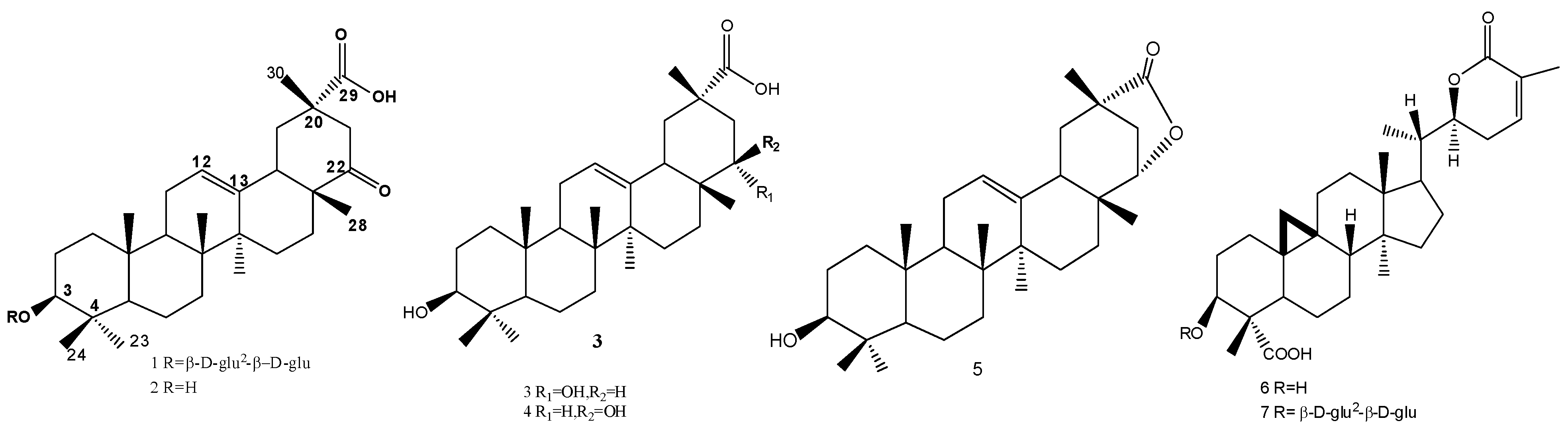
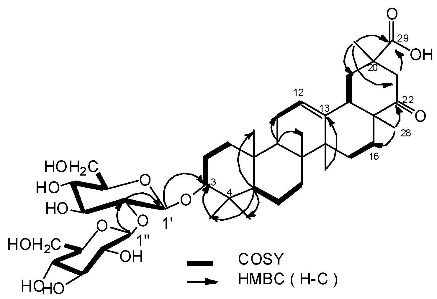
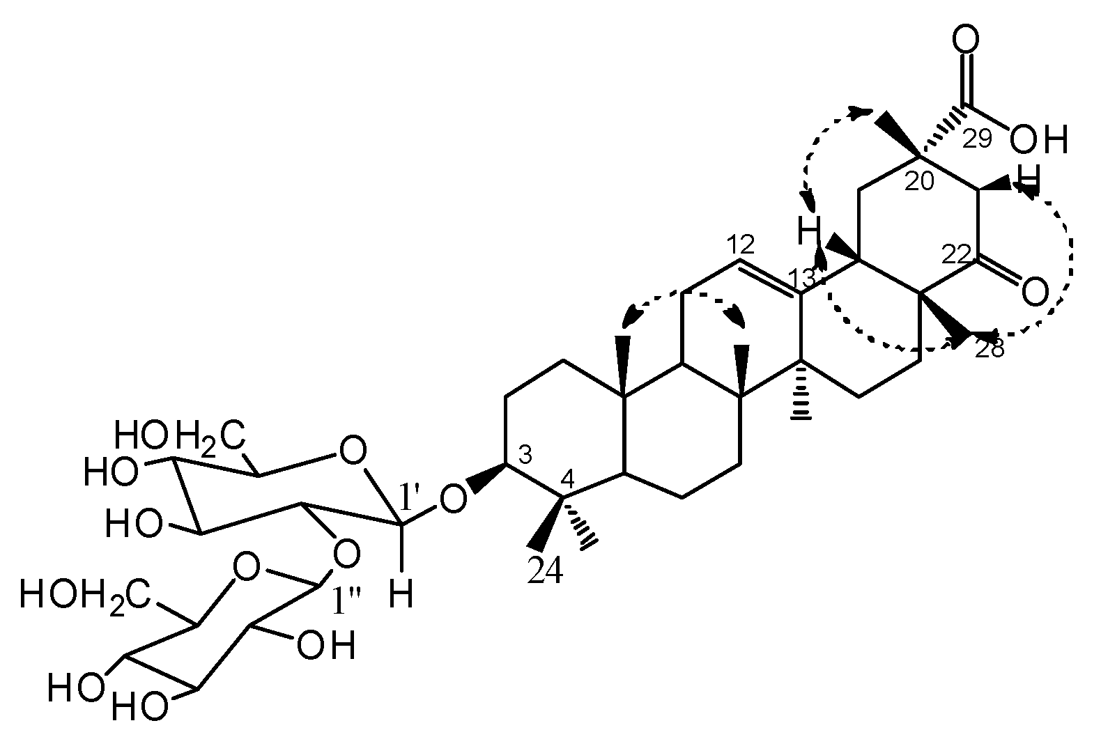
2. Results and Discussion

The aqueous EtOH extract of the Abrus precatorius was suspended in water, and then partitioned with petroleum ether, EtOAc, and n-butanol by liquid-liquid extraction respectively. The EtOAc and n-butanol fractions were successively subjected to repeated silica gel column, Sephadex LH-20 to yield compounds 17 (Figure 1).
Figure 1. Structures of compounds 17.
Molecules 17 00295 g001
Compound 1 had the molecular formula C42H66O14 as deduced from HRESI-MS m/z 817.4345 [M+Na]+ (calcd for C42H66O14Na, 817.4350) and NMR data (Table 1). The IR spectrum exhibited absorption bands at 3458 (OH), 1727(C=O), 1693(C=O) and 1624 (C=C) cm–1. Seven methyl groups (δH 1.42, 1.32, 1.27, 1.23, 1.14, 0.91 and 0.86), one oxygenated methine proton (δH 3.29, dd, J = 5.0, 11.0 Hz), and one olefinic proton (δH 5.31, br s) were observed in the 1H-NMR spectrum. The 13C-NMR and DEPT data confirmed the presence of seven methyl carbons (δC 28.1, 25.5, 21.6, 20.9, 16.8, 16.7, 15.6), two olefinic carbons (δC 124.7, 141.4), one oxygenated methine carbons (δC 89.0), a carbonyl carbon (δC 214.9) and a carboxylic carbon (δC 178.6) (Table 1). The 1H- and 13C-NMR spectra of 1 has the characteristic of Δ12 oleanene skeleton [16]. Comparison the 13C-NMR data of 1 with 2 except for the C-3 signal (δC 89.0) which shifted down field by 11 ppm, others were in accordance with that of 3β-hydroxy-22-oxo-12-oleanen-29-oic acid (2) [11]. The locations of carbonyl carbon and carboxylic carbon could be confirmed by the HMBC correlations from δH 1.23 (Me-28) to δC 214.9, 26.6 (C-16), and δH 1.42 (Me-30) to δC 178.6, 41.6 (C-19), 46.5 (C-21) (Figure 2). Moreover, the 1H and 13C-NMR spectra of 1 showed two sugar anomeric protons at δH 5.26 (1H, d, J = 7.0 Hz) and δH 5.03 (1H, d, J = 7.0 Hz) and carbons at δC 107.2 and 105.3 (Table 1). The monosaccharides were analysed as β-D-glucose with acetylated alditols derivatives by GC using authentic samples as references after hydrolysis of 1. This also could be validated by a combination of the coupling constants (J = 7.0 Hz for H-1″ and J = 7.0 Hz for H-1′) and 1D, 2D-NMR experiments. The signal at δC 89.0 in the 13C-NMR suggesting that the β-D-glucose moieties are linked to the oxygen at C-3 of the aglycon [17].This deduction and sequence of inter-glycosidic linkages were deduced from the following HMBC correlations: H-1′ (δH 5.03) of inner glucose with C-3 (δC 89.0) of sapogenin, H-1′′ (δH 5.26) of terminal glucose with C-2′ (δC 83.9) (Figure 2). The relative configuration of the hydroxylated carbon (C-3) was assigned as β form mainly on the basis of 1H-NMR coupling (1H, dd, J = 5.0, 11.0 Hz, H-3) [17] and by comparison with 2. In NOESY spectrum, the key NOE correlations of H-28/H-21β, H-28/H-18 and H-30/H-18, showed that H-30, H-28, H-21β, H-18 were on the same face, so the relative stereochemistry were determined (Figure 3).
Table 1. 1H- (500 MHz) and 13C-NMR (125 MHz) data of compound 1 (in Pyr-d5, δ in ppm, J in Hz).
No.δCδHKey HMBC (H to C)
138.7 CH21.40 (1H, m, H-1a)0.82 (1H, m, H-1b)C-2,C-3
227.3 CH22.24 (2H, m, H-2)C-1,C-3
389.0 CH3.29 (1H, dd, J = 5.0, 11.0 Hz, H-3)C-1′, 23, 24
439.5 qC
555.6 CH0.71 (1H, br d, J = 11.5 Hz, H-5)C-23,24,25
618.4 CH21.64 (1H, m, H-6a),C-24
1.46 (1H, m, H-6b)
732.8 CH21.48 (1H, m, H-7a),
1.29 (1H, m, H-7b)
839.9 qC
947.6 CH1.56 (1H, m, H-9)
1036.8 qC
1123.8 CH21.84 (2H, m, H-11)
12124.7 CH5.31 (1H, br s, H-12)
13141.4 qC
1441.9 qC
1525.5 CH21.68 (1H, m, H-15a),
0.98 (1H, m, H-15b)
1626.6 CH21.89 (1H, m, H-16a),C-28
1.28 (1H, m, H-16b)
1748.2 qC
1847.0 CH2.54 (1H, m, H-18)
1941.6 CH22.88 (1H, t, J = 13.5 Hz, H-19a),C-30
1.97 (1H, br d, J = 12.0 Hz, H-19b)
2044.6 qC
2146.5 CH23.46 (1H, d, J = 14.5 Hz, H-21a),C-30
2.71 (1H, br d, J = 14.0 Hz, H-21b)
22214.9 qC
2328.1 CH31.32 (3H, s, Me-23)C-3
2415.6 CH30.86 (3H, s, Me-24)C-3
2516.7 CH30.91 (3H, s, Me-25)
2616.8 CH31.14 (3H, s, Me-26)
2725.5 CH31.27 (3H, s, Me-27)C-13
2820.9 CH31.23 (3H, s, Me-28)C-22,C-16
29178.6 qC
3021.6 CH31.42 (3H, s, Me-30)C-29,C-19,C-21
glu
1′105.3 CH5.03 (1H, d, J = 7.0 Hz, H-1′)C-3
2′83.9 CH4.33 (1H, t, J = 8.0 Hz, H-2′)C-1′,C-1″,C-4′
3′77.7 CH 4.40 (1H, br d, J = 8.0 Hz, H-3′)
4′73.1 CH4.64 (3H, m, H-4′, 2″, 3″)
5′74.7 CH 4.20 (1H, br d, J = 9 Hz, H-5′)
6′61.3 CH24.44 (2H, m, H-6′)
glu
1″107.2 CH5.26 (1H, d, J = 7.0 Hz, H-1″)C-2′,C-3″
2″74.9 CH4.64 (3H, m, H-4′, 2″, 3″)C-4″
3″77.4 CH4.64 (3H, m, H-4′, 2″, 3″)
4″69.5 CH4.73 (1H, m, H-4″)
5″76.9 CH4.09 (1H, t, J = 6.1 Hz, H-5″)
6″61.3 CH24.67 (2H, m, H-6″)
Figure 2. Key HMBC and COSY correlations of 1.
Molecules 17 00295 g002
Figure 3. Key NOESY correlations of 1.
Molecules 17 00295 g003
All the above data identified 1 as 3-O-β-D-glucopyranosyl-(1→2)-β-D-glucopyranosyl subprogenin D. The structures of known compounds 27 were confirmed by detailed NMR data comparison with those in the literature [11,12,13,14,15].
The cytotoxicity of 17 against MCF-7, SW1990, Hela, Du-145 cancer cell lines were evaluated with 5-FU (5-Fluorouracil) and DOX (doxorubicine) as positive controls. Compound 5 showed moderate cytoxicity against SW1990, Hela, Du-145 cancer cell lines, and compound 6 showed moderate cytoxicity against MCF-7, SW1990, Du-145 cancer cell lines, whereas other compounds had no significant activity (Table 2).
Table 2. Cytotoxicity of 17 against four cancer cell lines.
Cytotoxicity (IC50 [μg/mL])(mean ± SD%)
MCF-7SW1990HelaDu-145
1-a---
2----
3----
4----
5-5 ± 0.3210 ± 0.895 ± 0.40
64 ± 0.182 ± 0.09-2 ± 0.08
7----
DOX1 ± 0.06 2 ± 0.16 1 ± 0.05
5-Fu 10 ± 0.95
a No significant activity at 10 μg/mL.

3. Experimental

3.1. General

1D and 2D NMR spectra were recorded on a Bruker-AV-500 spectrometer with TMS as internal standard. HRESIMS were measured with MAT 95XP mass spectrometer. IR were recorded on FT-IR Nicolet 6700. UV spectra were obtained on a Beckman DU-640 UV spectrophotometer. Optical rotations were measured with a Perkin-Elmer 341 plus. GC were run on a QP2010PLUS (Shimadzu Corporation) equipped with an ACQ mass spectrometer. For column chromatography (CC), silica gel (200–300 mesh) and GF254 for TLC were obtained from the Qingdao Marine Chemical Factory, Qingdao, China.

3.2. Plant Material

The leaves and stems of Abrus precatorius were collected in October 2010 from the mangrove wetlands of Hainan Island, China. The identification of the plant was performed by Professor Si Zhang. A voucher sample (No. 20101001) is maintained in the Key Laboratory of Marine Bio-Resources Sustainable Utilization, South China Sea Institute of Oceanology, Chinese Academy of Sciences, China.

3.3. Extraction and Isolation

The air-dried leaves and stems of Abrus precatorius (8 kg) were extracted with EtOH (95%, 20 L) three times (7 days each time) at room temperature. The combined extract was evaporated in vacuo, suspended in water, and then successively partitioned with petroleum ether, EtOAc, and n-butanol (800 mL × 3). The EtOAc and n-butanol fractions were concentrated to afford 53.7 g and 108 g of residues, resp. The EtOAc extract was subjected to silica gel CC using a gradient elution of CHCl3-MeOH (100:0–1:1) to afford ten fractions (Frs. AJ). Compound 3 (11 mg) was crystallizated in the bottle when eluted with the solvent CHCl3-MeOH 90:10 (Frs. B) and then purified with MeOH. Frs. B (7 g) was subjected to CC and eluted with CHCl3-acetone (30:1, 20:1, 15:1, 10:1, 5:1, each 1500 mL) to give six fractions (B1B6), B3 was purified by Sexphadex LH-20 (CHCl3-MeOH 1:1) to yield 2 (8 mg). Compound 4 (10 mg) was isolated from B1 by repeated chromatographic on Sephadex LH-20 (CHCl3-MeOH 1:1) and silica gel column (CHCl3-MeOH 80:1). Frs. C (4.4 g) was subjected to CC with gradient eluting of CHCl3-acetone (30:1, 20:1, 15:1, 10:1, 5:1, each 1,000 mL) to give six fractions (C1C6). Compound 5 (7 mg) was crystallizated from C2 when eluted with the solvent CHCl3-acetone 20:1, and then recrystallizated with MeOH. C4 was purified by Sexphadex LH-20 (CHCl3-MeOH 1:1) to give compound 6 (7 mg). Frs. G (2.36 g) was subjected to CC with gradient eluting of CHCl3-MeOH (30:1, 20:1, 15:1, 10:1, 5:1, each 500 mL) and purification on Sephadex LH-20 to yield 7 (20 mg). The n-BuOH extract (108 g) was subjected to CC on Amberlite XAD using MeOH-H2O (20%, 40%, 60% and 95%). The 40% extract part (7.23 g) was fractioned on silica gel column eluting with CHCl3-MeOH-H2O 9:1:0.1 to give ten fraction HQ. Fraction Q (0.4265 g) was purified by Sexphadex LH-20 (CHCl3-MeOH 1:1) to give compound 1 (5 mg). Yellow powder: Molecules 17 00295 i001 = −10 (c = 0.04, MOH), UV (MeOH) λmax 255 nm, IR (KBr) νmax: 3458, 2946, 1727, 1693, 1624, 1466, 1383, 1211, 1042 cm1; HRESI-MS m/z 817.4345 [M+Na]+ (calcd for C42H66O14Na, 817.4350). 1H and 13C-NMR data see Table 1.

3.4. Acid Hydrolysis of 1

Compound 1 (2 mg) was added into 3 N HCl (0.5 mL) and refluxed for 5 h in a water bath (100 °C). The solution was neutralized and extracted with EtOAc to afford the aglycon. The aglycon of 1 found to be identical with 2 by TLC. The sugars released were converted into acetylated alditols by reduction with NaBH4 followed by acetylation with acetic anhydride-pyridine mixture. The alditol acetates derivatives obtained were analyzed by GC using a GCMS-QP2010 Plus: The injector temperature was set at 250 °C and the column temperature program was as follows: The initial temperature of 200 °C was held constant for 2.5 min and then increased by 5 °C min to the final temperature of 250 °C. The detector temperature was set at 280 °C. MS-Scan: ACQ mode, event Time: 0.50 s with 1,000 scan speed. Alditol acetates were identified by comparison of their retention times with those of authentic samples [16].

3.5. Cytotoxicity Assays

Cytotoxicity was evaluated by the MTT [3-(4,5-dimethylthiazol-2-yl)-2,5-diphenyltetra-zolium bromide] method using MCF-7, SW1990, Hela and Du-145 cell lines. Details of the assays were described in a previous report [18].

4. Conclusions

The new compound 3-O-β-D-glucopyranosyl-(1→2)-β-D-glucopyranosyl subprogenin D (1) was isolated from Abrus precatorius, together with six known triterpenoids. Compounds 5 and 6 showed moderate cytoxicities against MCF-7, SW1990, Hela and Du-145. However, the new compound 1 and the other known ones had no significant activity.

Acknowledgements

The authors are grateful to the National Basic Research Program of China (973 Program) (2010CB833800) and the Chinese National Natural Science Fund (No. 40906076, 40906075) for financial support. The cytotoxicity assays were performed at the Department of Biochemistry and Molecular Biology, College of Basic Medical Sciences, Second Military Medical University, Shanghai, China.

References and Notes

  1. Ma, C.M.; Nakamura, N.; Hattori, M. Saponins and C-glycosyl flavones from the seeds of Abrus precatorius. Chem. Pharm. Bull. 1998, 46, 982–987. [Google Scholar] [CrossRef]
  2. Dnyaneshwar, J.T.; Ravindra, Y.P. Effect of Abrus precatorius leaves on milk induced leukocytosis and eosinophilia in the management of asthma. Asian Pac. J. Trop. Med. 2012, S40–S42. [Google Scholar]
  3. Ghosal, S.; Dutta, S.K. Alkaloids of Abrus precatorius. Phytochemistry 1971, 10, 195–198. [Google Scholar] [CrossRef]
  4. Markham, K.R.; Wallace, J.W.; Babu, Y.N.; Murty, V.K.; Rao, M.G. 8-C-Glucosylscutel larein 6,7-dimethyl ether and its 2″-O-apioside from Abrus precatorius. Phytochemistry 1989, 28, 299–301. [Google Scholar]
  5. Namcheol, K.; Darrick, S.H.L.K.; Kinghorn, D.A. New triterpenoids from the leaves of Abrus precatorius. Nat. Prod. Lett. 2001, 16, 261–266. [Google Scholar]
  6. Qing, S.C.; Hu, Z.B. Abruquinone A, B, D, E, F and G from the root of Abrus precatorius. Acta Bot. Sin. 1998, 40, 734–739. [Google Scholar]
  7. Anam, E.M. Anti-inflammatory activity of compounds isolated from the aerial parts of Abrus precatorius (Fabaceae). Phytomedicine 2001, 8, 24–27. [Google Scholar] [CrossRef]
  8. Yadava, R.N.; Sudhan Reddy, V.M. A new biological activity flavonol glycoside from the seeds of Abrus precatorius linn. J. Asian. Nat. Prod. Res. 2002, 4, 103–107. [Google Scholar] [CrossRef]
  9. Kuo, S.C.; Chen, S.C.; Chen, L.H.; Wu, J.B.; Wang, J.P.; Teng, C.M. Potent antiplatelet, anti-inflammatory and antiallergic isoflavanquinones from the roots of Abrus precatorius. Planta Med. 1995, 61, 307–312. [Google Scholar] [CrossRef]
  10. Dimetry, N.Z.; El-Gengaihi, S.; Reda, A.S.; Amer, S.A.A. Biological effects of some isolated Abrus precatorius L. alkaloids towards Tetranychus urticae Koch. J. Pest. Sci. 1992, 65, 99–101. [Google Scholar]
  11. Takeshito, T.; Yokoyama, K.; Ding, Y.; Kinjo, J.; Nohara, T. Four new and twelve known sapogenols from Sophorae subprostratae radix. Chem. Pharm. Bull. 1991, 39, 1908–1910. [Google Scholar] [CrossRef]
  12. Chiang, T.C.; Chang, H.M.; Mak, T.C. New oleanene-type triterpenes from Abrus precatorius and X-ray crystal structure of abrusgenic acid-methanol 1:1 solvate. Planta Med. 1983, 49, 165–169. [Google Scholar] [CrossRef]
  13. Zhang, C.P.; Zhang, Y.G.; Lv, X.Y.; Chen, Y.; Ma, P.C.; He, C.H.; Yu, D.Q.; Shen, F.L.; Yang, J.J.; Yang, J.; et al. Studes on triterpenoids of total glucosides of Tripterygium wilfordii (TII). Acta Acad. Med. Sin. 1989, 5, 322–325. [Google Scholar]
  14. Chang, H.M.; Chiang, T.C.; Thomas, C.W.M. Isolation and structure elucidation of abruslactone A: A new oleanene-type triterpene from the roots and vines of Abrus precatorius L. J. Chem. Soc. Chem. Commun. 1982, 20, 1197–1198. [Google Scholar]
  15. Choi, Y.H.; Hussain, R.A.; Pezzuto, J.M.; Kinghorn, A.D.; Morton, J.F. Abrusosides A–D, four novel sweet-tasting triterpene glycosides from the leaves of Abrus precatorius. J. Nat. Prod. 1989, 52, 1118–1127. [Google Scholar] [CrossRef]
  16. De Rosa, S.; Iodice, C.; Mitova, M.; Handjieva, N.; Popov, S.; Anchev, M. Triterpene saponins and iridoid glucosides from Galium rivale. Phytochemistry 2000, 54, 751–756. [Google Scholar] [CrossRef]
  17. Debellaa, A.; Haslingera, E.; Schmida, M.G.; Bucard, F.; Michlb, G.; Abebec, D.; Kunert, O. Triterpenoid saponins and sapogenin lactones from Albizia Gummifera. Phytochemistry 2000, 53, 885–892. [Google Scholar] [CrossRef]
  18. Wang, F.Z.; Tian, X.P.; Huang, C.G.; Li, Q.X.; Zhang, S. Marinactinones A–C, new γ-pyrones from marine actinomycete Marinactinospora thermotolerans SCSIO 00606. J. Antibiot. 2011, 64, 189–192. [Google Scholar] [CrossRef]
  • Sample Availability: Not availbale.

