2.1. Conjugation of an Anti-GRP78 scFv with Quantum Dot-625 Nanoparticle
We have conjugated scFv-GRP78-H19 [
10] to quantum dot-625 (Invitrogen) with an antibody conjugation kit according to the manufacturer’s instructions. The Q-dot- scFv-GRP78-H19 Conjugates(Qdot-GRP78) were then concentrated by ultrafiltration and purified by size exclusion chromatography with a molecular weight of over 220 kDa being observed (
Figure 1A). We have used both dot-blot and Western blot to test the specificity of the antibody to GRP78. In the dot-blot experiments, BSA and murine GRP78 (200 μg/mL) were spotted on a nitrocellulose filter and probed with 10 nM Qdot-GRP78 antibody overnight with PBSA solution in 1% nonspecific rabbit serum. We observed a very strong intensity of red dots under UV transilluminator (UVP gel document system, Cambridge, UK) on the GRP78 protein spot (
Figure 1B); no signal was detected on the control BSA spot.
Figure 1.
scFv-Grp78-H19 antibody was conjugated to a Qdot-625. (A) Coomassie blue-stained SDS-PAGE gel showing the Qdot labeled scFv; (B) Dot blot analysis of GRP78 protein binding by the Qdot625 labeled scFv; (C) Western blot analysis of GRP78 protein binding with Qdot625-GRP78 antibody with recombinant protein or whole cell extract (WCE) (upper panel). The same blot was hybridized with a goat-anti-GRP78 polyclonal antibody [N20, detected by horseradish peroxidase chemiluminescence (lower panel)]; (D) GRP78 protein was diluted in electrophoresis buffer and serial dilutions were then prepared and the western blot was probed with either anti-GRP78-Qdot625 scFv antibody (detected by UV illumination) or anti-GRP78-C20 polyclonal antibody (detected by horseradish peroxidase chemiluminescence).
We next performed a polyacrylamide gel electrophoresis on recombinant GRP78 protein and whole cell extracts (WCE) of human breast cancer MDA-MB-231/GFP cell samples. The gel was directly stained with 10 nM Qot-GRP78 antibody overnight. Again, we observed a very strong intensity red band in the lane which contained recombinant GRP78 protein under UV illumination. No signal was detected in the BSA lane (
Figure 1C, upper panel). We also detected the endogenous GRP78 signal from whole cell extracts of MDA-MB-231/GFP cells. It should be noted that the endogenous GRP78 band migrates more slowly than the recombinant GRP78 bands which contains the 6 HIS-tag. To corroborate the gel staining results, we made the Western blot with the same gel. As shown on the lower panel of
Figure 1C, the GRP78 band from the whole cell extract migrated fairly slower than that of the recombinant GRP78. To compare the sensitivity of the Qdot probe with a conventional Western blot, GRP78 protein was diluted in electrophoresis buffer and subjected to Western blot analysis with either Qdot-GRP78 antibody or the goat polyclonal anti-GRP78 antibody with conventional secondary antibody: HRP-conjugated Anti-Goat IgG (
Figure 1D). Both methods detected protein orders ranging from 0.25 μg to 0.03 μg, with Qdot antibodies only requiring a single step, while conventional ECL detection needs multiple steps and enzymatic amplification.
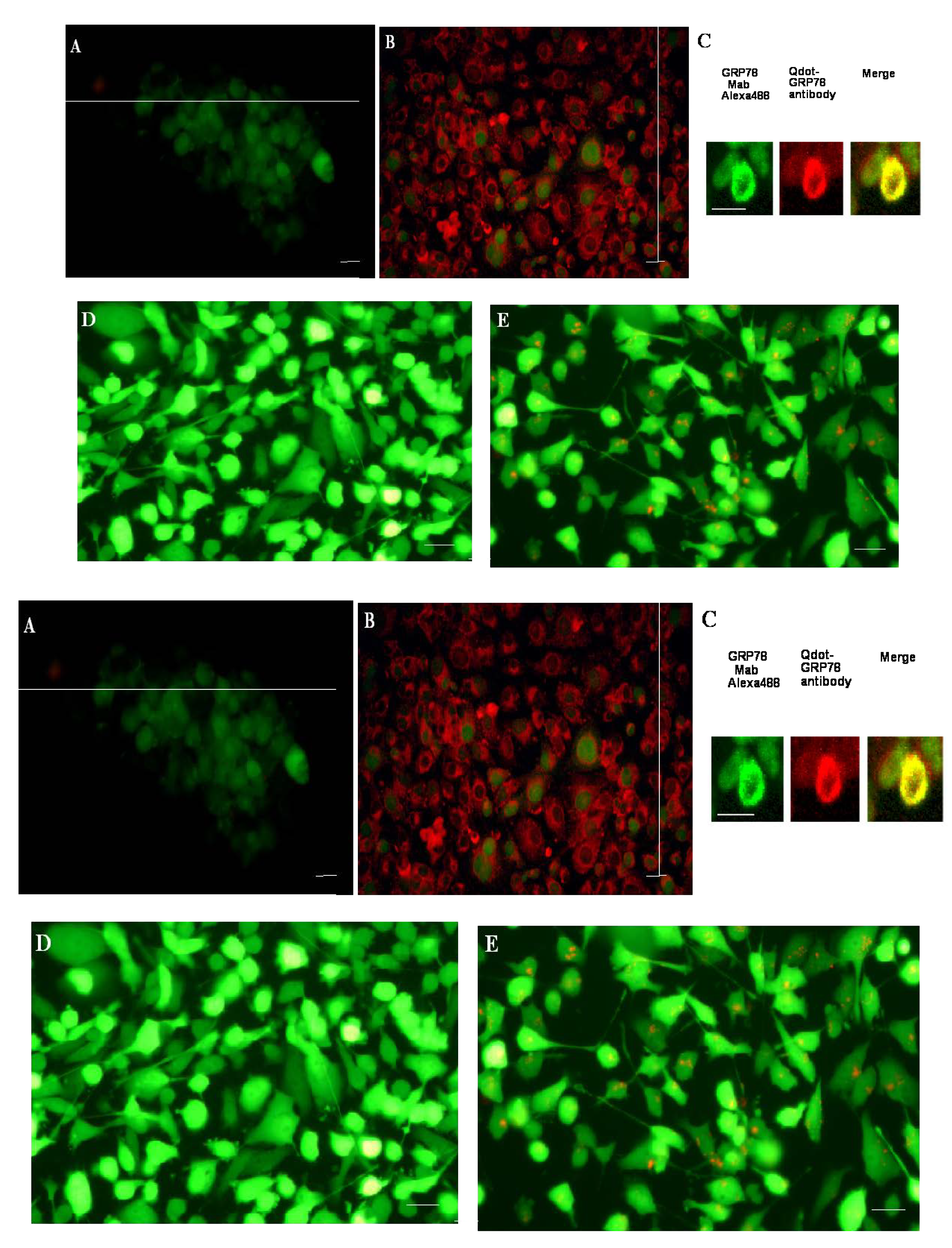
We then performed immunocytochemistry on breast cancer MDA-MB-231/GFP cells using Qdot-GRP78 antibody. As shown in
Figure 2B, the antibody stained both the membrane and ER in the breast cancer cells, with no specific staining in the control cells in which the unlabeled Qdot-625 nanobeads were used (
Figure 2A). To further confirm GRP78 staining of the antibody, we colocalized our Qdot antibody with an anti-GRP78 mouse monoclonal antibody in the LNCaP prostate cancer cell (
Figure 2C). Mouse anti-GRP78 can be detected with fluorescence Alexa488 labeled secondary Goat anti-mouse IgG (green), while our Qdot antibody can be detected by red fluorescence. Scan confocal microscopy imaging in LNCaP prostate cancer cell shows the overlapping between the two antibodies (yellow). In order to determine if Qdot-GRP78 antibody was capable of being internalized, the labeled antibody was incubated with MDA-MB-231/GFP cells for 24 h, and after washing with PBS cells were directly examined using a fluorescence microscope. Internalisation of the Qdot-GRP78 antibody (red dots) into the MDA-MB-231/GFP cells was observed in more than 30% cells (
Figure 2E), while no specific staining was observed in unlabeled Qdot-625 nanobeads (
Figure 2D), indicating efficient internalization of the Qdot-GRP78 antibody. Confocal microscopy was used to examine the details of the Qdot internalization. While no specific red fluorescence was detected in unlabeled Qdot-625 treated samples (
Figure 3F), the Qdot-GRP78 (red,
Figure 2G) was internalized in MDA-MB-231/GFP cells.
Figure 2H shows the same image of the 2G, with 3D reconstructed scanning confocal images. In Western blot analysis (
Figure 2I), we found that after treating cells with Qdot-GRP78 antibody for 16 h, the phosphorylated AKT-serine 473 expression level has significantly increased in comparison with that of unlabeled Qdot-625 treated samples, while pan-AKT level remains the same. To detect cell apoptosis, MDA-MB-231/GFP cells were incubated with various concentrations of Qot-GRP78 antibody for 24 h. The cells were then stained with Hoechst DNA dye H33342 (10). We found that there was a dose-dependent increase apoptosis in Qdot-GRP78 treated cells (
Figure 2J).
Figure 2.
Immunohistochemistry of Qdot-GRP78 antibody in MDA-MB-231/GFP breast cells and LNCaP prostate cancer cells. (A) and (B) Fluorescence images of cells staining with Qdot625-GRP78 antibody. (A) control cells staining with unlabeled nanobeads; (B) MDA-MB-231/GFP cells staining with Qdot-GRP78 antibody; (C) Scanned confocal microscopy imaging in LNCaP prostate cancer cell, stained with anti-GRP78 mouse monoclonal antibody with secondary Goat anti-mouse IgG labeled with Alexa488(green)and Qdot-GRP78(red dots) and overlapped with two probes (yellow); (D) and (E) Internalization of Qdot-GRP78 antibody by MDA-MB-231/GFP cells. Cells were incubated with unlabelled nanobeads. (D) or Qdot-GRP78 antibody; (E) at 37 °C for 16 h. Cells were then washed with PBS and analyzed by fluorescence microscopy; (F) Scanned confocal microscopy imaging in MDA-MB-231/GFP cell (green), treated with control unlabelled beads; (G) Qdot-GRP78(red dots) was detected inside MDA-MB-231/GFP cell; (H) 3D reconstruction of confocal Z stack with 0.8-μM, GFP cell showing in green channel, while Qdot-GRP78 showing in red channel. Scale bar represents 20 μm; (I) Western blot analysis of GRP78 protein in Qdot-GRP78 antibody treated cells. Cells either treated with control (unlabelled Qdot) or Qdot-labeled antibody. Phosphorylated Akt-ser473 protein was detected by an anti-anti-Akt-se473 antibody and Pan-Akt antibody was used as a loading control. The western blot represents three independent experiments. (J) MDA-MB-231/GFP cells were incubated with various concentrations of Qot-GRP78 antibody for 24h. Apoptotic nuclei or nuclear DNA strand breaks were visualized using Hoechst DNA dye H33342 (10). A minimum of 200 cells were counted in each sample and condensed or fragmented nuclei were expressed as a percentage of the total number of nuclei. Values are presented as mean ± S.D. of three determinations. * indicates significant difference (p < 0.05) between none treatment cells and antibody treatment cells.
![Molecules 17 00796 g002]()
2.2. QD scFv Bioconjugates Inhibit Breast Cancer Growth in a Xenograft Model
The advantage of the Qdot-conjugated antibody is that it can be easily used in multi-color experiments, where we can detect and monitor the antibody/antigen interactions
in vivo. In this study we used GFP labeled breast cancer MDA-MB-231/GFP cells, subcutaneously transplanted into the nude mice. When the tumor reached a volume of approximately 50–100 mm
3 within a two to three week period, Qdot-625-scFv-GRP78 (80 μL containing 80 nM antibody) was intratumorally injected into the pre-established GFP labeled tumors (
Figure 3). Tumor growth and antibody distribution can be monitored by visualization of multi-color fluorescence imaging (green representing tumor, red representing antibody) with a single excitation source (470 nm excitation filter) and single emission filter (515 nm viewing filter; LightTools Research, Encinitas, CA, USA) (
Figure 3A–E). A total of three injections were carried out at weekly intervals. Treatment of established tumors with the Qdot-GRP78 scFv antibody-conjugates significantly inhibits the breast tumour growth (191.1 ± 150 mm
3,
Figure 3E,G) in comparison with the samples treated only with unlabeled nanobeds (717 ± 335 mm
3,
Figure 3F,G.) seven weeks following implantation) which represents a 74% reduction in tumor volume (Students
t test,
p < 0.05).
At the end of seven weeks, three mice treated with Qdot-625-scFv-GRP78 antibody still showed a measurable tumor mass. The red fluorescence of scFv-GRP78 was detected in some of the cells inside tumors under fluorescence microscope (
Figure 4). It should be noted that we were unable to detect any red fluorescence cells in other organs (kidney, heart, liver, spleen and lung,
Supplementary Figure S1).
Figure 3.
Qdot-GRP78 antibody-conjugates inhibits breast cancer growth. Two-color fluorescence imaging of effect of the injected antibody(red) and GFP tumor(green). (A) before injection; (B) two days after injection; (C) five days after injection; (D) two weeks after injection; (E) five weeks after injection; (F) five weeks after unlabeled nanobeads only injection; (G) Total 2 × 10⁶ tumor cells were injected subcutaneously into each Balb/c nu/nu mouse. Each experiment used five female mice. In the treatment group, the Qdot-GRP78 antibody was intratumorally injected into pre-established tumors (on the second week) (white square). Then injected at weekly interval for three weeks. In control groups, mice received unlabelled nanobeads alone (black circle. * p < 0.05, two-tailed Student’s t test. Scale bar represents 5 mm.
Figure 4.
Qdot-GRP78 antibody detected in the tumor samples. The red fluorescence of Qdot-GRP78 antibody was detected in some of the cells inside tumor. Scale bar represents 20 μm.
Figure 4.
Qdot-GRP78 antibody detected in the tumor samples. The red fluorescence of Qdot-GRP78 antibody was detected in some of the cells inside tumor. Scale bar represents 20 μm.
2.3. Discussion
Recent groundbreaking work has established that GRP78 protein, a master switch in ER stress, plays a crucial role in cancer cell proliferation, angiogenesis and chemotherapy drug resistance [
7]. Cell surface Grp78 expression has been detected in many different cancers, such as breast, liver, and prostate cancer [
7,
11]. Previously we reported that nitric oxide induced coupling of mitochondrial respiration to ER-stress, inducing GRP78 expression and subsequent regulation [
9]. Furthermore, we have targeted membrane-bound GRP78 by subtractive screening of a single chain variable fragment (scFv) library and successfully isolated a panel of scFv specific for GRP78 [
10]. The isolated scFv-GRP78 detected breast cancer cell surface Grp78 (Figure S2A and membrane locations are indicated by arrows) on the breast carcinoma cells (Tx N1, stage II), as well as cancer cell surfaces in metastatic lymph nodes (Figure S2C,) in a high- density paraffin-embedded breast cancer microarray (Clinomics Biosciences, Inc). The epitope region of the antibody is on the N-terminus of GRP78 [
10]. Recent research has shown that both the C-terminus and N-terminus of GRP78 play important roles in ER-stress response signalling pathways. Misra
et al. demonstrated that antibodies directed against the COOH-terminal domain of GRP78 up-regulated the tumor suppressor protein p53 [
12], while several other studies have shown that antibody directed against the NH (2)-terminal domain regulated the prostate apoptosis response-4(par-4) signaling pathway [
13] and Cripto pathways [
14]. Burikhanov
et al. showed that antibody against the N-terminus of GRP78 neutralized cellular apoptosis induced by the
par-4, a cancer cell-selective pro-apoptotic protein [
13]. Furthermore, neutralizing antibody against the N-terminus of GRP78 inhibited oncogenic
Cripto signaling via MAPK/PI3K and Smad2/3 pathways [
14].
Recently, down-regulation of GRP78 by SiRNA in different human cell lines, such as JAR (human choriocarcinoma cell) and HUVECs (human umbilical vein endothelial cells, have shown p-AKT upregulation [
15]. p-AKT activation is well known to be involved in cell survival. But, as Yoeli-Lerner
et al. pointed out p-AKT also blocked cancer invasion in MDA-MB-435, MDA-MB-231 and SUM-159-PT breast cancer cells [
16]. Furthermore, activation of AKT has been found to suppress tumor invasion and metastasis in an
in vivo transgenic model [
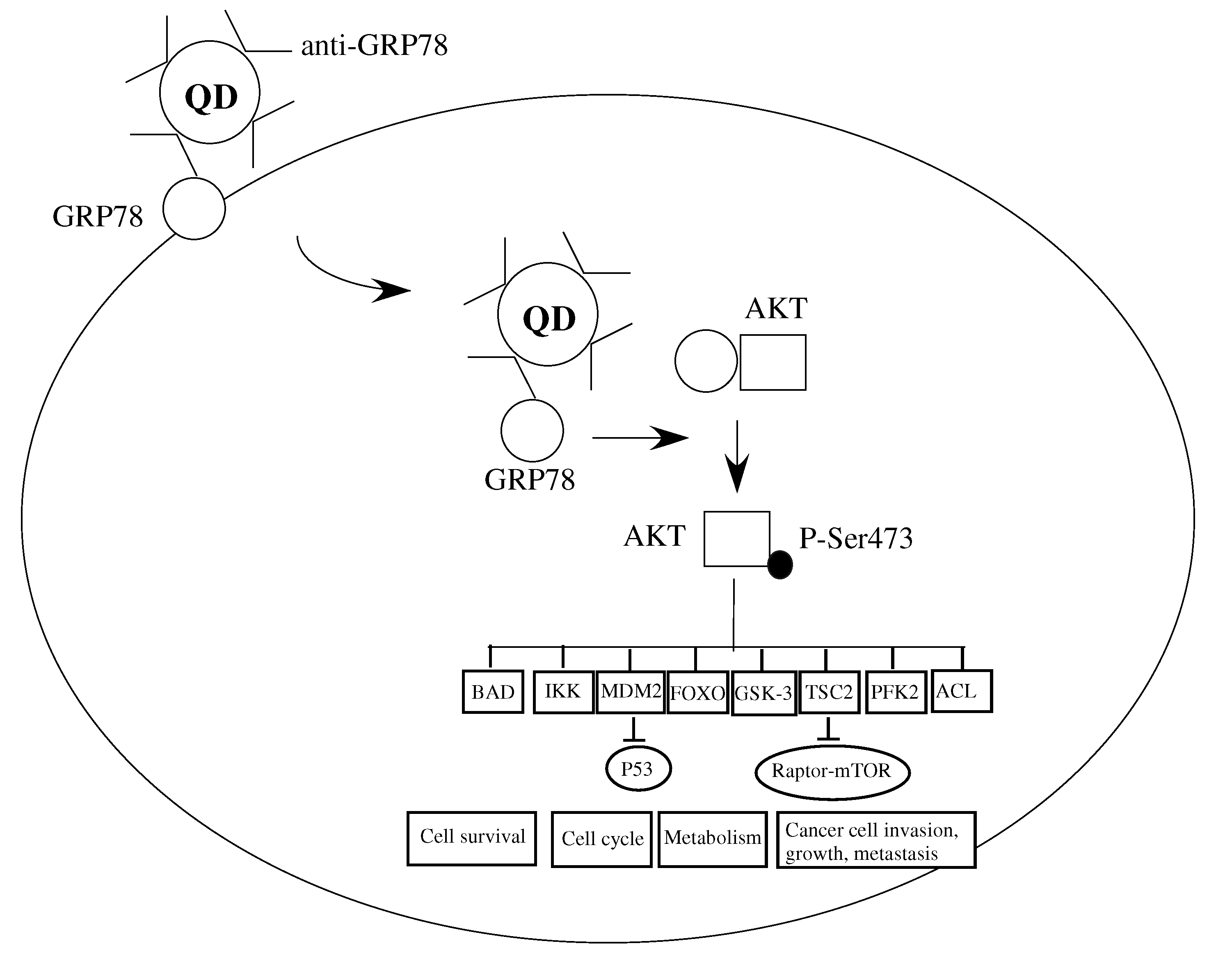
17]. We propose the following mechanism for cancer growth inhibition by our Qdot-GRP78 antibody. The antibody binds cell surface GRP78 and is transported into the cytoplasma in the complex form. The GRP78:Qdot-GRP78 antibody complex prevents GRP78 binding to AKT, leading the AKT-Ser473 phosphorylation, which in turn, regulates a wide range of downstream events, including cancer cell invasion, growth and metastasis (
Figure 5).
Figure 5.
Schematic illustration of the Qdot-GRP78-scFv and GRP78 complex. The complex enters the intracellular space through GRP78 movement from cell into cytoplasm. The Qdot-GRP78-scFv:GRP78 complex prevents GRP78 binds to AKT, freeing AKT to be phosphorylated at Ser374, thereby mediating its downstream target processing including cancer cell invasion and growth.
Recent rapid progresses in nanotechnology have had a great impact on cancer research and other clinical research [
18]. Quantum dots have relatively long half life and stability [
2], therefore conjugating QD with scFV could lead to scFv stabilisation. Most reports did not demonstrate the adverse effects of Qdots on cell viability, morphology and function over the duration of the experiments [
2], other share suggested at a relatively high dose, Qdots may affect organ development [
19]. Nevertheless the improved Qdot preparation will open up a new avenue for its therapeutic usage in medicine [
20].


