Synthesis and Anticancer Activity of Di(3-thienyl)methanol and Di(3-thienyl)methane

Abstract
:1. Introduction
2. Results and Discussion
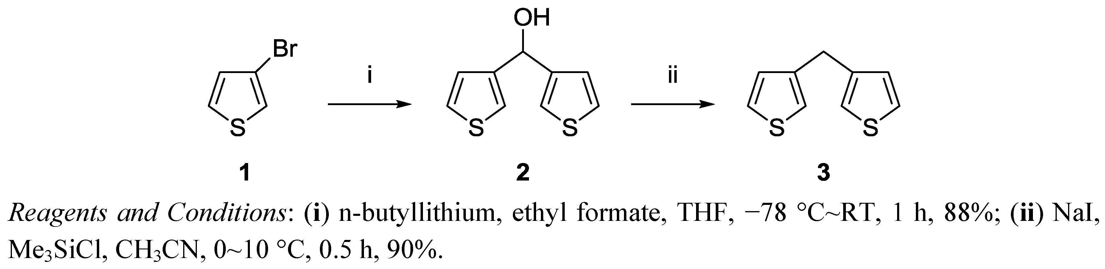
2.1. Synthesis
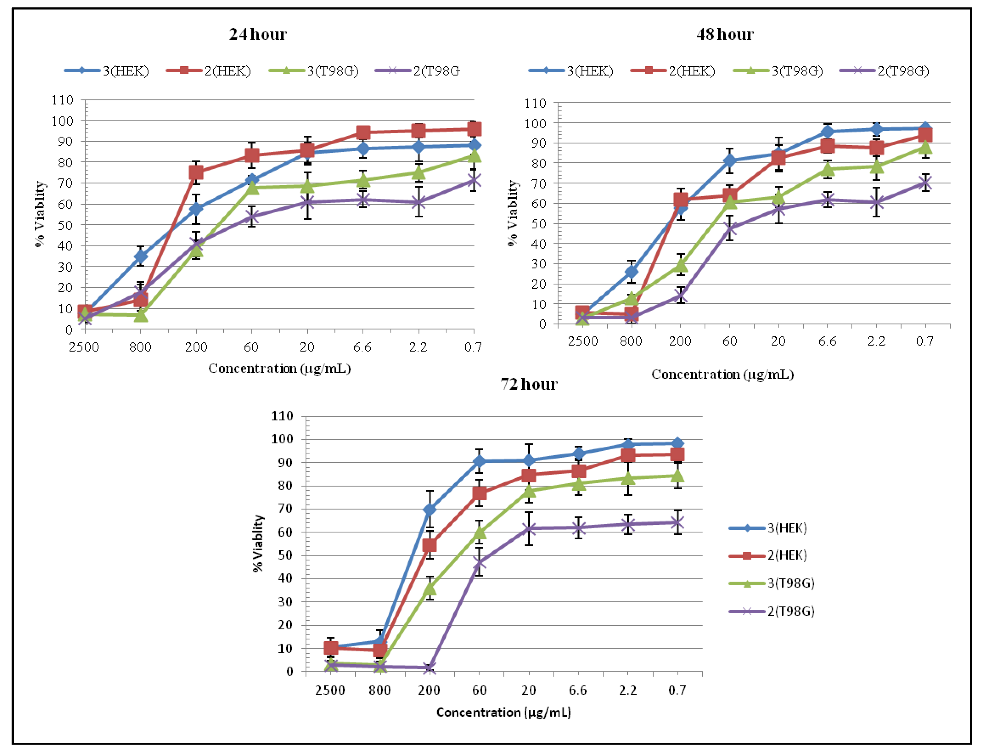
2.2. MTT Assay
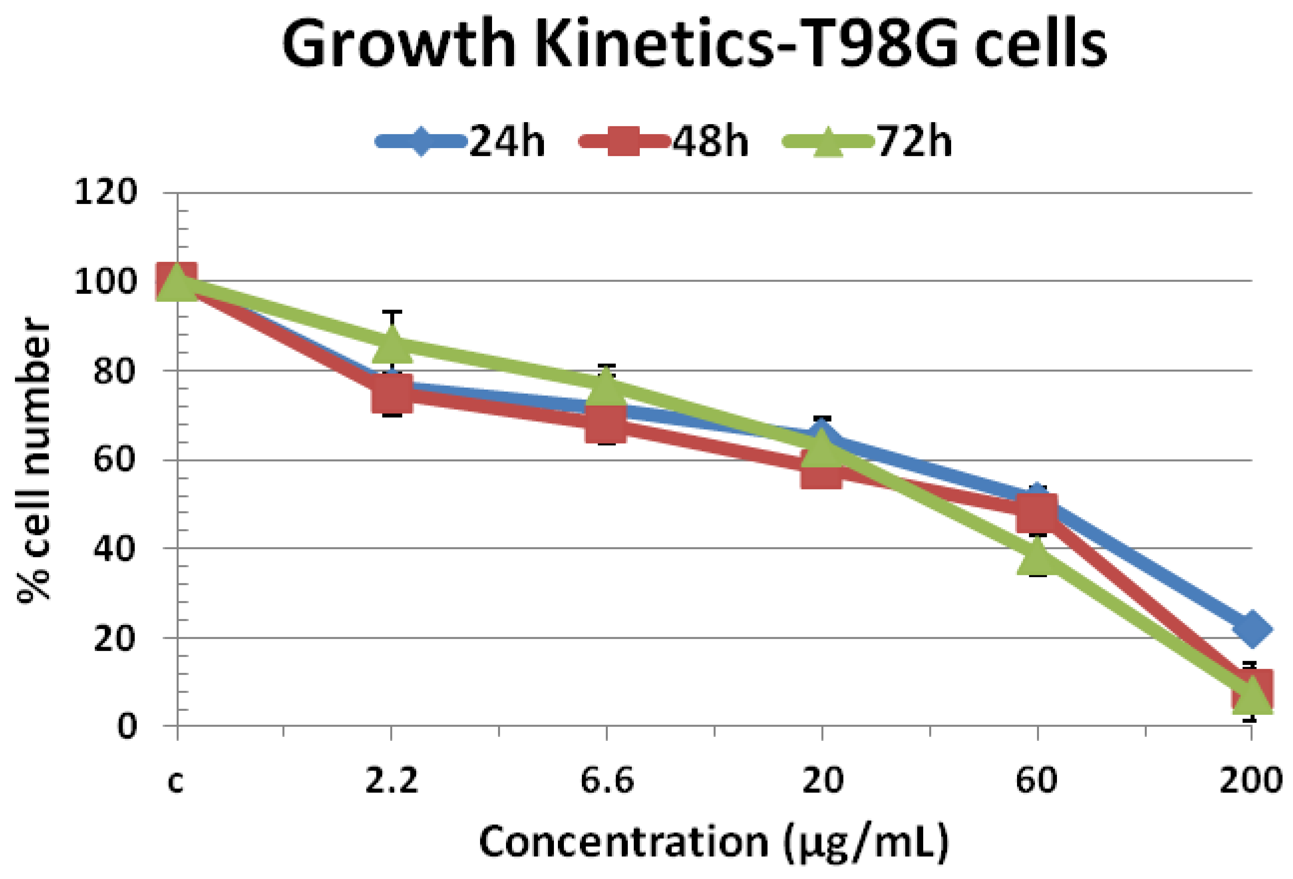
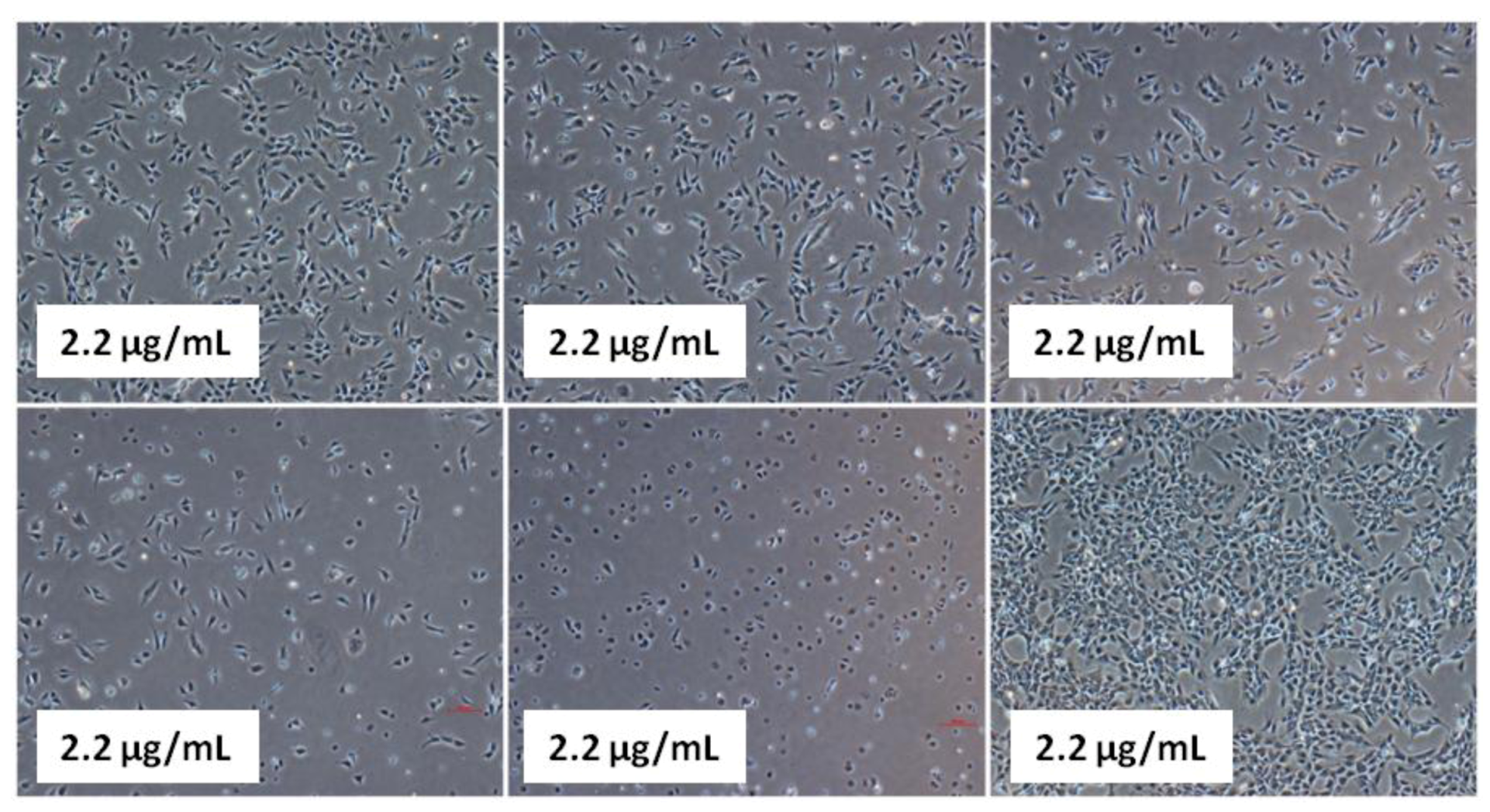
2.3. Growth Kinetics Assay

2.4. Clonogenic Assay

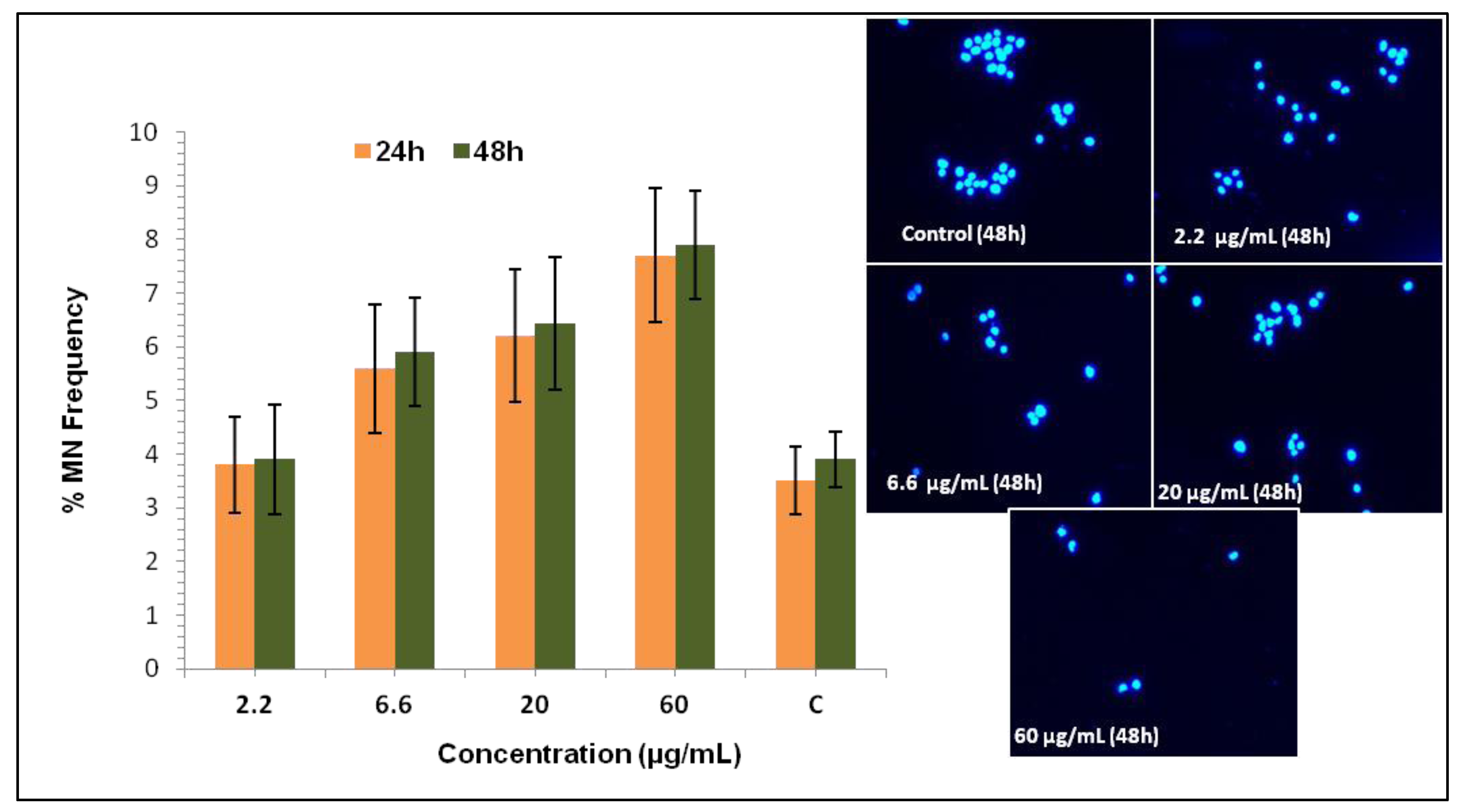
2.5. Micronucleus Assay
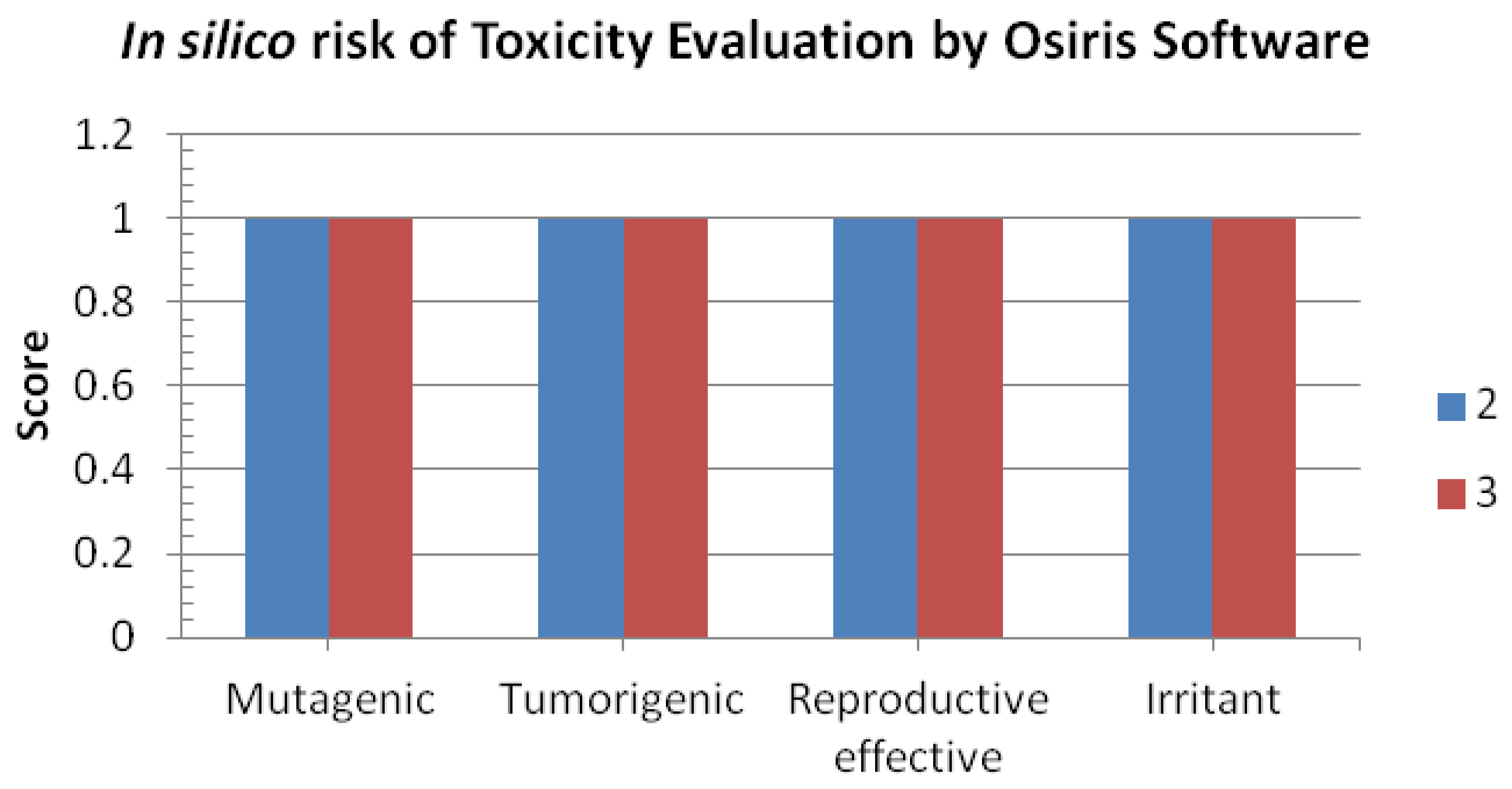
2.6. In Silico Pharmacokinetics


| Compounds | nHba | nHbd | nrotb | MW | cLog P | Druglikeness | Drug Score |
|---|---|---|---|---|---|---|---|
| 2 | 1 | 1 | 2 | 196 | 2.38 | −1.29 | 0.56 |
| 3 | 0 | 0 | 2 | 180 | 3.13 | −2.28 | 0.47 |
3. Experimental
3.1. General
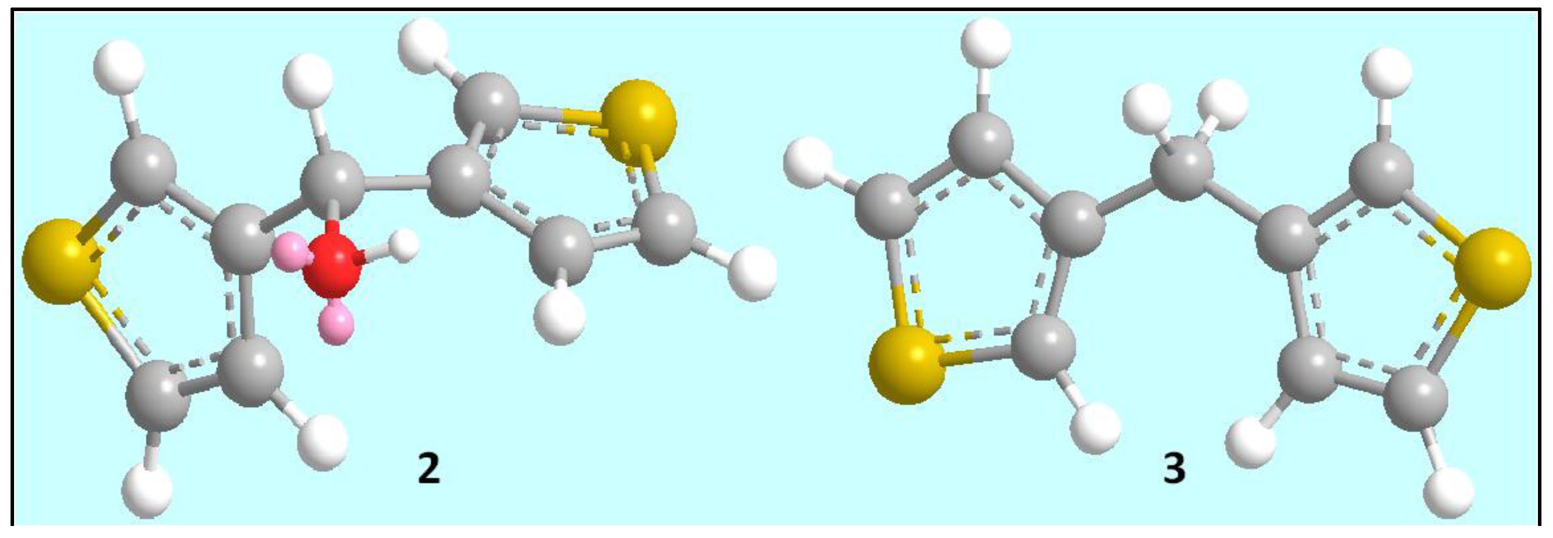
3.2. Preparation of Di(3-thienyl)methanol (2)
3.3. Preparation of Di(3-thienyl)methane (3)
3.4. Human Cell Culture
3.5. In Vitro Cell Viability Assay
3.6. Cell Growth Kinetics Assay
3.7. Clonogenic Survival Assay
3.8. Micronuclei Formation
3.9. In Silico Pharmacokinetic Screening
4. Conclusions
Acknowledgments
References
- Elsabee, M.Z.; Ali, E.A.; Mokhtar, S.M.; Eweis, M. Synthesis, characterization polymerization and antibacterial properties of novel thiophene substituted acrylamide. React. Funct. Polym. 2011, 71, 1187–1194. [Google Scholar] [CrossRef]
- Bonini, C.; Chiummiento, L.; Bonis, M.D.; Funicello, M.; Lupattelli, P.; Suanno, G.; Berti, F.; Campaner, P. Synthesis, biological activity and modelling studies of two novel anti HIV PR inhibitors with athiophene containing hydroxyethylamino core. Tetrahedron 2005, 61, 6580–6589. [Google Scholar]
- Ye, D.; Zhang, Y.; Wang, F.; Zheng, M.; Zhang, X.; Luo, X.; Shen, X.; Jiang, H.; Liu, H. Novelthiophene derivatives as PTP1B inhibitors with selectivity and cellular activity. Bioorg. Med. Chem. 2010, 18, 1773–1782. [Google Scholar] [CrossRef]
- Brandt, W.; Mologni, L.; Preu, L.; Lemcke, T.; Gambacorti-Passerini, C.; Kunick, C. Inhibitors of the RET tyrosine kinase based on a 2-(alkylsulfanyl)-4-(3-thienyl)nicotinonitrile scaffold. Eur. J. Med. Chem. 2010, 45, 2919–2927. [Google Scholar] [CrossRef]
- Abdel-Wahab, B.F.; Abdel-Gawad, H.; Ghada, E.A.A.; Badria, F.A. Synthesis, antimicrobial, antioxidant, antiinflammatory and analgesic activities of some new 3-(2′-thienyl)pyrazolebased heterocycles. Med. Chem. Res. 2012, 21, 1418–1426. [Google Scholar] [CrossRef]
- Meotti, F.C.; Silva, D.O.; Santos, A.R.S.D.; Zeni, G.; Rocha, J.B.T.; Nogueira, C.W. Thiopheneand furans derivatives: A new class of potential pharmacological agents. Environ. Toxicol. Pharmacol. 2003, 15, 37–44. [Google Scholar] [CrossRef]
- Lu, X.; Wan, B.; Franzblau, S.G.; You, Q. Design, synthesis and anti-tubercular evaluation of new 2-acylated and 2-alkylated amino-5-(4-(benzyloxy)phenyl)thiophene-3-carboxylic acid derivatives. Eur. J. Med. Chem. 2011, 46, 3551–3563. [Google Scholar] [CrossRef]
- Amr, A.E.G.E.; Sherif, M.H.; Assy, M.G.; Al-Omar, M.A.; Ragab, I. Antiarrhythmic, serotonin antagonist and antianxiety actvities of novel substitutedthiophene derivatives synthesized from 2-amino-4,5,6,7-tetrahydro-N-phenylbenzo[b]thiophene -3-carboxamide. Eur. J. Med. Chem. 2010, 45, 5935–5942. [Google Scholar] [CrossRef]
- Gonzalez, J.L.; Stephens, C.E.; Wenzler, T.; Brun, R.; Tanious, F.A.; Wilson, W.D.; Barszcz, T.; Werbovetz, K.A.; Boykin, D.W. Synthesis and antiparasitic evaluation of bis-2,5-[4-guanidinophenyl] thiophene. Eur. J. Med. Chem. 2007, 42, 552–557. [Google Scholar] [CrossRef]
- Romagnoli, R.; Baraldi, P.G.; Carrion, M.D.; Cara, C.L.; Cruz-Lopez, O.; Preti, D.; Tolomeo, M.; Grimaudo, S.; Cristina, A.D.; Zonta, N.; et al. Design, synthesis, and biological evaluation of thiophene analogues of chalcones. Bioorg. Med. Chem. 2008, 16, 5367–5376. [Google Scholar] [CrossRef]
- Diana, P.; Carbone, A.; Barraja, P.; Montalbano, A.; Martorana, A.; Dattolo, G.; Gia, O.; Via, L.D.; Cirrincione, G. Synthesis and antitumor properties of 2,5-bis(3'-indolyl)thiophenes: Analogues of marine alkaloid nortopsentin. Bioorg. Med. Chem. Lett. 2007, 17, 2342–2346. [Google Scholar] [CrossRef]
- Gouda, M.A.; Berghot, M.A.; Baz, E.A.; Hamama, W.S. Synthesis, antitumor and antioxidant evaluation of some new thiazole and thiophene derivatives incorporated coumarin moiety. Med.Chem. Res. 2012, 21, 1062–1070. [Google Scholar] [CrossRef]
- Scarpelli, R.; Marco, A.D.; Ferrigno, F.; Laufer, R.; Marcucci, I.; Muraglia, E.; Ontoria, J.M.; Rowley, M.; Serafini, S.; Steinkühler, C.; et al. Studies of the metabolic stability in cells of 5-(trifluoroacetyl)thiophene-2-carboxamides and identification of more stable class II histone deacetylase (HDAC) inhibitors. Bioorg. Med. Chem. Lett. 2008, 18, 6078–6082. [Google Scholar]
- Shaw, G.; Morse, S.; Ararat, M.; Graham, F.L. Preferential transformation of human neuronal cells by human adenoviruses and the origin of HEK 293 cells. FASEB J. 2002, 16, 869–871. [Google Scholar]
- Kaushik, N.K.; Kim, Y.H.; Han, Y.G.; Choi, E.H. Effect of jet plasma on T98G human brain cancer cells. Curr. Appl. Phys. 2013, 13, 176–180. [Google Scholar] [CrossRef]
- Mishra, A.; Jung, H.; Park, J.W.; Kim, H.K.; Kim, H.; Stang, P.J.; Chi, K.W. Anticancer Activity of Self-Assembled Molecular Rectangles via Arene-Ruthenium Acceptors and a New Unsymmetrical Amide Ligand. Organometallics 2012, 31, 3519–3526. [Google Scholar]
- Beyer, R.; Kalaji, M.; Burton, G.K.; Murphy, P.J.; Pereira, V.M.S.C.; Taylor, D.M.; Williams, G.O. Spectroelectrochemical and electrical characterization of low bandgap polymers. Synth. Metals 1998, 92, 25–31. [Google Scholar] [CrossRef]
- Nenajdenko, V.G.; Baraznenok, I.L.; Balenkova, E.S. Efficient One-Pot Synthesis of Dithieno(dibenzothieno)-Fused Cycloheptanones, Tropones, and Cyclooctanones. J. Org. Chem. 1998, 63, 6132–6136. [Google Scholar] [CrossRef]
- Wahab, R.; Kaushik, N.K.; Verma, A.K.; Mishra, A.; Hwang, I.H.; Yang, Y.B.; Shin, H.S.; Kim, Y.S. Fabrication and growth mechanism of ZnO nanostructures and their cytotoxic effect on human brain tumor U87, cervical cancer HeLa, and normal HEK cells. J. Biol. Inorg. Chem. 2011, 16, 431–442. [Google Scholar] [CrossRef]
- Kaushik, N.K.; Uhm, H.S.; Choi, E.H. Micronucleus formation induced by dielectric barrier discharge plasma exposure in brain cancer cells. Appl. Phys. Lett. 2012, 100, 084102:1–084102:4. [Google Scholar]
- Countryman, P.I.; Heddle, J.A. The production of micronuclei from chromosome aberrations in irradiated cultures of human lymphocytes. Mutat. Res. 1976, 41, 321–332. [Google Scholar] [CrossRef]
- Tekto, I.V. Computing chemistry on the web. Drug Discov. Today 2005, 10, 1497–1500. [Google Scholar] [CrossRef]
- Wenlock, M.C.; Austin, R.P.; Barton, P.; Davis, A.M.; Leeson, P.D. A comparison of physicochemical property profiles of development and marketed oral drugs. J. Med. Chem. 2003, 46, 1250–1256. [Google Scholar] [CrossRef]
- Sample Availability: Samples of the compounds Di(3-thienyl)methanol and Di(3-thienyl)methane are available from the authors.
© 2012 by the authors; licensee MDPI, Basel, Switzerland. This article is an open-access article distributed under the terms and conditions of the Creative Commons Attribution license (http://creativecommons.org/licenses/by/3.0/).
Share and Cite
Kaushik, N.K.; Kim, H.S.; Chae, Y.J.; Lee, Y.N.; Kwon, G.-C.; Choi, E.H.; Kim, I.T. Synthesis and Anticancer Activity of Di(3-thienyl)methanol and Di(3-thienyl)methane. Molecules 2012, 17, 11456-11468. https://doi.org/10.3390/molecules171011456
Kaushik NK, Kim HS, Chae YJ, Lee YN, Kwon G-C, Choi EH, Kim IT. Synthesis and Anticancer Activity of Di(3-thienyl)methanol and Di(3-thienyl)methane. Molecules. 2012; 17(10):11456-11468. https://doi.org/10.3390/molecules171011456
Chicago/Turabian StyleKaushik, Nagendra Kumar, Hong Seon Kim, Young June Chae, Young Nam Lee, Gi-Chung Kwon, Eun Ha Choi, and In Tae Kim. 2012. "Synthesis and Anticancer Activity of Di(3-thienyl)methanol and Di(3-thienyl)methane" Molecules 17, no. 10: 11456-11468. https://doi.org/10.3390/molecules171011456
APA StyleKaushik, N. K., Kim, H. S., Chae, Y. J., Lee, Y. N., Kwon, G.-C., Choi, E. H., & Kim, I. T. (2012). Synthesis and Anticancer Activity of Di(3-thienyl)methanol and Di(3-thienyl)methane. Molecules, 17(10), 11456-11468. https://doi.org/10.3390/molecules171011456