Article

Nitrogen-Containing Apigenin Analogs: Preparation and Biological Activity

1
Colleges of Science and Forestry, Northwest A&F University, Yangling 712100, Shaanxi, China
2
College of Agronomy and Life Sciences, Shanxi Datong University, Datong 037009, Shanxi, China
*
Authors to whom correspondence should be addressed.
Molecules 2012, 17(12), 14748-14764; https://doi.org/10.3390/molecules171214748
Submission received: 30 October 2012 / Revised: 30 November 2012 / Accepted: 5 December 2012 / Published: 11 December 2012
(This article belongs to the Section Natural Products Chemistry)

Abstract

:
A series of nitrogen-containing apigenin analogs 4aj was synthesized via Mannich reactions to develop anticancer, antibacterial, and antioxidant agents from plant-derived flavonoids. The chemical structures of these compounds were confirmed using 1H-NMR, 13C-NMR, and ESI-MS. The in vitro biological activities of the analogs were evaluated via assays of their antiproliferative, antibacterial, and antioxidant activities. The prepared apigenin analogs exhibited different antiproliferative activities against four human cancer cell lines, namely human cervical (HeLa), human hepatocellular liver (HepG2), human lung (A549), and human breast (MCF-7) cancer cells. Compound 4i showed the most favorable in vitro antiproliferative activity with IC50 values of 40, 40, 223, and 166 μg/mL against HeLa, HepG2, A549, and MCF-7, respectively. The 1,1-diphenyl-2-picrylhydrazyl (DPPH) free radical scavenging activity assay also showed that 4i had the most potent antioxidant activity, with the smallest IC50 value (334.8 μg/mL). The antibacterial activities of the analogs were determined using a two-fold serial dilution technique against four pathogenic bacteria, namely Staphylococcus aureus, Bacillus subtilis, Escherichia coli, and Pseudomonas aeruginosa. All the prepared apigenin analogs exhibited more potent activities than the parent apigenin. Compounds 4h and 4j, in particular, exhibited the best inhibitory activities against the Gram-positive bacteria Staphylococcus aureus and Bacillus subtilis with MIC values of 3.91 and 1.95 μg/mL, respectively.

Graphical Abstract

1. Introduction

Flavonoids are important components of various traditional Chinese medicines and phytomedicines that bear C6-C3-C6 skeletons [1,2]. In addition to their physiological roles in plants, these compounds also exhibit anticancer, antioxidant, anti-aging, and antibacterial activities [3,4,5]. Flavonoids display antibacterial activity by inhibiting nucleic acid synthesis [6,7,8], cytoplasmic membrane function [9,10,11], and energy metabolism [12]. They are generally classified as flavones, flavonols, flavanones, flavanonol, or isoflavones, based on the degree of oxidation of the carbon-3 bond (C-3) and the connection position of the B-ring [13]. Flavonoids are prevalent in higher plants as well as in the roots, stems, leaves, and flowers of ferns [14]. To date, over 4,000 flavonoids have been identified, and several of these compounds showed diverse pharmacological activities, such as antimicrobial [15], antiallergic [16], antidiabetic [17], anti-inflammatory [18], antiviral [19], antimutagenic [20], antithrombotic [21], antioxidant [22], anticarcinogenic [23], and hepatoprotective [24] effects.
Apigenin [2-(4-hydroxyphenyl)-5,7-dihydroxy-4H-chromen-4-one], which belongs to the flavone subclass of flavonoids, are found in fruits, vegetables, and traditional medicinal plants, such as parsley, onion, orange, tea, chamomile, wheat sprouts, and several seasonings [25]. Apigenin exhibits anti-inflammatory and anti-carcinogenic effects on the skin [26]. In some cells, apigenin displays a variety of anti-tumor effects, such as stimulation of gap junctional and intracellular communication [27] as well as inhibition of mutagenesis [28], transformation [29], angiogenesis [30], and tumorigenesis [31]. In addition, apigenin exhibits antibacterial activity against a number of bacterial species [32,33]. Previous studies have shown the potential of apigenin as a bioactive molecule for various clinical applications. However, compared with existing drugs, the anticancer, antibacterial, antioxidant, and other activities of apigenin hardly meet the requirements for clinical application. Considerable effort has thus been exerted to improve the activities of apigenin [34,35,36,37]. A simple and effective way is the study of the structure-activity relationships of apigenin analogs prepared by introducing various functional groups into different positions in the apigenin skeleton [38,39,40]. Gunnarsson et al. [40] synthesized sulfated apigenin analogs, which were all antithrombin activators to accelerate the inhibition of factor Xa. Mavel et al. [38] synthesized apigenin analogs with halogen on the 4'-position which showed cytotoxic activity and MDR-reversing capacity. However, all the current studies mainly focused on the 4'- or 7-position of apigenin. The 8-position of apigenin was scarcely examined.
The Mannich reaction is a classic method for the preparation of Mannich bases, namely, β-amino ketones and β-amino aldehydes. The Mannich reaction is also one of the very important reaction types and key reaction-steps in the synthesis of numerous drugs and natural products [41,42]. This reaction has been considered as an effective method in introducing aminomethyl substituents into the desired molecules to improve their biological activities [43]. Previous studies have demonstrated that Mannich bases offer a wide range of biological activities, such as anticancer [44], anti-inflammatory [45], analgesic [46], antibacterial, and antifungal activities [47]. The inhibitory activities of adriamycin salicylamide Mannich base against MCF-7 and PC-3 cells are four times better than that of adriamycin, while simultaneously reducing the clinical side effects [48]. In addition, several studies have shown that 8-aminomethylated oroxylin A analogs demonstrated significantly improved α-glucosidase inhibitory activity [49], nitrogen-containing baicalein/quercetin analogs exhibited potent CDK1/cyclin B inhibitory activity [50], and 8-aminomethylluteolin derivatives displayed potential anti-inflammatory activity [51].
Thus, in the present study, a series of new apigenin analogs with aminomethyl groups on the C-8 position was synthesized via Mannich reactions. In addition, the antiproliferative, antibacterial, and antioxidant activities of these analogs were investigated. The antiproliferative activities of the compounds were analyzed against four human cancer cell lines, namely the human cervical (HeLa), human hepatocellular liver (HepG2), human lung (A549), and human breast (MCF-7) cancer cells, using a standard 3-(4,5-dimethylthiazol-2-diphenyl-tetrazolium) bromide (MTT) assay. The antibacterial activities of the prepared apigenin analogs against four pathogenic bacteria, namely Staphylococcus aureus (S. aureus), Bacillus subtilis (B. subtilis), Escherichia coli (E. coli), and Pseudomonas aeruginosa (P. aeruginosa), were explored using a two-fold serial dilution technique. The antioxidant activities of the apigenin analogs were determined using the DPPH radical scavenging assay.

2. Results and Discussion

2.1. Chemistry
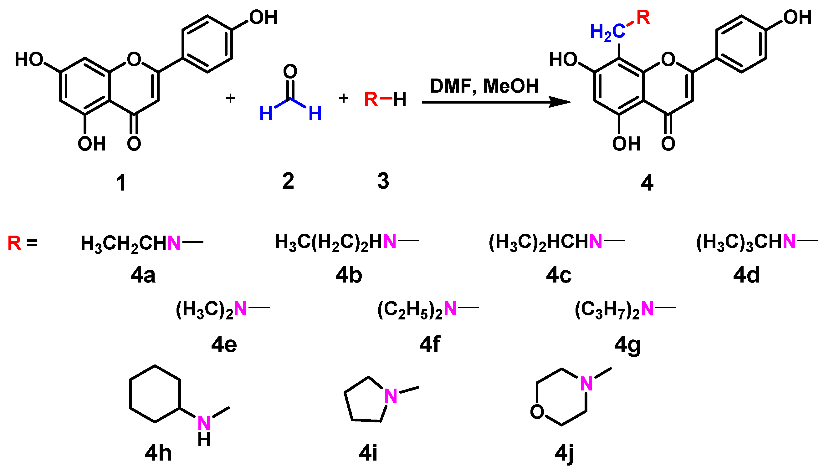
Ten new nitrogen-containing apigenin analogs, 4aj, were prepared via Mannich reactions (Scheme 1) to further explore their antiproliferative, antibacterial, and antioxidant activities. The hydrogen atom activity of apigenin in the C-8 position increases because of the p-π conjugative effect of the 5- and 7- hydroxy in the A-ring. Thus, apigenin easily reacts with formaldehyde and primary or secondary amines to produce different Mannich bases [52]. The reaction conditions, yields, and structures of the apigenin analogs are summarized in Table 1. Corresponding products were obtained with the introduction of different primary or secondary amines. The reaction temperatures had a little difference for different products, varied from 25 °C to 40 °C. Also, different times were required for these reactions since no or less byproducts and better yields were obtained under these conditions.
Scheme 1. Synthetic route of compounds 4a to 4j.
Molecules 17 14748 g003
Table 1. Physicochemical data of the nitrogen-containing apigenin analogs.
CompoundsRTime (h)Temperature (°C)Isolated yield (%)
4aEthylamino42540.5
4bPropylamino123038.7
4cIsopropylamino43561.7
4dTert-butylamino53552.3
4eDimethylamino42539.6
4fDiethylamino1.52560.9
4gDiisopropylamino122551.2
4hCyclohexylamino84041.8
4iPyrrolidinyl1.54042.1
4jMorpholinyl124043.4

2.2. Antibacterial Activity

A preliminary antibacterial screeningof the synthesized compounds was performed using the disk diffusion method [53]. Table 2 lists the screening results of the tested compounds against the Gram negative bacteria E. coli (ATCC 25922) and P. aeruginosa (ATCC 27853) and Gram positive bacteria S. aureus (ATCC 25923) and B. subtilis (ATCC 6633). The results showed that most of the compounds showed antibacterial activities against the microorganisms at a dose of 1000 μg/mL. Compounds showing inhibition of at least 10 mm were considered active and were further evaluated for their minimal inhibitory concentration (MIC) [54,55].
Table 2. Inhibition zones (IZ) in mm (Mean a ± S.D b) of the synthesized compounds 4a to 4j.
CompoundsS. aureusB. subtilisE. coliP. aeruginosa
ATCC 25923ATCC 6633ATCC 25922ATCC 27853
Apigenin11.1 ± 0.311.0 ± 1.010.1 ± 0.310.3 ± 0.2
4a17.5 ± 0.121.3 ± 0.610.3 ± 0.18.6 ± 0.1
4b17.9 ± 0.617.5 ± 0.410.9 ± 0.710.6 ± 0.2
4c14.1 ± 0.515.2 ± 0.112.5 ± 0.110.5 ± 0.4
4d14.8 ± 0.218.0 ± 0.312.8 ± 0.410.8 ± 0.1
4e12.1 ± 0.615.9 ± 0.610.4 ± 0.212.8 ± 0.5
4f18.3 ± 0.118.7 ± 0.514.8 ± 0.512.4 ± 0.3
4g15.0 ± 0.319.0 ± 0.111.0 ± 0.310.0 ± 0.7
4h22.1 ± 0.522.8 ± 0.215.3 ± 0.117.0 ± 1.0
4i18.8 ± 0.419.3 ± 0.413.0 ± 0.110.2 ± 0.8
4j25.9 ± 0.627.2 ± 0.815.6 ± 0.314.3 ± 1.0
Ampicillin37.3 ± 0.636.7 ± 1.528.9 ± 0.133.7 ± 0.5
Tetracycline26.3 ± 0.521.0 ± 1.021.7 ± 1.124.3 ± 1.1
a Mean value of measured diameters of zones of inhibition; b S.D. denotes the standard deviation.
The MIC values of all the compounds are presented in Table 3. All the prepared apigenin analogs showed relatively higher antibacterial activities than apigenin. Gram-positive strains S. aureus and B. subtilis showed relatively high sensitivities toward the synthesized compounds, with MIC values from 1.95 μg/mL to 15.63 μg/mL. Furthermore, 4h and 4j showed prominent activities with MIC values of 3.91 and 1.95 μg/mL against S. aureus and B. subtilis, comparable with the positive control tetracycline, although lower than ampicillin. Compounds 4a, 4b, 4f, and 4i showed moderate activities against S. aureus and B. subtilis with MIC values of 7.81 μg/mL. However, 4ce, and 4g exhibited weak antibacterial activities against S. aureus and B. subtilis with MIC values of 15.63 μg/mL to 31.25 μg/mL.
Table 3. In vitro antibacterial activity (MIC, μg/mL) and DPPH radical scavenging activity (IC50, μg/mL) of 4a to 4j.
CompoundsIC50 (μg/mL) aMIC (μg/mL) a
S. aureusB. subtilisE. coliP. aeruginosa
ATCC 25923ATCC 6633ATCC 25922ATCC 27853
Apigenin1094.7 ± 2.231.2531.2562.562.5
4a788.0 ± 1.67.813.9162.5125
4b782.4 ± 2.37.817.8162.562.5
4c670.1 ± 1.515.6315.6331.2562.5
4d539.5 ± 1.415.637.8131.2562.5
4e542.3 ± 1.331.2515.6362.531.25
4f434.8 ± 1.47.817.8115.6331.25
4g431.1 ± 1.115.637.8162.562.5
4h887.7 ± 2.53.913.9115.6315.63
4i334.8 ± 1.27.817.8131.2562.5
4j740.1 ± 1.81.951.9515.6315.63
Ampicillin--0.060.120.980.49
Tetracycline--1.953.913.913.91
Vitamin C44.2 ± 1.1
a Average of three parallel experiments.
To analyze further the structure-activity relationships, the prepared apigenin analogs were divided into two categories. One category includes the cyclic substituent-containing apigenin analogs, 4h to 4j, and the other includes the aliphatic chain substituent-containing apigenin analogs, 4a to 4g. From Table 3, we find that the apigenin analogs showed relatively higher antibacterial activities than apigenin. Furthermore, in the cyclic substituent-containing apigenin analogs, the order of the MIC values was 4j < 4h < 4i, indicating that the analogs with 6-membered rings had better antibacterial activities than those with 5-membered rings. Moreover, in the 6-membered ring compounds, compounds containing two heteroatoms (4j) showed stronger antibacterial activities than compounds containing one heteroatom (4i). For the aliphatic chain substituent-containing apigenin analogs, straight-chain primary amine-substituted apigenin analogs displayed stronger activities than the branched-chain substituted apigenin analogs.

2.3. Antioxidant Activity

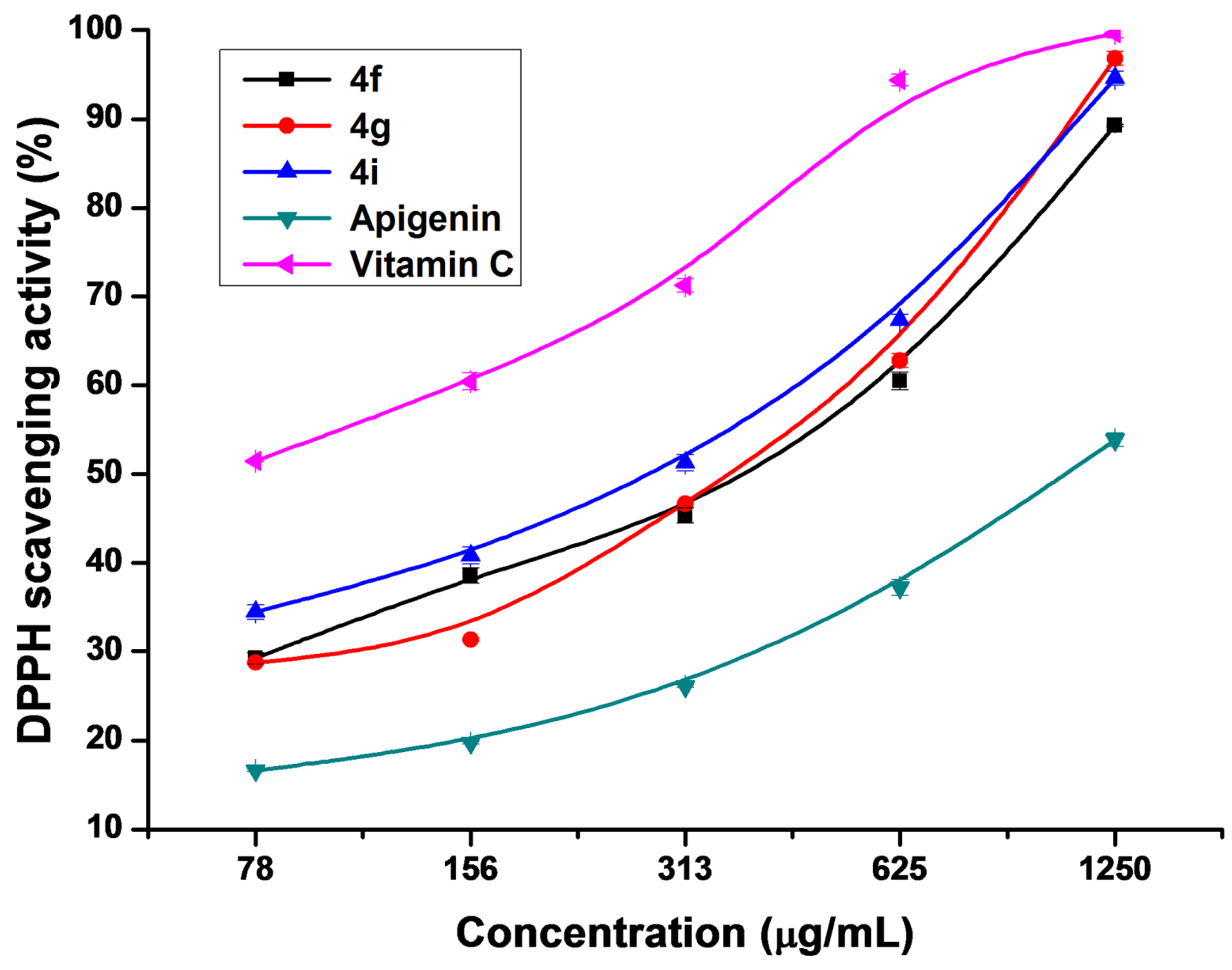
The DPPH radical is a stable free radical commonly used as a substrate to evaluate in vitro antioxidant activities of extracts from fruits, vegetables, and medicinal plants [56]. An antioxidant can scavenge the radical by hydrogen donation, which results in a decrease in DPPH absorbance at 517 nm [57]. Generally, flavonoids display different DPPH radical scavenging activities with their different structures. The radical scavenging activities of flavonoids are highly controlled by the number and configuration of phenolic hydroxyl groups in the molecules and also influenced by the configuration of other substituents [58]. Table 3 shows the DPPH radical scavenging activities of the prepared apigenin analogs compared with those of the parent apigenin and vitamin C. Most of the prepared compounds exhibited potential scavenging activities compared with the parent apigenin and moderate activities compared with vitamin C. Compound 4i was found to have significant antioxidant activity with the smallest IC50 value and compound 4h possessed the least activity among these prepared compounds with the largest IC50 value. IC50 value is negatively related to antioxidant activity such that the lower the IC50 value is, the higher the antioxidant activity will be [59]. Therefore, the order of DPPH radical scavenging abilities of the analogs was 4i > 4g > 4f > 4d > 4e > 4c > 4j > 4b > 4a > 4h > apigenin. Compared with the parent apigenin, 4f, 4g, and 4i exhibited significant antioxidant activities, with IC50 values of 434.8, 431.1, and 334.8 μg/mL, respectively. The concentration-dependent changes in the antioxidant activities of these compounds were also analyzed. The antioxidant activity was enhanced with the concentration of every sample was increased (Figure 1). When the concentration of 4f, 4g, and 4i reached 1,250 μg/mL, their scavenging effects were close to that of vitamin C.
Figure 1. DPPH radical scavenging activities of 4f, 4g, 4i, apigenin, and vitamin C.
Molecules 17 14748 g001
Apigenin analogs with branched-chain aliphatic substituents showed stronger antioxidant activities than those with straight-chain primary amine substituents or heteroatom-containing substituents, except for 4i. Moreover, the alkyl number of the substituent groups determined the antioxidant activity of the analogs. The more alkyl numbers existed in the substituted apigenin analogs, the higher were the displayed activities, as shown by 4f and 4g. All these results above suggested that substituted apigenin analogs exhibited antioxidant activities due to the introduction of different electron donations on the C-8 of apigenin [58], and the antioxidant activities of flavonoids could be modulated by the number and configuration of amine groups in the molecules.

2.4. Antiproliferative Activity

The antiproliferative activities of the prepared apigenin analogs 4aj were evaluated by performing in vitro assays of the inhibition ratios of these compounds to the proliferation of human cancer cells HeLa, HepG2, A549, and MCF-7. Figure 2 shows that the prepared apigenin analogs possessed different antiproliferative activities depending on the changes in their chemical structures, and high doses of these compounds showed more potent antiproliferative activities. Compound 4i exhibited the most significant antiproliferative activity against the four cancer cells. At 1000 μg/mL, the inhibition rates of 4i for HeLa, HepG2, A549, and MCF-7 cells were 89.97%, 92.60%, 93.33%, and 93.04%, respectively. All the prepared apigenin analogs possessed stronger inhibition effects on HeLa and HepG2 cells than on A549 and MCF-7 cells.
Figure 2. Inhibition ratios of apigenin and its analogs to the proliferation of HeLa (A), HepG2 (B), A549 (C), and MCF-7 (D) cell lines.
Molecules 17 14748 g002
Table 4 summarizes the IC50 values of apigenin analogs against HeLa, HepG2, A549, and MCF-7 cells. Compound 4i exhibited the strongest antiproliferative activity, and its IC50 values reached 40, 40, 223, and 166 μg/mL for HeLa, HepG2, A549, and MCF-7 cells, respectively. Moreover, compounds 4fh, and 4j showed better antiproliferative activities (with lower IC50 values) against HeLa and HepG2 cells than apigenin, with inhibition ratios reaching more than 50% at 250 μg/mL. However, no obvious antiproliferative activity was observed for 4a, 4b, and 4d, indicating the ineffectiveness of these compounds in suppressing the HeLa and HepG2 cell proliferation. Compounds 4b, 4e, and 4i displayed stronger activities toward A549 cells, whereas 4e, 4f, and 4i had better antiproliferative activities against MCF-7 cells, compared with apigenin. Moreover, the antiproliferative activities of the apigenin analogs against HeLa and HepG2 were enhanced with a short and more branched alkyl chain at the C-8 position of apigenin. The highest potency was obtained when a piperazin-1-yl-ethoxy group was present at the C-8 position of the apigenin. Combined with the structural features of the prepared analogs, the presence of nitrogen at the C-8 position of apigenin was critical to the antiproliferative activities of the apigenin analogs, which was supported by previous reports [60,61,62].
Table 4. Antiproliferative activities (IC50, μg/mL) of apigenin analogs against HeLa, HepG2, A549, and MCF-7 cell lines.
CompoundsIC50 (μg/mL)
HeLaHepG2A549MCF-7
1450 ± 2.0460 ± 2.21740 ± 3.4>2000 ± 3.6
4a450 ± 2.5470 ± 2,7>2000 ± 4.3>2000 ± 4.1
4b430 ± 1.9410 ± 2.21410 ± 3.0420 ± 2.3
4c270 ± 1.4270 ± 1.9>2000 ± 3.8280 ± 1.7
4d570 ± 2.0620 ± 3.1>2000 ± 4.0310 ± 2.1
4e230 ± 1.6270 ± 2.0380 ± 2.8210 ± 1.9
4f160 ± 1.5150 ± 1.7>2000 ± 3.7250 ± 2.0
4g210 ± 2.1190 ± 1.5>2000 ± 4.2610 ± 3.2
4h170 ± 1.7180 ± 1.4>2000 ± 3.5>2000 ± 4.0
4i40 ± 1.840 ± 1.5223 ± 2.1166 ± 1.8
4j180 ± 2.0220 ± 2.0>2000 ± 4.41010 ± 2.9

3. Experimental

3.1. General

Apigenin (>98%) was purchased from Xi’an Spring-chem Bio-tech Co., Ltd. (Xi’an, China). Tetracycline and DPPH were from Sigma-Aldrich (St. Louis, MO, USA). Ampicillin and MTT were obtained from Amersco Inc. (Solon, OH, USA). Yeast extract and tryptone were from Oxoid Ltd. (Basingstoke, Hampshire, UK). Dulbecco’s modified eagle medium, RPMI-1640 medium, and fetal bovine serum (FBS) were from Gibco Invitrogen Corporation (Carlsbad, CA, USA). HeLa and HepG2 cells were provided by the Cell Center of the Fourth Military Medical University (Xi’an, China). A549 and MCF-7 cells were from the Chinese Academy of Sciences (Shanghai, China). Vitamin C and primary or secondary amines were from Aladdin (Shanghai, China). N,N-Dimethylformamide (DMF) was purified by vacuum distillation over CaH2 before utilization. All solvents and other chemicals were supplied by local commercial suppliers and of analytical reagent grade, unless otherwise stated. Deionized water (Milli-Q, Millipore, Bedford, MA, USA) was used to prepare aqueous solutions. Column chromatography was performed with silica gel 60 (200 to 300 mesh). Thin-layer chromatography (TLC) and preparative TLC (PTLC) were used with silica gel 60 GF254 (Qingdao Haiyang Chem. Co., Ltd., Shandong, China). Product spots were detected under UV light using ZF-6 type III ultraviolet analyzer (Shanghai Jiapeng Technology Co., Ltd., Shanghai, China). Melting points were determined using a digital melting point apparatus (Zhengzhou Mingze Technology Co., Ltd., Zhengzhou, China) and were uncorrected. ESI mass spectra were obtained using a Thermo Scientific LCQ FLEET mass spectrometer equipped with an electrospray ion source and controlled by Xcalibur software (Thermo Fisher Scientific, Waltham, MA, USA). Proton nuclear magnetic resonance spectra (1H-NMR) and carbon-13 nuclear magnetic resonance spectra (13C-NMR) were obtained using a Bruker Avance DMX 500 MHz/125 MHz spectrometer (Bruker, Billerica, MA, USA), with chemical shifts reported in parts per million [in dimethyl sulfoxide (DMSO-d6), tetramethylsilane as an internal standard, J values were given in Hertz]. MIC was detected using a BIO-RAD 680 Microplate reader (Beijing Yuanye Bio. Co., Ltd., Beijing, China). The absorbance values in DPPH assays were recorded using a UV-1600PC spectrophotometer (Shanghai Mapada Instruments Co., Ltd., Shanghai, China).

3.2. General Procedure for the Synthesis of Compounds 4a to 4j

The apigenin analogs 4aj were synthesized via Mannich reactions [63]. A magnetic stirring bar and apigenin (1, 100 mg, 0.37 mmol) dissolved in anhydrous DMF (1 mL) were first placed into a two-necked flask. Then, methanol (15 mL) was added and a yellow solution was immediately obtained. The reaction system was then immersed in an oil bath while being stirred. Then, formaldehyde solution (0.028 mL, 37%) and ethylamine solution (0.031 mL, 70%) were added dropwise to the reaction mixture. Stirring was required to keep the reaction mixture homogenized during these operations. The reaction process was checked using TLC analysis. After 4 h reaction, the mixture was filtered, and the supernatant was collected. The solvent was removed under reduced pressure. The residue was then purified using PTLC with ethyl acetate-methanol (1:1, v/v) as developing agent to obtain 4a. Compounds 4b to 4j were prepared using similar methods, and their structures were confirmed via melting point determination, 1H-NMR, 13C-NMR, and ESI-MS. The data are listed as follows.
4',5,7-Trihydroxy-8-(ethylamino)-2-phenyl-4H-chromen-4-one (4a): yellow solid, yield 40.5%, m.p. >300 °C. 1H-NMR δ: 13.00 (s, 1H, 5-OH), 7.84 (d, 2H, J = 10.0 Hz), 6.92 (d, 2H, J = 10.0 Hz), 6.59 (s, 1H), 6.00 (s, 0.6H), 5.77 (s, 0.4H), 4.14 (s, 0.9H, CH2), 3.96 (s, 1.1H, CH2), 2.81 (m, 2H, -NH-CH2-CH3), 1.29 (t, 3H, -NH-CH2-CH3); 13C-NMR δ: 182.0, 169.9, 163.5, 162.0, 159.6, 159.2, 128.4, 128.4, 121.7, 116.6, 116.6, 103.1, 102.7, 102.3, 95.7, 41.7, 35.7, 17.6; ESI-MS (m/z): 325.9 [M−H].
4',5,7-Trihydroxy-8-(propylamino)-2-phenyl-4H-chromen-4-one (4b): yellow solid, yield 38.7%, m.p. >300 °C. 1H-NMR δ: 12.99 (s, 1H, 5-OH), 7.85 (d, 2H, J = 10.0 Hz), 6.90 (d, 2H, J = 10.0 Hz), 6.60 (s, 0.3H), 6.56 (s. 0.7H), 6.02 (s, 0.6H), 5.79 (s, 0.4H), 4.14 (s, 0.7H, CH2), 3.95 (s, 1.3H, CH2), 2.71 (t, J = 19.1 Hz, 2H, -NH-CH2-CH2-CH3), 1.54–1.58 (m, 2H, -NH-CH2-CH2-CH3), 0.91 (t, J = 6.7 Hz, 3H, -NH-CH2-CH2-CH3); 13C-NMR δ: 181.8, 169.9, 163.4, 161.5, 159.0, 157.0, 128.8, 128.8, 122.4, 116.7, 116.7, 103.7, 102.8, 102.1, 95.9, 42.3, 36.1, 27.5, 15.9; ESI-MS (m/z): 339.9 [M−H].
4',5,7-Trihydroxy-8-(isopropylamino)-2-phenyl-4H-chromen-4-one (4c): yellow solid, yield 61.7%, m.p. >300 °C. 1H-NMR δ: 12.99 (s, 1H, 5-OH), 7.86 (d, 2H, J = 10.0 Hz), 6.91 (d, 2H, J = 10.0 Hz), 6.59 (s, 0.6H), 6.56 (s, 0.4H), 6.01 (s, 0.4H), 5.79 (s, 0.6H), 4.14 (s, 1.1H, CH2), 3.96 (s, 0.9H, CH2), 3.07–3.14 (m, 1H, -NH-CH(CH3)2), 1.19 (d, 6H, -NH-CH(CH3)2); 13C-NMR δ: 181.9, 168.2, 163.4, 158.9, 158.0, 155.4, 128.4, 128.4, 121.1, 116.3, 116.3, 102.7, 102.4, 100.0, 95.8, 48.4, 40.4, 20.3; ESI-MS (m/z): 340.0 [M−H].
4',5,7-Trihydroxy-8-(tert-butylamino)-2-phenyl-4H-chromen-4-one (4d): yellow solid, yield 52.3%, m.p. >300 °C. 1H-NMR δ: 12.98 (s, 1H, 5-OH), 7.84 (d, 2H, J = 10.0 Hz), 6.92 (d, 2H, J = 10.0 Hz), 6.57 (s, 0.4H), 6.54 (s, 0.6H), 6.00 (s, 0.6H), 5.77 (s, 0.4H), 4.14 (s, 1.1H, CH2), 3.96 (s, 0.9H, CH2), 1.27 (s, 9H, (CH3)3C-); 13C-NMR δ: 180.8, 168.7, 162.9, 161.5, 158.0, 155.4, 128.6, 128.6, 125.6, 116.7, 116.7, 102.6, 102.4, 101.5, 95.9, 54.0, 49.1, 26.6; ESI-MS (m/z): 353.9 [M−H].
4',5,7-Trihydroxy-8-(dimethylamino)-2-phenyl-4H-chromen-4-one (4e): yellow solid, yield 39.6%, m.p. >300 °C. 1H-NMR δ: 13.03 (s, 1H, 5-OH), 7.94 (d, 2H, J = 10.0 Hz), 6.96 (d, 2H, J = 10.0 Hz), 6.77 (s, 1H), 6.11 (s, 1H), 3.97 (s, 2H, CH2), 2.43 (s, 6H, -N(CH3)2); 13C-NMR δ: 181.9, 168.2, 163.4, 161.5, 161.1, 155.4, 128.8, 128.8, 121.9, 116.5, 116.5, 103.0, 102.7, 100.0, 99.9, 53.4, 43.9; ESI-MS (m/z): 325.8 [M−H].
4',5,7-Trihydroxy-8-(diethylamino)-2-phenyl-4H-chromen-4-one (4f): yellow solid, yield 60.9%, m.p. >300 °C. 1H-NMR δ: 13.47 (s, 1H, 5-OH), 7.89 (d, 2H, J = 8.0 Hz), 6.92 (d, 2H, J = 8.0 Hz), 6.71 (s, 1H), 6.29 (s, 1H), 3.92 (s, 2H, CH2), 2.75 (q, 4H, J = 10.0Hz, -N(CH2CH3)2), 1.11 (t, 6H, J = 10.0 Hz, -N(CH2CH3)2; 13C-NMR δ: 181.8, 169.3, 163.6, 161.6, 159.0, 157.2, 128.7, 128.7, 121.8, 116.4, 116.4, 103.6, 102.9, 102.2, 94.9, 48.5, 46.3, 10.7; ESI-MS (m/z): 354.0 [M−H].
4',5,7-Trihydroxy-8-(diisopropylamino)-2-phenyl-4H-chromen-4-one (4g): yellow solid, yield 51.2%, m.p. >300 °C. 1H-NMR δ: 13.45 (s, 1H, 5-OH), 10.37 (s, 1H, 7-OH), 7.91 (d, 2H, J = 10.0 Hz), 6.93 (d, 2H, J = 10.0 Hz), 6.72 (s, 1H), 6.25 (s, 1H), 3.96 (s, 2H, CH2), 3.18–3.25 (m, 2H,-N(CH(CH3)2)2, 1.15(d, 12H, J = 5.0 Hz, -N(CH(CH3)2)2; 13C-NMR δ: 181.8, 169.9, 163.6, 161.5, 158.3, 157.0, 128.8, 128.8, 121.9, 116.4, 116.4, 104.0, 103.0, 102.1, 94.9, 49.5, 41.9, 19.2; ESI-MS (m/z): 381.8 [M−H].
4',5,7-Trihydroxy-8-(cyclohexylamino)-2-phenyl-4H-chromen-4-one (4h): yellow solid, yield 41.8%, m.p. >300 °C. 1H-NMR δ: 13.01 (1H, s, 5-OH), 7.85 (d, 2H, J = 10.0 Hz), 6.89 (d, 2H, J = 10.0 Hz), 6.58 (s, 0.6H), 6.55 (s, 0.4H), 6.04 (s, 0.4H), 5.80 (s, 0.6H), 4.15 (s, 1.2H, CH2), 3.97 (s, 0.8H, CH2), 2.74 (s, 1H, cyclohexylamine-CH), 1.24–1.97 (m, 10H, cyclohexylamine-CH2); 13C-NMR δ: 181.0, 169.0, 162.8, 162.6, 161.1, 158.7, 128.5, 128.5, 121.2, 116.7, 116.7, 102.4, 101.0, 100.7, 95.5, 55.5, 55.2, 31.2, 25.7, 24.5; ESI-MS (m/z): 379.9 [M−H].
4',5,7-Trihydroxy-8-(pyrrolidin-1-ylmethyl)-2-phenyl-4H-chromen-4-one (4i): yellow solid, yield 42.1%, m.p. >300 °C. 1H-NMR δ: 13.53 (1H, s, 5-OH), 7.89 (d, 2H, J = 5.0 Hz), 6.91 (d, 2H, J = 5.0 Hz), 6.69 (s, 1H), 6.28 (s, 1H), 3.95(s, 2H, CH2), 2.80 (s, 4H, pyrrolidine-N(CH2)2), 1.83 (s, 4H, pyrrolidine-(CH2)2); 13C-NMR δ: 181.5, 170.1, 163.4, 161.7, 159.0, 157.4, 128.7, 128.7, 121.7, 116.5, 116.5, 104.6, 102.7, 101.5, 95.0, 53.2, 49.9, 23.6; ESI-MS (m/z): 351.9 [M−H].
4',5,7-Trihydroxy-8-(morpholinomethyl)-2-phenyl-4H-chromen-4-one (4j): yellow solid, yield 43.4%, m.p. >300 °C. 1H-NMR δ: 12.98 (s, 1H, 5-OH), 8.03 (d, 2H, J = 5.0 Hz), 6.91 (d, 2H, J = 5.0 Hz), 6.83 (s, 1H), 6.58 (s, 1H), 4.11 (s, 2H, CH2), 3.81 (s, 2H, morpholine-O-CH2), 3.75 (s, 2H, morpholine-O-CH2), 2.52 (s, 4H, morpholine-N(CH2)2); 13C-NMR δ: 182.5, 165.0, 164.0, 161.9, 160.7, 154.9, 128.9, 128.9, 121.7, 116.6, 116.6, 104.3, 103.3, 102.9, 101.5, 66.6, 53.3, 50.9; ESI-MS (m/z): 368.0 [M−H].

3.3. Antibacterial Assay

The bacterial strains were kept in liquid nitrogen (−196 °C) in a Luria-Broth (LB) medium (5 g/L yeast extract, 10 g/L bactopeptone, and 10 g/L sodium chloride) containing 15% glycerol. Prior to the experiment, the bacterial strains were grown on LB agar plates at 37 °C. A single colony of bacteria from an overnight culture was inoculated into 150 mL of fresh LB medium. Each bacterial colony was then continuously incubated for 12 h at 37 °C in a rotary shaker set to 200 rpm. The cell density was determined via the normal plate count method [64]. The inocula were then diluted with 0.9% sterile normal saline to obtain a cell density of approximately 1.5 × 10⁸ CFU/mL to match the 0.5 McFarland standard [65].
Antibacterial activity was done by the disk diffusion method according to the National Committee for Clinical Laboratory Standards (NCCLS) [66]. Ten μL of bacterial suspension was mixed with sterile LB agar medium (10 mL) at 40 °C and poured onto an agar plate. Sterile paper discs (6.0 mm in diameter) impregnated with compound dissolved in dimethylsulfoxide (DMSO) at concentrations of 1000 μg/mL were prepared. Then, the impregnated paper discs were placed on the surface of the media inoculated with the microorganism. Ampicillin and tetracycline were used as positive controls. DMSO poured disk was used as negative control. The diameter of zone of growth inhibition around the disc was measured after 18 h of incubation at 37 °C. An average of three independent determinations was recorded.
The MICs for the synthesized compounds were also determined using the National Committee for Clinical Laboratory Standards in a LB medium in 96-well tissue culture plates [67]. All the tested compounds were dissolved in DMSO and diluted in the growth media, resulting in the final concentration ranging from 0.06 μg/mL to 500 μg/mL. Afterward, 100 μL of this solution was pipetted into the first well of each line in the 96-well tissue culture plate, which contained 100 μL of the LB medium. The solution was then serially diluted to obtain two-fold serial dilutions of the test compounds and positive controls in the subsequent wells, which contained 100 μL LB medium. The 0.5 McFarland-matched bacterial suspension (1 mL) was then diluted with 100 mL of the medium. The diluted bacterial suspension (100 μL) was added to each well and then kept for incubation. One well containing bacterial cells and DMSO without any test compounds (growth control), and one well containing only growth medium (sterility control), were used as controls. Ampicillin and tetracycline were used as positive controls. The maximum concentration of the test compounds was 125 μg/mL. The sealed microtiter plates were incubated at 37 °C in a moist, dark chamber. MIC values were recorded spectrophotometrically at 630 nm after 24 h incubation. All compounds were tested in triplicate. The experiments were repeated at least thrice.

3.4. Antioxidant Assay

DPPH free radical scavenging activity was measured to evaluate the antioxidant activity via the previously reported method [68]. The chemical compounds were dissolved in methanol to obtain a final concentration ranging from 78 μg/mL to 1,250 μg/mL to determine IC50 (a high concentration results in 50% inhibition of DPPH color). A methanol solution of DPPH (0.2 mM) was incubated with a methanol solution of each of the test samples (78 μg/mL to 1250 μg/mL) for 30 min at room temperature (25 °C) in the dark. The DPPH radical scavenging activity was determined by measuring the absorbance at 517 nm using a spectrophotometer. The DPPH radical scavenging activity of Vitamin C was also assayed for control, whereas that of methanol was used as a blank. The percentage of DPPH radical scavenging effect was calculated as follows:
[(AcontrolAtest)/Acontrol] × 100%
where Acontrol is the absorbance of the control (DPPH solution without any test sample) and Atest is the absorbance of the test sample (DPPH solution plus test sample). All tests were performed in triplicate.

3.5. Antiproliferative Assay

The inhibition effects of 4aj on the HeLa, HepG2, A549, and MCF-7 cell proliferation were evaluated in vitro via MTT staining according to the procedures reported previously [69,70], with slight modification. The four selected cancer cell lines, HeLa, HepG2, A549, and MCF-7 were routinely cultured using suitable medium supplemented with 10% FBS, 100 U/mL penicillin, and 100 μg/mL streptomycin at 37 °C and 5% CO2 with 95% humidity. The cells were normally passaged at a ratio of 1:3 every 3 days to maintain them in the exponential growth phase. Before use, the cells were harvested through trypsinization with 0.25% trypsin in Ca2+- and Mg2+-free Hanks’ balanced salt solution at 37 °C. Trypsinization was stopped through the addition of fresh supplemented medium. The cell suspension was centrifuged at 1000 rpm for 5 min. The cells were then resuspended in supplemented medium (1.0 × 10⁶ cells/well in 6-well plates) for further use. Then, the cells were seeded into 96-well microtiter plates (4.0 × 10³ cells per well) with fresh medium (150 μL). After 24 h incubation, the tested compounds (150 μL, final concentrations of 62.5, 125, 250, 500, 1000, or 2000 μg/mL in the culture medium) were added to each well and continuously cultured for another 24 h. Afterward, MTT (20 μL, 5 mg/mL) was added to each well, which was then cultured for another 4 h under similar conditions. Finally, DMSO (150 μL) was added to terminate the reaction. The survival rate of the cancer cells was evaluated by measuring the optical density (A) on a microplate reader (model 680, BIO-RAD, Hercules, CA, USA) at 490 nm. All in vitro results were expressed as the cancer cell proliferation inhibition ratio according to the formula below:
[(AcontrolAtest)/Acontrol] × 100%
where Acontrol and Atest are the optical densities of the control and the test groups, respectively. All assays were done in triplicate.

4. Conclusions

In summary, we have synthesized a series of 8-aminomethylated apigenin analogs. Bioactivity assays showed that all the synthesized compounds exhibited greater potential antiproliferative, antibacterial, and antioxidant activities compared with the parent apigenin. Among these apigenin analogues, compound 4j was found to be the most active, with MIC values of 1.95, 1.95, 15.63, and 15.63 μg/mL against S. aureus, B. subtilis, E. coli, and P. aeruginosa, respectively. Compound 4i exhibited the strongest antiproliferative activities against HeLa, HepG2, A549, and MCF-7 cells. Compound 4i may also act as an antioxidant agent because of its potent DPPH scavenging activity.
Previous reports [49,50,51,60,61] have shown that the bioactivity increased when aminomethyl groups were incorporated at the C-8 position. Our results are consistent with this. The above screening results also showed that the increased biological activities were due to the introduction of aminomethyl groups into the C-8 position of the parent apigenin. However, further pharmaceutical studies on the mechanisms of the antiproliferative, antibacterial, and antioxidant activities of the apigenin analogs are still necessary. In addition, the synthesis yields of these apigenin analogs should be improved. All these studies are currently under way in our laboratory.

Acknowledgments

This work was financially supported by the State Forestry Administration of the People’s Republic of China (Grant No. 200904004), the National Natural Science Foundation of China (Grant Nos. 20975082, 21175107, and 31100726), the Ministry of Education of the People’s Republic of China (Grant No. NCET-08-0464), and the Northwest A&F University.

References

  1. Ross, J.A.; Kasum, C.M. Dietary flavonoids: Bioavailability, metabolic effects, and safety. Annu. Rev. Nutr. 2002, 22, 19–34. [Google Scholar] [CrossRef]
  2. Beecher, G.R. Overview of dietary flavonoids: Nomenclature, occurrence and intake. J. Nutr. 2003, 133, 3248S–3254S. [Google Scholar]
  3. Ren, W.Y.; Qiao, Z.H.; Wang, H.W.; Zhu, L.; Zhang, L. Flavonoids: Promising anticancer agents. Med. Res. Rev. 2003, 23, 519–534. [Google Scholar] [CrossRef]
  4. Carlo, G.D.; Mascolo, N.; lzzo, A.A.; Capasso, F. Flavonoids: Old and new aspects of a class of natural therapeutic drugs. Life Sci. 1999, 65, 337–353. [Google Scholar] [CrossRef]
  5. Tripoli, E.; Guardia, M.L.; Giammanco, S.; Majo, D.D.; Giammanco, M. Citrus flavonoids: Molecular structure, biological activity and nutritional properties: A review. Food Chem. 2007, 104, 466–479. [Google Scholar] [CrossRef]
  6. Ohemeng, K.A.; Schwender, C.F.; Fu, K.P.; Barrett, J.F. DNA gyrase inhibitory and antibacterial activity of some flavones (1). Bioorg. Med. Chem. Lett. 1993, 3, 225–230. [Google Scholar] [CrossRef]
  7. Cushnie, T.P.; Lamb, A.J. Antimicrobial activity of flavonoids. Int. J. Antimicrob. Agents 2005, 26, 343–356. [Google Scholar] [CrossRef]
  8. Bernard, F.X.; Sable, S.; Cameron, B.; Provost, J.; Desnottes, J.F.; Crouzet, J.; Blanche, F. Glycosylated flavones as selective inhibitors of topoisomerase IV. Antimicrob. Agents Chemother. 1997, 41, 992–998. [Google Scholar]
  9. Ikigai, H.; Nakae, T.; Hara, Y.; Shimamura, T. Bactericidal catechins damage the lipid bilayer. BBA Biomembranes 1993, 1147, 132–136. [Google Scholar]
  10. Tsuchiya, H.; Iinuma, M. Reduction of membrane fluidity by antibacterial sophoraflavanone G isolated from Sophora exigua. Phytomedicine 2000, 7, 161–165. [Google Scholar] [CrossRef]
  11. Mirzoeva, O.K.; Grishanin, R.N.; Calder, P.C. Antimicrobial action of propolis and some of its components: The effects on growth, membrane potential and motility of bacteria. Microbiol. Res. 1997, 152, 239–246. [Google Scholar] [CrossRef]
  12. Haraguchi, H.; Tanimoto, K.; Tamura, Y.; Mizutani, K.; Kinoshita, T. Mode of antibacterial action of retrochalcones from Glycyrrhiza inflata. Phytochemistry 1998, 48, 125–129. [Google Scholar]
  13. Shahidi, F.; Ho, C.T. Phenolics in food and natural health products: An Overview. In Phenolic Compounds in Foods and Natural Health Products; ACS Symposium Series; American Chemical Society: Washington, DC, USA, 2005; pp. 1–8. [Google Scholar]
  14. Bandele, O.J.; Osheroff, N. Bioflavonoids as poisons of human topoisomerase IIα and IIβ. Biochemistry 2007, 46, 6097–6108. [Google Scholar] [CrossRef]
  15. Proestos, C.; Boziaris, I.S.; Nychas, J.E.; Komaitis, M. Analysis of flavonoids and phenolic acids in Greek aromatic plants: Investigation of their antioxidant capacity and antimicrobial activity. Food Chem. 2006, 95, 664–671. [Google Scholar] [CrossRef]
  16. Kawai, M.; Hirano, T.; Higa, S.; Arimitsu, J.; Maruta, M.; Kuwahara, Y.; Ohkawara, T.; Hagihara, K.; Yamadori, T.; Shima, Y.; et al. Flavonoids and related compounds as anti-allergic substances. Allergol. Int. 2007, 56, 113–123. [Google Scholar] [CrossRef]
  17. Jung, M.; Park, M.; Lee, H.C.; Kang, Y.H.; Kang, E.S.; Kim, S.K. Antidiabetic agents from medicinal plants. Curr. Med. Chem. 2006, 13, 1203–1218. [Google Scholar] [CrossRef]
  18. García-Lafuente, A.; Guillamón, E.; Villares, A.; Rostagno, M.A.; Martínez, J.A. Flavonoids as anti-inflammatory agents: Implications in cancer and cardiovascular disease. Inflamm. Res. 2009, 58, 537–552. [Google Scholar] [CrossRef]
  19. Sanchez, I.; Gómez-Garibay, F.; Taboada, J.; Ruiz, B.H. Antiviral effect of flavonoids on the dengue virus. Phytother. Res. 2000, 14, 89–92. [Google Scholar] [CrossRef]
  20. Park, K.Y.; Jung, G.O.; Lee, K.T.; Choi, M.Y.; Kim, G.T.; Jung, H.J.; Park, H.J. Antimutagenic activity of flavonoids from the heartwood of Rhus verniciflua. J. Ethnopharmacol. 2004, 90, 73–79. [Google Scholar] [CrossRef]
  21. Anton, R.; Beretz, A. Flavonoids: Antithrombotic agents or nutrients. Bull. Acad. Natl. Med. 1990, 174, 709–714. [Google Scholar]
  22. Rice-Evans, C.A.; Miller, N.J.; Bolwell, P.G.; Bramley, P.M.; Pridham, J.B. The relative antioxidant activities of plant-derived polyphenolic flavonoids. Free Radic. Res. 1995, 22, 375–383. [Google Scholar] [CrossRef]
  23. Hertog, M.G.L.; Hollman, P.C.H.; Kayan, M.B. Content of potentially anticarcinogenic flavonoids of 28 vegetables and 9 fruits commonly consumed in the Netherlands. J. Agric. Food Chem. 1992, 40, 2379–2383. [Google Scholar] [CrossRef]
  24. Oh, H.; Kim, D.H.; Cho, J.H.; Kim, Y.C. Hepatoprotective and free radical scavenging activities of phenolic petrosins and flavonoids isolated from Equisetum arvense. J. Ethnopharmacol. 2004, 95, 421–424. [Google Scholar] [CrossRef]
  25. Alscher, R.G.; Hess, J.L. Antioxidants in Higher Plants; CRC Press: Boca Raton, FL, USA, 1993; pp. 135–139. [Google Scholar]
  26. Kim, H.P.; Mani, I.; Iversen, L.; Ziboh, V.A. Effects of naturally-occurring flavonoids and biflavonoids on epidermal cyclooxygenase and lipoxygenase from guinea-pigs. Prostag. Leukotr. Ess. 1998, 58, 17–24. [Google Scholar] [CrossRef]
  27. Chaumontet, C.; Droumaguet, C.; Bex, V.; Heberden, C.; Gaillard-Sanchez, I.; Martel, P. Flavonoids (apigenin, tangeretin) counteract tumor promoter-induced inhibition of intercellular communication of rat liver epithelial cells. Cancer Lett. 1997, 114, 207–210. [Google Scholar] [CrossRef]
  28. Miyazawa, M.; Hisama, M. Antimutagenic activity of flavonoids from Chrysanthemum morifolium. Biosci. Biotechnol. Biochem. 2003, 67, 2091–2099. [Google Scholar] [CrossRef]
  29. Kuo, M.L.; Yang, N.C. Reversion of vH-ras-trasformed NIH 3T3 cells by apigenin through inhibiting mitogen-activated protein kinase and its downstream oncogenes. Biochem. Biophys. Res. Commun. 1995, 212, 767–775. [Google Scholar] [CrossRef]
  30. Fotsis, T.; Pepper, M.S.; Aktas, E.; Breit, S.; Rasku, S.; Adlercreutz, H.; Wähälä, K.; Montesano, R.; Schweigerer, L. Flavonoids, dietary-derived inhibitors of cell proliferation and in vitro angiogenesis. Cancer Res. 1997, 57, 2916–2921. [Google Scholar]
  31. Wei, H.C.; Tye, L.; Bresnick, E.; Birt, D.F. Inhibitory effect of apigenin, a plant flavonoid, on epidermal ornithine decarboxylase and skin tumor promotion in mice. Cancer Res. 1990, 50, 499–502. [Google Scholar]
  32. Basile, A.; Giordano, S.; Lopez-Saez, J.A.; Cobianchi, R.C. Antibacterial activity of pure flavonoids isolated from mosses. Phytochemistry 1999, 52, 1479–1482. [Google Scholar]
  33. Sato, Y.; Suzaki, S.; Nishikawa, T.; Kihara, M.; Shibata, H.; Higuti, T. Phytochemical flavones isolated from Scutellaria barbata and antibacterial activity against methicillin-resistant Staphylococcus aureus. J. Ethnopharmacol. 2000, 72, 483–488. [Google Scholar] [CrossRef]
  34. Zhang, L.; Wei, G.H.; Du, Y.G. Total synthesis of apigenin-4'-yl 2-O-(p-coumaroyl)-β-D-glucopyranoside. Carbohydr. Res. 2010, 345, 2714–2717. [Google Scholar] [CrossRef]
  35. Chen, P.; Han, J.J.; Liu, T.; Chen, L.Y. Synthesis of water-soluble apigenin. Food Sci. 2009, 30, 67–70. [Google Scholar]
  36. Zheng, X.; Meng, W.D.; Qing, F.L. Synthesis of gem-difluoromethylenated biflavonoid via the Suzuki coupling reaction. Tetrahedron Lett. 2004, 45, 8083–8085. [Google Scholar] [CrossRef]
  37. Al-Maharik, N.; Botting, N.P. Synthesis of lupiwighteone via a para-Claisen–Cope rearrangement. Tetrahedron 2003, 59, 4177–4181. [Google Scholar] [CrossRef]
  38. Mavel, S.; Dikic, B.; Palakas, S.; Emond, P.; Greguric, I.; Gracia, A.G.; Mattner, F.; Garrigos, M.; Guilloteau, D.; Katsifis, A. Synthesis and biological evaluation of a series of flavone derivatives as potential radioligands for imaging the multidrug resistance-associated protein 1 (ABCC1/MRP1). Bioorg. Med. Chem. 2006, 14, 1599–1607. [Google Scholar] [CrossRef]
  39. Rasku, S.; Wähälä, K. Synthesis of deuterium labeled polyhydroxy flavones and 3-flavonols. Tetrahedron 2000, 56, 913–916. [Google Scholar] [CrossRef]
  40. Gunnarsson, G.T.; Riaz, M.; Adams, J.; Desai, U.R. Synthesis of per-sulfated flavonoids using 2,2,2-trichloroethyl protecting group and their factor Xa inhibition potential. Bioorg. Med. Chem. 2005, 13, 1783–1789. [Google Scholar] [CrossRef]
  41. Arend, M.; Westermann, B.; Risch, N. Modern Variants of the Mannich Reaction. Angew. Chem. Int. Ed. 1998, 37, 1044–1070. [Google Scholar] [CrossRef]
  42. Song, J.; Wang, Y.; Deng, L. The mannich reaction of malonates with simple imines catalyzed by bifunctional cinchona alkaloids: Enantioselective synthesis of β-amino acids. J. Am. Chem. Soc. 2006, 128, 6048–6049. [Google Scholar]
  43. Karthikeyan, M.S.; Prasad, D.J.; Poojary, B.; Bhat, K.S.; Holla, B.S.; Kumari, N.S. Synthesis and biological activity of Schiff and Mannich bases bearing 2,4-dichloro-5-fluorophenyl moiety. Bioorg. Med. Chem. 2006, 14, 7482–7489. [Google Scholar]
  44. Holla, B.S.; Veerendra, B.; Shivananda, M.K.; Poojary, B. Synthesis characterization and anticancer activity studies on some Mannich bases derived from 1,2,4-triazoles. Eur. J. Med. Chem. 2003, 38, 759–767. [Google Scholar] [CrossRef]
  45. Sujith, K.V.; Rao, J.N.; Shetty, P.; Kalluraya, B. Regioselective reaction: Synthesis and pharmacological study of Mannich bases containing ibuprofen moiety. Eur. J. Med. Chem. 2009, 44, 3697–3702. [Google Scholar] [CrossRef]
  46. Malinka, W.; Swiatek, P.; Filipek, B.; Sapa, J.; Jerierska, A.; Koll, A. Synthesis, analgesic activity and computational study of new isothiazolopyridines of Mannich base type. II Farmaco 2005, 60, 961–968. [Google Scholar] [CrossRef]
  47. Ashok, M.; Holla, B.S.; Poojary, B. Convenient one pot synthesis and antimicrobial evaluation of some new Mannich bases carrying 4-methylthiobenzyl moiety. Eur. J. Med. Chem. 2007, 42, 1095–1101. [Google Scholar] [CrossRef]
  48. Cogan, P.S.; Fowler, C.R.; Post, G.C.; Koch, T.H. Doxsaliform: A novel N-Mannich base prodrug of a doxorubicin formaldehyde conjugate. Lett. Drug Des. Discov. 2004, 1, 247–255. [Google Scholar]
  49. Babu, T.H.; Subba, R.V.R.; Tiwari, A.K.; Babu, K.S.; Srinivas, P.V.; Ali, A.Z.; Madhusudana, R.J. Synthesis and biological evaluation of novel 8-aminomethylated oroxylin A analogues as α-glucosidase inhibitors. Bioorg. Med. Chem. Lett. 2008, 18, 1659–1662. [Google Scholar]
  50. Zhang, S.X.; Ma, J.G.; Bao, Y.M.; Yang, P.W.; Zou, L.; Li, K.J.; Sun, X.D. Nitrogen-containing flavonoid analogues as CDK1/cyclin B inhibitors: Synthesis, SAR analysis, and biological activity. Bioorg. Med. Chem. 2008, 16, 7127–7132. [Google Scholar]
  51. Zhou, M.R.; Li, Y.; Dou, H.S.; Fan, C.H.; Gao, N.; Yin, S.F. Synthesis of derivatives of 8-aminomethylluteolin and their anti-inflammatory activity. Chem. Res. Appl. 2008, 20, 10–15. [Google Scholar]
  52. Dimmock, J.R.; Kandepu, N.M.; Hetherington, M.; Quail, J.W.; Pugazhenthi, U. Cytotoxic activities of Mannich bases of chalcones and related compounds. J. Med. Chem. 1998, 41, 1014–1026. [Google Scholar] [CrossRef]
  53. Prabuseenivasan, S.; Jayakumar, M.; Ignacimuthu, S. In vitro antibacterial activity of some plant essential oils. BMC Complement. Altern. Med. 2006, 6, 39–46. [Google Scholar] [CrossRef]
  54. Hassan Hilmy, K.M.; Khalifa, M.M.A.; Allah Hawata, M.A.; AboAlzeen Keshk, R.M.; El-Torgman, A.A. Synthesis of new pyrrolo[2,3-d]pyrimidine derivatives as antibacterial and antifungal agents. Eur. J. Med. Chem. 2010, 45, 5243–5250. [Google Scholar] [CrossRef]
  55. Lv, P.C.; Li, H.Q.; Xue, J.Y.; Shi, L.; Zhu, H.L. Synthesis and biological evaluation of novel luteolin derivatives as antibacterial agents. Eur. J. Med. Chem. 2009, 44, 908–914. [Google Scholar]
  56. Molyneux, P. The use of the stable free radical diphenylpicrylhydrazyl (DPPH) for estimating antioxidant activity. Songklanakarin. J. Sci. Technol. 2004, 26, 211–219. [Google Scholar]
  57. Sharma, O.P.; Bhat, T.K. DPPH antioxidant assay revisited. Food Chem. 2009, 113, 1202–1205. [Google Scholar]
  58. Cai, Y.Z.; Sun, M.; Xing, J.; Luo, Q.; Cork, H. Structure–radical scavenging activity relationships of phenolic compounds from traditional Chinese medicinal plants. Life Sci. 2006, 78, 2872–2888. [Google Scholar]
  59. Williams, W.B.; Cuvelier, M.E.; Berset, C. Use of a free radical method to evaluate antioxidant activity. LWT-Food Sci. Technol. 1995, 28, 25–30. [Google Scholar]
  60. Hu, K.; Wang, W.; Ren, J. Synthesis and antitumor activities of mannich base derivatives of chrysin. J. Shenyang Pharm. Univ. 2010, 27, 448–452. [Google Scholar]
  61. Ren, J.; Pan, S.S.; Cheng, H.; Hu, K. Synthesis and antitumor activities of mannich base derivatives of luteolin. Chin. J. New Drugs 2011, 20, 743–747. [Google Scholar]
  62. Wang, X.B.; Liu, W.; Yang, L.; Guo, Q.L.; Kong, L.Y. Investigation on the substitution effects of the flavonoids as potent anticancer agents: a structure–activity relationships study. Med. Chem. Res. 2012, 21, 1833–1849. [Google Scholar]
  63. Li, J.J. Name Reactions a Collection of Detailed Reaction Mechanisms, 2nd ed; Springer: Berlin, Germany, 2003; pp. 246–247. [Google Scholar]
  64. Jung, W.K.; Koo, H.C.; Kim, K.W.; Shin, S.; Kim, S.H.; Park, Y.H. Antibacterial activity and mechanism of action of the silver ion in Staphylococcus aureus and Escherichia coli. Appl. Environ. Microbiol. 2008, 74, 2171–2178. [Google Scholar]
  65. Jorgensen, J.H.; Crawford, S.A.; McElmeel, M.L.; Fiebelkorn, K.R. Detection of inducible clindamycin resistance of Staphylococci in conjunction with performance of automated broth susceptibility testing. J. Clin. Microbiol. 2004, 42, 1800–1802. [Google Scholar] [CrossRef]
  66. National Committee for Clinical Laboratory Standards (NCCLS), Performance Standards for Antimicrobial Disk Susceptibility Tests, 7th ed; NCCLS: Wayne, PA, USA, 2000; Approved standard, M2–A7.
  67. National Committee for Clinical Laboratory Standards (NCCLS), Performance Standards for Antimicrobial Susceptibility Testing; Document M100–S12; NCCLS: Wayne, PA, USA, 2002.
  68. Liu, Z.Q. Chemical methods to evaluate antioxidant ability. Chem. Rev. 2010, 110, 5675–5691. [Google Scholar]
  69. Liu, J.Y.; Pang, Y.; Chen, J.; Huang, P.; Huang, W.; Zhu, X.Y.; Yan, D.Y. Hyperbranched polydiselenide as a self assembling broad spectrum anticancer agent. Biomaterials 2012, 33, 7765–7774. [Google Scholar]
  70. Xie, J.H.; Liu, X.; Shen, M.Y.; Nie, S.P.; Zhang, H.; Li, C.; Gong, D.M.; Xie, M.Y. Purification, physicochemical characterisation and anticancer activity of a polysaccharide from Cyclocarya paliurus leaves. Food Chem. 2013, 136, 1453–1460. [Google Scholar]
  • Sample Availability: Contact the authors.